Note : Les descriptions sont présentées dans la langue officielle dans laquelle elles ont été soumises.
CA 02352809 2001-05-29
WO 00/32134 PCT/US99/28304
IN THE UNITED STATES PATENT AND TRADEMARK OFFICE
BIOROOT ENDOSSEOUS IMPLANT
Peter S. Wahrle
1. FIELD OF INVENTION
The present invention relates generally to the field of implant dentistry, and
more
particularly to the design of one- and two-stage endosseous implants.
2. BACKGROUND OF THE INVENTION
Endosseous, i.e., intra boney, implants are commonly used to support fixed or
removable prostheses where a patient's natural roots have been lost, and as a
consequence,
support is lacking to provide an adequate foundation onto which the dentist
can rebuild a
dentition. As the aging population retains more of their natural teeth, and as
the younger
generations want to take advantage of more conservative approaches offered by
implant
dentistry, e.g., using a single implant rather than cutting down adjacent
teeth to support a
short span bridge to replace a missing tooth, implant dentistry has gained
more and more
popularity and has moved into the mainstream of dentists worldwide.
The current implant design is based on an endosseous fixture, a titanium screw
that
acts as an artificial root. Brinemark, Tissue-Integrated Prostheses (1985).
Modifications
made to the endosseous fixture have centered on the macro structure of the
implant (e.g., by
exchanging the screw with a press-fit/cylindrical implant, a stepped screw or
cylinder, or a
tapered screw or cylinder), (Brunski J.B., Biomechanics Of Oral Implant,.
Future Research
Directions NIH Consensus Development Conference on Dental Implants, 1988;
Kirsch A. et
al., The IMZ Osseointegrated Implant System, Dent. Clin. North Am. 1989 (4),
33:733-79 1;
Niznick G.A., A Multimodal Approach To Implant Prosthodontics, Dent. Clin.
North Am.
1989 (4), 33:869-878; Wennerberg A. et al., Design And Surface Characteristics
Of 13
Commercially Available Oral Implant Systems, Id 1993:8:622-633; Siegele D. et
al.,
Numerical Investigations Of The Influence Of Implant Shape On Stress
Distribution In The
Jaw Bone, Id., 1989:4:333-340; Olsson M. et al., tLlkll-a Modified Self-
Tapping Brknemark
Implant: 3-Year Results, Id at 1995:10:15-21; Langer B. et al., The Wide
Fixture: A
Solution For Special Bone Situations And A Rescue For The Compromised Implant,
Part 1,
Id, 1993:8:400-408; Schnitman P.A. et al., Implants For Partial Edentulism,
NIH
CA 02352809 2001-05-29
WO 00/32134 PCT/US99/28304
Consensus Development Conference On Dental Implants, 1988), on the micro
structure
(e.g., surface modifications such as use of machined titanium, blasted
titanium, titanium
alloy, acid-etched titanium, plasma-sprayed titanium and hydroxyappatite
coating such as
growth factors and proteins), (Baier R.E. et al., Future Dfrections In Surface
Preparation Of
Dental Implants, NIH Consensus Development Conference On Dental Implants,
1988;
Young F.A., Future Directions In Dental Implant Materials Research, Id;
Krauser J.,
Hydroxylappatite-Coated Dental Implants, Dent. Clin. North Am. 1989, 33:4:879-
903;
Buser D. et al., Tissue Integration Of One-Stage ITllmplants: 3-Year Results
OfA
Longitudinal Study With Hollow-Cylinder And Hollow-Screw Implants, Int. J.
Oral
Maxillofac. Implants, 1991:6:405-412), on one-vs-two-stage designs, (Weber
H.P. et al.,
Comparison Of Healed Tissues Adjacent To Submerged And Non-Submerged Unloaded
Titanium Dental Implants, Clin. Oral Impl. Res. 1996:7:11-19; Busser D. et
al., Tissue
Integration Of One-Stage ITI Implants: 3-Year Results Of A Longitudinal Study
With
Hollow-Cylinder and Hollow-Screw Implants, Int. J. Oral Maxillofac Implants
1991:6:405-
412), and on modifying the connection between the implant and its abutment
(e.g., either
internal hex, external hex, standard hex, tall hex, wide hex, etc.), (U.S.
Pat. No. 4,960,381;
U.S. Pat. No. 5,407,359; U.S. Pat. No. 5,209,666; U.S. Pat. No. 5,110,292).
Irrespective of the design variables discussed above, current systems have two
general characteristics in common: First, the abutment-implant interface is
planar; and
second, the area intended for bone apposition, i.e., osseointegration,
terminates parallel to
the abutment-implant interface, 360 degrees around the implant.
Traditionally, endosseous implants were designed for treatment of the fully
edentulous patient. In general, this particular patient population exhibits
reduced bone-
tissue volume, both in height and width when compared to the partially
edentulous patient
with recent or impending tooth loss. However, the bone-tissue morphology of
partially
edentulous patients significantly differs from that of fully edentulous
patients, in that the
naturally occurring supporting bone structures reveal a scalloped architecture
around the
tooth.
Currently available implant technology does not take the different bone-tissue
morphologies into consideration. Heretofore use of an implant with an intended
bone-tissue
apposition surface parallel to a flat abutment-implant interface has led to
either (1)
placement of soft-tissue intended parts of the implant within bone-tissue,
leading to bone-
tissue resorption in these areas, and/or (2) exposure of hard-tissue intended
surfaces to the
soft tissue, resulting in possible peri-implant infections due to bacterial
colonization around
the rough surface and potential loss of the implant.
-2-
CA 02352809 2001-05-29
WO 00/32134 PCT/US99/28304
3. SUMMARY OF THE INVENTION
The present invention is directed towards novel endosseous implants, which are
structured to better maintain hard and soft-tissue in the area where the
implant exits from
the bone-tissue and transverses the soft-tissue. More particularly, the
implants of the
present invention are designed so that areas intended for hard- and soft-
tissue apposition
exhibit a scalloped appearance, including convex and/or concave patterns,
which
approximate the naturally occurring bone morphology. Thus, the implants of the
present
invention provide substantially increased attachment possibilities for both
bone-tissue and
soft-tissue, thereby facilitating bone-tissue and soft-tissue preservation and
maintenance.
The present invention will enable the surgeon to place an implant into
residual bone
with the surface of the implant intended for bone-tissue contact and
apposition (machined or
roughened, surface coated or textured, altered with biologic modifiers such as
proteins and
growth factors, or any combination thereof) being substantially in contact
with bone-tissue,
and with the surface intended for soft-tissue apposition (polished/treated
with soft tissue
specific surface modifications) being substantially in contact with soft-
tissue.
More specifically, the implant, according to an embodiment of the present
invention,
is a substantially cylindrical shaft made from a biocompatible material having
a distal end
and a proximal end. A bone-tissue/soft-tissue transition region and a abutment-
implant
interface are both disposed towards the proximal end of the shaft. The bone-
tissue/soft-
tissue transition region is defined as the approximate region of the shaft
and/or the
abutment-implant interface where the implant exits the bone-tissue and
transverses into the
soft-tissue. The bone-tissue/soft-tissue transition region has a bone-tissue
apposition
surface configured to approximate the physiological contours of the alveolar
bone. In a
two-stage implant, the abutment-implant interface may be either substantially
planar,
approximately 90 to the longitudinal axis of the shaft, or contoured to
approximate the
contour of the alveolar bone. In a one-stage implant the abutment is
permanently attached
to the abutment-implant interface, or an integrat part of the implant itself.
The abutment, in
both one-and two-stage implants, has an abutment-crown interface, which is
either
substantially planar or contoured to approximate the contour of the alveolar
bone, and a
chimney onto which the crown is secured.
An implant constructed according to the principles of the present invention
facilitates hard- and soft-tissue maintenance, increases longevity of the
implant and
improves its aesthetic appearance. As will be readily apparent to the skilled
artisan, the
present invention may be applied to numerous prosthetic applications, such as,
but not
limited to, a single tooth replacement, an abutment for a bridge (fixed
partial denture)
-3-
CA 02352809 2001-05-29
WO 00/32134 PCT/US99/28304
regardless of the nature of the other abutment (natural tooth or implant), a
pier abutment or
an over denture abutment.
4. BRIEF DESCRIPTION OF THE DRAWINGS
FIG. 1 depicts a frontal view of a prior art implant;
FIG. 2 depicts an interproximal view of the prior art implant in FIG. 1;
FIG. 3 depicts a frontal view an implant according to an embodiment of the
present
invention;
FIG. 4 depicts an interproximal view of the implant in FIG. 3;
FIG. 5A depicts a three-dimensional top frontal view of the implant in FIG. 3;
FIG. 5B depicts a three-dimensional interproximal top view of the implant in
FIG. 3
FIG. 6 depicts a frontal view of an implant according to another embodiment of
the
present invention;
FIG. 7 depicts an interproximal view of the implant in FIG. 6;
FIG. 8 depicts a three-dimensional top view of the implant in FIG. 6;
FIG. 9 shows a frontal view of an implant according to another embodiment of
the
present invention;
FIG. 10 depicts an interproximal view of the implant in FIG. 9;
FIG. 11 depicts a frontal view of an implant according to another embodiment
of the
present invention; and
FIG. 12 depicts an interproximal view of the implant in FIG. 11.
5. DETAILED DESCRIPTION OF THE PREFERRED EMBODIMENTS
FIGS. 1 and 2 show prior art implant 10, abutment-implant interface 12,
abutment
14 and crown 16 constructed according to the current state of the art. Implant
10, according
to the current state of the art, has a bone apposition surface 17, typically
threads or
otherwise roughened surface, extending into alveolar bone 18. Abutment-implant
interface
12 extends partially into the alveolar bone and has polished surface 20, which
is not suitable
for bone apposition. Use of implant 10, constructed according to the current
state of the art,
results in bone-tissue resorption in bone-tissue/soft-tissue transition region
22 because
polished surface 20 contacts bone-tissue, which as discussed, leads to bone
resorption. Any
loss of natural bone structure or topography is highly undesirable from both
structural and
aesthetic perspectives. Even the smallest bone-tissue loss between the tooth
and an implant
will lead to soft-tissue shrinkage due to lack of boney support, resulting in
"black triangles"
(open spaces) between the teeth-a highly unaesthetic situation.
-4-
CA 02352809 2001-05-29
WO 00/32134 PCT/US99/28304
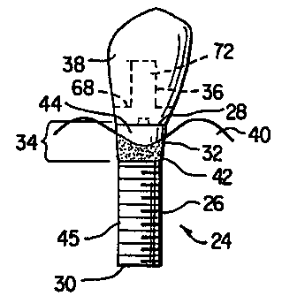
FIGS. 3 and 4 show a two-stage implant according to an embodiment of the
present
invention. Implant 24 has shaft 26, substantially planar abutment-implant
interface 28,
distal end 30, proximal end 32 and bone-tissue/soft-tissue transition region
34. Abutment
36 and crown 38 are attached to implant 24 using means well known to the
skilled artisan
for two-stage implants. Implant 24 is made from a biocompatible material,
including but
not limited to, metal, ceramic, glasses or any combination thereof. Preferably
implant 24 is
made from titanium or an alloy thereof.
Bone-tissue/soft-tissue transition region 34 has a scalloped bone-tissue
apposition
surface 42, which approximately follows the naturally occurring contours of
existing bone
40, and a scalloped soft-tissue apposition surface 44, which approximately
follows the
naturally occurring contours of the existing soft-tissue (not shown). Thus,
there are two
distinctive scalloped tissue-attachment surfaces: bone-tissue apposition
surface 42 to
maintain the naturally occurring bone-tissue morphology; and soft-tissue
apposition surface
44 to maintain the naturally occurring soft-tissue morphology. The degree of
scalloping or
the height of the convex and concave regions depends on, inter alia, the
degree of existing
bone-tissue resorption, the size of the implant, the implant location within
the arch, the bone
morphology and the soft-tissue morphology. The dimensions are similar to the
scalloped
appearance of the cemento-enamel (CE) junction observed on natural teeth. The
vertical
difference between the highest and lowest point of the scalloped margin ranges
from less
than 1mm on posterior teeth to approximately 3-5mm on anterior teeth. By way
of example,
bone-tissue apposition surface 42 can be obtained by machining, application of
textured
surfaces, acid etching, blasting with particles, applying growth factor,
applying protein, or
other materials that promote, enhance, and/or maintain bone-tissue growth
and/or
apposition. Also by way of example, soft-tissue apposition surface 44 can be
achieved by
polishing or other treatment that leaves a surface to promote, enhance, and/or
maintain soft-
tissue growth and/or apposition. Below the bone-tissue/soft-tissue transition
region 34,
shaft 26 has threads 45, or other means well known in the art, to anchor the
implant into the
alveolar bone.
In use, the surgeon inserts distal end 30 into the alveolar bone such that
bone-tissue
apposition surface 42 and soft-tissue apposition surface 44 approximately
mirror the
existing bone- and soft-tissue morphology respectively. The implant should be
aligned such
that the highest points of bone apposition surface 42 are substantially
aligned with the
interproximal areas of the bone-tissue and such that the lowest points are
substantially
aligned with the buccal and lingual area of the bone-tissue. In a two-stage
process, the
surgeon sutures tissue over the implant, waits several months for the bone to
adhere to the
-5-
CA 02352809 2001-05-29
WO 00/32134 PCT/US99/28304
implant, opens the tissue, attaches abutment 36 to abutment-implant interface
28 and
attaches crown 38 to abutment 36. Bone-tissue apposition surface 42 and soft-
tissue
apposition surface 44 maintain bone- and soft-tissue attachment levels and
facilitate
prevention of peri-implant infections, which occur due to increased peri-
implant pocket
depths frequently observed with. the prior art implant designs. Therefore,
implants
constructed according to the present invention increase the longevity of the
implant and
improve the aesthetic appearance of the restoration.
Referring to FIGS. 5A and 5B, abutment-implant interface 28 has substantially
planar upper surface 25, which is approximately 90 to the longitudinal axis
of shaft 26, and
connecting means 46 for connecting abutment 36 (FIGS. 3 and 4) to abutment-
implant
interface 28. Connecting means 46 is well known in the art and includes, but
is not limited
to, internal hex, external hex, standard hex, tall hex, wide hex or camlog. In
an alternative
embodiment of the present invention, as shown in FIGS. 6-8, abutment-implant
interface 48
has at least its edges contoured to approximate the contours of the alveolar
bone, thereby
defming a contoured upper surface 50 (FIG. 8) surrounding connecting means 46.
Also
provided in this alternative embodiment is abutment 52, which has lower
contoured surface
54 configured to substantially mate with contoured upper surface 50. The upper
and lower
contoured surfaces provide additional lateral support between abutment 52 and
abutment-
implant interface 48. Additionally, contoured upper surface 48 of this
alternative
embodiment results in a narrower depth between gum line 54 and abutment-
implant
interface 48 (FIGS. 6 and 7), thus enhancing longevity of the restoration as a
result of
decreased pocket depths.
A skilled artisan will readily recognize that the principles of the present
invention
can be equally applied to one-stage as well as two-stage processes. For
example, FIGS. 9
and 10 show one-stage implant 58, according to another embodiment of the
present
invention: Implant 58 includes shaft 60, distal end 62, proximal end 64 and
bone-
tissue/soft-tissue transition region 66 with scalloped bone-tissue apposition
surface 42 and
scalloped soft-tissue apposition surface 44, as substantially described above.
Abutment 69
is permanently attached to the one-stage implant 58 as is well know in the
art.
One-or two-stage implants, according to alternative embodiments of the present
invention, may include either a planar abutment-crown interface 68 (FIGS. 3,
4, 9 and 10) or
a contoured abutment-crown interface 70 (FIGS. 6, 7, 11 and 12), the latter of
which
substantially matches the natural contour of the alveolar bone. Contoured
abutment-crown
interface 70 allows for crown 38, in both one-and two-stage implants, to
extend further
towards the gum line, thereby resulting in a more aesthetically pleasing
restoration.
-6-
CA 02352809 2001-05-29
WO 00/32134 PCT/US99/28304
Chimney 72, or other means well known to the skilled artisan, is provided in
both one-and
two-stage implants according to the present invention for attaching crown 38
to the
abutment.
Although various embodiments of the present invention have been described, the
descriptions are intended to be merely illustrative. Thus, it will be apparent
to the skilled
artisan that modifications may be made to the embodiments as described herein
without
departing from the scope of the claims set forth below.
15
25
35
-7-