Note : Les descriptions sont présentées dans la langue officielle dans laquelle elles ont été soumises.
CA 02353876 2001-06-04
- WO 80%37918 PCT/DK99/00720
A METHOD AND AN APPARATUS FOR CUTTING OF TISSUE BLOCKS
The present invention relates to a method and an apparatus for cutting of a
tissue
block for pathological examination. The invention further relates to a method
an
apparatus for preparing a tissue block for sectioning in such an apparatus by
use of
such method. The invention also relates to a tissue embedding obtained by said
method and apparatus for preparing a tissue block.
The cutting of larger tissue blocks for pathological examination has normally
been
performed by hand. This technique involves a special pathology knife that is
used for
cutting slices of parenchymateous organs such as brain, liver, kidney and
heart. This
cutting technique is quick and sufficient for the daily qualitative
examinations on a
pathological institute. The technique, however, result in tissue sections with
highly
variable form and thickness, just as the hand cutting does not prevent
deformation of
i 5 the organ.
From US 5,148,729 a biological tissue slicer is known that can produce thin
slices of
live tissue for biochemical, pharmacological or toxicological studies. With
this
machine a thin slice of tissue can be peeled off the tissue block one at the
time by a
reciprocally cutting blade. The slices obtained hereby are completely
inadequate for
pathological examination purposes.
US 4,820,504 discloses a method of preparing a rnulti-specimen tissue block
and
sections thereof, where a plurality of different anitigenically reactive
tissue
specimens are formed into a rods and embedded in a medium and then sliced off
into
sections which each contain a cross-section of the rod. With this technique,
the
resulting tissue slices are inaccurate in form due to possible deformation
during the
formation and during the slicing of a rod. Moreover, these sections are only
usable as
check samples of the different tissues in the mufti-specimen tissue block from
which
the section is sliced off.
CONFIRMATIOW CQPIf
CA 02353876 2001-06-04
WO OOj37918 PCT/DK99/00720
2
The present techniques for preparing tissue sections for pathological
examination are
in accurate and does prevent deformation of the tissue block to an acceptable
degree.
It is the object by the present invention to circumvent the problems that lead
to bias
when quantitative organ or tissue examinations are desired. Another object of
the
invention is to provide for new quantitative unbiased stereological techniques
which
require cutting of the tissue in question in sections with equal thickness and
orientation.
These objects are achieved by a method for cutting of a tissue block in slices
with a
predetermined orientation in the tissue block preferably corresponding to
orientation
of the plane of a scanning, such as a CT, MR or PET scanning, wherein the
tissue
block, such as an internal organ or other internal anatomical structures, is
placed with
a predetermined position and then simultaneously sliced into a multiple of
sections.
In a second aspect, the invention involves an apparatus for cutting of a
tissue block in
slices with a predetermined orientation in the tissue block for obtaining a
direct
correlation of CT, MR or PET images for pathological examination, said
apparatus
comprising a support surface for receiving a tissue block, sectioning means
comprising a multiple of cutting members, and driving means for moving the
sectioning means towards the support surface for slicing a tissue block into
sections.
The invention circumvents the problems that lead to bias when quantitative
organ or
tissue examinations are desired. The invention is ideally suited for the new
quantitative unbiased stereological techniques which require cutting of a
tissue block
in sections with equal thickness and orientation. The invention also allows
the
resulting organ or tissue sections to be directly correlated to corresponding
scanning
planes from imaging modalities such as e.g. computerised tomography (CT),
magnetic resonance imaging (MRI) and positron emission tomography (PET).
In a third and fourth aspects, the invention involves a method and an
apparatus for
preparing a tissue block for sectioning in the slicing machine. By this
preparation, the
organ or tissue is embedded in an alginate plastic polymer mould that together
with
CA 02353876 2001-06-04
WQ~00/37918 PCT/DK99100720
3
the embedded tissue subsequently can be sectioned in the tissue slicing
machine.
finally the invention also involves a tissue embedding prepared by the use of
such
method.
In a first embodiment of the method and apparatus for cutting of tissue blocks
invention, the sectioning means comprise a multiple of parallel cutting
members
arranged in a cutting frame. Hereby, the sections obtained by the simultaneous
sectioning of the tissue block can be easily produced by lowering a frame with
cutting members down to and through the underlying tissue block.
In a preferred embodiment the distance between the cutting members can be
adjusted. Hereby sections of a predetermined thickness can be obtained.
The tension of the cutting members can preferably also be adjusted. Hereby,
the risk
of causing a deformation of the tissue during the cutting action.
In a first embodiment the cutting members are razor blades. This ensures a
sharp and
accurate cut without deforming the tissue block during the slicing.
In an alternative embodiment the cutting members can be wires. Hereby, a more
simple and less expensive solution can be provided where appropriate.
In the preferred embodiment of the invention, the support surface is provided
with
positioning means for allowing accurate positioning of a tissue block,
preferably
embedded in an embedding having predetermined reference surfaces. This ensures
that the tissue can be positioned relative to the cutting members in such a
way that
the resulting sections correspond to scanning planes used in a scanning.
In the preferred embodiment, the support surface is provided with vacuum
supply
means for retaining the tissue block in a predetermined position. Hereby, a
simple
and hygienic and stable retention means is provided.
CA 02353876 2001-06-04
-WO~ 00/37918 PCT/DK99/00720
4
In the preferred embodiment centring means with a laser pointer are provided
for
accurate positioning of the tissue block on the support surface. The laser can
be used
for accurate position of the tissue block relative to the cutting members by
assisting
the positioning of the tissue block in the centre of the support surface.
This positioning could also comprise concentric centring marking circles in
the
support surface and possibly supplemented with an aiming crossing lines. This
could
e.g. be in the form of concentric recesses in the support surface.
In particular, concentric circular suction rings are provided that can be
supplied with
vacuum from the vacuum supply means for retaining the tissue block. This is
particularly advantageous since the vacuum can be used not only for the
retention but
also for the aligning or centring of the tissue block.
I S The cutting members are preferably connected to vibration means for
vibration
during the slicing action, in order to facilitate the cutting action and
prevent
deformation of the tissue during the cutting action.
The vibration means could advantageously comprise a pneumatic vibrator that is
connected to pneumatic supply means.
The vacuum in the vacuum supply means could preferably be generated by vacuum
generating means connected to the pneumatic supply means. Hereby, only the
number of control or supply systems needed can be reduced.
In a preferred embodiment, the driving means comprise pillar guiding means
provided on the support surface and linear actuation means for linear movement
of
the sectioning means towards the support surface along the path defined by the
pillar
guiding means. This allows an accurate and smooth linear movement of the
cutting
frame up and down relative to the support surface for cutting the tissue. By
the use of
a die set for the guiding means, the travel of the cutting frame can be
carried out
virtually without slack whereby an accuracy in the sectioning is achieved. The
linear
CA 02353876 2001-06-04
12V0 80137918 PCT/DK99/00720
actuation means preferably comprise a threaded driving spindle parallel to the
guide
means and a corresponding threading in the cutting frame.
In a first embodiment, the threaded driving spindle is provided with a handle
for
5 manual operation. This offers a simple apparatus for carrying out the
sectioning.
However, in an alternative embodiment, the driving spindle can be
pneumatically or
electrically driven.
In order to ensure a good positioning of the tissue block in the apparatus and
to
prevent deformation during the cutting, the invention also relates to method
and an
apparatus for preparing a tissue block. This method comprises the steps of
filling a
moulding form with an appropriate amount of non-toxic, biologically inert
polymer
moulding material, said form having at least one reference surface, and
positioning a
tissue block in said polymer moulding material in a predetermined position
relative
to said at least one reference surface, while the polymer moulding material is
in its
soft state.
By this method, the tissue block is provided with regular outer surfaces. that
due to
the form of the mould can adapted to the support surface of the sectioning
apparatus.
The tissue block is in a preferred embodiment positioned in the polymer
material
with an orientation that corresponds to the orientation of the tissue block in
vivo.
Hereby, a correlation between scanning images and the sections can be ensured.
The tissue block is embedded in a bottom mould part and a top mould is formed
in a
top moulding form that is filled with polymer moulding material and placed on
the
top of the lower moulding part with a partly encased tissue block, so that the
tissue
block is completely encased by in the moulding. This provides an effective
insurance
against the otherwise free top part of the tissue to be deformed by the
cutting
members.
CA 02353876 2001-06-04
WO 00137918 PCT/DK99/00720
6
The tissue block is in a preferred method of preparation fixed to a reference
moulding of predetermined dimensioned and that said reference moulding is
pivoted
into a predetermined position in one or more directions, and then moulded into
at
least a bottom moulding. Hereby, the orientation of the tissue block can be
vary
accurately embedded relative to the reference surfaces.
The polymer material that is preferably used, is a cold polymerisate that
polymerises
by addition of water, such as an algino plastic polymer.
The apparatus and the details of the functions of the apparatus can be
appreciated in
the dependent claims 33 to 36.
Finally, the invention also relates to a tissue embedding comprising a tissue
block
made by this preparation method and apparatus. This tissue embedding providing
the
I S tissue block with regular reference surfaces ensures an accurate cutting
of slices of
the block for pathological and other purposes. It is realised that this
technique of
embedding the tissue block in an alginate or similar suitable moulding
material can
advantageously be used prior to any cutting action, whether a slice at the
time is cut
or - as it is the case in the first aspect of the invention - that the slices
are cut
simultaneously.
The invention will be described more detailed below with reference to the
accompanying drawings, in which
Fig. 1 is a perspective view of an apparatus for sectioning a tissue block
according to the invention,
fig. 2 shows a cutting frame of said apparatus,
fig. 3a and 3b show the tissue block embedded in an alginate bottom and with
an
alginate top mould,
fig. 4a to 4g show the embedding apparatus and the steps in the oriented
alginate
embedding procedure, and
fig. S is a top view of the embedding apparatus of figs. 4a-4g.
CA 02353876 2001-06-04
WO 0037918 PCT/DK99/00720
7
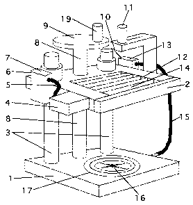
Referring to fig. l, a preferred embodiment of the tissue slicing machine is
shown.
The tissue slicing machine stand on an aluminium or steel base plate 1 that
preferably rest on a rubber pad or rubber knobs attached to the base plate 1.
The base
S plate i is connected to the aluminium or steel top plate 2 by pillar guiding
means
comprising two the pillars 3 which through operation of the crank and spindle
8, 9
allow lowering and elevation of the top plate 2 in relation to the base plate
1. In the
centre of the top plate 2 a rectangular hole leaves room for attachment of the
cutting
frame 12. The cutting frame 12 is fixed in place by screws or a handle on the
side of
the cutting frame 12. The cutting frame 12 comprises a number of cutting
members
14 (see fig. 2), preferably in the form of thin razor blades of hardened
steel. The
razor blades 14 are spaced by spacing blocks 38 that can be made of metal or
plastic.
In the preferred embodiment the cutting frame 12 is exchangeable as a whole
when
the razor blades 12 are worn out and has lost their sharpness. In another
embodiment
the knife frame 12 allow changing or removal of individual blades 14, just as
spacing
blocks 38 of different thickness can be used. On the side of the top plate 2 a
pneumatic or electric operated vibrator 4 is placed. It will when activated
set the
sectioning means comprising the top plate 2 and knife frame 12 into vibrations
along
the long axis of the razor blades 14. This facilitates the cutting procedure
by lowering
of the friction as the knives 14 pass through the tissue 20 and alginate block
25 (see
figs. 3a and 3b). On the side of the top plate 2 there is also placed a
pneumatic valve
7 for pressurised air to operate the embodiment with the pneumatic vibrator 4.
The
pneumatic vibrator 4 is connected to the pneumatic air valve 7 through a
pneumatic
hose S. The pneumatic air valve 7 is connected to a pressurised air source at
the
pneumatic intake 6. On the other side of the top plate 2 or on the base plate
1 in
another embodiment a valve 13 for vacuum with vacuum outtake 10 is placed. In
one
embodiment the vacuum outtake 10 is connected to a vacuum pump (not shown). In
a second embodiment the vacuum is produced by a second pneumatic air flow
valve
(not shown). The vacuum hose 15 connects the vacuum valve 13 to a recess and
associated apertures 16 for retention and vacuum fixation of alginate and
tissue block
25. In the support surface of the base plate 1 concentric circles 17 and a
cross hair cut
allow centring of alginate and tissue block 25. To further aid the centring of
the
CA 02353876 2001-06-04
r WO.OU/37918 PCT/DK99/00720
8
tissue and alginate block 25 a laser pointer 11, that point to the central
vacuum hole
of the vacuum apertures 16, is provided on the top plate.
Fig. 2 show an embodiment of the cutting frame 12 consisting of knives 14 that
are
angled in relation to the horizontal plane of the base plate 1. This
embodiment will
reduce friction and deformation during the cutting of the tissue alginate
block 25. In
one embodiment the knife frame 12 consist of razor blades 14 that cannot be
replaced, and the whole frame 12 must be changed when the blades become dull.
In
another embodiment the knife frame allow exchanging of individual blades and
use
of spacing blocks 38 with different thickness.
The bearing construction of the tissue slicing machine comprises a pillar
guided base
and top plate 1 and 2. The top plate 2 contains a set of parallel oriented
knives 14
positioned in a frame 12. The knives 14 are mounted in a "knife frame set''
and the
distance between the knives 14 are spaced by high tolerance spacing blocks 38
with
an equal thickness. Changing between different knife frame sets can vary the
knives
distance. The frame of the tissue slicing machine is equipped with a pneumatic
vibrator 4 that make the knife frame set vibrate along its longitudinal axis,
i.e. along
the cutting edge of the knives. The vibration 4 of the knife frame 12
diminishes
friction as it moves through alginate and tissue block 25. The knife frame
with
vibrator is mounted on a columnar lead equipped with a crank 19 that by
turning
allow movement of the knife frame 12 in the vertical plane. For fixation of
alginate
and tissue block 25 the base plate 1 of the tissue slicing machine is equipped
with a
suction pad that is activated by opening a vacuum valve after placement of the
alginate tissue block. The concentric rings of the suction pad also serve to
centre the
alginate tissue block, just as a laser pointer identifies the centre.
Refernng to figure 3a and 3b, any tissue block 20 or organ can be embedded
into an
alginate plastic mould 25. In one embodiment of this invention the tissue 20
is first
embedded in an alginate bottom mould 22. This can be done by pouring the
mixture
of alginate powder and water in to a moulding form 21, such as a plastic jar,
followed by placement of the tissue 20 into the still soft alginate-water
mixture in the
CA 02353876 2001-06-04
WO Oti%37918 PCT/DK99/00720
9
mould 22. When the alginate bottom 22 has hardened an alginate top mould 23
can
be cast in a similar fashion by placing a second moulding form 24, such as a
second
plastic jar 24. This top mould 23 can subsequently be removed for better
placement
of the alginate bottom 22 in the tissue slicing machine as shown in fig. 1 by
then use
S of anatomical landmarks. In a second embodiment the tissue 20 can be cast
entirely
into alginate followed by CT or MRI scanning of the tissue and alginate block.
When
placed in the tissue slicing machine in same way as in the CT or MRI scanner
the
resulting tissue sections will correspond to the scanning planes. In a third
embodiment of the embedding procedure, alginate embedded tissue can be cut on
prior art tissue sectioning machines, such as cryostats, vibratomes and
microtomes.
For tissue embedding alginate plastic polymer from Bayer Dental was used. The
alginate is a non-toxic cold polymerisate that polymerises after addition of
water.
The alginate powder is stirred into the water and then poured into a plastic
jar or
other moulding form 21 of appropriate size for the tissue block in question.
The
organ or tissue block 20, such as a pig brain, is then placed in the still
soft polymer
and hold in place until the alginate hardens. The embedding is the crucial
step in the
process and care must be taken to orient the tissue 20 in the alginate as it
is oriented
in vivo. For less accuracy this can achieved by the use of an angle protractor
34-36
and anatomical landmarks on the tissue 20 in question. For high accuracy the
tissue
embedder must be used. A further option is to cast another alginate mould 24on
top
of the tissue and alginate bottom 22. This done in order to support the tissue
20
during the cutting procedure and avoid tissue deformation. In the following
this will
be described as a tissue and alginate bottom 22 and an alginate lid 24.
An alternative strategy that can be used, if no scanning is needed before
pathological
extraction of the organ, is to embed the organ 20 in alginate and then perform
the
desired computer assisted scanning modality on tissue and alginate block 25
followed by the sectioning as described by the first aspects of the present
invention.
This strategy abolishes the need for orientation of the tissue block 20 as the
resulting
digital image scanning planes will correspond to the histological sections
provided
CA 02353876 2001-06-04
VSO 00/37918 PCT/DK99/00720
that the tissue and alginate block 25 is placed in the cutting machine in the
same
fashion as in the CT, MRI or PET scanner.
In the apparatus for cutting of tissue blocks, the tissue block 25 with the
embedded
5 tissue 20, is placed on the suction pads 16 of the support surface and
centred in
relation to the cutting frame 12 by use of the concentric circles 17 of the
tissue
slicing machine base plate 1 and the laser pointer 11. Following the centring
the
alginate tissue block 25 is fixed by activation of the vacuum valve 13 and is
now
ready for the cutting. This can be done with or without the alginate lid 24.
An
10 opening of the pneumatic valve 7 activates the pneumatic vibrator 4 and the
cutting
frame 12 starts vibrating. By a steady rotating movement, the crank 19 of the
columnar lead is turned and the cutting frame 12 is lowered through the
alginate and
tissue block 25. The cutting results in a set of alginate and tissue slabs
(not shown)
that are of equal thickness and oriented corresponding to the scanning plane
of the
I 5 given computerised scanning modality.
Referring to figures 4a to 4g a method and an apparatus for preparing a tissue
block
by embedding the tissue block 20 in alginate plastic polymer. This can be done
in
such a way that the tissue 20 is oriented to existing CT, MRI or PET scanning
planes.
Fig. 4a shows the embedding apparatus that comprises of a central plastic rod
28
with a spheric top end 28a and two concentric plastic cylinders, i.e. an inner
cylinder
29 and an outer cylinder 30. The rod 28 is fixed to a plastic base plate 31,
where
upon also the cylinders 29, 30 rest in their retracted positions. On top of
the outer
cylinder 30 four plastic pins 33 with or without a screw thread are placed at
90
degrees interval and orthogonal to the long axis of the cylinder 30 (see fig.
5).
A reference moulding form 27 is placed on plate means 32 comprising two half
parts
placed on the inner cylinder 29 on each side of the rod 28. Hereby, a
reference
moulding form is defined. This form is filled with polymer moulding material
26 in
which the tissue block 20 is placed. This means that the tissue block 20 is
embedded
CA 02353876 2001-06-04
VYO 00%37918 PCT/DK99/00720
11
in a reference mould 26, such as shown in fig. 4b, where the plate means 32
and the
form 27 is removed.
Fig. 4c shows a tissue and alginate reference mould 26 placed in the embedding
apparatus. The alginate mould 26 is first fixed with two plastic pins 33
facing each
other at 180 degrees. This plane can be defined as the X plane. The tilt angle
of the
tissue and alginate reference mould 26 in relation to the horizontal Z plane,
as
determined from the desired CT, MRI or PET scanning, is determined by an angle
protractor 36 (see fig. 4d) in one embodiment. In a second embodiment the
angle
protractor 36 is equipped with a laser guide 34 directing a beam 35 at the
mould for a
measure of the angle of inclination. When the tissue and alginate reference
mould 26
is fixed in the desired angle in the X plane, it is then fixed in a similar
way by the
two plastic pins 33 placed orthogonally in the Y plane.
Fig. 4e shows the casting of the second alginate bottom 22. The shape of the
bottom
mould 22 is adapted to fit into the tissue slicing machine of fig. 1. First,
the outer
cylinder 30 is raised and a second base plate 37 is slid into a corresponding
horizontal opening in the outer plastic cylinder 30, followed by casting of
the second
alginate bottom 22, as the outer cylinder 30 forms the side part of the
moulding form.
Fig. 4f shows the placement of a top form 21 on the outer cylinder 30 followed
by
casting of an alginate top mould 24 on the top of the bottom mould 22 and the
tissue
block 20.
Fig. 4g shows the embedded tissue block 20 with its alginate moulding 25 -
i.e. the
reference mould 26, bottom mould 22 and top mould 24 - free of the embedding
apparatus after the outer cylinder 30 has been slid back towards the base
plate 31.
Fig. 5 shows a top view of the outer cylinder 30, inner cylinder 29, centre
rod 28 and
plastic pins 33 for pivoting the reference mould 26 of the tissue block.
CA 02353876 2001-06-04
WO OOI37918 PCT/DK99/00720
12
This embedding apparatus allows accurate three dimensional orientation of a
tissue
reference moulding 26 in relation to CT, MRI or PET scans. Following the
accurate
orientation of the tissue reference moulding 26, a second moulding is
performed to
produce a second alginate bottom 22 with an outer surface that will fit into
the tissue
slicing machine. If desired a final alginate lid 24 can be cast on top of the
tissue 20
and alginate bottom 22 in order to avoid tissue deformations during the
cutting
procedure. The embedding apparatus comprises a circular rod 29 of transparent
plastic, such as plexi-glass or similar material, two outer concentric plastic
cylinders
29 and 30, fixation pins 33, semicircular plastic plates32 and an insertable
base plate
37 that preferably also is made from a plastic material.