Note : Les descriptions sont présentées dans la langue officielle dans laquelle elles ont été soumises.
CA 02353901 2002-O1-17
we omssii rc-i.,usooiz~ss~,
METIiOD AND APPARATUS FOR GUIDING ABLATIVE THERAPY OF
ABNORMAL BIOLOGICAL ELECTRICAL EXCITATION
GOVERNMENT SUPPORT
The U.S. Government has a paid-up license in this invention and the right in
limited circumstances to require the patent owner to license othtrs on
reasonable terms as
provided for by the terms of Grant No. NAGS-4989 awarded by the National
Aeronautics
and Space Administration.
BACKGROUND OF THE INVENTION
The electrical activity generated in certain organs in the human body is
intimately
related to their function. Abnormalities in cardiac and brain electrical
conduction
processes are principal causes of morbidity and mortality in the developed
world.
Appropriate treatment of disorders arising from such abnon:nalities frequently
requires a
determination of their location. Such localization of the site of origin of an
abnormal
r~~~ excitation is typically achieved by painstaking mapping of the electrical
activity
on the inner surface of the heart or the brain from electrodes or a catheter.
Often, this
recording must be done while the abnormal biological electrical excitation is
ongoing.
Radio frequency catheter ablation procedures .have evolved in recent years to
become an established treatment for patients with a variety of
supraventricular [Lee,
1991; Langberg, 1993] and ventricular arrhythmias [Stevenson, 199?; Stevenson,
1998].
1
SUBSTITUTE SHEET (RULE26)
CA 02353901 2002-O1-17
we omssiz rcrnrsooms~
However, in contrast to supraventricular tachycardia ablation, which is highly
successful
because the atrio-ventricular node anatomy is known, ventricular tachycardia
ablation
remains difficult because the site of origin of the arrhythmia could be
anywhere in the
ventricles.
Sustained ventricular tachycardia is often a difficult arrhythmia to manage.
One of
the most common indications for radio frequency catheter ablation of
ventricular
tachycardia is arrhythmia refractory to drug therapy that results in frequent
discharges
from an implantable cardioverter-dcftbrillator. Radio Frequency ablation is
also indicated
when the ventricular tachycardia (VT) is too slow to be detected by the
implantable
cardioverter-defibrillator or is incessant [Strickbtrger, 1997].
Selection of the appropriate target sites for ablation is usually based on a
combination of anatomical and electrical criteria. The ability of the
physician to deliver
radio frequency . energy through a catheter at the reentry site is restricted
by the
limitations of the current technology that is employed to guide the catheter
to the
appropriate ablation site. The pri~ipal limitation of the radio frequency
ablation
technique is the determination of the correct site for delivery of the radio
frequency
~ergy. Conventionally, this determination is achieved by painstaking mapping
of the
electrical activity on the inner surface of the heart from electrodes on the
catheter. Often,
this recording must be done while the arrhythmia is ongoing. This is a major
problem,
especially for those arrhythmias which compromise hemodynamic function of the
patient.
Many arrhythmias for this reason are not presently amenable to radio frequency
ablation
treatment.
2
SUBSTITUTE SHEET (RULE26)
CA 02353901 2002-O1-17
WO ~1II3~2 PCTIU9102?664
The acute lesion created by radio frequency current consists of a central zone
of
coagulation necrosis surroundod by a zone of hemorrhage and inflammation.
Arrhythmias may recur if the target tissue is in the border zone of a lesion
instead of in
the central area of necrosis. If the inflammation resolves without residual
necrosis,
arrhythnuas may recur several days to several weeks after an apparentl~~
successful
ablation [I,angberg, 1992]. Conversely, an arrhythmia site of origin that was
not initially
successfully ablated may later become permanently nonfunctional if it lies
within the
border zone of a lesion and if microvascular injury and inflammation within
this zone
result in progressive necrosis [Math, 1994]. Thus, the efficacy and long term
outcome of
catheter ablation depend on accurate determination of the site of origin of
the arrhythmia.
Catheter ablation of sustained monomorphic ventricular tachycardia laic after
myocardial infarction has been challenging. Tl~se anrhythmias arise from
reentry circuits
that can be large and complex, with broad paths and narrow isthmuses, and that
may
traverse sube~ndocardial, intramural, and epicardial regions of the myocardium
[deBakker,
1991; Kaltenbrunner, 1991].
Mapping and ablation are further complicated by the frequent presence of
multiple reentry circuits, giving rise to several morphologically different
VTs [Wilbur,
1987; Waspel, I985]. In some cases, different reentry circuits form in the
same abnormal
region. In other cases, reentry circuits form at disparate sites in the
infarct area. The
presence of multiple morphologies of inducible or spontaneous VT has been
associated
with antiarrhythmic drug ine~cacy [Mitrani, 1993] and failure of surgical
ablation
[Miller, 1984].
3
SUBST1TUTE SHEET (RULE2b)
CA 02353901 2002-O1-17
we omsern
rcT~rsoomss4
Several investigators have reported series of studies of patients selected for
having one predominant morphology of VT ("clinical VT") who were treated with
radio
frequency catheter ablation [Morady, 1993; Kirn, 1994]. It is likely that this
group of
patients represents less than 10% of the total population of patients with VT
[Kim, 1994].
The patient must remain hemodynamieally stable while the arrhythmia is induced
and
maintained during mapping. The mapping procedure may take many hours during
which
the arrhythmia must be maintained. Thus, currently radio frequency catheter
ablation is
generally limited to "slow" ventricular tachycardia 0130 bpm) which is most
likely to be
hemodynamically stable.
Ablation directed towards the ~"clinical tachycardia" that did not target
other
inducible VTs successfully abolished the "clinical VT" in 71 % to 76% cases.
However,
during followup up to 31% of those patients with successful ablation of the
"clinical VT"
had arrhythmic recurrences, some of which were due to different V'f
raorphologies from
that initially targeted for ablation.
Furthermore, there are several difficulties in selecting a dominant, "clinical
VT"
for ablation. Often it is not possible to determine which VT is in fact the
one that has
occurred spontaneously. In most cases, only a limited recording of one or a
few ECG
leads may be available. In patients with implantable defibrillators VT is
typically
. ,
terminated by the device before an ECG is obtained. Even if one VT is
identified as
predominant, other VTs that are inducible may subsequently occur
spontaneously. An
alternative approach is not to consider the number of VT morphologies in
deternzining
eligibility for catheter ablation but rather to attempt ablation of all
induc~ble VTs that are
sufEciently tolerated to allow mapping [Stevenson, 1998b; Stevenson, 1997].
However,
4
SUBSTITUTE SHEET (RULE26)
CA 02353901 2002-O1-17
WO 01IZS822 rcrrosooms~t
this approach requires that the patient be hetnodyaamically stable during the
VT mapping
procedure.
The use of fluoroscopy (digital bi-plane x-ray) for the guidance of the
ablation
catheter for the delivery of the curative radio frequency energy is common to
clinical
catheter ablation strategies. However, the use of fluoroscopy for these
purposes may be
problematic for the following reasons: (1) It may not be possible to
accurately associate
intracardiac electrograms with their precise location within the heart; (2)
The endocardial
surface is not visible using fluoroscopy, and the target sites can only be
approximated by
their relationship with nearby structures such as ribs and blood vessels as
well as the
position of other catheters; (3) Due to the limitations of two-dimensional
fluoroscopy,
navigation is frequently inexact, time consuming, and requires multiple views
to estimate
the three-dimensional location of the catheter; (4) It may not be possible to
accurately
return the catheter precisely to a previously mapped site; (5) It is desirable
to minimize
exposure of the patient and medical personnel to radiation; and (6) Most
importantly,
fluoroscopy cannot identify the site of origin of an arrhythmia and thus
cannot be used to
specifically direct a catheter to that site.
Electro-anatomic mapping systoma (e.g., Carto, Biosense, Marlton, NJ~ provide
an electro-anatomical map of the heart. This method of nonfluoroscopic
catheter mapping
is based on an activation sequence to track and localize the tip of the
mapping catheter by
magnetic localization in conjunction with electrical activity recorded by the
catheter. This
approach has been used in ventricular tachyardia [Nademanee, 1998; Steveason,
1998],
atrial flutter [Shah,1997; Nakagawa, 1998], and atrial tachycardia ablation
[Natale,1998;
ICottkamp, 1997]. The ability to localize in space the tip of the catheter
while
SUBSTITUTE SHEET (RULE26)
CA 02353901 2002-O1-17
we einsszZ renusoon»t
simultaneously measuring the electrical activity may facilitate the mapping
process.
However, this technique fundamentally has the Limitation that it involves
sequentially
sampling endocardial sites. The mapping pmcess is prolonged while the patients
must be
maintained in VT. Also, the localization is limited to the cndocardial surface
and thus
sites of origin within the myocatr3ium cannot be accurately localized.
The basket catheter technique employs a non-contact 64-electrode basket
catheter
(Endocardial Solutions Inc., St. Paul, MN) placed inside the heart to
electrically map the
heart. In the first part of, this procedure high frequency current pulses are
applied to a
standard catheter used in an ablation procedure. The tip of this catheter is
dragged over
the endocardial surface, and a basket catheter is used to locate the tip of
the ablation
catheter and thus to trace and reconstruct the endocardial surface of the
ventricular
chamber. Then the chamber geometry, the known locations of the
basket~catheter, and the
non-contact potential at each etocrmde on the basket catheter are combined in
solving
Laplace's equation, and electrognams on the endocardial surface are computed.
This
technique bas been used in mapping atriat and ventricular arrhythmias
[Schitling, 1998;
Gornick, 1999]. One of the drawbacks of this methodology is that the
ventricular
geometry is not fixed but varies during the cardiac cycle. In addition, the
relative
movement between the constantly eondracting heart and the electrodes affects
the
.. .
mapping. While the inter-electrode distances an each sidearm of the basket
catheter are
fixed, the distances between the actual recording sites on the endocardium
decrease
during systole. This leads to relative movement between the recording
electrode and the
tissue, significantly limiting the accuracy of the mapping method. Also, the
localization
6
SUBSTITUTE SHEET (RULE26)
CA 02353901 2002-O1-17
wo otns~ti
is limited to the endocardial surface, and thus sites of origin within the
myocardium
cannot be accurately localized.
What is needed is a means of efficiently directing the tip of a catheter to a
site of
origin of an arrhythmia in the heart (whether on the endocardial surface or
witlun the
myocardium itself), without the need to introduce additional passive
electrodes into the
heart, so that energy can be delivered through the catheter to ablate the site
of origin. It
would be advantageous to be able to accomplish this task without having to
maintain the
arrhythmia while advancing the catheter to the site of origin, so that the
patient does not
suffer the ill effects of the arrhythmia for a prolonged period. This
consideration is
particularly important in the case of rapid arrhythmias that compromise
hemodynamic
function.
SZJMIvLARY 4F THE INVENTION
The present invention provides methods and apparatus for localizing an
electrical
source within the body. The invention further provides methods and apparatus
for
delivering ablative electrical energy in the vicinity of an electrical source
within the body.
The electrical source may be located anywhere within the body. For example,
the
electrical source may be within. the heart and may be the site of origin of a
cardiac
arrhythmia. The electrical source may be a focus of elxtrical activity within
the brain,
such as a site involved in triggering an epileptic seizure, or may be located
in other
neurological tissue.
Cardiac arrhythmias are frequently treated by delive~cing elxtrical energy to
the
site of origin of the arrhythmia in an effort to ablate the site. To
effectively perform this
7
SUBSTITUTE SHEET (RULE26)
CA 02353901 2002-O1-17
we oiasszi
procedure, accurate localization of both the site of origin of the arrhythmia
and the
energy delivery device (e.g., the tip of a catheter) is necessary. As used
herein, the term
"localization" refers to determining either an absolute or a relative
location. The present
invention provides techniques for accurately performing such locali2ation. The
nunimally
invasive and fast aspects of certain embodiments of the invention, as
disclosed herein, are
particularly important.
In preferred embodiments the methods of the present invention involve placing
passive electrodes on the body surface, placing active electrodes) in andlor
on the body,
acquiring from the passive electrodes signals emanating from the electrical
source,
processing the signals emanating from the electrical source to determine the
relative
location of the electrical source, delivering electrical energy to the active
electrode(s),
acquiring from the passive electrodes the signals emanating from the active
electrode(s),
processing the signals emanating from the active electrodes) to determine the
relative
location of the active electrode(s), and positioning the active electrodes) to
locali2e the
electrical source. In another embodiment at least one of the passive
electrodes is placed
within the body, for example within the heart. The positioning step of the
present
invention may involve approximating the relative locations of the active
electrodes) and
the electrical source. In preferred embodiments of the method the energy
delivering step,
the second acquiring step, the second processing step and the positioning step
are
performed iteratively.
In a preferred embodiment the first processing step is used to determine the
relative location of the electrical source at a multiplicity of time epochs
during the cardiac
cycle, and the positioning step localizes the electrical source at one of the
time epochs. At
8
SUBSTITUTE SHEET (ItULEZ6)
CA 02353901 2002-O1-17
wo oinssiz rcT~soon»ca
least one criterion may be used to choose the time epoch. In a particularly
preferred
embodiment at least one of the processing steps involves fitting the acquired
signals to a
moving dipole model. In a particularly preferred embodiment of the invention
the
second processing step includes determining the relative Location of a moving
dipole that
is approximately parallel to the moving dipole fitted in the first processing
step to signals
emanating from the electrical source. In one embodiment, such determination is
made
using a multiplicity of active electrodes.
Another preferred, embodiment of the invention involves delivering ablative
energy in the vicinity of the location of an electrical source within the body
by delivering
ablative energy in the vicinity of the location of the active electrode(s).
The active
electrodes) may be located on a catheter, and the ablative energy may be
delivered
through the catheter. In a preferred embodiment the ablative energy is radio
frequency
energy.
The methods of the present invention may further include displaying various
parameters. Among the parameters of interest are the relative location of the
electrical
source and measures of the size, strength, and/or uncertainty in the relative
location of the
electrical source.
Other features and advantages of the invention will become apparent from the
following description, including the drawing, and from the claims.
BRIEF DESCRIPTION OF THE DRAWITiG
FIG. 1 is a flow chart of the method for localizing an electrical source
within the body.
9
SUBSTITUTE SHEET (RU1LE26)
CA 02353901 2002-O1-17
WO Al/2S81Z
FIG. 2 is a flow chart of a procedure for fitting single equivalent moving
dipole
parameters to ECG potentials.
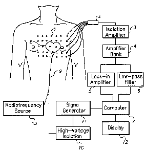
FIG. 3 is a schematic diagram of an apparatus for localizing a site of origin
of an
arrhythmia, guiding the delivery of ablative therapy, and delivering ablative
therapy to
the site of origin of the arrhythnua.
DESCRIPTION OF PREFERRED EMBODIMENTS
The present invention encompasses the finding that by employing a moving
dipole model it is possible to accurately localize a source of electrical
energy within the
body relative to the location of an active electrode. If one can localize the
site of origin
of an arrhythmia during the cardiac cycle, it is possible to ablate the site
through delivery
of ablative electrical energy. The present invention provides methods and
apparatus for
localizing an electrical source within the body. The invention further
provides methods
and apparatus for localizing and ablating the site of origin of a cardiac
arrhythmia.
The concept of considering the heart as a single dipole generator originated
with
Einthoven [Einthoven, 1912], and its mathematical basis was established by
Gabor and
Nelson [Gabor, 1954]. Several investigators [Mirvis, 1981; Gulrajani, 1984],
Tsunakawa, 1987] have studied the cardiac dipole in clinical practice and
attempted to
determine the dipolar nature of the ECG. The advantages of the use of the
equivalent
cardiac dipole are: (1) It permits quantification of source strength in
biophysical terms
that are independent of volume conductor size (classic electroeardiography),
and (2) The
active equivalent source can be loealizal and assigaed a location, something
that cannot
be done using classical electrocardiography.
SUBSTITUTE SHEET (RULE26)
CA 02353901 2002-O1-17
)
For many arrhythmias, the electrical activity within the heart is highly
localized
for a portion of the cardiac cycle. During the remainder of the cardiac cycle
the electrical
activity may become more diffuse as the waves of electrical activity spread.
It is not
possible to construct the three-dimensional distribution of cardiac electrical
sources from
a two-dimensional distribution of ECG signals obtained on the body surface.
However,
if it is known that a source is localized, then this localized source can be
approximated as
a single equivalent moving dipole (SEMD), for which one can compute the dipole
parameters (i.e., location and momeats) by processing electrocardiographic
signals
acquired from passive electrodes placed on the body surface or in the body.
Fitting the dipole parameters to body surface ECG signals provides a solution
(referred to herein as the inverse solution) for the dipole location (as well
as for its
strength and orientation). The location of the dipole at the time epoch when
the electrical
activity is confined to the vicinity of the site of origin of an arrhythmia
should coincide
with the site of origin of the arrhythmia. In contrast to standard mapping
techniques, the
inverse solution can be computed from only a few beats of the arrhythmia,
thereby
eliminating the need for prolonged maintenance of the arrhythmia during the
localization
process. In addition, if one delivers low-amplitude bipolar current pulses to
the tip of an
ablation catheter and acquires tire resulting body surface signals, the tip of
the catheter
_.
may likewise be modeled as a single equivalent moving dipole. Therefore, the
same
inverse algorithm may be employed to localize the tip of the catheter. Using
this
information one can guide the tip of the catheter to the site of origin of the
arrhythmia.
The confounding factors of the SEMD method involve the fact that, as described
above, the method does not consider boundary conditions and inhomogeneities in
tissue
11
SUBSTITUTE SHEET (RULE26)
CA 02353901 2002-O1-17
wo oirissaa
conductivity. Furthermore, even the exact position of the passive acquiring
electrodes in
three-dimensional space may not be accurately determined. Thus the inverse
solution
obtained is distorted. However, as long as the site of origin of the
arrhythmia and the tip
of the catheter are identified using the same algorithm, then when the two are
brought
together, both their positions will be distorted by the same amount. In other
words, when
the algorithm identifies that the site of origin of the arrhythmia and the
catheter tip are at
the same location, then they are at the same location. Thus the distortion due
to the above
factors should not significantly affect the accur~y by which one can make the
tip of the
ablation catheter and the site of origin of the arrhythmia coincide.
Therefore, although the
SEMD method described herein may not establish the absolute locations of the
site of
origin of the arrhythmia and the tip of the catheter, it can effectively
identify their relative
locations.
Figure 1 shows a flowchart of the method according to the present invention
for
localizing an electrical source within the body. The method includes placing
passive
electrodes in or on the body to acquire electrical signals. The signals from
the passive
electrodes are processed to determine the relative location of the electrical
source within
a given short time epoch. The processing steps are repeated for multiple
sequential time
epochs, and the location of the source corresponding to the site of the origin
of the
arrhythmia is obtained. As used herein, the phrase "sequential time epochs"
does not
necessarily imply immediately successive tixne epochs.
As further shown in Figure 1, electrical energy is delivered from at least one
active electrode placed within or on the body, and the signals emanating fram
the at least
one active electrode (e.g., at the tip of a catheter) are acquired from the
passive
12
SUBSTITUTE SHEET (RULE26)
CA 02353901 2002-O1-17
wo oinst;zz
electrodes. Signals emanating from the at least one active electrode are
processed to
determine the relative location of the at least one active electrode within a
given short
time epoch. Thereafter, the processes of delivering electrical energy and
determining the
relative location of at least one active elxtrode are repeated until the
active electrode is
superposed to the relative location of the eloctrical source. In other words,
in a preferred
embodiment of the invention, the processes of delivering electrical energy to
the at least
one active electrode, acquiring from the passive electrodes the signals
emanating from
the at least one active electrode, processing the signals emanating from the
at least one
active electrode, and positioning the at least one active eiectmde are
performed
iterazively. This repetition of steps 4 through 7 of Figure 1 may be
terminated when the
relative locations of the electrical source and the active electrode are
within a
predetermined distance (not shown on Figure 1).
The present invention provides method and apparatus for guiding ablative
therapy
within au organ system either from body surface electrodes or from internal
electrodes.
This technique explicitly recognizes that one cannot uniquely reconstruct from
a two-
dimensional array of electrodes a throe-dimensional distribution of sources.
The present
invention models bio-elxtrical activity as a single equivalent moving dipole
(SEM13),
which is a valid model when the bio-electrical activity is highly Localized.
In the cardiac
context, the evolution of the SEl~ during the cardiac cycle provides a three-
dimensional
picture of cardiac electrical activity.
The basic theory of the present invention derives from electromagnetic theory.
The potential due to a dipole in an infinite homogeneous volume conductor is
given by
the equation below.
Z3
SUBSTITUTE SHEET (RULE Z6)
CA 02353901 2002-O1-17
wo omse~i
. (~' r -r 7 (Eq. 1)
IT _ r~i3
where,
r' is the three-dimensional vector representing the i~, observation location,
p is the three-dimensional vcctar representing the dipole moment, end
r' is the three-dimensional vector representing the dipole location.
One may obtain the dipole parameters, i.e., p and r', from potential
measurements
through minimization of an objective function. One may use chi-square (x1) as
the
objective function to obtain the dipole parameter estimates. x2 is given by
equation 2
below:
I
xz _ ~ (~ _ ~~~Z (>~1. 2>
iW a
where ~ is the potential at the i,n electmde because of the specific dipole
components,
~,~ is the measured potential at the i~a electrode,
d is a noise measurement at the i,~ electrode, and
I is the number of electrodes.
Because of the linear dependence of the potential (Eq. I) on the dipole moment
parameters, the latter can be separated from the spatial dipole parameters.
Consequently,
14
SUBSTITUTE SHEET (RULE26)
CA 02353901 2002-O1-17
we oi»
any optimization method may be applied to the spatial parameters only, while
an analytic
optimization procedure rosy be perfonztod to obtain the optimal fitting dipole
moment
parameters for a specific set of dipole spatial parameters. We coin the term 3
plus 3
parameter optimization for this algorithm.
Using the x2 as an objective fimetioa the optimal dipole moment components
(px,
py, p=) at each dipole location can be obtained by solving the following
system of
equations:
~k
_ ~ axZ ~~
1
la)
~ f r~ _r.
= 2~ ~ -9~~ x r ~ k =1,2,3 (Eq. 3)
aG..~W'' I r~ -r~ h
and after substituting Eq. 1 into Eq. 3, we obtain
3
~Pp,k~ _ ~t (Eq. 4)
SUBSTITUTE SHEET (RULE26)
CA 02353901 2002-O1-17
WO DZ t
WhCI'C
irk-rk~~~j-r~l
(Eq. 5)
ki . ~ Ira _ r16
and
~~rk-rx~ ~4~6)
Ir _ r_1..1"
Thus, the potential is now given by an equation of the form ~l = ~t(p(r'),
r'), where p(r')
represents the optimal dipole moment at the location r'. This equation can now
be solved
for all dipole moment components p~.
In a preferred embodiment of the invention the moving dipole model presented
above is employed to localize a source of electrical activity within the body
{e.g., a site of
origin of a cardiac arrhythn>ia) and to localize at least one active electrode
(e.g., the tip of
a cathetar). According to the invention, passive elxtrodes are used to acquire
ECG
potentials from the body. Following data acquisition, each of the body surface
ECG
channels is examined to secure good data quality. Inadmissible data could
occur, for
example, due to (1} lack of contact of the electrodes to the skin or other
body tissue, or
(2) electrode failure during the procedure.
Following confirmation that the data quality is adequate, in preferred
embodiments of the invention the data is preprocessed. In a preferred
embodiment, the R
wave in the QRS complex is identified in each ECG beat and for each channel
and
subsequently the baseline of each beat is adjusted relative to an identified
PR segment.
16
SUBSTITUTE SHEET (RULE26)
CA 02353901 2002-O1-17
WO 01/:S822 PCTNStIt1/Z7664
Baseline correction includes estimation of the baseline in the isoclectric PR
segment by
averaging successive samples in this time window for each lead and
subsequently
subtracting this estimate before construction of the vector magnitude. The
resulting
annotations and RR intervals may be displayed and graphically examined for
evidence of
spurious and/or undetected events. Then, Ftducial points are determined by an
adaptive
QRS template matching scheme to refine initial fiducial point estimates. For
this
refinement phase the vector magnitude waveform of the QRS complex from
standard
ECG leads (I, D, IIl) is calculated for each beat. This is performed by
calculating the
square root of the sum of the squares of each of the three baseline-corrected
standard
leads. The average vector magnitude QRS complex is then calculated with the
use of
initial fiducial point estimates. With this average as a template, the
fiducial points
corresponding to each QRS complex are shifted to maximize the cross-
correlation
between each beat and the template [Smith, 1988). Next, a median beat is
created to
represent each data segment by aligning each beat within the data segment
according to
the R wave, and identifying the median value on a time epoch-by-time epoch
basis within
the beat. After estimation of the median beat for each channel, a noise
estimate will be
obtained from each median beat (and channel) u~ a predefined noise window.
After completion of the preprocessing of the data, an algorithm to fit the
single
equivalent moving dipole (SEMD) parameters to the ECG potentials is applied
for every
time epoch in the cardiac cycle. In a preferred embodiment of the invention
the
algoritlun shown in Figure 2 is employed. This algorithm utilizes a multiple
seed value
search (e.g., a maximum of ten seeds). A spatial criterion is imposed to
elinunate
solutions that land outside a predefined volume (e.g., the volume of the
body). The
I7
SUBSTITUTE SHEET (RULE26)
CA 02353901 2002-O1-17
WO O1/Z5812 P~.~
distance, D~, between the location of the solution resulting from each secd
(i.e., the
current location) and the location of the best previous solution is
determined. if the
distance (Dr) is not Iess than 0.1 cm and the y' of the current solution
(y~~,~) is less than
the x2 of the best solution obtained thus far (xZ~,), then the current
solution becomes the
new best solution. Alternatively, if D~ < 0.1 cm then the final solution is
set to whichever
of either the current solution or the best previous solutions has the lower
x2. T'he
algorithm is terminated when two solutions are found to be closer than
approximately 0.1
cm. Note that the two solutions need not necessarily be successive solutions.
A solution
obtained for a particular time epoch in a given cardiac cycle serves as the
initial seed for
the next time epoch. If, after all seeds are used, no solutions have been
found within a
given time epoch that satisfy the spatial criterion and are closer together
than
approximately 0.1 cm, the algorithm outcome will be considered nonconvergent
for that
time epoch.
In another embodiment, an algorithm able to perforce beat-to-beat analysis
(continuous analysis) across all channels can be employed in the processing
step. This
algorithm has the ability to select a baseline segment before each QRS complex
for
individual beat noise estimation. Since, for each time epoch, the algorithm
obtains N
w ' solutions that cowesspond to the best solutions of the same time epoch for
the N beats, the
selection of the solution with the smallest error in predicting the potentials
measured on
the electrodes is chosen to be the best solution for that time epoch.
In a preferred embodiment of the present invention the 3-point derivative and
the
maximum absolute value of the slope given by equation 7 below will be used to
identify
the curliest activation in the surface unipolar ECG signal, to identify the
time epoch
18
SUBSTITUTE SHEET (RULE Z6)
CA 02353901 2002-O1-17
we oinsssZ rc~riusoom~s
during the cardiac cycle that corresponds to the first indication of the site
of the origin of
the arrhythmia on the body surface
dY(tr) = Y(t~,~)- Y(t,_, ) (Eq. ~)
de 2*SI
where SI is the sampling interval.
The same model and algorithm used to identify the region of localized
electrical
activity in the organism afe also used to identify the location of the tig of
the catheter.
An oscillatory low amplitude electrical signal is applied at the catheter tip,
which penaits
the invention to discriminate between the catheter signal and the bio-electric
signal. The
signal from each electrode in the body is a sum of two componayts: the low
frequency
bio-electric signal and the high frequency catheter tip signal. The inv~ation
prefecraably
utilizes a low-pass filter to select that component of the signal originating
from bio-
electric activity and a lock-in amplifier to select that component of the
signal originating
from the catheter tip signal.
The lock-in amplifier demodulates the signal from each electrode by the known
catheter tip signal. This process has two consequences: (1 ) The effects of
the bio-electric
signal are removed, and (2) The signal from the catheter tip is altered so
that it can be
treated as a simple direct current (DC) dipole is the same manner as the bio-
electric
signal.
In order to detect the bio-electric signal of interest in preferred
embodiments of
the invention, the signal from each electrode is first low-peas filtered to
remove the high
frequency signal due to the catheter tip. The low-pass filter cut-off'
frequency is adjusted
19
SUBST1TUTE SHEET (RULEZG)
CA 02353901 2002-O1-17
WO OI/25g'12
PCTIUS00/Z7f64
so that it will remove that componaat of the signal due to the catheter tip
signal, while
leaving that component of the signal due to bio-electrical activity unaltered.
The catheter signal amplitude is chosen such that it is sufficiently low
(e.g., less
than 10 microamperes) that it will not induce unwanted bio-electric activity,
yet high
enough that it can be readily detected. The catheter signal frequency is
sufficiently high
that it ties far outside the bandwidth of the bio-electric signal of interest.
At the same
time, the catheter signal frequency is not so high that the frequency-
dependent tissue
impedance will differ signi6eantly between the catheter frequency and the bio-
electric
signal frequency. A typical frequency is approximately 5 kHz. It should be
understood
that numerical values for the catheter signal amplitude and frequency are
presented for
illustrative purposes and are not intaxiod to limit the scope of the
invention.
Figure 3 shows a preferred embodiment of the apparatus for localizing an
eicctiical source (e.g., the site of origin of an arrhythmia) within the
heart, guiding the
delivery of ablative energy, and delivering ablative energy to the vicinity of
the electrical
source. A multiplicity of passive electrodes are placed on the body surface of
a subject
(0) such that the heart may be viewed from the anterior, left lateral, right
lateral, and
posterior chest, Each electrode position is provided by the operator to
analysis software
included among the application programs of computer (7).
Signals from the passive electrodes (1) are carried in a mufti-lead cable (2)
through an isolation amplifier (3) to an amplifier bank (4) with adjustable
gain and
frequency response. A lock-in amplifier (5) is used to identify signals that
are generated
by the signal generator, emanate from the active electrode(s), and are
acquired from the
passive electrodes. A low-pass filter (6) is used to identify signals acquired
from the
SUBSTITUTE SHEET (RULE26)
CA 02353901 2002-O1-17
we otr~s~z
rcT~soomss~
passive electrodes that arise due Io bio-electrical activity within the body.
A computer
(7) equipped with a multiplexor and an analog to digital conversion card
digitizes and
processes the signals emanating from a bio-electrical source within the body
and the
signals emanating from the active electrode. As described in detail above, in
a preferred
embodiment of the invention the processing step utilizes a single equivalent
moving
dipole (SEMD) model to localize both the bio-electrical source and the active
electrode at
a series of time epochs. In a preferred embodiment of the invention computer
(7)
includes application prqgrams containing code (l.c., routines) for performing
the
algorithms and computations described above. The computer also creates an
electronic
representation of the signals acquired from each electrode, stores the
signals, and displays
the signals on a display (12). To inspect the signals the operator may display
them off
line from storage at a rate slower than real time. The position, magnitude,
and orientation
of the SF.I~ attributed to cardiac electrical activity at each time epoch are
displayed in a
three-dimensional view of the heart. The uncertainty in the position of the
SEMD and the
goodness-of fit value of the estimation of the SEMD parameters may also be
displayed
for each time epoch.
The ablation catheter (9) with its at least one active electrode is placed in
the heart
. (g) of the subject. The catheter is connected through a hiSh-voltage
isolation stage (10)
that serves as an automatic switch (the switch automatically turns off the
signal generator
circuit after sensing the radiofrequ~cy source) to a signal generator ( 11 ),
which is
controlled by the computer. The position, magnitude, and orientation of the
dipole
attributed to the tip of the catheter are displayed for each time epoch. The
uncertainty in
the position of the dipole attributed to the tip of the catheter and the
goodness-of fit value
21
SUBSTITUTE SHEET (RULE26)
CA 02353901 2002-O1-17
wo oinssta
of the estimation of the dipole paramaexs attributed to the tip of the
catheter are also
displayed for each time epoch. A radio frequency source (13) controlled by the
operator
is also attached to the ablation catheter. After localization of the source of
abnormal
electrical activity (i.e., the site of origin of the aahythmia) and
positioning of the tip of
the catheter at the source of abnonnai electrical activity as described above,
the catheter
is used to deliver ablative radio frequency energy to the vicinity of the site
of origin of
the arrhythmia.
As discussed abo~re, the location of the moving dipole, estimated by
processing
the electrical signals detectod from the passive electrodes, is subject to
distortion due to
boundary effects and inhamogeneities in tissue conductivity. This distortion
may in tum
depend on the orientation (direction in 3-dimensional space) of the dipole. If
the moviag
dipole generated by an active electrode located on the tip of the ablation
catheter has a
different orientation than the moving dipole generated by the electrical
source, then when
the estimatod locaxions of the two moving dipoles are superposed by
positioning of the
active electrode, the actual locations of the two moving dipoles may still be
offset. In one
preferred embodiment, in order to reduce this offset a multiplicity of
active.electmdes are
placed on the tip of the ablation catheter. Current is driven sequentially
through different
pairs of these active electrodes and resulting sets of potentials are dctectod
on the passive
electrodes. The different sets of potentials are then combinod linearly and
the location
and moments of the moving dipole conxsponding to the linear combination are
computed. This procedure is continued (e.g., by computing the location and
moments of
the moving dipole conresponding to different linear combinations of the
potentials) until
the moving dipole associated with a particular linear combination of the
different sets of
22
SUBSTITUTE SHEET (RULE~6)
CA 02353901 2002-O1-17
wo o»aa
potentials is approximately parallel to the moving dipole estimated for the
electrical
source.
In one preferred embodiment pulses are delivered through three different pairs
of
active electrodes respectively, resulting in sets of potential cp,, cpi, and
cp3 respectively.
Dipoles with moments [Px~, PY~, Pz~), [PxZ, PYZ, Pzi]. [Px3, PY3~ Pz3) ~~
~umated from
the three respective sets of potentials measurod on the passive electrodes.
?he values of
these moments constitute a matrix M. If the moments of the estimated dipole
corresponding to the electrical source are [P~; , PY , Pi ] then a linear
combination of the
three sets of potentials ~ given by
~ = M-~ ~ ~R~ 8)
where M't is the inverse of the matrix M and cp is the column vector with
elements cpt, cpi,
and cp3, will provide an initial linear combination of the three sets of
potentials from
which a dipole approximately parallel to the dipole estimated for the
electrical source is
computed. Further adjustments in this combination may improve the degree of
parallelness.
It is recognized that modifications and variations of the present invention
will
occur to those skilled in the art, and it is intended that all such
modifications and
variations be included within the scope of the appended claims.
23
SUBSTITUTE SHEET (RULE Z6)
CA 02353901 2002-O1-17
wo omsst,~ pcTnrsoom6w
REFERENCES
De Bakker JCT, van Capelle FJL, Janse MJ, van Hemel NM, Hauer RNW, Defauw J,
Venmeulen F,de Wekker P. Macroreentry in the infarcted human heart: mechanism
of
ventricular tachycardias with a focal activation pattern. J Am Coll CardioL,
1991;18;
1005-1014.
Einthoven W. The different forms of the human electrocardiogram and their
signification, Lancet, 1912; 853-861.
Gabor D and Nelson CV. Determination of the resultant dipole of the heart from
measurements on the body surface. J Appl Physics, 1954; 25:413-416.
Gornick CC, Stuart SW, Pederson B, Hauck J, Budd J, Schweitzer J. Validation
of a new
noncontact catheter system for electroanatomic mapping of left ventricular
endocardium.
Circulation, 1999; 99,829-835.
Gepstein L, Hayam G, Ben-Haim SA. Activation-repolarization coupling in the
normal
swine endocardium. Circulation, 1997; 96:4036-4043.
Gulrajani RM, Pham-Huy H, Nadem RA, et al. Application of the single moving
dipole
inverse solutions to the study of the WPW syndrome in man. J Electrocardiol,
1984;
17:271-288.
24
SUBSTITUTE SHEET (RULE26)
CA 02353901 2002-O1-17
wo otnssiz rcT~soom~
Kalienbrunner W, Cardinal R, Dubuc M, Shenasa M, Nadeau R, Tremblay G,
Vermeulen
M, Savard P, Page PL. Epicardial and endocardial mapping of ventricular
tachycardia in
patients with myocardial infarction: is the origin of the tachycardia always
subendocardially localized. Circulation, 1991; 84,1058-1071.
Kim YH, Sosa-Suarez G, Tmuton TG, O'Nunain SS, Osswald S, McGovern BA, Ruskin
JN, Garan H. Treatment of ventricular tachycardia by transcatheter
radiofrequency
ablation in patients with ischanic heart disease. Circulation, 1994; 89:1094-I
102.
Kottkamp H, Hindricks G, Breithardt G, Borggrefe M. Three-Dima~sional
Electromagnetic Catheter Technology: Electroanatomical Mapping of the Right
Atrium
and Ablation of Ectopic Atrial Tachycardia. J Cardiovasc Electrophysiol, 1997;
8:1332-
1337.
Langberg JJ, Catkins H, Kim YN, et al. Recurrence of conduction in accessory
atrioventricular connections after initialiy successful radiofrequency
catheter ablation. J
Am Coll Cardiol,1992; 19:1588-1592.
Langberg JJ, Harvey M, Catkins H, el-Atassi R, Kalbfleish SJ, Morady F.
Titration of
power output during radiofrequency catheter ablation of atrioventricular nodal
reentrant
tachycardia. Pacing Clin Electrophysiol, 1993; 16;465-470.
SUBSTITUTE SHEET (RULE26)
CA 02353901 2002-O1-17
WO U1/258I= PCTIUSIN27664
Lee MA, Morady F, Kadish A, et at. Catheter modification of the
atrioventruclar
junction with radiofrequency energy for control of atrioventricular nodal
reentry
tachycardia. Circulation, 1991; 83:827-835.
Mirvis DM, Hollrook MA. Body surface distributions of repolarization
potentials after
acute myocardial infarction. III. Dipole ranging in normal subjects and in
patients with
acute myocaridial infarction. J Electrocardiol, 1981; 14:387-98.
Mitrani RD, Biblo LA, Carlson MD, Gatzoylis ICA, Hentorn RW, Waldo AL.
Multiple
monomorphic ventricular tachycardia configurations predict failure of
antiatrhythmic
drug therapy guided by electrophysiologic study. J Am Coll Cardiol, 1993;
22:1117-
1122.
Miller JM, ICienzle M, Harken AH, Josephson ME. Subendoeardial resection for
ventricular tachycardia: predictors of surgical success. Circulation. 1984;
?0:624-631.
Morady F, Harvey M, Kalbfleisch SJ, El-Atassi R, Catkins H, Langberg JJ.
Radiofrequency catheter ablation of ventricular tachycardia in patients with
coronary
artery disease. Circulation, 1993; 87:363-372.
Nademanee K, Kosar EM. A Nonfluoroscopic Caxheter-Based Mapping Technique to
Ablate Focal Ventricular Tachycardia. PACE, 1998; 21:1442-1447.
26
SUBSTITUTE SHEET (RULE26)
CA 02353901 2002-O1-17
wo oinssii ~,~s~n»
Nakagawa H, Jaclcman WM. Use of a Three-Dimensional, Nonfluoroscopic Mapping
System for catheter ablation of Typical Atrial Flutter. PACE, 1998;21:1279-
1286.
Natale A, Breeding L, Tamassoni G, Rajkovich K, Richey M, Beheiry S, Martinez
K,
Cromwell L, Wides B, Leonelli F. Ablation of Right and Left Ectopic Atrial
Tachycardias Using a Three-Dimensional Nonfluoroscopic Mapping System. Am J
Cardiol, 1998;82:989-992.
Nath S, Whayne JG, Kaul S, Goodman NC, Jayaweera AR, Haines DE. Effects of
radiofrequency catheter ablation on regional myocardial blood flow: possible
mechanism
for late electrophysiological outcome. Circulation, 1994;89:2667-2672.
Paul T, Moak JP, Moms C, et al. Epicardial mapping: How to measure local
activation?
PACE, 1990;13 :285.
Shah DC, Jais P, Haissaguerre M, Chouairi S, Takahashi A, Hocini M, Garrigue
S,
Clementy J. Three-dimensional Mapping of the Common Atrial Flutter Circuit in
the
Right Atrium. Circulation, 1997;96:3904-3912.
Schilling RJ, Davies DW, Peters NS. Characteristics of Sinus Rhythm
Electrograms at
Sites of Ablation of Ventricular Tachycardia Relative to All Other Sites: A
Noncontact
27
SUBSTITUTE SHEET (RULE26)
CA 02353901 2002-O1-17
WO 01115822 PCTNS00127664
Mapping Study of the Entire Left Ventricle. J Cardiovasc Electrophysiol,
1998;9:921-
933.
Smith JM, Clancy EA, Valeri CR, Ruskin JN, Cohcn RJ. Electrical alternans and
cardiac
electrical instability. Circulation, 1988;77:110-121.
Shpun S, Gepstein L, Hayam G, Ben-Haim SA. Guidance of radiofrequency
cndocardial
ablation with real-time three-dimensional magnetic navigation system.
Circulation,
1997;96:2016-2021.
Stevenson WG, Friedman PL, Ganz LI. Radiofrequency Catheter Ablation of
Ventricular
Tachycardia Late After Myocardial Infarction. J Cardiovasc Electrophysiol,
1997;8:1309-1319.
Stevenson WG, Delacretaz E, Friedman PL, Ellison KE. Identification and
Ablation of
Macroreentrant Ventricular Tachycardia with the CARTO Electroanatomical
Mapping
System. PACE, 1998;21:1448-145b.
Stevenson WG, Friedman PL, Kocovic D, Sager PT, Saxon LA, Pavri B.
Radiofrequency catheter ablation of ventricular tachycardia after myocardial
infarction.
Circulation, 1998;98:308-314.
28
SUBSTITUTE SHEET (RULE26)
CA 02353901 2002-O1-17
wo omssiz rc~rrt~soon~ss~
Strickberger SA, Man KC, Daoud fiG, et al. A prospective evaluation of
catheter
ablation of ventricular tachycardia as adjuvant therapy in patients with
coronary artery
disease and an implantable cardioverter-defibrillator. Circulation,
1997;9b:1525-1531.
Tsunakawa H, Nishiyama G, Kanesaka S, Harumi K. Application of dipole analysis
for
the diagnosis of myocardial infarction in the presence of left bundle branch
block. J Am
Coll Cardiol, 1987;10:1015-21.
Waspel LE, Brodman R, Kim SG, Matos JA, Johnston DR, Scavin GM, Fisher JD.
Activation mapping in patients with coronary artery disease with multiple
ventricular
tachycardia configurations: occutnnce and therapeutic implications of widely
separate
apparent sites of origin. J Am Coll Cardiol, 1985;5:1075-1086.
Wilber DJ, Davis MJ, Rosenbaum D$, Ruskin JN, Garan H. Incidence and
determinants
of multiple morphologically distinct sustained ventricular tachycardias. J Am
Call
Cardiol, 1987;10:589-591.
29
SUBSTITUTE SHEET (RULE26)