Note : Les descriptions sont présentées dans la langue officielle dans laquelle elles ont été soumises.
CA 02360917 2009-01-12
CONFOCAL MICROSCOPY APPARATUS AND METHOD
BACKGROUND OF THE INVENTION
Technical Field
The present invention relates to a confocal microscopy apparatus and
method and in particular to confocal microscopy apparatus that enables real-
time
imaging to be performed.
Description of the Related Prior Art
Confocal microscope systems were originally designed to generate
a substantially pure confocal image by scanning a diffraction limited spot of
light across the object being imaged. Light reflected from separate
scanned points of the object was sequentially received by a series of
photodetectors to gradually build up a 2-D confocal image of the object.
Such confocal systems have the disadvantage that a very bright light
source such as a laser is necessary and real-time imaging could not easily
be performed because of the time required to scan over the surface of the
object.
More recent developments have focused on ways of increasing the
speed with which confocal images can be generated to enable real-time
imaging. In W097/31282 a confocal microscope is described in which a
mask is used to generate a combined confocal and non-confocal image.
The non-confocal image is subsequently subtracted from the combined
image to provide a substantially pure confocal image. The mask in
W097/31282 consists of different, separated regions with a first region
being substantially transparent to incident light and unpatterned and a
second region being patterned, for example with an irregular array of spots
that are opaque to incident light. Light that passes through the first region
illuminates the object to generate a conventional image of the object. On
the other hand, light that passes through the second region produces a
pattern of the mask on the surface of the object that in turn generates a
combined confocal and conventional image of the object. The mask is
spun so that the first and second regions of the mask afternately transmit
the illumination to the object and so generate alternate images of the
object. The alternate images are then subtracted from one another so that
the conventional image is removed from the combined image to leave only
CA 02360917 2009-01-12
2
the confocal image.
An alternative theoretical design of a confocal microscope is
described in a paper that appeared in the Journal of Microscopy, VoI. 189,
Pt 3, March 1998 entitled "Theory of confocal fluorescence imaging in the
programmable array microscope (PAM)", Verveer et al. The programmable
array microscope described in this paper consists of a spatial light
modulator (SLM) in the form of a digital micromirror device (DMD). The
individual pixels of the SLM are programmed to generate an arbitrary
pattern of conjugate illumination and detection apertures and two separate
cameras are used, one positioned to view active ('on') micromirrors of the
SLM and to receive the confocal reflected light from the object and one to
view inactive ('off) micromirrors and to receive a nonconjugate image of
the object. However, significant difficulties have been encountered in
developing the theoretical design to a working model and the design has
never been made to work convincingly.
SUMMARY OF THE INVENTION
The present invention seeks to provide improved confocal microscopy
apparatus that enables real-time imaging of an object and generates images
containing less noise than is generally possible with current systems.
The present invention provides a confocal microscopy apparatus
comprising: one or more light sources; means for directing light to a
specimen; a
mask for encoding light incident on the specimen and for decoding light from
the
specimen, the mask having an opaque patterning on a first surface; and one or
more image detectors for detecting images of the specimen, wherein the mask
has a reflective patterning on a second surface, and wherein the reflective
patterning is substantially identical to the opaque patterning on the first
surface of
the mask, and wherein one of the first and second surfaces of the mask
generates a first image of the specimen consisting of a confocal image
superimposed on a non-confocal image, and wherein the other of the first and
second surfaces of the mask generates a second image of the specimen
consisting of a non-confocal image from which a confocal image has been
removed.
CA 02360917 2009-01-12
3
Ideally, analysing means are provided for generating a confocal image of
the specimen using the first and second images.
In a preferred embodiment a single mask is provided that encodes light
incident on the specimen and decodes light from the specimen. The encoding
means may consist of reflective patterning on one surface of the mask.
Preferably, the two light sources may be provided to illuminate each side
of the mask respectively. Furthermore, the detection means may detect both the
first and second images.
Alternatively, a second detection means may be provided for detecting the
second image, the first and second detection means being located on opposite
sides of the mask. The second detection means may be located to receive light
reflected from said reflective patterning.
The mask may be patterned with equidistant light transmissive stripes and
light barring stripes or with a random patterning. Also, the mask may be
mounted
for rotation about the optical axis of the apparatus.
In an alternative aspect the present invention provides a confocal
microscopy method comprising: providing illumination from a first light
source;
encoding light incident on a specimen and decoding light from the specimerr by
means of a mask having an opaque patterning on a first surface; and detecting
images of the specimen, wherein the mask has a reflective patterning on a
second surface that is substantially identical to the opaque patterning on the
first
surface of the mask and generating a first image of the specimen consisting of
a
confocal image superimposed on a non-confocal image by means of one of the
first or second surfaces of the mask and generating a second image of the
specimen consisting of a non-confocal image from which a confocal image has
been removed by means of the other of the first or second surfaces of the
mask.
Ideally, the method further comprises generating a confocal image
from said first and second images. Preferably, the second image is
subtracted from the first image to extract the confocal image.
In a preferred embodiment a single mask is used to encode light to
the specimen and to decode light from the specimen. Also, the second
image may be generated by reflecting light off reflective patterning on one
surface of said single mask. The mask may be rotatable.
CA 02360917 2009-01-12
4
BRIEF DESCRIPTION OF THE DRAWINGS
Embodiments of the present invention will now be described by way of example
with reference to the accompanying drawings, in which:
Figure 1 is a schematic diagram of a first embodiment of confocal
microscopy apparatus in accordance with the present invention;
Figure 2 is a schematic diagram of a second embodiment of
confocal microscopy apparatus in accordance with the present invention;
Figure 3 is a schematic diagram of a third embodiment of confocal
microscopy apparatus in accordance with the present invention;
Figure 4 is a schematic diagram of a fourth embodiment of confocal
microscopy apparatus in accordance with the present invention;
Figures 5a and 5b are combined confocal and conventional images
of a transistor at a first focal plane produced using the apparatus of Figure
1;
Figures 5c and 5d are respectively a confocal image and a
convention image extracted from the images of Figures 5a and 5b;
Figures 6a and 6b are combined confocal and conventional images
of a transistor at a second focal plane produced using the apparatus of
Figure 1; and
Figures 6c and 6d are respectively a confocal image and a
convention image extracted from the images of Figures 6a and 6b.
DESCRIPTION OF THE PREFERRED EMBODIMENT
Microscopy apparatus suitable for use in generating real-time
confocal images is shown in Figure 1. The microscopy apparatus includes
a first light source 1 with an associated collimating lens 2 and a beam
splitter 3 for directing light from the first light source 1 through an
objective
lens 4 to an object 0 or specimen supported on a mount 5. The beam
splitter 3 may be a semi-silvered mirror, as shown in Figure 1.
Alternatively, a polarising beam splitter may be used in combination with a
quarter waveplate located between the mask 6 and the object 0 so as to
minimise the effect of spurious reflections. In the case of fluorescence
microscopy, a dichroic beam splitter may conveniently be used.
CA 02360917 2009-01-12
4a
Between the beam splitter 3 and the objective lens 4 a mask 6 is
provided across the main optical axis X of the microscopy apparatus such
that a first surface of the mask 6a is illuminated by the first light source
1.
The mask 6 is encoded with a predetermined pattern that modulates
CA 02360917 2001-07-10
WO 00/43819 PCT/GBOO/00142
spatially in the plane of the mask the light from the first light source 1.
The
modulation may be intensity, phase or polarisation modulation. In the
following description reference will be made to intensity modulation,
however, it will be understood that polarisation may be substituted for
5 intensity or phase throughout. The mask 6 has transparent and opaque
patterning that extends through the mask from its first surface 6a to its
opposing second surface 6b. The patterning covers substantially all of the
first and second surfaces of the mask. For example, as indicated in Figure
1, the patterning may be in the form of regular stripes of substantially
opaque material separated by stripes of substantially transparent material.
Alternatively the patterning may be formed by an array of small spots or a
wholly irregular pattern. In order to ensure substantially zero cross-
correlation between individual points or pixels of the mask, the patterning
may be determined using finite length binary time sequences such as time-
shifted complementary Golay sequences with the number of sequences. in
the set limited to the sequence length. In the case of the mask shown in
Figure 1, the ratio of the transparent and opaque patterning is preferably
1:1 so as to maximise the light budget of the microscopy apparatus. Where
the ratio is other than 1:1, the difference in the light transmitted/blocked
by
the patterning should be accommodated by normalisation factors employed
in the extraction of the confocal image. The size of the patterning of the
mask 6 is determined by the optical arrangement of the microscopy
apparatus to the extent that the patterning must be resolvable on the object
0.
The mask 6 is mounted with its normal at a small angle, for example
a few degrees, to the main optical axis X of the apparatus. The angle is
sufficiently small that it has only a nominal effect to the final imaging of
the
mask pattern on the object. Where the patterning of the mask is fixed,
preferably the mask is mounted on an axle (not shown) for rotation about
its normal. However, the mask 6 may be fixed so that no rotation is
permitted depending upon the type of patterning employed for the mask 6
and in particular where the patterning can be cyclically altered such where
CA 02360917 2001-07-10
WO 00/43819 PCT/GBOO/00142
6
a spatial light modulator (SLM) is used as the mask.
The beam splitter 3 is positioned to reflect light from the light source
1 towards the mask 6 and to allow light from the mask 6 to pass through to
a camera 7. The camera 7 is preferably a CCD camera, however,
alternative two dimensional image detectors may be employed such as a
simple array of photodetectors.
A second light source 8 and an associated collimating lens 9 are
also provided off the main optical axis of the apparatus so as to illuminate
the second surface 6b of the mask that faces towards the object O. The
second surface 6b of the mask bears reflective patterning that is
substantially identical to the opaque patterning of the first surface of the
mask. Preferably, where the opaque patterning of the mask extends
through the thickness of the mask, the opaque patterning is made reflective
at least on the second surface 6b of the mask. In this way it is ensured that
the reflective patterning is identical to the opaque patterning which encodes
light from the first light source 1. Hence, the second surface 6b of the
mask 6 presents a reflective patterning to light from the second light source
8.
With the apparatus shown in Figure 1, ideally the first and second
light sources 1, 8 illuminate the mask 6 with substantially identical
intensity.
Where necessary, though, predetermined normalisation factors can be
employed during extraction of the confocal image to accommodate any
differences between the intensities of the two light sources. Although
Figure 1 shows two separate light sources alternatively, a single light
source may be used that can be re-directed to illuminate either the first or
second faces of the mask 6. This has the advantage of ensuring matched
illumination of the mask.
The light sources are preferably in the form of light emitting diodes
(LEDs). However, alternative light sources such as incandescent lamps,
arc lamps or lasers may be employed. Arc lamps are particularly desirable
where fluorescent imaging of biological samples is to be performed.
In use, the object 0 is alternately illuminated by the first and second
CA 02360917 2001-07-10
WO 00/43819 PCT/GBOO/00142
7
light sources 1 and 8. In the case of light from the first light source 1, the
light is reflected by the beam splitter 3 and is encoded by the mask 6 as it
passes through the mask. The light thus forms an image of the opaque
patterning of the mask on the object 0 at a predetermined plane. The
encoded light is then reflected by the object 0 back through the mask 6 to
the beam splitter 3. As the reflected light returns through the mask 6, the
light is decoded by the patterning of the mask. The decoded light then
passes through the beam splitter 3 to the camera 7.
The image, referred to as the first or positive image, received by the
camera 7 is thus a combination of a conventional image Iconõ superimposed
with a confocal image Iconf with the confocal image being produced from the
encoded light that is focused on the object at the focal plane and is
accurately decoded by the mask 6. On the other hand, light from the
second light source 8 is reflected by the reflective patterning of the mask 6
at the second surface 6b to the object 0 to produce a reverse image of the
opaque patterning on the object 0 at the focal plane. Thus, the encoding
of the light from the second light source 8 is the reverse of the encoding of
the light from the first light source 1. The light encoded by the reflective
patterning is reflected by the object 0 back to the second surface 6b of the
mask 6 and thence to the camera 7. The image, referred to as the second
or negative image, received by the camera 7 is thus a conventional image
Iconv minus the confocal image Iconf as the confocal image has not been
transmitted by the mask to the camera7.
The positive and negative images produced using the first and
second light sources are then transferred to an analyser 10 which subtracts
the negative image from the positive image, i.e. (Iconv+Iconf)-(Iconv-Iconf)=
The
resultant image 2 Iconf is a confocal image having twice the intensity of
confocal images produced using previous confocal microscopy systems.
This in turn significantly increases the signal to noise ratio in the confocal
images produced using the microscopy apparatus and method described
above. The conventional image Iconõ may also be extracted by the analyser
10 by the addition of the positive and negative images. The conventional
CA 02360917 2001-07-10
WO 00/43819 PCT/GBOO/00142
8
image is particularly useful in navigating around the object being imaged.
As mentioned above, the positive and negative images may be normalised
where necessary to accommodate differences in the intensity of the
illuminating beams.
It will, of course, be apparent that where real-time imaging is
required and alternate positive and negative images of the object are
generated, each positive image and each negative image can be used
twice with its corresponding neighbours in time. Thus, the microscopy
apparatus is particularly suited to the confocal imaging of dynamic systems.
Figure 5a is a positive image, (Iconv+Iconf), of a semiconductor
transistor using the microscopy apparatus and method shown in Figure 1
using a polarising beam splitter and LEDs as light sources. Figure 5b is the
negative image, (Iconv-Iconf), of the same transistor at the same focal plane.
In Figure 5c the confocal image extracted from the positive and negative
images of Figures 5a and 5b is shown and Figure 5d shows the extracted
conventional image. It will be immediately apparent from Figure 5c that the
microscopy apparatus describe above is able to produce a particularly clear
and strong confocal image. Figures 6a to 6d are further positive, negative,
confocal and conventional images respectively of the same transistor at a
second focal plane.
The images shown in Figures 5 and 6 were produced using a
rotating disc having alternate transparent and opaque stripes, each stripe
being approximately 80 m wide. The mask was positioned with its normal
at an angle of approximately 100 to the optical axis of the apparatus. In
order to produce the confocal images at video rate, for example 25 Hz,
each light source was operated at a frequency of 25/2 Hz. Of course the
object could be illuminated at faster speeds, if necessary or at much slower
speeds where real-time imaging is not required. Depending upon the type
of light source used, the light source itself may be turned on and off at the
desired speed or alternatively, the output of the light source may be
shuttered to control the illumination of the object. Rotation of the disc at
high speed enables the pattern of stripes on the disc to be removed from
CA 02360917 2001-07-10
WO 00/43819 PCT/GBOO/00142
9
the final positive and negative images as the images can be integrated to
average out the pattern of stripes.
In an alternative microscopy apparatus as shown in Figure 2, a
single light source 1 is used instead of two light sources. As in the previous
embodiment the second surface 6b of the mask is reflective and the normal
of the mask is set at a small angle, for example between 2 and 15 , to the
main optical axis X of the apparatus. However, in this case the second
light source is replaced by a second camera 11. As before a positive
image is recorded by the first camera 7 however the negative image is
simultaneously recorded by the second camera 11 from light reflected from
the second surface of the mask 6. Both images are transmitted to the
analyser 10 which then extracts the confocal image from the positive and
negative images. This arrangement has the advantage that both images
are produced simultaneously and so the confocal images can be produced
approximately twice as fast. Also, the light budget is maximised as all
available light is recorded either by the first or second cameras. However,
this arrangement introduces greater difficulties with alignment as compared
with the two light source system.
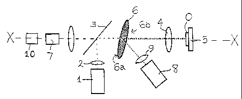
In Figure 3 a further alternative embodiment is shown in which two
light sources are used. This embodiment is similar to the first embodiment
except that the camera 7 is moved so that it only receives images reflected
from the second surface 6b of the mask. With this arrangement the first
light source 1 generates the negative image and it is the second light
source 8, on the same side of the mask as the camera 7, that generates
the positive images. A fourth embodiment is shown in Figure 4 which is
similar to Figure 2 in that the apparatus includes two cameras 7 and 11.
With this alternative arrangement the first camera 7 receives the negative
image and the second camera 11, on the same side of the mask 6 as the
light source 8 receives the positive image.
Although reference herein has been to the generation of a two-
dimensional confocal image of an object, it will be appreciated that three-
dimensional confocal images may be produced using the microscopy
CA 02360917 2001-07-10
WO 00/43819 PCT/GBOO/00142
apparatus and method described above by creating a plurality of two-
dimensional confocal images at different focal planes that are used to
construct the three-dimensional image.
It will be apparent that the microscopy apparatus and method
5 described above is suitable for most situations where confocal images are
required. However, the apparatus and method is particularly suited for
real-time imaging of biological processes and quality inspections for
example of semiconductor chips. Although reference has been made
throughout to light being reflected by the object, in many cases the object is
10 not highly reflective in which case light scattered from the object is used
to
extract the confocal image. Also in the imaging of biological processes the
incident light is not reflected by the object but rather is used to stimulate
an
equivalent pattern of fluorescence. The incident light is encoded in the
manner described above and this pattern of light incident on the biological
material stimulates fluorescence. The pattern of fluorescent light produced
by the biological material is then decoded and positive and negative
images are produced that can be subtracted to extract the confocal image
in exactly the same way as described above.
Also, the confocal microscopy apparatus may be implemented in a
conventional microscope through the addition of the encoded mask and
either a second light source or a second camera.
It will of course be understood that alternative arrangements of
optical elements, in particular alternative or additional lens arrangements,
may be employed without departing from the spirit of the present invention.
Furthermore, although reference has been made herein to a single mask
having a patterned reflective surface it will be apparent that two or more
transmission masks may be employed with one of the masks having a
patterning that is the reverse of the patterning of one of the other masks.