Note : Les descriptions sont présentées dans la langue officielle dans laquelle elles ont été soumises.
CA 02370215 2003-12-04
WO 00/67021 . PCT/US00/12I27
1 PRODUCTS AND METHODS FOR SINGLE PARAMETER AND MULTIPARAMETER
2 PHENOTYPING OF CELLS
3
4
6
7
. 8
9 FIELD OF THE INVENTION
The present invention relates generally to phenotyping and immunophenotyping
of cells
11 and more particularly to single parameter and multiparameter phenotyping
and immunophenotyping
12 of cells.
13
14 BACKGROUND OF THE INVENTION
Immunophenotyping of cells and tumors, particularly hematopoietic tumors, is
often of
16 critical importance for clinical evaluation of cancer patients. However;
currently available
17 methodologies, particularly flow cytometry, are expensive and require a
high degree of suspicion at
18 the time of biopsy. . All too often, even before the diagnosis of cancer is
made, precious tissue
19 must be set aside for possible immunophenotyping. 1f tissue is not set
aside and there is cancer
present, the correct subtyping of the tumor (and proper assignment to
treatment protocols) cannot
21 be done after the fact. Methods that do not require forethought, such as
immunostaining of
22 paraffin blocks, are far less sensitive and do not work well in
laboratories that do not perform these
23 stains frequently. Flow cytometry is the currently accepted "gold standard"
for
24 immunophenotyping of hematopoietic cell types. however, there are several
problems with the
method. The expense of establishing and maintaining these laboratories is
perhaps the most severe
26 problem. Generally large hospitals, academic centers, or commercial
reference Laboratories are the
27 only institutions capable of establishing flow cytometry laboratories.
These laboratories often
28 charge a preonium for their services, and transportation of specimens to
laboratories is not a trivial
29 problem. Since flow cytometry requires live cells, specimens must be
handled under sterile
conditions.. In laboratories where the technology is unavailable, a fresh
specimen has to be
31 prepared and shipped to a flow cytometry laboratory under sterile
conditions for evaluation.
32 Uncontrollable factors such as temperature variations, rough handling,
bacterial contamination, or
33 shipping delays may render samples unsuitable for analysis. In addition,
flow cytometry requires
34 technologists who have specialized training and their time must often be
dedicated solely to the
technology itself, further increasing the expense of the procedure. Relatively
large volumes of cells
36 must be analyzed in order to obtain statistically meaningful results during
analysis, In addition, red
CA 02370215 2001-11-O1
WO 00/67021 PCT/US00/12127
1 cells must be removed from the sample prior to analysis. This is because the
number of red cells
2 in blood and bone marrow samples is far greater than other cells types, and
shear numbers alone
3 would overwhelm the sensitive detectors of the machines. The sample
preparation method
4 therefore requires Ficoll-Hypaque separation, followed by multiple washes,
followed by a lysis step
to lyse remaining red cells. This method virtually eliminates megakaryocytes
from most analysis
6 and frequently destroys delicate malignant cells (particularly from the
relatively common tumors
7 such as large cell lymphoma and Hodgkin's disease). It is in these
situations that the great
8 sensitivity and complexity of flow cytometry may work to its disadvantage.
9 Despite the problems described above, however, flow cytometry can very
accurately and
with great sensitivity identify the presence of malignant cells and
characterize the kind of malignant
11 cells. Without the information that flow cytometry provides, cases can be
frequently incorrectly
12 diagnosed with catastrophic consequences for the patient. This is
particularly true in the setting of
13 a type~of biopsy called fine needle aspiration where examination of a slide
alone by light
14 microscopy may be quite difficult.
What would be very useful to the average hospital pathologist or to any
physician in an
16 outpatient or remote setting is a device or kit that would allow the same
kind of single parameter or
17 multiparameter analysis of samples using cheaper, more readily available
materials. This would
18 eliminate the need for specialized laboratories and technologists dedicated
solely to the flow
19 cytometry technology itself and would allow any well trained clinical
laboratorian ready access to
the same kind of analysis. Furthermore, if the need for live cells could be
eliminated, cells could
21 be preserved by appropriate fixatives which would broaden the availability
of immunophenotyping
22 data.
23 Over the last 20 years there has been a tremendous growth in the
identification and
24 characterization of molecules expressed by blood cells on their cell
membranes (called cell surface
antigens). This growth in understanding has been accompanied by the refinement
of technologies
26 that allow the rapid and sensitive identification of these molecules on the
surfaces of live cells.
27 However, the overwhelming majority of these cell surface antigens are not
unique to one type of
28 cell. There is only rarely a single diagnostic marker to identify a cell
type. Instead, most cell
29 populations must be characterizing by analyzing multiple parameters at the
same time.
Antibodies are proteins produced by the body's immune system that have the
property that
31 they bind to a singe specific molecule (referred to as an antigen). Antigen-
antibody completes are
32 formed when an antibody binds its respective antigen. Normally, these
complexes are then cleared
33 by the immune system to rid the body of an infection. However, the immune
system has a
34 virtually limitless capacity to produce unique antibodies, which can be
tailored to identify particular
substances, even when present in very small quantities. Antibodies are now
commercially
2
CA 02370215 2001-11-O1
WO 00/67021 PCT/US00/12127
1 produced to literally hundreds of different antigens. Furthermore,
antibodies can be easily tagged
2 with marker molecules, such as fluorescent molecules, dyes, or other
substances that make
3 identifying the presence of an antigen-antibody complex a relatively simple
matter. This well-
4 known biochemical reaction has been used to develop a methodology called
flow cytometry. In
flow cytometry, intact cells are treated with antibodies that bind specific
markers on the cell
6 surface. The antibodies are, in turn, labeled with a fluorescent molecule
and the cell suspension
7 then flows past a light beam with a light detector which counts the number
of fluorescent cells
8 versus the other cells present. This technology has proved tremendously
useful in identifying
9 malignant cell populations in blood and tissue samples from patients.
In flow cytometry, a cell suspension is treated with antibodies labeled with
fluorescent
11 molecules (fluorochromes), washed, and placed in the machine. The cell
suspension is "focused"
12 using buffer solutions so that the cells pass through the flow detector in
a single file. When each
13 cell passes through the flow detector, a beam of laser light is passed
through the cell. Some of the
14 light passes through the cell (called forward light scatter) and some is
refracted at an angle (called
side scatter). Forward scatter increases with a cell's size and side scatter
increases with a cell's
16 internal complexity (mostly granules within the cytoplasm). Thus using just
these two
17 measurements, individual cell types can be roughly categorized. However,
there are also light
18 detectors, which, by using appropriate color filters, can specifically
detect the fluorescence given
19 off by the antibodies that are attached to the cell surface. Since current
state of the art machines
have up to four different color detectors (referred to as four-color flow
cytometry), up to four
21 different antibodies can be added to the same tube. Samples from individual
patients are usually
22 divided into multiple tubes, each of which contains multiple antibodies.
Data analysis is therefore
23 quite complex, and requires computers that are capable of simultaneously
displaying multiple plots
24 from each tube. This is referred to as multiparameter analysis. This
simultaneous analysis of
multiple parameters is necessary to first electronically isolate and then
characterize cell populations.
26 Therefore, even though modern flow cytometers analyze up to 6 simultaneous
parameters (forward
27 scatter, side scatter, and four antibodies) 3 of the parameters must be
used for electronic isolation
28 of cell types (forward scatter, side scatter, and CD45 staining intensity).
Broad categories of cells
29 present in hematologic samples are known in the art and include myeloid
cells, monocytes,
lymphocytes, megakaryocytes, and red cells. 1n other words, these 3 parameters
must be used to
31 roughly mimic what the human eye does so effortlessly: identify or
characterize broad categories of
32 cells. Indeed, laboratories commonly hire technicians with 2 years of
training (only part of which
33 is in the area of hematology) who can, with a very reasonable degree of
accuracy and precision,
34 identify or characterize different cell types present in blood samples.
With some additional
training, they can also correctly enumerate cell types within bone marrow
aspirate samples. Thus
3
CA 02370215 2001-11-O1
WO 00/67021 PCT/US00/12127
1 if the human eye were also equipped with the means to also identify cell
surface antigens, there
2 would be no need for flow cytometry for this purpose. Furthermore, of the
remaining 3
3 parameters available for analysis on the flow cytometry, only 2 can be
displayed in any one plot
4 although new software exists that can display 3 dimensional plots. While 3
dimensional plots add
to convenience and are applicable in limited situations, two parameter
analysis is quite sufficient in
6 most cases. This last point is critical, since any method that seeks to
supplant flow cytometry must
7 have the ability to characterize at least 2 cell surface markers
simultaneously.
g Analysis of cell populations by flow cytometry is not a trivial process and
requires highly
9 trained personnel as outlined above. Both single parameter and
multiparameter analysis can be
performed. If data is analyzed as histogram plots of fluorescence of a single
marker versus cell
11 number, then one parameter analysis is being performed. Analyzing two such
histograms of a
12 single gated cell population could then be referred to as simultaneous
single parameter analysis.
13 An example of simultaneous single parameter analysis would involve the use
of such plots to
14 identify cell surface expression of both the B-cell marker CD20 and the
light chain kappa.
Analysis of the binding of each set of antibodies is independent of the other.
In multiparameter
16 analysis, the binding of the two antibodies are linked and are not
independent. Analytical methods
17 require the binding of both antibodies simultaneously brought together in a
single histogram such as
18 fluorescence 1 versus fluorescence 2. Characterization of the target cell
population is best
19 performed by analysis of this fluorescence 1 vs. fluorescence 2 plot and
analyzing the binding
characteristics of each of these antibodies together. This decreases the
possibility of an error that
21 would incorrectly analyze two overlapping cell populations as a single cell
population.
22 Finally, with the limited exception of DNA ploidy analysis,
characterization of solid
23 tumors and non-hematopoietic tumors is quite limited by flow cytometry.
Often there are not well
24 developed protocols for developing cell suspensions. In addition, tumor
cells may be delicate and
may not survive processing. In addition, many markers used for solid tumors
such as vimentin or
26 smooth muscle actin are intracytoplasmic antigens and may be difficult
to.assay by flow cytometry.
27 In addition, most available markers for these other tumors are not specific
markers for the tumors
28 and many normal cells, including cells present in the background of the
available sample, may be
29 strongly positive for the same markers. Therefore, interpretation of these
kinds of samples without
specific morphologic correlation is hazardous at best.
31 An object of the invention is to provide a cheaper, more accessible method
for single
32 parameter and multiparameter analysis of cell populations. This analysis is
not limited to just cell
33 surface markers but also optionally includes identifying active receptor
sites on cell surfaces, loss
34 of cell surface proteins, intracellular proteins, and intracellular nucleic
acid sequences. One of the
features of this invention is that the target cell population is being
analyzed by preserving
4
CA 02370215 2001-11-O1
WO 00/67021 PCT/US00/12127
1 morphologic characteristics of the cells for analysis. In addition, it is
also possible to count events
2 to obtain specific cell numbers in relation to specific sample volume. Due
to the many preparatory
3 steps of flow cytometry, obtaining absolute cell numbers is not possible --
only percentages of cells
4 analyzed.
6 SUMMARY OF THE INVENTION
7 A method of characterizing cells is provided, comprising the steps of a)
providing a
8 suspension of cells in a liquid medium, said cells including first cells, b)
adding to said suspension
9 a group of substantially identical first beads, each of said first beads
being coated with a binding
substance or being magnetic such that each first bead is adapted to bind to a
first cell, c) incubating
11 said first beads in said suspension for a period of time effective to
permit said first beads to bind to
12 said first cells to form first bead-first cell complexes, each first bead-
first cell complex comprising
13 a first bead and a first cell, d) separating said first bead-first cell
complexes from said suspension
14 by filtration, and e) examining said separated first bead-first cell
complexes and characterizing said
first cells. A kit is also provided, comprising at least one group of
substantially identical
16 first beads, each of said first beads being coated with a binding substance
or being magnetic such
17 that each first bead is adapted to bind to a first cell, said kit further
comprising a set of instructions
18 effective to instruct a technician in how to use said first beads to
perform single parameter or
19 multiparameter analysis on a suspension containing first cells. An
apparatus for performing single
parameter or multiparameter analysis on a suspension of cells is provided. The
apparatus
21 comprises a sample loader, a plurality of reaction chambers, and a
filtration chamber.
22
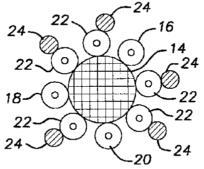
23 BRIEF DESCRIPTION OF THE DRAWINGS
24 Fig. 1 is a schematic illustration showing a cell bound to an antibody
which is bound to or
coated on a bead.
26 Fig. 2 is a schematic illustration showing a number of cells bound to a
bead.
27 Fig. 3 is a schematic illustration showing a number of cells bound to a
bead in the center,
28 and five smaller beads bound to five of the cells.
29 Fig. 4 is a schematic illustration of an automated device for performing
phenotypic or
immunophenotypic analysis in accordance with the present invention.
31 Fig S. is a schematic illustration of a single slide for use with the
automated device of Fig.
32 4.
33
34 DETAILED DESCRIPT10N OF THE PREFERRED EMBODIMENTS OF THE INVENTION
As used herein, when a preferred range such as 5-25 is given, this means
preferably at
5
CA 02370215 2001-11-O1
WO 00/67021 PCT/US00/12127
1 least 5 and, separately and independently, preferably not more than 25. The
cells herein are
2 preferably human cells. If a first group of cells does not include members
of a second group of
3 cells, and the second group of cells does not include members of the first
group of cells, the two
4 groups do not overlap. "Visually distinguishable" includes visually
distinguishable via light
microscopy. Quantitate includes to estimate or enumerate or count the number
of. Phenotyping
6 includes immunophenotyping and genotyping.
7 The invention uses beads. As used herein, beads means small particles or
support
8 surfaces, preferably microspheres, more preferably plastic microspheres,
more preferably
9 polystyrene microspheres (also referred to as latex beads or spheres or
microspheres), which have
preferably been coated with a binding substance or which are magnetic. As used
in the claims,
11 "first beads" includes beads which have been coated with a binding
substance or which are
12 magnetic. The bead may be any solid support surface or particle that can be
suspended in an
13 appropriate solution. Preferred beads are available as polystyrene
microshperes from Bangs
14 Laboratories, Fisher, IN. The bead sizes are preferably greater than 5
microns diameter,
preferably 5.5-10.3 microns, less preferably at least 5.5 or 10 or 12 microns
and preferably not
16 more than 12, 15, 20 or 30 microns diameter, far less preferably less than
5 microns, such as at
17 least 1, 2 or 3 microns diameter. 5.5 and 10.3 micron beads are preferred.
The beads can also be
18 colored, such as red or blue, less preferably green, purple, orange, brown,
yellow, or any other
19 color. The beads are preferably coated with a binding substance, such as
antibodies or
immunoreactive proteins, or any molecule that can bind to, or interact with a
cell surface in such a
21 way as to bring the cell and the bead into contact or adherence with each
other or to bind with each
22 other; alternatively, the bead can contact or bind with the cell surface
through electrostatic charge
23 interactions or magnetic interaction; all of these concepts being covered
by the terms "binding to"
24 or "bind to". When a bead binds to a cell, it forms a bead-cell complex. In
the invention the cell-
bead interaction forms a large enough complex to inhibit the passage through a
filter containing
26 pores of appropriate dimensions. The filters are preferably sized and
selected such that unbound
27 cells and beads will pass through the pores but bound cell-beads do not.
These complexes are then
28 transferred to a glass slide and stained with a variety of stains so as to
render the complexes visible
29 by routine microscopy. The complexes and cells are examined and the cells
are characterized.
Examples of binding substances or reactive substances that may be used to coat
the bead surface
31 include, but are not limited to: antibodies to specific cell surface
proteins, small molecules that bind
32 receptors or other cell surface molecules such as IL-2 or GM-CSF, avidin,
biotin, or beads may
33 remain uncoated in suspension that can interact by other means with cells.
The antibodies that can
34 be used are those known in the art. Many such antibodies are available from
commercial
companies, such as Zymed Inc., South San Francisco, CA and Dako Corp.,
Carpinteria, CA.
6
CA 02370215 2003-12-04
1 Beads or microspheres can be made from a variety of substances including
gold, ferritin,
2 polyacrylamide, or polystyrene. The latter is among the preferred substances
as beads can be
3 made precisely to any size specification and can be uniformly conjugated to
both molecular linker
4 arms and reactive binding substances. Polystyrene microspheres (also known
as "latex
microspheres") may be prepared by methods known in the art. Binding substances
that can be
6 used include monoclonal antibodies, polyclonal antibodies, antibody
"cocktail" mixtures,
7 antibody fragments (such as Fc portions or Fab or Fab' fragments in either
monovalent or divalent
8 forms), small molecules that bind specific cell surface receptors, covalent
and non-covalent
9 linkers, and indirect adherence such as utilizing electrostatic or magnetic
or paramagnetic
attraction.
11 The prior art includes U.S. Pats. 5,554,505; 5,348,859; 5,340,719;
5,231,005; 5,260,192;
12 5,338,689; 5,256,532; and 5,501,949. These patents include discussions of
using certain
13 microspheres or beads for identification of cells. It is known in the art
how to provide a
14 suspension of cells in a liquid medium for analysis.
A second feature of a preferred embodiment of the present invention is that it
16 concentrates cells by using an appropriate filter without added
manipulation of the cell suspension
17 by cell lysis or added incubation steps of submicroscopic paramagnetic
microspheres. The filters
18 to be used in the invention can be any of those known in the art, such as
gynecological filters
19 from Cytec Corp., Boxborough, MA. The filters preferably have a pore size
laxger than the beads
being used so that all or most or substantial amounts of unbound beads and
unbound cells pass
21 through, but the pore size is preferably small enough so that all or most
or substantial amounts of
22 beads bound to cells are trapped on the filter, such as the filter pore
size being about 1, 2, 3, 4, 5,
23 6, 8, 10 or 12 microns larger than the bead size. Preferred filter pore
sizes include 10-15, less
24 preferably 7-20, 7-30 or 7-40 micron pore sizes. Alternatively the filter
pore sizes can be at least
15 or 20 microns. The filter is preferably mounted on a solid support, such as
at the end of a tube
26 through which the suspension can drain.
27 ~ A cell suspension is preferably prepared from a peripheral blood sample,
a bone marrow
28 aspirate, a fine needle aspirate, a lymph node biopsy, or a body site
specimen. In the method
29 described herein, single parameter, simultaneous single parameter, and true
multiparameter
analysis is possible which compares to the level of sophistication of analysis
possible by flow
31 cytometry. Beads that can be easily distinguished from each other optically
either by size, color,
32 or both can be added to a cell suspension either simultaneously or
sequentially. Positive binding
3,3 by the target cell population results in a bead-cell complex that has a
significantly larger physical
34 size than either unbound cells or beads. These complexes can be then easily
concentrated and
separated from the
7
CA 02370215 2001-11-O1
WO 00/67021 PCT/US00/12127
1 rest of the solution using an appropriately sized filter containing pores of
sufficient size to let
2 unbound cells and beads to pass through while complexes remain on the
filter. The method may
3 also be used in reverse, in that abnormal cell populations may fail to bind
beads while normal cells
4 bind strongly. An example of this latter method can be found with the
myelodysplastic disorders
S (MDS), which currently cannot be diagnosed by flow cytometry with any degree
of reliability.
6 Normal human myeloid cells strongly express surface markers such as CDllb,
CD13, CD15,
7 CD16 and CD33. However, in MDS, these cell populations lose expression of
these markers.
8 However, as normal cells degenerate from prolonged storage or poor specimen
handling such as
9 temperature extremes, which may occur in specimen transport, they also lose
expression of these
markers. Flow cytometry cannot distinguish between these two conditions.
However, degenerated
11 cells are easily recognized morphologically from the dysplastic cells of
MDS. Loss of binding by
12 beads coated with antibodies to these markers could easily be identified
(with a slide made of the
13 cells passing through the filter as well as those trapped on the filter).
Therefore, this method is the
14 first easily available method to diagnose MDS, heretofore only diagnosable
in those minority of
cases showing abnormal cytogenetics or persistent hematologic abnormalities
after prolonged
16 clinical follow up. As an example of MDS analysis, one can look at a
peripheral smear. If the
17 cells are degenerated, get a new sample. If the cells are not degenerated,
incubate the cell
18 suspension with large beads coated with anti-CD13. Then add small beads
coated with anti-CD15
19 and let react. Then filter (can be small pore size to trap both bound and
unbound cells, or large
pore size to trap bound cells only, in which case unbound cells are collected
from what went
21 through the filter). If the result is lots of complexes like Fig. 3 and few
unbound cells, this
22 indicates normal cells. If there are few bound cells and many unbound
cells, this suggests MDS.
23 Immature cells also have weak binding, but this can be seen
morphologically. The same procedure
24 can be done with CD 11 b and CD 16.
One can also count beads trapped on the filter prior to transfer to the glass
slide. Using
26 methods such as light scattering, reflectance, fluorescence, or
electrostatic field changes, the
27 number of beads trapped on the filter can be counted. An average number of
cells bound to each
28 bead can be obtained and an estimate of the number of cells in the original
sample volume
29 obtained.
With reference to Fig. 1 there is shown, not to scale, a bead 2, such as a
polystyrene
31 microsphere, which has coated thereon and bound thereto a binding substance
4 such as an
32 antibody. There is also a cell 6, such as a target cell, which has a cell
surface marker 8. The
33 binding substance 4 or antibody binds to the cell surface marker 8 on the
target cell 6. Fig. 2
34 illustrates how this kind of reaction may appear on a glass slide; a group
of cells or target cells 12
have bound to a bead 10. This shows single parameter binding of cells to
beads. The ratio of
8
CA 02370215 2001-11-O1
WO 00/67021 PCT/US00/12127
1 beads to cells should be adjusted properly for effective results. The actual
number of cells binding
2 the bead is variable, ranging from a single cell to numerous cells crowding
the bead's surface.
3 With reference to Fig. 3 there is shown a large bead 14 coated with a
binding substance
4 which has bound to eight cells 16, 18, 20, 22. Small beads or different
colored beads 24 coated
with a different binding substance have bound to the cells 22 but not to the
cells 16, 18, 20. This
6 provides positive identification of target cells 22. This illustrates
multiparameter analysis. Cells 22
7 is a subset of cells 16, 18, 20, 22. A variable number of beads 24 can bind
to each cell 22. In
8 some cases each cell bound to bead 14 will be bound to one or more beads 24,
or each cell bound
9 to bead 14 may be unbound to small beads. Note that different kinds of cells
may bind to the large
bead 14 that can in some cases be distinguished morphologically. Preferably
the large bead 14 is
11 added first to the cell suspension so that a plurality of cells can bind to
its surface. Then the small
12 beads 24 are added to bind to the periphery of the complex. Alternatively
small beads 24 can be
13 added first or small beads 24 and large beads 14 can be added
simultaneously. The order of
14 addition is dependent in large part upon the relative concentrations and
surface areas of the beads
and the cells. For example, you would not want to add beads 14 or 24 in such
concentrations that
16 they completely cover or obscure the surface area of the target cells and
thus prevent access thereto
17 by the other beads. Preferably there is an excess of target cells to fully
coat the bead. Optionally
18 the suspension can be filtered after the first complex is formed, to trap
the first complex and
19 resuspend it before the second beads are added. Thus a group of complexes
can be filtered and
resuspended before a subsequent set of beads is added; this can lead to more
certain and distinct
21 results by removing materials which would provide interference. The beads
may be distinguishable
22 in size or color or both. Further levels of multiparameter analysis can
also be carried out, such as
23 by adding to Fig. 3 another set of different sized or different colored
beads which would bind to a
24 first subset of cells 22 but not the remaining cells 22, thus providing
positive identification of said
first subset of cells 22. In this manner subsequent or additional levels of
multiparameter analysis
26 can be carried out.
27 There is a wide variety of available beads that can be used, and those
selected would
28 depend on the specific application. In multiparameter analysis beads that
can be easily
29 distinguished by either size or color are preferable. For example, two sets
of colorless beads sized
10 and 5 microns respectively can be used to isolate a population of B cells
using 10 micron beads
31 coated with an anti-pan B cell antigen such as CD19 and S micron beads
coated with anti-kappa.
32 Multiparameter analysis that cannot be easily mimicked by flow cytometry is
available by a minor
33 variation of this example. Colorless 10 micron beads are used to bind B
cells by using anti-CD19
34 coated beads. 5-micron colorless beads are coated with anti-kappa while
dark blue 5-micron beads
are coated with anti-lambda. Similarly, a blast cell population can be
analyzed using anti-CD34
9
CA 02370215 2001-11-O1
WO 00/67021 PCT/US00/12127
1 coated 10 micron beads and anti-CD19 coated colorless 5 micron beads.
Colored 5-micron beads
2 coated with anti-CD13 are simultaneously added for rapid characterization of
most blast cell
3 populations.
4 Preferred methods:
I) Substantially identical beads are purchased commercially precoated with
strepavidin (Bangs
6 Laboratories, Fishers, IN). A small quantity is suspended in any buffered
salt solution such as
'7 phosphate buffered saline or commercially available antibody diluent. The
beads are incubated with
8 biotinylated goat anti-mouse antibodies for 30 minutes (however, any
biotinylated anti-allogeneic
9 antibody may be used). The suspension is centrifuged and the supernatant
drawn off. The
incubation is repeated two times to ensure coating of as much of the available
surface area of the
11 beads as possible. The beads are then washed three times using the same
buffer. The suspension
12 is then incubated with specific mouse anti-human antibodies for 1 hour (or
any non-biotinylated
13 anti-allogeneic antibody specific for the target cell population may be
used). The suspension is
14 again washed three times and diluted to the desired concentration. The
resulting suspension can be
refrigerated at 4 degrees Centigrade until use. Alternatively, biotinylated
primary antibodies may
16 be used without the use of secondary antibodies. The beads produced by this
technique are
1'7 substantially identical.
1 g 2) Beads are precoated with anti-Fc receptor antibodies (Bangs
Laboratories, Fishers, IN)
19 such as goat anti-mouse IgG Fc receptor antibodies. These beads can then be
suspended in a
solution of antibodies which would spontaneously bind to the anti-Fc receptor
sites on the beads.
21 In the example cited above, mouse anti-human antibodies would be bound to
the beads followed by
22 appropriate washing steps similar to that described above.
23 3) Binding substances such as any protein, peptide, or nucleotide sequence
may be bound by
24 other chemical or specific binding methods. For example, polystyrene
microspheres are "naturally"
left coated with sulfate surface groups after manufacture. These ligands can
be used to link
26 proteins and peptides directly to the surface of the beads. Examples of
such functional surface
2'7 groups that can be coated on the surface includes, but is not limited to,
aldehyde, aliphatic amine,
28 amide, aromatic amine, carboxylic acid, chloromethyl, epoxy, hydrazide,
hydroxyl, sulfonate; and
29 tosy (toluene sulfonyl) reactive ligands. These can then be used in tum to
link peptides, proteins,
oligonucleotides, and other biochemical ligands to the surface. These ligands
or binding substances
31 would in turn be used to bind specific sites on cell surfaces which would
link the cell to the surface
32 of the bead. For example, a small molecule such as the hormone IL-2 could
be used by one of the
33 above methods to coat beads with the intention of binding IL-2 receptor
sites (CD25) on cell
34 surfaces. This could be used to bind cells such as T-cells, monocytes, and
neoplastic cells such as
hairy cell leukemia.
CA 02370215 2001-11-O1
WO 00/67021 PCT/US00/12127
1 Other methods:
2 Submicroscopic paramagnetic microspheres (preferably less than 1 micron in
diameter) are
3 bound to any reactive biomarker of interest. The binding that is used could
be any of the above
4 methods. Cells are then permeabilized and fixed using a variety of
detergents and weak fixative
solutions such as 1 % paraformaldehyde. Alternatively a number of commercially
available
6 permeabilizing kits are available for this purpose such as IntraStain (Dako
Corp., Carpinteria, CA).
7 The reactive biomarker, such as antimyeloperoxidase antibodies, anti-
terminal deoxytidyl
g transferase antibodies, or specific RNA or DNA probes, is then incubated
with the cell suspension.
9 The biomarkers and paramagnetic particles get inside the cell and, for
example, the probe binds to
the intracellular target. The cells are then washed and resuspended in a
suitable buffer such as PBS
11 or RPMI. The suspension is then incubated with magnetic beads or
microspheres of a size or color
12 easily visualized, such as 1 to 20 or 3-15 or 5-10 or 10-20 microns. The
magnetic beads bind to
13 the cell surface, but cannot cross the membrane, to create a cell-bead
complex that is easily trapped
14 such as via filtration.
In one example, abnormal blasts in a bone marrow suspension can be
permeabilized and
16 incubated with anti-myeloperoxidase antibodies bound to submicroscopic
paramagnetic
1~ microspheres. The suspension is then washed three times in buffered salt
solution and resuspended
lg and incubated with large magnetic beads of a preferred size of 5-15 micron
diameter to create cells
19 bound to large beads.
In another example, specific DNA sequences (probes) are bound to
submicroscopic
21 paramagnetic microspheres using methods such as avidinated microspheres and
biotinylated probes.
22 Cells from a patient with chronic myelogenous leukemia are permeabilized
and incubated with
23 probes binding to the specific bcr-abl translocation that is diagnostic for
the disease. The
24 suspension is then washed and incubated with large magnetic beads of a
preferred size of 5-15
micron diameter to create cells bound to large beads.
26 Detection and analysis:
2'7 The cell-bead complexes (cells bound to beads) provided or obtained as
described above
2g are then passed through a solid support filter having a porosity of
sufficient size to allow unbound
29 cells and beads to pass through. The suspension is passed through the
filter using a variety of
acceptable methods which includes gravity, suction (applied vacuum), positive
pressure on the fluid
31 side, or wicking the fluid through the filter using a porous absorbable
material such as gauze pads.
32 Various devices that can be used include pistons, syringes, or suction
methods to create a negative
33 pressure to pass fluid through the filter. In a preferred embodiment, a
single solid filter with a
34 pore size of 10-15 microns is used. Cell-bead complexes remain trapped on
the filter and the layer
is then transferred to a glass slide by direct contact with the slide and
applying gentle pressure.
11
CA 02370215 2001-11-O1
WO 00/67021 PCT/US00/12127
I The resulting slide preparation can be stained using a variety of
commercially available stains such
2 as hematoxylin and eosin, Papanicolau stain, or any Romanowsky stain. In a
preferred
3 embodiment, the cells remain suspended in a compatible buffer such as PBS,
RPMI, or
4 commercially available antibody diluent and the resulting slide is stained
with Wright-Giemsa stain.
Alternatively, cells may be suspended in ethanol or a commercially available
fixative such as
6 Cytolyte (Cytyc Corp., Boxborough, MA). The resulting slide is then stained
with Papanicolau or
7 hematoxylin and eosin stains. The complexes are examined and the cells are
characterized under
8 routine light microscopy.
9 The invention can be used to perform single parameter analysis correlated
with
morphology, simultaneous single parameter analysis, or multiparameter
analysis. In single
11 parameter analysis, (depicted in Figs. 1 and 2) a single bead type is added
to a suspension of cells
12 in a liquid medium so that after filtration the slide is provided with an
enriched single cell
13 population. This is useful as a simple screen to determine if a cell
population has a particular
14 characteristic such as distinguishing monocytes from monocytoid B
lymphocytes as cited in
Example 1 below. In this configuration, cells bind to beads and are visible on
the glass slide for
16 analysis. Alternatively, a B cell population can be assayed for expression
of kappa or lambda by
17 using two separate slides or slide wells each of which contain a single
bead type (anti-kappa or anti-
18 lambda). Another variant of this analysis is to add simultaneously to the
cell suspension two
19 different bead types, one anti-kappa and a second anti-lambda. This is an
example of simultaneous
single parameter analysis since binding of each bead type is independent of
the other but the results
21 are analyzed together. An analogous situation occurs in flow cytometry
analysis when fluorescence
22 is displayed vs. cell number to obtain a single histogram. In kappa and
lambda analysis, a
23 monoclonal population can only be detected by simultaneous analysis of both
histograms and
24 looking for single peaks of fluorescence. Finally, multiparameter analysis
can be performed by
linking detection of two different characteristics so that analysis is
performed together. In this
26 case, binding of one set of beads occurs, followed by a second and
optionally more sets of beads
27 (see Figure 3). Analysis looks for simultaneous binding of more than one
set of beads to the target
28 cell population (as depicted in Example 2 below).
29 The invention can be used to detect abnormal loss of binding when strong
binding would
be expected. For example, normal myeloid cells such as mature granulocytes and
monocytes in the
31 peripheral blood would be expected to strongly express the surface markers
CD13, CD33, CDllb,
32 and CD16. In a bone marrow sample there would be a continues range of
increasing expression of
33 these markers as the cell matures. However, cells showing abnormal
maturation, as seen in
34 myelodysplasia, would show diminished expression of these markers. This
phenomenon has been
previously described by Davis, et al. and can be seen in flow cytometry
analysis as abnormal
12
CA 02370215 2001-11-O1
WO 00/67021 PCT/US00/12127
1 patterns of expression on appropriate histograms. However, a similar loss of
expression is seen
2 when normal cells die and degenerate as occurs in specimen mishandling or
aging. Since
3 morphologic correlation is less than optimal by flow cytometry, the
phenomenon has limited
4 diagnostic usefulness, particularly when the specimen has been transported
long distances. In the
present invention, cells can be visualized on the glass slide to confirm their
viability. Normal cells
6 would strongly bind beads coated with these markers but there would be
decreased binding of beads
7 in cells with myelodysplasia. In the low grade myelodysplasias such as
refractory anemia and
8 refractory anemia with ringed sideroblasts, there are often no objective
diagnostic criteria for
9 confirming the diagnosis. Current state of the art in such cases requires
prolonged follow up and
diagnosis by exclusion of other possible entities such as ethanol toxicity or
megaloblastic anemia
11 from vitamin B12 or folate deficiency. The invented method provides a much
needed positive
12 diagnostic test.
13 A complementary detection method is that prior to transfer of the cells to
a glass slide, the
14 filter is gently rinsed and scanned using a light beam of either a white
light beam or a specific
wavelength to correspond to the excitation wavelength of fluorescent beads.
The number of events
16 is counted electronically and the cells are then transferred to a glass
slide and stained. The average
17 number of cells per microsphere is then obtained manually and an estimate
of the total number of
18 target cells in the sample can be estimated (assuming that a known volume
of sample is used).
19 Preferred applications:
1) Single parameter analysis of tumors and other specific cell populations. A
suspected tumor
21 with a known immunophenotype can be analyzed to confirm the presence of a
single marker as
22 outlined in Examples 1 and 3 below. This is most useful in settings where a
single issue regarding
23 cell phenotype needs to be settled. In Example 1 below, knowing that the
abnormal cell population
24 is of B cell origin is sufficient information to proceed with further
studies, since this suggests (but
does not prove) malignancy. In Example 3 below, knowing that the lymphoid
population is of T
26 cell origin suggests that the patient has a reactive infiltrate rather than
a malignant infiltrate. If this
27 assay had been clinically available in both of these unusual cases, the
results of the simple study in
28 Example 1 would justify further expense of additional evaluation. The
results of Example 3 justify
29 not performing flow cytometry and proceeding to treatment for meningitis.
Other applications of
these kinds of analysis can be useful in other kinds of tumors such as MN/CA9
screening for
31 cervical cancer, identifying specific tumor types in malignant infiltrates
such as melanoma (using
32 markers such as HMB-45), or identifying micrometastic disease in lymph
nodes and bone marrows.
33 In addition, single parameter analysis can be used in genetic phenotypic
and genotypic analysis.
34 For example, a peripheral blood sample can be permeabilized and treated
with a specific probe to
the bcr-abl translocation. The probe can be labeled with paramagnetic
submicroscopic
13
CA 02370215 2001-11-O1
WO 00/67021 PCT/US00/12127
1 microspheres. The cells can then be treated with large, magnetic beads to
identify the presence of
2 the translocation that would be diagnostic of chronic myelogenous leukemia.
Alternatively, a
3 similar method can be used to identify the presence of intracellular
proteins or RNA sequences
4 using appropriate antibodies or nucleotide sequences, for example, the
expression of the
intracellular protein terminal deoxyribonucleotidyl transferase (1'dT) using
an antibody also labeled
6 with paramagnetic microspheres and detecting the reaction using large
surface magnetic beads.
7 Finally, the use of CD64 expression has been proposed as a rapid diagnostic
test for clinically
8 significant acute inflammatory reaction (Lab. Hematol. 1995; 1:3-12). For
reasons described
9 above, flow cytometry is too expensive and difficult to use as a screening
procedure for common
conditions. The invented method allows rapid, inexpensive single parameter
analysis for CD64
11 expression in peripheral granulocytes.
12 2) Simultaneous single parameter analysis is where there is simultaneous
analysis of markers
13 that are independent of each other. Most commonly, this is used in a B cell
lymphoid population to
14 determine expression of either kappa or lambda light chain restriction by
expressed surface
immunoglobulins. This can either be done by using similar beads as used in two
separate glass
16 slides analyzed simultaneously or by using a single slide using two sets of
beads which can be
17 easily distinguished based on size, color, or both. This is extremely
useful as an inexpensive, rapid
18 screen for B cell monoclonality. Other useful types of simultaneous single
parameter analysis are
19 in the setting of a malignant tumor of unknown origin where a cell
suspension can be analyzed,
either by using multiple separate slides or a single slide containing multiple
sets of beads that can
21 be distinguished by size, color, or both. In this example, these sets of
beads typically include
22 beads marking for CD45 (leukocyte common antigen), HMB-45 (melanoma), and a
general
23 cytokeratin marker (often AEI and AE3 cocktail for epithelial tumors). A
third type of this kind of
24 analysis is to screen a population of lymphocytes to determine whether this
population is composed
of B cells, T cells, other cells, or any combination of these types.
26 3) The invention also includes multiparameter analysis where expression of
markers are
27 analyzed in conjunction with other markers. A simple, but common, example
of this kind of
28 analysis is depicted in Example 2 below. In Example 2, the positive binding
reaction by the anti-
29 CD20 coated beads which isolates the B cells is linked to kappa or lambda
light chain expression.
Multiparameter analysis enhances analysis since correctly identifying certain
cell populations
31 requires logical association of multiple subsets of markers. A case of
acute leukemia serves as a
32 useful example of this kind of analysis. Morphologic examination is one of
the best methods for
33 identifying the abnormal blast cells, but it does not characterize the kind
of blasts present.
34 Combining morphologic analysis with the present invention would yield the
following typical kind
of analysis. Anti-CD34 coated beads are combined with anti-HLA-DR coated beads
to confirm
14
CA 02370215 2001-11-O1
WO 00/67021 PCT/US00/12127
1 expression of both of these markers in the malignant cell population.
Positive expression of both of
2 these markers supports the diagnosis of acute leukemia. The cells can then
be analyzed with anti-
3 CD13 and anti-CD33 coated beads in conjunction with anti-CD19 and anti-CD2
coated beads to
4 determine if the cells are myeloid or lymphoid in origin. If they bind to
CD13, CD33, or both,
this confirms the myeloid derivation of the cells. The cells can also be
analyzed with anti-CD15,
6 anti-CD14, anti-CD56, anti-CD7, and anti-CD4 to determine subtype (myeloid,
monocytic, or both)
7 and to yield prognostic information. Of particular interest is successful
analysis of acute
8 promyelocytic leukemia (FAB subtype M3). Analysis of this tumor type by flow
cytometry is
9 fraught with errors and the tumor can be missed since it is composed of
maturing myeloid cells.
Using the present invention, morphologic analysis would confirm the presence
of excess numbers
11 of promyelocytic cells. In addition, the promyelocytes would usually be HLA-
DR negative and
12 could also be analyzed for the translocation of chromosomes 15 and 17
(t(15;17)) which is
13 diagnostic of the disease. This kind of analysis is particularly useful in
the microgranular variant
14 of the disease in which the cells may resemble monoblasts. Monocytic
leukemias can also be
analyzed for additional monocytic markers such as CD36. Similar kinds of
analyses can be
16 performed for other hematologic malignancies, other tumor types, and other
specific cell
17 populations. In addition, the method can be used in reverse to offer a
diagnostic test for
18 myelodysplasia. Normal myeloid cells strongly bind the myeloid markers
CDllb, CD13, CD16,
19 and CD33. Among the changes seen in myelodysplasia, is decreased expression
of these markers
by flow cytometry. However, degenerating cells, as occurs in excessive sample
age, temperature
21 extremes, or other forms of specimen mishandling also causes decreased
expression of these
22 markers. Since morphologic correlation with flow cytometry is so poor, this
form of analysis has
23 not gained significant clinical acceptance since flow cytometry cannot
reliably distinguish between
24 degenerated normal cells and myelodysplastic cells. In the invented method
there is excellent
morphologic correlation, and trained observers will easily recognize
degenerated cells. Therefore,
26 normal cells can easily be distinguished from dysplastic cells, as normal
cells will avidly bind beads
27 coated with antibodies to these markers and dysplastic cells will not.
2g Another similar application can be used for analysis of breast cancer to
determine
29 prognostic factors such as Her2/neu overexpression. Current state of the
art utilizes primarily
immunohistochemistry to localize actual tumor from surrounding breast tissue
by visual methods.
31 Her2/neu cytoplasmic membrane expression is estimated by the observer
visually on a scale
32 expressed as 0+ positive (no expression) to 4+ positive (strongest possible
expression). There are
33 no objective quantitative methods to estimate the level of Her2/neu
overexpression. Alternatively,
34 Her2/neu expression can be more objectively estimated by using fluorescent
in-situ hybridization
(FISH) which labels each gene copy with a fluorescent dot. The number of gene
copies in each
CA 02370215 2001-11-O1
WO 00/67021 PCT/US00/12127
1 cell can be estimated by merely counting the dots within the nucleus of each
cell. However,
2 because cells cannot be easily counter-stained and observed, it is difficult
to tell a malignant breast
3 epithelial cell from an admixed benign one or even a stromal cell from the
breast supporting
4 matrix. Therefore, analysis by FISH has less acceptance in the clinical
setting. More recently,
Her2/neu expression can be performed by flow cytometry, however, like FISH
there is no method
6 for evaluating whether the analyzed cell is a malignant cell or a benign
one. Using multiparameter
7 analysis as described in the present invention, epithelial cells in a cell
suspension can be
8 distinguished from stromal cells by using large (10 micron) beads coated
with anti-cytokeratin
9 antibodies. Only epithelial cells would bind to this bead. Small 5 micron
beads coated with an
appropriate anti-Her2/neu antibody is then added to the mixture and the
suspension filtered.
11 Her2/neu expression can be analyzed objectively by several methods. In one
method, the filter
12 itself can be analyzed to determine the quantity of 5 micron beads present
on the filter by using
13 methods such as fluorescence (if the 5 micron beads are fluorescent),
electrostatic assessment, or
14 other of a variety of known counting methods. 1n an alternative method, the
suspension is
transferred to a glass slide after filtration and the slide stained. Benign
cells can be distinguished
16 from malignant ones by morphologic assessment and the average number of
beads binding to
17 malignant cells can be estimated. This can either be performed manually by
the observer or in a
18 semi-automated manner using an electronic visual analysis to count the
number of beads bound to
19 each cell identified by the observer as malignant.
4) Signal amplification of weakly expressed antigens. One of the major
advantages of flow
21 cytometry is its ability to detect weakly expressed antigens on the surface
of cells. Many antigens
22 fall under this category and cannot be easily detected using alternative
means such as routine
23 immunostains using standard colorimetric detection methods such as
diaminobenzadine (DAB).
24 This problem in immunostains has been partially overcome using signal
amplification methods such
as tyramide signal amplification which is commercially available such as the
Catalyzed Signal
26 Amplification kit (Dako Corp., Carpinteria, CA). In the method, the primary
antibody is
27 conjugated to peroxidase enzyme (usually horseradish peroxidase or HRP) and
oxygen free radicals
28 are generated. In the presence of tyramide, the tyramide molecules
themselves become free
29 radicals and are short lived, highly reactive species. They readily
conjugate to nearby molecules
and are fixed in the immediate area of the primary antibody. The signal
amplification derives from
31 the ease in which tyramide is conjugate either to a fluorescent molecule or
peroxidase. This added
32 peroxidase is used to generate additional DAB signal and thus the signal is
amplified. This signal
33 amplification technique can also be applied to the invented method
described herein. In one
34 example, primary antibodies are conjugated to HRP to generate biotinylated
tryamide free radicals
as per the manufacturer's directions. Avidinated beads then readily and
spontaneously bind to the
16
CA 02370215 2001-11-O1
WO 00/67021 PCT/US00/12127
1 cell surface at the appropriate sites. An alternative method uses
submicroscopic beads that are
2 invisible by routine light microscopy which are coated with the antibody of
interest that also have a
3 peroxide free radical generator such as HRP bound either to the antibody or
to the surface of the
4 bead. Biotinylated tyramide free radicals are generated as per the
manufacturer's directions and
S then the cells are washed (or filtered) and treated with avidinated large
beads that are easily visible
6 by light microscopy (typically beads in the 5-20 micron size range). This
method of signal
7 amplification greatly enhances otherwise weak binding of beads when only
rare antigens are present
8 on the cell surface. Single amplification can also be achieved using (1) the
dual-labelled Envision
9 polymer system available from Dako Corp., Carpinteria, CA. or (2) the mirror
image
complimentary antibodies technique, a kit for which is available from The
Binding Site Company,
11 Birmingham, England.
12 5) An alternative method of multiparameter analysis can be performed by
first using a single
13 set of beads to isolate the target cell population. The second parameter
can then be detected by
14 using routine or conventional immunohistochemical techniques such as
immunflouresence,
colorometric methods such as peroxide reduced DAB or alkaline phosphatase
methods, or
16 immunogold/silver enhancement. This second antibody detection system can be
applied either in
17 the cell suspension or after the slide is made but before it is stained.
The choice of method and
18 detection method would be dependent on the desired stain in the final
product and the particular
19 antibody to be used. Since this method bypasses fixation and processing
used in paraffin embedded
tissue sections, antibodies that cannot be used in these paraffin can be used
here such as CD10,
21 CD2, or CD19.
22 The following Examples further illustrate various aspects of the invention,
including single
23 parameter and multiparameter analysis.
24 Example 1
A 30 year old man presented with pancytopenia and splenomegaly. Examination of
the
26 peripheral smear confirmed the pancytopenia. In addition, scattered cells
were present that showed
27 bland cytological characteristics, with a monocytoid appearance. The nuclei
of these cells were
28 round to oval, with a single intermediate nucleolus. There was abundant
blue-gray cytoplasm that
29 showed numerous cytoplasmic projections. A bone marrow examination revealed
a hypocellular
aspirate with similar cells present. Small clusters of abnormal cells were
present on the core
31 biopsy. A buffy coat sample of the peripheral smear was suspended in anti-
CD20 coated 10-
32 micron colorless beads to distinguish the abnormal cells from monocytes.
The suspension was
33 passed through an appropriate filter and the cells were then transferred to
a glass slide and stained.
34 A schematic of the resulting slide preparation is demonstrated in Figure 2.
Positive binding of the
abnormal cell population to the 10-micron beads was a suspicious finding and
suggested an
17
CA 02370215 2001-11-O1
WO 00/67021 PCT/US00/12127
1 abnormal B cell population. Flow cytometry performed on the bone marrow
aspirate revealed a
2 monoclonal population of monocytoid B cells expressing CD19, CDllc, CD103,
and kappa light
3 chain restriction confirming the diagnosis of hairy cell leukemia.
4 Example 2
$ A 68 year old man with a known history of chronic lymphocytic leukemia (CLL)
presented
6 for routine follow up examination. Clinical examination revealed that the
patient had a peripheral
7 white cell count of 435,500 cells/ml (normal range 4,300-11,000 cells/ml)
which included 87'Y
8 lymphocytes. Morphologic examination of the peripheral blood smear revealed
predominantly an
9 abnormal population of small lymphocytes with a small but significant
population of large
transformed cells. A suspension of cells in a liquid medium was provided. This
sample was
11 analyzed using anti-CD20 coated 10-micron beads, anti-kappa coated
colorless 5-micron beads and
12 anti-lambda coated colorless 5-micron beads in two separate tubes. In the
procedure, the same
13 sample was placed into each of 2 tubes. To each tube was added anti-CD20
coated 10-micron
14 beads. These strongly bound the B cells. The question then was whether the
B cells were kappa,
lambda or a combination of both. Therefore, the 5 micron anti-kappa beads were
added to the first
16 tube and the 5 micron anti-lambda beads were added to the second tube. The
results were then
17 analyzed after filtering and placing on a glass slide. The cells strongly
bound to the 10-micron
18 beads and showed no binding to the anti-lambda beads and scattered binding
to the anti-kappa beads
19 (ie, like Fig. 3, except only 1-2 small beads per complex). These results
are typical of CLL since
this tumor strongly expresses CD20 but weak light chain restriction when
analyzed by flow
21 cytometry. As an alternative procedure, the 5 micron anti-kappa beads could
be red and the S
22 micron anti-lambda beads could be blue. The procedure could still be in 2
tubes as described
23 above, or the kappa and lambda beads could be added simultaneously to the
first tube. Analysis of
24 this latter result would show a complex like Fig. 3 with blue only around
the periphery (indicating
monoclonal lambda), red only around the periphery (indicating monoclonal
kappa), or a
26 combination of red and blue around the periphery (indicating polyclonal B
cells).
27 Example 3
28 A 19 year old man presented with headache and stiff neck to the emergency.
His
29 evaluation included obtaining a sample of cerebral spinal fluid for which
emergency pathologist
evaluation of the fluid was requested to rule out the presence of "blasts".
Evaluation showed a
31 relatively uniform population of small lymphocytes, and a diagnosis of
viral meningitis was
32 suggested. The patient's physician requested flow cytometry to completely
rule out the possibility
33 of malignancy. Since excess fluid was available, a small sample was treated
with anti-CD20 coated
34 10-micron beads and anti-kappa and anti-lambda coated 5-micron beads in two
separate tubes using
essentially the same procedure as described in Example 2 above. The majority
of cells did not bind
18
CA 02370215 2001-11-O1
WO 00/67021 PCT/US00/12127
1 to either the anti-CD20, anti-kappa, or anti-lambda beads, suggesting that
the lymphoid population
2 was composed predominantly of T cells. Flow cytometric analysis received two
days later
3 confirmed approximately 60 ~O T cells and 40 ~O B cells with normal T cell
subsets and polytypic B
4 cells consistent with viral meningitis.
$ A major advantage of the invention is that analysis of cell populations can
now be
6 performed by simple inspection of the glass slide by any physician or
technologist. This kind of
7 analysis can be used on any type of cell population bearing specific cell
surface markers and in a
8 wide variety of conditions (lymphoma is one example). Malignant clones from
patients with acute
9 leukemia can be similarly analyzed (using different types of markers), as
can cell populations from
patients with acquired immune deficiency syndrome. Finally, as tumor markers
for solid
11 neoplasms become available, this kind of analysis can also be performed in
a similar fashion. For
12 example, the new MN/CA9 antibody appears to be specifically expressed by
dysplastic and
13 malignant uterine cervical squamous cells. Since these cells may be
suspended in a sea of normal
14 cells, they may be difficult to identify even by routine
immunohistochemistry. This method of
analysis may both identify these cells and enrich a cytological preparation
for them so that they can
16 be more easily analyzed.
17 The present invention also provides a kit for practicing the invention. The
kit contains one
18 or more sets of beads as described above. Each set of beads is preferably
in a container such as a
19 sealed test tube. In some cases of simultaneous single parameter or
multiparameter analysis, two
or more sets of beads can be premixed, but typically they are kept separated.
The kit also
21 preferably contains one or more appropriate filters as described above and
preferably a set of
ZZ instructions.
23 The methodology described herein can be automated and condensed. An example
of a
24 semiautomated device 25 for the performance of this kind of analysis is
depicted in Figure 4. A
sample is prepared to make a cell suspension. The sample is then loaded into
the machine 25 in
26 the sample loader 26 and the machine 25 is programmed for the kind of
analysis desired
27 (lymphoma screen, acute leukemia analysis, myelodysplasia, etc.). The
sample is divided into the
28 appropriate number of reaction chambers 28 (for example, 2, 4, 6, 8, 10 or
12) and a
29 preprogrammed number of bead sets (for example, 1, 2, 3, 4, etc. bead sets)
added sequentially or
simultaneously to each reaction chamber. The beads are incubated in the cell
suspension and
31 allowed to bind to the cells and all reaction chamber samples are then
transferred to a filtration
32 chamber 30 where each reaction chamber sample is filtered. The resulting
filters or filtered
33 materials are arranged so that all of them are simultaneously transferred
to a single glass slide such
34 as glass slide 32. The resulting slide contains a series of wells, each
well corresponding to a
reaction chamber sample. The multi-well slide can be stained, then scanned
under a microscope.
19
CA 02370215 2001-11-O1
WO 00/67021 PCT/US00/12127
1 Each well can correspond to a multiparameter analysis, which is performed in
minutes. Figure 5 is
2 a schematic for a suggested lymphoma panel slide using such a procedure.
Fig. 5 shows 6 wells,
3 each having run a 3-bead set as shown for multiparameter analysis. In the
upper left hand corner
4 is "CD20/kappa/lambda". This indicates a well where the machine ran the
CD20/kappa/lambda
analysis described earlier herein. The other 5 wells give antibody information
for running similar
6 analyses as known in the art. Optionally a fourth or fifth set of beads can
be added for further
7 levels of analysis. Preferably after the single parameter or multiparameter
incubation and filtration
8 is carried out, the resulting complexes (such as in Fig. 3) are stained by
immunohistochemistry or
9 in-situ hybridization and then evaluated. Coated glass slides are preferred,
to increase
adhesiveness. Preferably, the slides are stained, coverslipped and examined by
routine light
11 microscopy to assess binding. Cells bound to beads are preferably assessed
to characterize and
12 ensure cell type.
13 In the present invention cells in suspension in fixative or tissue media
can be phenotyped
14 by antibody coated beads and isolated from the surrounding milieu by the
use of a filter of proper
pore size. These bound cells, thus separated from the sea of other cells, can
be transferred to a
16 glass slide and stained with a variety of stains for visualization. In
addition, if immunophenotyping
17 is not desired, a routine cytologic preparation using a variety of methods
such as cytospin, cell
18 block, or ThinPrep can be prepared.
19 Single parameter analysis can be used to phenotype cells of interest, such
as enumerating
relative numbers of kappa and lambda-bearing B lymphocytes. Another
application is the isolation
21 of MN/CA9 positive cervical epithelial cells.
22 Certain cell surface markers can be semi-quantitated by first isolating
cells of interest and
23 then enumerating the average number of beads bound to the surface.
24 It should be evident that this disclosure is by way of example and that
various changes
may be made by adding, modifying or eliminating details without departing from
the fair scope of
26 the teaching contained in this disclosure. The invention is therefore not
limited to particular details
27 of this disclosure except to the extent that the following claims are
necessarily so limited.