Sélection de la langue

Search

Sommaire du brevet 2373691 

Énoncé de désistement de responsabilité concernant l'information provenant de tiers

Une partie des informations de ce site Web a été fournie par des sources externes. Le gouvernement du Canada n'assume aucune responsabilité concernant la précision, l'actualité ou la fiabilité des informations fournies par les sources externes. Les utilisateurs qui désirent employer cette information devraient consulter directement la source des informations. Le contenu fourni par les sources externes n'est pas assujetti aux exigences sur les langues officielles, la protection des renseignements personnels et l'accessibilité.

Disponibilité de l'Abrégé et des Revendications

L'apparition de différences dans le texte et l'image des Revendications et de l'Abrégé dépend du moment auquel le document est publié. Les textes des Revendications et de l'Abrégé sont affichés :

  • lorsque la demande peut être examinée par le public;
  • lorsque le brevet est émis (délivrance).
(12) Brevet: (11) CA 2373691
(54) Titre français: PROCEDE POUR LA GENERATION D'IMPLANTS SPECIFIQUES AUX PATIENTS
(54) Titre anglais: METHOD FOR GENERATING PATIENT-SPECIFIC IMPLANTS
Statut: Périmé et au-delà du délai pour l’annulation
Données bibliographiques
(51) Classification internationale des brevets (CIB):
  • G05B 19/4099 (2006.01)
  • A61F 2/28 (2006.01)
  • A61F 2/30 (2006.01)
(72) Inventeurs :
  • HENNING, TORSTEN (Allemagne)
  • BEINEMANN, JORG (Allemagne)
  • FRIED, WOLFGANG (Allemagne)
  • LINSS, WERNER (Allemagne)
  • LITSCHKO, PETER (Allemagne)
(73) Titulaires :
  • 3DI GMBH
(71) Demandeurs :
  • 3DI GMBH (Allemagne)
(74) Agent: BORDEN LADNER GERVAIS LLP
(74) Co-agent:
(45) Délivré: 2007-03-20
(86) Date de dépôt PCT: 2000-05-10
(87) Mise à la disponibilité du public: 2000-11-16
Requête d'examen: 2003-11-25
Licence disponible: S.O.
Cédé au domaine public: S.O.
(25) Langue des documents déposés: Anglais

Traité de coopération en matière de brevets (PCT): Oui
(86) Numéro de la demande PCT: PCT/EP2000/004166
(87) Numéro de publication internationale PCT: WO 2000068749
(85) Entrée nationale: 2001-11-09

(30) Données de priorité de la demande:
Numéro de la demande Pays / territoire Date
199 22 279.7 (Allemagne) 1999-05-11

Abrégés

Abrégé français

L'invention concerne un procédé pour la génération d'implants spécifiques à un patient à partir des résultats d'examens existants sur le patient concerné provenant de procédés d'imagerie médicale. L'invention vise à générer un implant adapté plus exactement fonctionnellement et esthétiquement au défaut du patient, quelles que soient la taille, la forme et la complexité de ce défaut, et qui puisse être fabriqué et mis en oeuvre opératoirement sur le patient plus rapidement et à moindres frais. A cet effet, on compare un modèle tridimensionnel virtuel du patient, constitué de manière connue en soi à partir de données d'image (bidimensionnelles) enregistrées existantes du patient, à des données de référence médicales réelles. On sélectionne ou on réalise à partir de cette comparaison effectuée, par exemple, au moyen d'une base de données de volontaires, l'objet modèle de référence convenant le mieux au patient ou un objet modèle de référence le plus proche du modèle du patient et on génère un modèle d'implant virtuel selon cet objet. Des données de commande numérique par ordinateur pour la fabrication d'implants commandée par programme sont alors immédiatement générées à partir du modèle d'implant généré virtuellement se trouvant dans l'ordinateur.


Abrégé anglais


The invention relates to a method for generating patient-specific
implants from the results of an examination of a patient arising
from an imaging method in medical technology. The aim of the
invention is to generate an implant which is functionally and
aesthetically adapted to the patient with a greater degree of
precision, irrespective of the size, form and complexity of the
defect, whereby said implant can be produced and operatively
inserted into the patient over a short time period and in a simple
manner. According to the invention, a virtual three-dimensional
model of the patient which is formed from existing recorded
(two-dimensional) image data of a patient known per se is
compared with real medical reference data. Said comparison
which is, for example, carried out using a data bank with test
person data enables a reference model object which is most
suited to the patient or closest to the patient model to be selected
or formed and a virtual implant model is generated accordingly.
CNC control data is directly generated from the implant model
which is generated virtually in the computer for program-assisted
production of said implant.

Revendications

Note : Les revendications sont présentées dans la langue officielle dans laquelle elles ont été soumises.


CLAIMS:
1. Method for manufacturing a patient-specific implant, comprising:
obtaining medical two-dimensional image data of a defect area in a
patient requiring an implant and an environment thereof for a patient by
computer tomography (CT) or nuclear magnetic resonance (NMR)
tomography;
using a mathematical image processing algorithm to form a surface
using the two-dimensional image data;
performing a segmentation to detect bones and hard tissue ranges;
generating a virtual three-dimensional model from the image data
of at least the defect area in the patient requiring an implant and the
environment thereof;
comparing the virtual three-dimensional model to real medical
reference data;
selecting from the real medical reference data a set of said
reference data best suited for the patient and forming a three-dimensional
reference model object therefrom, the step of selecting the set of said
reference data best suited for the patient and forming a reference model
object therefrom comprising:
first selecting a plurality of sets of the reference data and
forming a corresponding plurality of three-dimensional reference
model objects therefrom most resembling the patient considering
mathematical, functional, medical and aesthetic parameters; and
then selecting one of said plurality of three-dimensional
reference model objects best suited for the patient;
generating a virtual implant model from said selected one of said
plurality of three-dimensional reference model objects by superimposing
12

said selected one of said plurality of three-dimensional reference model
objects with the virtual three-dimensional model; and
manufacturing the implant by computer numeric control based on
data from the virtual implant model.
2. Method as claimed in claim 1, wherein the real medical reference
data comprise a database.
3. Method as claimed in claim 1 or 2, wherein the real medical
reference data comprises data from the patient.
4. Method as claimed in claim 1, wherein the virtual implant model is
a three-dimensional virtual implant model.
5. Method as claimed in claim 1, wherein the selection of one of said
plurality of three-dimensional reference model objects best suited for the
patient is made in consideration of an expert medical opinion.
6. Method for manufacturing a patient-specific implant, comprising:
obtaining medical two-dimensional image data of a defect area in a
patient requiring an implant and an environment thereof for a patient by
computer tomography (CT) or nuclear magnetic resonance (NMR)
tomography;
using a mathematical image processing algorithm to form a surface
using the two-dimensional image data;
performing a segmentation to detect bones and hard tissue ranges;
generating a virtual three-dimensional model from the image data
of at least the defect area in the patient requiring an implant and the
environment thereof;
13

comparing the virtual three-dimensional model to real medical
reference data;
selecting from the real medical reference data a set of said
reference data best suited for the patient and forming a three-dimensional
reference model object therefrom, the step of selecting the set of said
reference data best suited for the patient and forming a reference model
object therefrom comprising:
first selecting a plurality of three-dimensional reference
model objects similar to the virtual three-dimensional model
considering mathematical, functional, medical and aesthetic
parameters; and
then selecting one of said plurality of three-dimensional
reference model objects best suited for the patient;
generating a virtual implant model from said selected one of said
plurality of three-dimensional reference model objects by superimposing
said selected one of said plurality of three-dimensional reference model
objects with the virtual three-dimensional model; and
manufacturing the implant by computer numeric control based on
data from the virtual implant model.
7. Method as claimed in claim 6, wherein the real medical reference
data comprise a database.
8. Method as claimed in claim 6 or 7, wherein the real medical
reference data comprises data from the patient.
9. Method as claimed in claim 6, wherein the virtual implant model is
a three-dimensional implant model.
14

10. Method as claimed in claim 6, wherein the selection of one of said
plurality of three-dimensional reference model objects best suited for the
patient is made in consideration of an expert medical opinion.

Description

Note : Les descriptions sont présentées dans la langue officielle dans laquelle elles ont été soumises.

CA 02373691 2001-11-09
METHOD FOR GENERATING PATIENT-SPECIFIC
IMPLANTS
BACKGROUND OF THE INVENTION
s The present invention relates to the generation of patient-specific
implants based on the examination findings on a patient obtained by
imaging methods in medical technology.
It has been possible for long to use, to a limited degree, exogenic
i o material (implants) to close organ defects. The recent state of art is to
generate hard-tissue implants specifically adapted to a patient either by
obtaining the implants during a surgical operation under use of existing
intermediate models or, more recently, under use of CAD/CAM
technologies as an aid in computer-aided reconstruction. Imaging
i s methods of the medical technology, such as computer tomography,
nuclear magnetic resonance tomography, and sonography increasingly
form the basis for generation.
It is common medical practice (refer to, for example, US 4,097,935 and
US 4,976,737) to use as implants plastic and workable, respectively,
Zo metal webs and metal plates, easily to form materials that have a short
curing time (for example, synthetic resin) and endogenic material from
the patient by which the defects are closed during the surgical
operation, i. e. the implant is obtained during the operation, formed and
adapted to the defect. However, metallic implants such as webs and
Zs plates etc. can be very disturbing at later diagnosises on the patient and
can even render impossible to carry out future special methods of
1

CA 02373691 2001-11-09
examination, in particular, when larger defect areas are concerned. The
progress of operation is usually dependent on the situation of treatment
itself, and the experience of the surgeon. In such cases it is scarcely
possible to have a specific operation planning for the insertion of the
s implant in advance. Therefore the operated on patient occasionally has
to undergo follow-up treatments that are an additional physical and
psychological strain for the patient. Moreover, some materials, such as
synthetic materials which are easily to form and/or can be produced at
comparatively low expenditures, can only be utilized in a limited
to degree with respect to their loadability and endurance. Additionally,
there is the desire of the patient to get an aesthetic appearance which in
many cases is very hard to realize.
Furthermore, "Stereolithographic biomodelling in cranio-maxillofacial
surgery, a prospective trial", Journal of Cranio-Maxillofacial Surgery,
is 27, 1999 or US 5.370.692 or US 5.452.407 or US 5.741.215), it is
possible to start the design of the implant by generating a physical
three-dimensional intermediate model, for example, by
stereolithographic methods based on medical imaging methods
mentioned at the beginning. Then the implant is manually modeled in
zo the defect site by use of plastic workable materials and only then the
implant is finally manufactured from the implant material. Thereby the
implant preferably is produced from materials of a higher strength, such
as titanium.
Furthermore, there is known ("Schadelimplantate - computergestiitzte
Zs Konstruktion and Fertigung", Spektrum der Wissenschaft, Febniar
1999; "Die Rekonstruktion kraniofazialer Knochendefekte mit
2

CA 02373691 2001-11-09
individuellen Titanimplantaten", Deutsches Arzteblatt, September
1997), to generate a simple three-dimensional CAD patient model from
the data obtained by applying imaging methods on a patient, and to use
these data to manually design the implant by computer under use of
s simple design engineering methods. Subsequently the implant is
manufactured for the surgical operation by a computer numeric control
{CNC) process.
The methods mentioned hereinbefore, however, have the common
disadvantage that the result of the implant-modelling predominantly
io depends on the experience, the faculty and the "artistic" mastership of
the person generating respectively producing said implant. The
manufacturing, starting from the data obtained and up to the
operationally applicable and mating implant, requires high expenditures
of time and cost which are still increased when there is manufactured a
is so-called intermediate model. The manufacture of an implant during an
operation requires correspondingly high expenditures of time and
executive routine for the surgical intervention and, thus, means a very
high physical and psychological strain, last not least for the patient.
Moreover, it is still more difficult to operatively and form-fittingly
Zo insert an implant, non-mating to the defect site on the patient, while
attending to medical and aesthetic aspects. Also here the special skill
and experience of the surgeon very often will be decisive for the
outcome of the operation. Practically and in the frame of the clinical
routine, the first-mentioned methods can only be used with narrow
?s and/or lowly structurized defects and they will very soon reach their
3

CA 02373691 2001-11-09
technological limits with complicated defects and implants as concerns
shaping and fitting.
The operative expenditure is greatly dependent on the adaptability of
the implant to the defect site. But with a manufacture of an implant via
s intermediate models this precision can be additionally deteriorated due
to copying the intermediate model.
In complicated cases the implants have to be manufactured in lengthy
and extremely time-consuming processes and, if necessary, via a
plurality of intermediate stages. Within this comparatively long period
Io the defect area on the patient can possibly change in the meantime.
These changes, in practice, cannot be sufficiently taken into
consideration as concerns the adaptability of the implants and
additionally increase the operation expenditures.
From the viewpoint of the surgeon as well as of the patient it will be
is desirable that the implants should be manufactured in the shortest
possible time, also with respect to the surgical intervention, and with a
high adaptability to the defect site on the patient. Concrete infom~ation
not only about the defect site on the patient but also to the size and
shape of the implant to be inserted should be available to the attending
Zo surgeon for planning the operation in advance and before the
intervention on the patient.
SUMMARY OF THE INVENTION
It is an object of the present invention to provide an implant generated
as to be functionally and aesthetically more precisely adaptable to a defect
site on a patient, said implant being independent of the size, shape and
4

CA 02373691 2001-11-09
complexity of said defect site, whereby said implant can be
manufactured and inserted into a patient in one operation process in a
shorter time and with less expenditures. The method should be
applicable with a same accuracy for all shapes, sizes and for all suitable
s implant materials.
The present invention provides a virtual three-dimensional model of the
patient which is compared to actual medical reference data, whereby
the model is formed from known (two-dimensional) image data taken at
to least from its implant area and environment. The model best suited for
the patient and a reference model object, respectively, best resembling
the model of the patient are selected from this comparison and a virtual
implant model is generated according to this model. From the virtual
implant model data present in the computer, computer numeric
i s controlled (CNC) data are on-line produced for a program controlled
manufacture of the implant. The real-medical reference data can be
compared in a database to the medical data taken from a number of
probands (third persons) as well as to the data from the patient
hirn/herself, whose body symmetry (in particular mirror-symmetrical
Zo body regions, doubly present) is taken into consideration with respect
to the selection for and generation of, respectively, a patient reference
model. Even data, which do not show this defect or are indicative of
changes in the same, can be used for this comparison.
In this way the implant is virtually customized modeled in a very short
?s time under aesthetic and functional aspects and only by computational
expenditures (software). Furthermore, the implant is very accurately

CA 02373691 2001-11-09
adapted to the shape of the defect site on the patient as concerns any
desired form, size and degree of complexity of the required implant. By
means of the virtual implant model, which has been generated and
adapted to the defect site and to typical reference data, respectively, by
s CAD/CAM, the attending surgeon can obtain very concrete data for a
physical planning of the operation on the virtual model. He can already
simulate the progress of an operation in advance of the intervention so
that the proper operation and its progress can be better prepared,
carried out and its success evaluated more realistically and, if
i o necessary, to have it discussed with the patient a priori and agreed
upon. The implant model is extracted from the virtual reference model
of the patient by employing mathematical algorithms. 'hherefrom the
control data for the implant, which has to fill respectively to close the
defect site, are on-line deduced.
is Thus, the implant can be physically and program controlled
manufactured on-line by exploiting the advantages of CNC which ~is
known per se. Thereby it is not necessary to have any intermediate
models or test models (in particular for copying, for tests, for
improvements and for corrections as well as for a new manufacture, if
Zo necessary). Implants of nearly any desired form and size as well as
made of any desired material, including ceramics and titanium, can be
manufactured by computer numerical controlled (CNC) production
machines into which the data input is computer aided. Thus the implant
can be selected for each patient with respect to the required properties
~s (function, strength, absorbability, endurance, aesthetic appearance,
biological compatibility etc.). The generation of the implant, which is
6

CA 02373691 2001-11-09
thus obtained in a very short time and which can be repeated just as
quickly under new or changed aspects of the operative intervention,
thus reduces the time and routine schedule in the clinical work.
Furthermore, the stress for the surgeon and the health risk for the
s patient are reduced, too. In particular, from the viewpoint of the patient
it is a further advantage that a high aesthetic of the implanted defect
area is obtained by the accurate adaptation of the implant to be
generated to the defective range, and that surgical corrections,
refinements as well as other follow-up operations are avoided or at
to least reduced as to their extent and number.
DETAILED DESCRIPTION OF THE INV ELATION
In the following, the invention will be explained in more detail by virtue
of the embodiments by reference to the following drawings, in which:
is
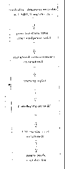
Fig. 1 is a general view of the method according to the present
invention,
Fig. 2 shows more detail of the general view of the method
according to the present invention,
Zo Fig. 3 shows the preparation of medical two-dimensional image
data,
Fig. 4 shows the generation of a three-dimensional patient model,
Fig. 5 shows an inversion model,
Fig. 6 shows a three-dimensional reference model, and
zs Fig. 7 shows a three-dimensional implant model.
7

CA 02373691 2001-11-09
As an example, the case of a patient will be illustrated who has a
complicated large area defect (for example, resulting from an accident,
a tumor etc.) in the upper half of the cranium. In Fig. 1 and 2 there are
represented both, a general block diagrarnmatical overview and a
s detailed block diagrammatical overview to illustrate the method
according to the present invention.
For a precise diagnosis and for a later implant generation medical two-
dimensional image data 1 (two-dimensional tomograms) of a defect
area 5 and of the environment of the same (refer to Fig. 3) are taken
io from a patient in' a radiological hospital department (for example by
computer tomography or by nuclear magnetic resonance tomography).
By use of a mathematical image processing algorithm at first a contour
detection is made in the two-dimensional image data 1 and
subsequently a segmentation is carried out with the airn to detect the
is hard tissue ranges (bones). As a result of the contour detection and
segmentation two-dimensional image data 2 are obtained via which, by
a respective spatial arrangement, a virtual three-dimensional patient
model 3 (dotted model) is formed at least of the defect area 5 and
environment.
Zo In a cooperation between a physician and a design engineer the defect
area is precisely defined and marked in this virtual three-dimensional
patient model 3 by utilizing user-specific computer programs especially
applicable for this purpose.
Zs In the next step, the implant design engineer has several methods at his
disposal for generating precisely fitting implants. These methods are:
8

CA 02373691 2001-11-09
1. When in a three-dimensional patient model 4 (shown in a cross-
sectional view in Fig. 5), the defect area 5 is completely located in
one body half, that is, entirely in one head side, then the data of this
body side with the defect area 5 can, by inversion, be reconstructed,
s making use of the bilateral symmetry of the human body, from the
data of the undamaged side 7 of the three-dimensional patient model
4 (imaging of the undamaged side 7 at the plane of symmetry 6).
After inversion, an extraction of a virtual implant model 9 is carried
out by use of mathematical algorithms which will here not be
io referred to in more detail.
2. When in a three-dimensional patient model IO (shown in a lateral
view in Fig. 6), the defect area 5 is located in the plane of symmetry
of the human body or the data of the undamaged side cannot be
is utilized, somehow or other, then the virtual implant model 9 can be
generated via a three-dimensional reference model 11. To this end,
specific features of the three-dimensional patient model 10 are
compared to a reference database and a selection of similar models
is made under consideration of mathematical, functional, medical
Zo and aesthetic aspects. Then, the three-dimensional reference model
11 is selected from this range of models, preferably under particular
consideration of the medical expert opinion. By superimposing the
three-dimensional reference model 11 and the three-dimensional
patient model 10 to one another, a virtual three-dimensional patient
?s model 12 will be obtained, from which, in turn, the virtual implant
model 9 will be generated by computer, as described under item 1.
9

CA 02373691 2001-11-09
3. In special cases, when for example the defect partially lies in the
plane of symmetry, both methods {inversion according to item 1 and
database comparison according to item 2) can be used one after the
s other and the results will be combined to a three-dimensional
reference model for the implant modeling.
The selection and/or the shaping of the three-dimensional reference
model after at least one of the methods mentioned hereinabove and the
io generation of the virtual implant model from the three-dimensional
reference model are performed merely by computation. By this
processing both, a very rapid and a very precisely fitting generation and
subsequent manufacture of the implant for the operative insert on the
patient is given.
is The present virtual implant model 9 (Fig. 7) is subjected to various
procedures after its generation. Said procedures may include, for
example, strength calculations, simulations for the medical operation
planning and the manufacture, as well as providing markings (bore
holes, fixings or the like), quality control etc.
?o . After designing the virtual implant model 9, a generation/simulation of
the CNC-data for the physical implant manufacture and the transfer of
the virtual implant model into a usable implant are carried out.

CA 02373691 2001-11-09
LIST OF
REFERENCE
NUMERALS
1 - medical two-dimensional image data
2 - contour detection and segmentation two-dimensional
image data
3 - three-dimensional patient model (dotted model)
s 4 - three-dimensional patient model (cross-section)
- - defect area
6 - plane of symmetry of human body
7 - undamaged side of the three-dimensional patient
model
8 - inversion of undamaged side 7
io 9 - (virtual) implant model
- three-dimensional patient model (lateral view)
11 - three-dimensional reference model
12 - . (virtual) three-dimensional patient model
11

Dessin représentatif
Une figure unique qui représente un dessin illustrant l'invention.
États administratifs

2024-08-01 : Dans le cadre de la transition vers les Brevets de nouvelle génération (BNG), la base de données sur les brevets canadiens (BDBC) contient désormais un Historique d'événement plus détaillé, qui reproduit le Journal des événements de notre nouvelle solution interne.

Veuillez noter que les événements débutant par « Inactive : » se réfèrent à des événements qui ne sont plus utilisés dans notre nouvelle solution interne.

Pour une meilleure compréhension de l'état de la demande ou brevet qui figure sur cette page, la rubrique Mise en garde , et les descriptions de Brevet , Historique d'événement , Taxes périodiques et Historique des paiements devraient être consultées.

Historique d'événement

Description Date
Le délai pour l'annulation est expiré 2016-05-10
Lettre envoyée 2015-05-11
Accordé par délivrance 2007-03-20
Inactive : Page couverture publiée 2007-03-19
Inactive : Taxe finale reçue 2006-12-28
Préoctroi 2006-12-28
Un avis d'acceptation est envoyé 2006-10-18
Lettre envoyée 2006-10-18
Un avis d'acceptation est envoyé 2006-10-18
Inactive : Approuvée aux fins d'acceptation (AFA) 2006-09-20
Modification reçue - modification volontaire 2006-02-08
Inactive : Dem. de l'examinateur par.30(2) Règles 2005-10-31
Lettre envoyée 2004-01-09
Lettre envoyée 2004-01-07
Inactive : Supprimer l'abandon 2003-12-19
Requête d'examen reçue 2003-11-25
Exigences pour une requête d'examen - jugée conforme 2003-11-25
Toutes les exigences pour l'examen - jugée conforme 2003-11-25
Inactive : Abandon. - Aucune rép. à lettre officielle 2003-11-12
Inactive : Correspondance - Transfert 2003-10-22
Inactive : Renseignement demandé pour transfert 2003-08-12
Inactive : Correspondance - Transfert 2003-04-28
Lettre envoyée 2003-04-11
Inactive : Renseignement demandé pour transfert 2003-04-11
Inactive : Renseign. sur l'état - Complets dès date d'ent. journ. 2003-03-27
Inactive : Rétablissement - Transfert 2003-03-04
Exigences de rétablissement - réputé conforme pour tous les motifs d'abandon 2003-03-04
Inactive : Abandon. - Aucune rép. à lettre officielle 2003-02-13
Inactive : Page couverture publiée 2002-05-03
Inactive : Lettre de courtoisie - Preuve 2002-04-30
Inactive : Notice - Entrée phase nat. - Pas de RE 2002-04-29
Demande reçue - PCT 2002-03-27
Exigences pour l'entrée dans la phase nationale - jugée conforme 2001-11-09
Demande publiée (accessible au public) 2000-11-16

Historique d'abandonnement

Il n'y a pas d'historique d'abandonnement

Taxes périodiques

Le dernier paiement a été reçu le 2006-01-17

Avis : Si le paiement en totalité n'a pas été reçu au plus tard à la date indiquée, une taxe supplémentaire peut être imposée, soit une des taxes suivantes :

  • taxe de rétablissement ;
  • taxe pour paiement en souffrance ; ou
  • taxe additionnelle pour le renversement d'une péremption réputée.

Veuillez vous référer à la page web des taxes sur les brevets de l'OPIC pour voir tous les montants actuels des taxes.

Titulaires au dossier

Les titulaires actuels et antérieures au dossier sont affichés en ordre alphabétique.

Titulaires actuels au dossier
3DI GMBH
Titulaires antérieures au dossier
JORG BEINEMANN
PETER LITSCHKO
TORSTEN HENNING
WERNER LINSS
WOLFGANG FRIED
Les propriétaires antérieurs qui ne figurent pas dans la liste des « Propriétaires au dossier » apparaîtront dans d'autres documents au dossier.
Documents

Pour visionner les fichiers sélectionnés, entrer le code reCAPTCHA :


Pour visualiser une image, cliquer sur un lien dans la colonne description du document. Pour télécharger l'image (les images), cliquer l'une ou plusieurs cases à cocher dans la première colonne et ensuite cliquer sur le bouton "Télécharger sélection en format PDF (archive Zip)" ou le bouton "Télécharger sélection (en un fichier PDF fusionné)".

Liste des documents de brevet publiés et non publiés sur la BDBC .

Si vous avez des difficultés à accéder au contenu, veuillez communiquer avec le Centre de services à la clientèle au 1-866-997-1936, ou envoyer un courriel au Centre de service à la clientèle de l'OPIC.


Description du
Document 
Date
(aaaa-mm-jj) 
Nombre de pages   Taille de l'image (Ko) 
Dessin représentatif 2002-05-02 1 6
Page couverture 2002-05-03 2 53
Abrégé 2001-11-09 1 36
Revendications 2001-11-09 3 90
Description 2001-11-09 11 487
Revendications 2006-02-08 4 122
Dessin représentatif 2007-02-23 1 6
Page couverture 2007-02-23 2 51
Dessins 2001-11-09 5 217
Rappel de taxe de maintien due 2002-04-29 1 111
Avis d'entree dans la phase nationale 2002-04-29 1 194
Demande de preuve ou de transfert manquant 2002-11-13 1 105
Courtoisie - Lettre d'abandon (lettre du bureau) 2003-03-20 1 167
Avis de retablissement 2003-04-11 1 168
Accusé de réception de la requête d'examen 2004-01-07 1 188
Courtoisie - Certificat d'enregistrement (document(s) connexe(s)) 2004-01-09 1 125
Avis du commissaire - Demande jugée acceptable 2006-10-18 1 161
Avis concernant la taxe de maintien 2015-06-22 1 170
PCT 2001-11-09 10 399
Correspondance 2002-04-29 1 26
Correspondance 2003-03-04 8 305
Correspondance 2003-04-11 1 17
Correspondance 2003-08-12 1 26
Correspondance 2004-01-09 1 14
Correspondance 2006-12-28 1 31