Note : Les descriptions sont présentées dans la langue officielle dans laquelle elles ont été soumises.
CA 02416571 2003-O1-17
WO 02/005719 PCT/USO1/22406
METHOD AND DEVICE FOR URETHRAL-VESICLE ANASTOMOSIS
Backnround of the Invention
Field of the Invention
The present invention generally relates to the reconnection of the urethra and
bladder after a radical
retropubic prostatectomy. Specifically, the invention relates to a method and
device for performing a urethral-vesicle
anastomosis.
Description of the Related Art
In a radical retropubic prostatectomy, the surgeon removes all or most of the
patient's prostate. Because the
urethra travels through the prostate immediately before reaching the bladder,
the upper part of the urethra is removed
in the surgery. In order to restore proper urinary functions, the bladder and
the urethra must be reconnected.
Heretofore, surgeons would execute painstaking suturing operations with tiny,
fine needles to reconnect
these anatomical bodies. It has been found that the use of sutures for this
purpose has caused certain problems in
recovery. These problems include necrosis of the sutured tissues, stricture of
the urethra which impedes the flow of
fluid through it, and a urethra-bladder connection which is not fluid-tight.
In addition, when suturing the urethra to the
bladder the surgeon often inadvertently pierces the nearby neurovascular
bundle, which can cause incontinence or
impotence.
The suturing process itself has also been found to be cumbersome, requiring
the surgeon to grasp and stretch
the bladder and urethra together before making the fine sutures.
With radical retropubic prostatectomies becoming more common, a quicker and
simpler way to reconnect the
bladder and the urethra is needed.
Summary of the Invention
One aspect of the present invention is an improved method for the anastomosis
of the urethra to the bladder
following a prostatectomy.
A further aspect of the present invention is an anastomosis procedure that
eliminates the use of sutures in
. the urethra-bladder junction.
A still further aspect of the present invention is an anastomosis procedure
with an improved means of
grasping the urethra and bladder, bringing them together and holding them for
the connection process.
A method and device are provided for the anastomosis of the urethra and
bladder after radical retropubic
prostatectomy. The surgeon inserts a trocar into the urethra and secures the
bladder to the trocar with an external
ring, or, alternatively, with at least one prong associated with the trocar.
The surgeon then inserts a sheath into the
bladder and secures the bladder to the sheath with at least one prong. The
trocar and the sheath are then advanced
toward each other, and fit together in an end-to-end fashion. When the
urethral tissue and the bladder tissue are in
close proximity, the urethra and the bladder are reconnected using at least
one clip. The urethra is secured to the
bladder
CA 02416571 2003-O1-17
WO 02/005719 PCT/USO1/22406
In accordance with one preferred embodiment, a method is provided for securing
the urethra to the bladder of
a patient. The method comprises the steps of inserting a first approximation
device into the urethra, securing the
urethra to the first approximation device, inserting a second approximation
device into the bladder and securing the
bladder to the second approximation device. The method further comprises the
step of advancing the second
approximation device toward the first approximation device so that a distal
end of the urethra comes in close proximity
to a distal end of the bladder. The final step of the method comprises
securing the urethra to the bladder.
In accordance with yet another preferred embodiment, a method for securing the
urethra to the bladder of a
patient comprises the steps of inserting a first approximation device into the
urethra, securing the urethra to the first
approximation device, inserting a second approximation device into the bladder
and securing the bladder to the second
approximation device. The method further comprises advancing the first
approximation device and the second
approximation device toward one another so that a distal end of the urethra
comes in close proximity to a distal end of
the bladder. Finally, the urethra is secured to the bladder.
In accordance with still another preferred embodiment, there is provided a
system for securing the urethra of
a patient to the bladder of the patient. This system comprises a first
approximation device adapted to be inserted into
the urethra of the patient and a ring. The ring is suitable for placement on a
exterior of the urethra for securing the
urethra to the first approximation device. The system further comprises a
second approximation device adapted to be
inserted into the bladder. The second approximation device has at least one
prong on a cannula of the second
approximation device. The prang secures the second approximation device to the
bladder. The system further
comprises at least one clip. The clip is suitable to secure the urethra to the
bladder once the urethra and bladder are
within close proximity.
In accordance with still another preferred embodiment, there is provided a
system for securing the urethra of a
patient to the bladder of the patient. The system comprises of first
approximation device that has a generally rigid cannula
and at least one prong. The prong is moveable from a retracted position to an
extended position on a exterior surface of
the cannula to secure the urethra to the first approximation device. The
system also comprises a second approximation
device that has a generally rigid cannula and at least one prang. The prong is
moveable from a retracted position to an
extended position on an exterior surface of the cannula to secure the bladder
to the second approximation device.
Brief Description of the Drawings
FIG.1 is a schematic view of a trocar and sheath as used to join the bladder
and urethra in accordance with
the present invention;
FIG. 2 is a crass-sectional view of the trocar and sheath, and the juncture of
the bladder and urethra;
FIG. 3 is a side elevation view of a sheath;
FIG. 4 is a perspective view of a sheath;
FIG. 5 is a close-up perspective view of the distal end of a sheath;
FIG. 6 is a side cross-sectional view of a sheath;
FIG. 7 is a detail cross-section view of the proximal end of a sheath;
-2-
CA 02416571 2003-O1-17
WO 02/005719 PCT/USO1/22406
FIG. 8 is a second detail cross-section view of the proximal end of a sheath,
oriented 90° to the view in FIG.
7;
FIG. 9 is a detail cross-section view of the distal end of a sheath;
FIG.10 is a perspective view of another embodiment of a trocar in accordance
with the invention;
FIG. 11A and 11B are cross-sectional views of the joining of the bladder and
urethra tissues, employing
another embodiment of the trocar and sheath;
FIG.12 is a perspective view of a dual approximator;
FIG. 13 is a perspective view of a dual approximator, with the bladder
everting device displaced in the distal
direction;
FIG.14 is a crass-sectional view of a dual approximator;
FIG.15 is a detail cross-sectional view of the proximal end of a dual
approximator;
FIGS. 16A-16C are side elevation, side cross-section, and perspective views of
a bushing and everting wire
assembly for use with a dual approximator;
FIG.17 is a detail cross-sectional view of the distal end of a dual
approximator; and
FIG. 18 is a cross-sectional view of the use of the dual approximator to join
the bladder to the urethra.
Detailed Description of the Preferred Embodiment
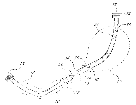
FIG. 1 depicts, among other things, the relevant anatomical structures of a
patient following a radical
retropubic prostatectomy. The urethra 10 has been separated from the bladder
12 by virtue of the removal of the
prostate (not shown). The urethra 10 must therefore be re-attached to the
bladder 12 at the bladder outlet 14.
To rejoin the bladder and urethra, a urethra approximation trocar 16,
comprising a proximal end 18 and a
tapered distal end 20, may be inserted into the urethra 10 via the urethral
outlet in a manner known to those skilled in
the art. The trocar 16 is preferably constructed of a stiff plastic or metal
to provide sufficient rigidity despite a cross-
sectional area small enough to permit the trocar 16 to pass through the
urethra. The trocar 16 is advanced within the
lumen of the urethra 10 so that the tapered or rounded distal end 20 of the
trocar 16 emerges from the urethral
passage. The urethra 10 is then secured to the trocar 16 in a manner which
will prevent the urethra 10 from sliding
backwards, away from the tapered distal end 20 of the trocar 16, when the
trocar is subsequently advanced toward
the bladder 12. Preferably, this is accomplished by a removable external ring
22 placed around the urethra 10 near the
distal end 20, securing the urethra 10 to the trocar 16. Another method to
secure the urethra with respect to the
trocar is by means of one or more everting prongs extendable from the outer
surface of the trocar 16 near the distal
end 20. (This is similar to the everting prongs 30 extendable from the sheath
24, the operation of which will be
discussed in greater detail below.) The prongs evert the urethra tissue from
the trocar shaft, pushing it out radially to
facilitate attachment.
FIG. 1 also depicts a urethra approximation sheath 24 having an everting knob
26 on the proximal end 28
and multiple everting prongs 30 near the distal end 32. The distal end 32 also
forms a cavity 34 which is sized so as
to snugly receive the tapered end 20 of the trocar 16 (see FIG. 2). As with
the trocar 16, the sheath 24 is preferably
-3-
CA 02416571 2003-O1-17
WO 02/005719 PCT/USO1/22406
constructed of a stiff plastic or metal to provide sufficient rigidity despite
a cross-sectional area small enough to
permit the sheath 24 to pass through the bladder outlet 14.
To insert the sheath 24, the surgeon first makes an abdominal incision 36 to
gain access to the bladder 12.
The sheath 24, with everting prongs 30 in a retracted position, is inserted
into the incision 36 and is advanced toward
the bladder outlet 14 so that the distal end 32 of the sheath 24 emerges from
the bladder outlet 14. By manipulation
of the everting knob 26, the everting prongs 30 are extended from the sheath
24 and positioned inside the bladder 12
such that they engage the bladder tissue near the bladder outlet 14, securing
the bladder 72 with respect to the
sheath 24. The everting prongs 30 thus prevent the bladder 12 from sliding
backward on the sheath 24, away from
the distal end 32 of the sheath 24, when the sheath 24 is subsequently
advanced toward the urethra 10. In addition,
the everting prongs 30 pull the tissue of the bladder 12 both longitudinally
and radially to facilitate the eventual
application of one or more clips to the junction of the bladder and urethra
(see FIG. 2).
With further reference now to FIG. 2, the surgeon advances the sheath 24
toward the trocar 16, stretching
the bladder 12 in the process. In one embodiment, the surgeon also moves the
trocar 16 toward the sheath 24,
stretching the urethra 10 in the process. When the trocar 16 and the sheath 24
meet, the tapered distal end 20 of the
trocar 16 enters the cavity 34 in the distal end 32 of the sheath 24, to an
extent sufficient to enable the urethral
tissue and the bladder tissue to press together as shown. Fit together in this
manner, the trocar 16 and the sheath 24
can retain the tissues in this orientation suitable for the connection
process, in a "hands-free' manner. The tissues of
the urethra 10 and the bladder 1 Z are subsequently clamped together using one
or more external clips 38, around the
circumference of the urethra-bladder attachment. The application of the clips
may effect disengagement of the
bladder tissue 12 from the everting prongs 30. In one embodiment, VCS clips
are used to secure the urethral tissue to
the bladder. The clips 38 may be applied either individually, or
simultaneously in a "one-shot" fashion.
After the application of the clips 38, the external ring 22 is removed,
releasing the trocar 16 from the
urethra 10. The surgeon is now able to remove the trocar 16 via the urethral
outlet in a manner known to those
skilled in the art. Similarly, the sheath 24 may be moved in the proximal
direction, after retracting the evening prangs
30 by manipulation of the everting knob 26. The sheath 24 exits the bladder 12
through the incision 36.
FIGS. 3-9 show the components of the sheath 24 in detail. The sheath 24 has an
elongated cannula 40
with a cavity 34 in the distal end 32 and an everting knob 26 near the
proximal end 28. FIGS. 3, 4 and 6 show a
sheath 24 which is straight; advantageously, the sheath may be curved as seen
in FIG. 1, to promote ease of insertion
and use. (Similarly, the straight instruments seen in FIGS. 10, 12. 13 and 14
may also be curved, to obtain the same
advantages.) The everting knob 26 engages threads 42 near the proximal end of
the sheath so that rotating the
everting knob 26 causes it to advance in the desired direction (either
distally or proximally) along the threaded portion
of the sheath 24. Knurling 44 is provided on both the cannula surface and the
everting knob to facilitate easy gripping
of the knob and sheath during surgery. Best shown in FIG. 5, a number
(preferably 4-6) of openings 46 are distributed
radially about the circumference of the cannula 40, near the distal end 32.
The openings 46 permit everting prongs
(not shown) to extend from, or retract into, the cannula 40 when the everting
knob 26 is rotated.
-4-
CA 02416571 2003-O1-17
WO 02/005719 PCT/USO1/22406
As seen in FIG. 6, an everting tube 48 is disposed within a lumen 50 of the
cannula 40 and is coaxial with
the cannula 40. The everting tube 48 fits snugly within the lumen 50 but can
easily move longitudinally within the
cannula 40 in both the distal and proximal directions. Near its proximal end
the lumen 50 widens at a neck 52 to take
on a larger-diameter cross section proximal of the neck 52. Correspondingly,
the everting tube 48 widens to form a
stub 54 disposed within the larger-diameter portion of the lumen 50. The neck
52 coacts with the stub 54 to limit the
travel of the everting tube 48 in the distal direction.
FIGS. 7 and 8 show the proximal end 28 of the sheath 24 in detail. Note that
FIGS. 7 and 8 are oriented
90° with respect to one another, so that FIG. 7 may be considered a
side view and FIG. 8 a top view. A longitudinal
slot 56 is formed in the wall of the cannula 40 near the proximal end 28. The
slot 56 permits an alien screw 58 to
extend from a threaded hole 60 in the stub 54 beyond the external wall of the
cannula 40 and into a space 62 formed
by a radial groove 64 in the everting knob 26, between distal and proximal
walls 66, 68.
With the screw 58 in place, one can cause the everting tube 48 to move in
either the distal or proximal
direction by manipulating the everting knob 26. If the everting knob 26 is
rotated so as to advance in the distal
direction, the proximal wall 68 of the radial groove 64 bears on the screw 58
as the everting knob advances distally,
causing the everting tube 48 to move distally within the lumen 50. Similarly,
if the everting knob 26 is rotated so as
to advance in the proximal direction, the distal wall 66 of the radial groove
64 will bear on the screw 58, causing the
everting tube 48 to move proximally within the lumen 50.
Referring momentarily to FIG. 6, it can be seen that the distal end of the
everting tube 48 is connected to a
bushing 70, which is disposed within the lumen 50 and is moveable both
distally and proximally therein. Best seen in
FIG. 9, the bushing 70 forms a longitudinal socket 72 and two threaded holes
74 intersecting the socket 72. The
socket 72 receives the proximal ends of a number of everting wires 76, and
screws 78 threaded into the holes 74
clamp the everting wires 76 into the bushing 70.
The everting wires 76 extend distally from the bushing 70 into angled channels
80 that correspond to the
openings 46 in the distal end of the cannula 40. The angled channels 80 force
the distal ends of the everting wires,
when moved distally, to extend from the cannula so as to form everting prongs
30 see FIG. 2). Similarly, the everting
wires 76 retract into the angled channels 80 when moved proximally.
Thus it can be seen that rotation of the everting knob 26 in the desired
direction will extend or retract the
everting prongs 30. When the everting knob 26 is rotated in a direction
causing the everting tube 48 to move distally,
the everting tube 48 pushes the bushing 70 in the distal direction, forcing
the everting wires 76 to extend from the
openings 46 and form everting prongs. By rotating the everting knob 26 in the
opposite direction, the everting tube 48
moves proximally and pulls the bushing 70 proximally as well, causing the
everting wires 76 to retract into the angled
channels 80.
FIG. 10 shows an alternative embodiment of the trocar 16, which employs the
same everting-prong
mechanism as the sheath discussed above. This type of trocar also has a
tapered distal tip 20 which fits snugly into
the cavity formed in the distal end of the sheath.
-5-
CA 02416571 2003-O1-17
WO 02/005719 PCT/USO1/22406
FIG. 11A depicts the use of the averting-prong mechanism of the sheath 24 with
the bladder tissue 12.
Additionally, FIG. 11A shows the use of that version of the trocar 16
employing a similar mechanism, with the urethra
10. After positioning the distal end 32 of the sheath 24 near the bladder
outlet 14, the surgeon extends the averting
prongs 30, which engage the bladder tissue 12, averting the bladder outlet 14
and holding it in a suitable position for
attachment to the urethra 10. When using a trocar 16 equipped with averting
prongs 30, the surgeon inserts the
trocar 16 into the urethra 10 and positions the distal end 20 near the opening
of the urethra 10. In a similar manner
the averting prongs 30 are extended so as to avert the tissue near the end of
the urethra 10 in the desired position for
reattachment.
After averting both the bladder and urethra tissue, the surgeon brings the
trocar 16 and sheath 24 together
so that the tapered distal end 20 of the trocar 16 fits into the cavity 34 of
the sheath, and the bladder and urethra
tissue meet. Upon joining the trocar and sheath, the surgeon has both hands
free to perform final alignment of the
bladder and urethra tissue, and apply the clips 38 as shown in FIG.11 B.
FIGS. 12-14 show yet another embodiment of the instruments to be used in the
present invention. This
embodiment enables a surgeon to perform the operation without making an
incision in the bladder (otherwise needed to
insert the sheath) by combining the functions of the trocar and the sheath in
a dual approximator 100 to be used
transurethrally.
The dual approximator 100 has an elongated cannula 102 with a rounded distal
end 104, two sets of
openings 106 in the surface of the cannula 102 for the bladder and urethra
averting prongs 108, 110, and a bladder
averting knob 112 and a urethra averting knob 114 near the proximal end. As
seen in FIG. 13, The cannula 102 is
separable at a point 116 between the two sets of openings 106, into a bladder
averting unit 118 and a urethra
averting unit 120. This separation feature permits the bladder averting unit
118 to move distally, into the bladder
opening as necessary. Preferably, the bladder and urethra averting prongs 108,
110 are radially staggered with
respect to one another so that the two sets of prongs will not "collide' when
extended.
The urethra averting unit 120 resembles the sheath described above, with some
additions best seen in FIG.
14. A central channel 122 runs along the centerline of the urethra averting
unit 120, through the distal end 124,
bushing 126, averting tube 128 and stub 130. A bladder averting knob 112 is
located proximal of a urethra averting
knob 914, and engages threads on the outer surface of the cannula 102 so that
rotation of the bladder averting knob
112 causes it to advance in the desired direction (either distally or
proximally) along the threaded portion of the
cannula 102. Best seen in FIG. 15, a radial channel 134, longitudinal slot
136, screw 138, and block 140 coast in a
manner similar to that disclosed above with respect to the averting knob 26 on
the sheath 24, to cause the block 140
to move longitudinally within the lumen 142 of the cannula 102 in response to
rotation of the bladder averting knob
112 in the desired direction.
Attached to the block 140 is a bladder averting rod 144 which runs through the
central channel 122 and out
the distal end 124, continuing into the bladder averting unit 118 (see FIG.
14). To accommodate the central channel
122 and bladder averting rod 144, the bushing 126 is modified as shown in
FIGS.16A-16C, and 17. Urethra averting
-6-
CA 02416571 2003-O1-17
WO 02/005719 PCT/USO1/22406
wires 146 are bent 90° at the proximal ends and are received in slots
148 formed at the distal end of the bushing 126.
The central channel 122 and the bladder averting rod 144 (best seen in FIG.
17) pass through the bushing 126, and
the bladder averting rod 144 continues distally through a space 150 formed
between the urethra averting wires 146.
This arrangement of the bushing 126 and urethra averting wires 146 permits the
bladder averting rod 144 and urethra
averting wires 146 to move freely with respect to each other within the
cannula 102 without interference.
Referring again to FIGS. 1214, the bladder averting unit 118 is located at the
distal end of the dual
approximator 100, and has a relatively short cannula 152 with a lumen 154 and
a rounded distal tip 104. The
proximal end 156 is tapered, in the same way as the distal end of the trocar
16, to fit within the cavity 158 formed in
the distal end 124 of the urethra averting unit 120. Near the proximal end 156
are located a number (preferably 4-6)
of openings 106 distributed radially about the circumference of the cannula
152. As seen in FIG. 17, the central
channel 122 continues from an opening 160 in the proximal tip, along the
longitudinal axis of the bladder averting unit
118, to the proximal end of the lumen 154.
A bushing 162 is disposed within the lumen 154 and is moveable both distally
and proximally therein. The
bladder averting rod 144 passes through fihe central channel 122, into the
lumen 154, and to the bushing 162. The
bushing 162 forms a longitudinal socket 164 and two threaded hales 166 which
intersect with the socket 164. The
socket 164 receives the distal ends of a number of bladder averting wires 168
and the bladder averting rod 144, and
screws 170 threaded into the holes 166 clamp the wires 168 and rod 144 into
the bushing 162.
The bladder averting wires 168 extend proximally from the bushing 162 into
angled channels 172
corresponding to the openings 106 in the proximal end of the bladder averting
unit 118. The angled channels 172
force the proximal ends of the averting wires 168, when moved proximally, to
extend from the cannula 152 so as to
form averting prongs 108. Similarly, the bladder averting wires 168 will
retract into the angled channels 172 when
moved distally.
Thus, by reference especially to FIGS. 14 and 17, it can be seen that rotation
of the bladder averting knob
112 in the desired direction will extend or retract the bladder averting
prongs 108. When the bladder averting knob
112 is rotated in a direction causing the bladder averting rod 144 to move
proximally, the bladder averting rod 144 will
pull the bushing 162 in the proximal direction, forcing the bladder averting
wires 168 to extend from the openings 106
and form bladder averting prongs 108. By rotating the bladder averting knob
112 in the opposite direction, the bladder
averting rod 144 moves distally and pushes the bushing 162 distally as well,
causing the bladder averting wires 168
to retract into the angled channels 172.
The bladder averting knob 112 also expands or contracts the distance between
the urethra averting unit 120
and the bladder averting unit 118. When the bushing 162 in the bladder
averting unit 118 remains relatively immobile,
rotation of the bladder averting knob 112 so as to move the bladder averting
rod 144 distally or proximally, causes a
corresponding distal or proximal movement of the bladder averting unit 118.
FIG. 18 details the use of the dual approximator 100 in performing the
anastomosis procedure. The surgeon
inserts the dual approximator 100 into the lumen of the urethra 10, through
the urethral outlet, in a manner known to
.7.
CA 02416571 2003-O1-17
WO 02/005719 PCT/USO1/22406
those skilled in the art. The dual approximator 100 is advanced within the
lumen of the urethra 10 until the distal end
of the dual approximator 100, including the bladder everting unit 118, emerges
from the opening. Next the surgeon
rotates the bladder everting knob so as to move the bladder everting unit 118
distally and create a suitable gap
between the bladder everting unit 118 and the urethra everting unit 120. The
bladder everting unit 118 is then
inserted into the bladder opening 14, to a point where the openings 106 in the
bladder everting unit 118 are properly
aligned within the bladder 12. The surgeon then rotates the bladder everting
knob to extend the bladder everting wires
108 from the openings 106, forming everting prongs 108, until the tips of the
prongs 108 contact and evert the
bladder tissue 12. Similarly, the surgeon rotates the urethra everting knob to
evert the end of the urethra 10 as
desired. The surgeon then brings the everted bladder and urethra tissue 12, 10
together by further rotating the
bladder everting knob until the tapered proximal end 156 of the bladder
everting unit 118 meets the cavity 158 in the
distal end of the urethra everting unit 120. At this point the surgeon will
have both hands free to perform final
alignment of the bladder and urethra tissue 12. 10, and apply the clips 38 in
a similar manner as shown in FIG. 11B.
After applying the clips 38, the surgeon rotates the bladder everting knob to
retract both sets of everting wires, and
then withdraws the dual approximator 100 from the urethra 10.
The clips 38 perform a holding function, in a manner similar to sutures but
without penetration of the vessel
walls. One example of a suitable clip for use in this procedure is disclosed
in U.S. Patent No.4,983,176, titled
DEFORMABLE PLASTIC SURGICAL CLIP, the entirety of which is hereby incorporated
herein by reference.
The present invention utilizes a simple, effective mechanical arrangement for
reconnecting the bladder to the
urethra. By eliminating the painstaking, cumbersome suturing techniques,
urethral-vascular anastomosis techniques
are improved. Furthermore, in the disclosed procedure, there is provided
improved apparatus for grasping and everting
the urethra and bladder tissues, leaving the surgeon's hands free for
performing the reconnection step of the
anastomosis process.
By utilizing the disclosed techniques and apparatus, the number of steps in
the anastomosis procedure is
decreased, minimizing cost and reducing the required time for the procedure.
The present invention eliminates many
complications associated with other anastomosis techniques, such as stapling
or suturing. Because the clips do not
penetrate the vessel walls, there is a decreased likelihood of clotting, which
may cause stricture. The clips also reduce
the occurrence of necrosis, which occurs when insufficient blood is supplied
to the joined tissues. In addition, the use
of clips eliminates the possibility of piercing the neurovascular bundle with
the suture needle(s), which piercing can
cause impotence andlor incontinence.
Although this invention has been disclosed in the context of certain preferred
embodiments and examples, it will
be understood by those skilled in the art that the present invention extends
beyond the specifically disclosed embodiments
to other alternative embodiments andlor uses of the invention and obvious
modifications and equivalents thereof. Thus, it
is intended that the scope of the present invention herein disclosed should
not be limited by the particular disclosed
embodiments described above, but should be determined only by a fair reading
of the claims that follow.
.g.