Note : Les descriptions sont présentées dans la langue officielle dans laquelle elles ont été soumises.
CA 02417341 2003-O1-23
WO 02/12896 PCT/US00/25381
METHODS FOR MANIPULATING MOIETIES IN MICROFLUIDIC
SYSTEMS
Technical Field
This invention relates generally to the field of moiety or molecule
manipulation in a chip format. In particular, the invention provides a method
for
manipulating a moiety in a microfluidic application, which method comprises:
a) coupling a moiety to be manipulated onto surface of a binding partner of
said
moiety to form a binding partner-moiety complex; and b) manipulating said
binding
partner-moiety complex with a physical force in a chip format, wherein said
manipulation is effected through a combination of a structure that is external
to said
chip and a structure that is built-in in said chip, thereby said moiety is
manipulated.
Background Art
Intensive research efforts in developing microfluidic systems have been
pursued by academic and industrial institutions over recent years. These
microfluidic
devices and apparatus are developed for perfornling various fluidics-related
functions,
processes and activities. Almost all microfluidic devices involve
manipulating,
handling, and processing molecules and particles. However, up to now, there is
not a
general method for manipulating molecules in microfluidic devices. Some
examples
of physical methods for manipulating molecules used in biochips include
electric field
based electrophoresis, optical radiation force related optical tweezers and
others. All
these methods have many limitations. Electrophoresis utilizes direct current
(DC)
electrical field. Generating sufficient DC field in aqueous solutions without
causing
undesired effects, e.g., surface electrochemistry, gas bubble generation, is
very
difficult. Electric field can only guide molecules either with or against with
the field
direction. There won't be any force induced if the molecule charges are small.
Most
importantly, the DC electrical field cannot be readily structured to generate
manipulation forces in a versatile way. Also, electrode polarization
determines that
over 80% of the applied DC voltage is dropped across the electrode-solution
double
layer and there is only a very small percent of the applied voltage that is
actually
across the bulk solution. Optical radiation force can operate on large
molecules, e.g.,
CA 02417341 2003-O1-23
WO 02/12896 PCT/US00/25381
DNA molecules, but there are certain difficulties in generating 3-D, flexible,
optical
manipulation forces.
Despite the existence of a number of physical forces applicable to molecule
manipulation, several key difficulties exist. First, many physical forces are
proportional to the volume of the particles that are manipulated. Direct
manipulation
of many types of molecules with these forces requires extremely high field
strength
because of the relative small dimensions of molecules, and effective
manipulation of
molecules is almost impossible. High field strengths tend to induce undesired
fluid
motion for manipulation forces such as dielectrophoresis or traveling-wave-
dielectrophoresis. Secondly, certain types of physical forces can be generated
on
molecules, but the 3-D distributions of these physical forces cannot be
readily
structured for flexible, versatile handling and manipulation of molecules.
Thirdly,
there is still no general method for manipulating and handling molecules in
microfluidic systems and devices.
Microparticles have been used for manipulating molecules in biological fields.
One example is the use of magnetic microparticles to harvest and isolate
nucleic acid
molecules, e.g., mRNAs or DNAs, from a solution suspension. Typically, the
separation process takes place in an Eppendorf tube in which paramagnetic
particles
are mixed with solutions containing target nucleic acid molecules. The
modification
of the paramagnetic particles' surface molecules allows the binding of the
target
molecules to paramagnetic particles' surfaces. After incubation of the
magnetic
particles with nucleic acid molecules in the Eppendorf tube, the nucleic acid
molecules are bound to the paramagnetic particles. An external magnetic field
is then
applied to the Eppendorf tube from one side by using a permanent magnet. All
the
magnetic particles are collected onto the regions of the tube wall, which are
closest to
the magnet. Micropipette is then used to pipette out the solutions while the
magnetic
particles being retained on the tube wall by the magnetic field. This step
leaves all the
magnetic particles in the tube. New buffer solutions are then introduced into
the
Eppendorf tube, which is taken away from the magnet. After resuspending
magnetic
particles into the solution, the new buffer may allow the bound nucleic acid
molecules
to de-couple from the magnetic particle surfaces. Then a magnet may be applied
to
attract and trap magnetic particles on the tube wall. Micropipette is then
used to
pipette solutions out of the tube and to collect the nucleic acid molecules.
Recently,
2
CA 02417341 2003-O1-23
WO 02/12896 PCT/US00/25381
similar methods have been used on a chip using paramagnetic beads and an
externally
applied, off chip permanent magnet (Fan et al., Ahal. Chem., 71 21 :4851-9
(1999)).
This method has certain limitations. Reducing such permanent magnet size and
handling a large number of these small permanent magnets automatically for
' manipulation of particles in a chip format will be a very difficult, if not
impossible,
challenge. Thus, the method cannot be readily miniaturized and automated.
Furthermore, the permanent magnet-based methods are not applicable to many
steps
in bioanalytical procedures. Thus, the biochip-system integration based this
method
will be difficult, if not impossible.
' U.S. Patent No. 5,653,859 discloses a method of analysis or separation
comprising: treating a plurality of original, particles to form a subplurality
of altered
particles from at least some of said plurality of original particles, said
subplurality of;.
altered particles having travelling wave field migration properties distinct
from those
of said plurality of original particles; and producing translatory movement of
said
subplurality of altered particles and/or said plurality of original particles
by travelling
wave field migration using conditions such that said translatory movement of
said ,
subplurality of altered particle differs from said translatory movement of
said plurality
of original particles under the same conditions. The physical force used in
the
methods of U.S Patent No. 5,653,859 is limited to the force effected via
travelling
wave field. In addition, to be used in the methods of U.S Pafent No.
5,653,859, the
original particles have to be partially, but not completely, converted into a
subplurality of altered particles because the methods are based upon detecting
different translatory movement of the altered particles and the original
particles. ,
In summary, the currently available manipulation methods suffer from the
following deficiencies: (1) it is difficult to directly apply effective,
physical
manipulation forces to many types of molecules because of the relative small
dimensions of molecules; and (2) some physical forces that can be generated on
molecules often have limitations in 3-D structuring of the force distribution
and (3) it
is difficult to use currently available biochip-based methods for developing
fully
automated, miniaturized and integrated biochip systems.
The present invention addresses these and other related needs in the art. It
is
an objective of the present invention to provide a general method for
manipulating a
variety of moieties including molecules. It is another objective of the
present
3
CA 02417341 2003-O1-23
WO 02/12896 PCT/US00/25381
invention to make full use of a number of force mechanisms effectively for
manipulating the moieties. It is still another objective of the present
invention to
provide for standardized on-chip manipulation procedure, leading to
simplification
and standardization of the design of microchips and the associated systems. It
is yet
another objective of the present invention to expand and enhance the
capabilities of
molecule manipulation with the choice of microparticles with special physical
properties. It is yet another objective of the present invention to provide a
general,
effective procedure for on-chip molecule manipulation that allows for fully
integration of biochip-based analytical systems and processes.
Disclosure of the Invention
This invention relates. generally to the field of moiety or molecule
manipulation in a chip format. In one aspect, the invention is directed to a
method for
manipulating a moiety in a microfluidic application, which method comprises:
a) coupling a moiety to be manipulated onto surface of a binding partner of
said
moiety to form a binding partner-moiety complex; and b) manipulating said
binding
partner-moiety complex with a physical force in a chip format, wherein said
manipulation is effected through a combination of a structure that is external
to said
chip and a structure that is built-in in said chip, thereby said moiety is
manipulated.
The present invention .provides a general method for handling, processing and
manipulating a variety of moieties including molecules in a chip format for
numerous
microfluidic applications. For biomedical applications, moieties such as
cells,
organelles, marcromolecules,, small molecules and molecule aggregates may be
manipulated for various bioanalytical procedures. Target moiety types may be
separated, concentrated, transported, selectively manipulated. Using numerous
types
of binding partners, multiple target moieties (e.g., certain mRNA and protein
molecules from cell lysate) may be isolated and selectively manipulated from a
moiety mixture. Molecules or certain moiety types that cannot be manipulated
directed by chip-generated physical forces may now be handled and processed
through the use of the binding-partner for forming the binding partner-moiety
complexes. With the present invention, for example, small protein molecules
that can
not be effectively manipulated by dielectrophoresis forces because of the
small
4
CA 02417341 2003-O1-23
WO 02/12896 PCT/US00/25381
volume may be now handled by on-chip generated dielectrophoresis forces
through
the procedure of coupling them onto the surfaces of microbeads and
manipulating the
protein-bead complexes with the built-in electrodes on a chip. Thus, the
present
invention addresses one critical limitation in current biochip application,
i.e., the lack
of general method for manipulation of a variety of moieties especially
molecules.
The present invention provides a method for handling and manipulating a
variety of moieties in a chip format by utilizing a number of force
mechanisms.
Coupling the moiety onto the binding partners expands the possibility of
available
force mechanisms for manipulating moieties. For example, cells that can not be
directly manipulated by magnetic forces because of the lack of certain
magnetic
properties may now be processed by on-chip generated magnetic forces through
the
procedure of coupling them onto the surfaces of magnetic beads and
manipulating the
magnetic bead-cell complexes with the built-in electromagnetic units on a
chip. Thus,
the present invention improves significantly the flexibility and easiness for
manipulating a variety of moieties in a chip format.
The present invention provides for the standardized on-chip manipulation
procedure and allows for simplification and standardization of the design of
microchips and the associated systems. The manipulation and processing of
target
moiety types is an essential requirement involved in almost all bioanalytical
processes, procedures and steps. The present invention may be utilized for all
these
processes and steps, leading to additional advantages of fully integration of
biochip-
based analytical systems and processes.
Generally, biochip-based applications are divided into sample preparation,
biochemical reactions and result-detection. Sample preparation refers to the
isolation
and preparation of certain target moiety (or moieties) from a mixture sample.
Biochemical reactions refer to the reaction processes involving the prepared
moiety
(or moieties) for the follow-on detection and quantification. The result-
detection
refers to the detection and/or quantification steps to analyze the reaction-
generated
products. An example of these steps is the separation of target cancer cells
from body
fluid and the isolation of target mRNA molecules from the separated cancer
cells, the
reverse-transcription of mRNA to cDNA followed by cDNA amplification and
detection. The present invention may be used in all these steps. Micorbeads
with
antibodies on the bead surfaces that are specific for target cancer cells may
be used to
CA 02417341 2003-O1-23
WO 02/12896 PCT/US00/25381
isolate cancer cells through selective manipulation of microbead-cell
complexes in a
chip format. After the cancer cells are lysed to obtain cellular molecules,
microbeads
that allows for the specific hybridization of target mRNA molecules may be
used to
separate the mRNA molecules on a chip through selective manipulation of mRNA-
bound microbeads from cell lysate mixture. The mRNA-bound microbeads may be
further transported to a location on the chip for further reverse-
transcription of mRNA
to cDNA followed by cDNA amplification. The amplified cDNA molecules may
then be manipulated using the present invention in a procedure of coupling the
cDNA
onto microbead surfaces and manipulating the cDNA-microbead complexes in a
chip
format.
Because the present invention can handle and process molecules and other
moieties.in a chip format and is applicable to all steps of bioanalytical
steps and
procedures, the method allows for a number of bioanalytical processes
integrated on a
chip and/or a number interconnected chips. Such integrated devices and systems
have
advantages in terms of automation,. simplicity, flexibility, integration,
reduced
consumption of reagents, result accuracy and minimum contamination. Thus, the
present invention addresses another critical limitation in current biochip
application,
i. e., the' lack of integration capability. Currently, many biochip-based
methods can be
applied only to certain steps in bioanalytical procedures. Furthermore,
certain biochip
methods exploit physical forces generated using the external structures that
are not
incorporated in chip, imposing limitations for miniaturization, automation and
integration of biochip-based systems. Both these shortcomings are addressed by
the
present invention.
The present invention further expands and enhances the capabilities of
molecule manipulation in a chip-format with the choice of binding partners,
e.g.,
microparticles, with special physical properties. By utilizing different types
of
microparticles with unique physical properties, the molecule manipulation can
be
achieved using a variety of physical force generation mechanisms. In addition,
different particles having different physical properties can be used
simultaneously to
handle and manipulate multiple types of moieties (e.g., DNAs, proteins, mRNAs
and
other biomolecules) because these particles can be selectively manipulated.
The present methods can be used for manipulating any types of moieties when
the moieties are involved in certain processes, such as physical, chemical,
biological,
6
CA 02417341 2003-O1-23
WO 02/12896 PCT/US00/25381
biophysical or biochemical processes, etc., in a chip format. The moieties
include the
ones that can be manipulated directly by various physical forces and
preferably, the
ones that cannot be manipulated directly by various physical forces and have
to be
manipulated through the manipulation of their binding partners. In specific
embodiments, moieties to be manipulated are cells, cellular organelles,
viruses,
molecules or an aggregate or complex thereof. Non-limiting examples of
manipulatable cells include animal, plant, fungus, bacterium, recombinant
cells or
cultured cells. Non-limiting examples of manipulatable cellular organelles
include
nucleus, mitochondria, chloroplasts, ribosomes, ERs, Golgi apparatuses,
lysosomes,
proteasomes, secretory vesicles, vacuoles or microsomes. Manipulatable
molecules
can be inorganic molecules such as ions, organic molecules or a complex
thereof
Non-limiting examples of manipulatable ions include sodium, potassium,
magnesium,
calcium, chlorine, iron, copper, zinc, manganese, cobalt, iodine, molybdenum,
vanadium, nickel, chromium, fluorine, silicon, tin, boron or arsenic ions. Non-
limiting examples of manipulatable organic molecules include amino acids,
peptides,
proteins, nucleosides, nucleotides, oligonucleotides, nucleic acids, vitamins,
monosaccharides, oligosaccharides, carbohydrates, lipids or a complex thereof.
Any binding partners that both bind to the moieties with desired affinity or
specificity and are manipulatable with the desired physical forces) can be
used in the
present methods. Unlike the moieties to be manipulated, which can or cannot be
manipulated directly by the physical forces, the binding partners must be
directly
manipulatable with the desired physical force(s). One type of binding partner
can
possess properties that make it manipulatable by various physical forces. The
binding
partners can be cells such as animal, plant, fungus, bacterium or recombinant
cells;
cellular organelles such as nucleus, mitochondria, chloroplasts, ribosomes,
ERs, Golgi
apparatuses, lysosomes, proteasomes, secretory vesicles, vacuoles or
microsomes;
viruses, natural microparticles, synthetic microparticles or an aggregate or
complex
thereof. The micropaxticles used in the methods could have a dimension from
about
0.01 micron to about ten centimeters. Preferably, the microparticles used in
the
methods have a dimension from about 0.1 micron to about several thousand
microns.
Microparticles could have any compositions, shapes and structures, provided
that they
properties that make them manipulatable by physical forces. Examples of
microparticles that can be used in the methods include, but not limited to,
plastic
7
CA 02417341 2003-O1-23
WO 02/12896 PCT/US00/25381
particles, polystyrene microbeads, glass beads, magnetic beads, hollow glass
spheres,
metal particles, or particles of complex compositions, microfabricated free-
standing
microstructures. In utilizing the present inventions, it is necessary that the
choice of
the binding partners in terms of physical properties, e.g., size, shape,
density,
structural composition, dielectric characteristics, magnetic properties,
acoustic
impedance, optical refractive index, should match the choice of the type of
the
manipulation forces and manipulation methods. In the case of utilizing
multiple types
of binding partners for simultaneous manipulation of multiple types of
moieties,
physical properties of each binding partner should be chosen so that they can
be
selectively manipulated in a chip format.
The moiety to be manipulated can be coupled to the surface of the binding
partner with any methods known in the art. For example, the moiety can be
coupled
to the surface of the binding partner directly or via a linker, preferably, a
cleavable
lii~lcer. The moiety can also be coupled to the surface ~of the binding
partner via a
covalent or a non-covalent linkage. Additionally, the moiety can be coupled to
the
surface of the binding partner via a specific or a non-specific binding.
Preferably, the
linkage between the moiety and the surface of the binding partner is a
cleavable
linkage, e.g., a linkage that is cleavable by a chemical, physical or an
enzymatic
treatment: The coupling step or the decoupling step, if there is one, can be
carried out
on or off the chip.
Any physical forces can be used in the present methods. For instances, a
dielectrophoresis force or a traveling-wave dielectrophoresis force such as
the ones
effected on electrically polarized particles via electrical fields generated
by
microelectrodes energized with AC (alternating current) electric signals, a
magnetic
force such as one effected on magnetic particles via magnetic fields generated
by
ferromagnetic material or by a microelectromagnetic unit, an acoustic force
such as
one effected on many types of particles via a standing-wave acoustic field, a
traveling-wave acoustic field generated by a piezoelectric material energized
with
electrical signals, an electrostatic force such as one effected on charged
particles via a
DC electric field, a mechanical force such as fluidic flow force, an optical
radiation
force such as one effected on various types of particles via laser tweezers,
or a
thermal convection force, can be used. In utilizing the present inventions, it
is
necessary that the choice of the type of the manipulation forces and
manipulation
CA 02417341 2003-O1-23
WO 02/12896 PCT/US00/25381
methods should match the choice of the binding partners in terms of physical
properties and manipulation methods are realized in a chip format.
The present methods can be used in any chip format. For example, the
methods can be used on silicon, silicon dioxide, silicon nitride, plastic,
glass, ceramic,
photoresist or rubber chips. In addition,~the methods can be used on a
chemchip, i.e.,
on which chemical reactions are carried out, a biochip, i. e., on which
biological
reactions are carried out, or a combination of a biochemchip. The chip used
for the
present invention has the built-in structures that can be energized by an
external
energy source and can produce physical forces to act on the binding partners
and
binding partner-moiety complexes. In many cases, the built-in structures are
fabricated on or in a chip substrate. For example, microfabricated spiral
electrode
structures on a glass chip may be used for isolating, concentrating and
manipulating
microparticles.
The physical force used in the present methods are effected through a
combination of the structure that is external to the chip and the structure
that is built-
in on the chip. The external structures are energy sources that can be
connected to the
built-in structures for energizing the built-in structures to generate a
physical force
such as dielectrophoresis force, magnetic force, acoustic force, electrostatic
force,
mechanical force or optical radiation force. The built-in structures can
comprise a
single unit or a plurality of units, each unit is, when energized and in
combination
with the external structure, capable of effecting the physical force on the
binding
partner. In the case of a plurality of units, the built-in structure may
further comprise
the means for selectively energizing any one of the plurality of units.
The present methods can 'be used for any type of manipulations. Non-limiting
examples of the manipulations include transportation, focusing, enrichment,
concentration, aggregation, trapping, repulsion, levitation, separation,
fractionation,
isolation or linear or other directed motion of the moieties. Of particular
importance
is the selective manipulation, e.g., separation, isolation, fractionation,
enrichment, of
one or more target moieties from a mixture.
In another aspect, the invention is directed to a method for manipulating a
moiety which further comprises a step of decoupling the moiety from the
surface of
the binding partner after the moiety is manipulated. The nature of the
decoupling step
depends on the nature of the moiety, the binding paxtner, the surface
modification of
9
CA 02417341 2003-O1-23
WO 02/12896 PCT/US00/25381
the partner and the manipulation step. Generally, the condition of the
decoupling step
is the opposite of the conditions that favor the binding between the moiety
and the
binding partner. For example, if a moiety binds to the binding partner at a
high salt
concentration, the moiety can be decoupled from the binding partner at a low
salt
concentration. Similarly, if a moiety binds to the binding partner through a
specific
linkage or a linker, the moiety can be decoupled from the binding partner by
subjecting the linkage to a condition or agent that specifically cleaves the
linkage.
In a specific embodiment, the moiety to be manipulated is a DNA, the binding
partner is a porous bead and the DNA is reversibly absorbed onto the surface
of the
porous bead in a buffer containing high salt concentration. Alternatively, the
DNA
specifically binds to the surface of a binding partner (e.g., polystyrene
beads) via
sequence specific hybridization or binding to an anti-DNA antibody.
In another specific embodiment, the moiety to be manipulated is a mRNA and
the mRNA specifically binds to the surface of a binding partner (e.g.,
polystyrene
beads and magnetic beads) that is modified to contain oligo-dT polynucleotide.
Iri still another specific embodiment, the moiety to be manipulated is a
protein
and the protein non-specifically binds to the surface of a binding partner
that is
modified with a detergent, e.g., SDS. Alternatively, the protein specifically
binds to
the surface of a binding partner that is modified with an antibody that
specifically
recognizes the protein.
In still another specific embodiment, the moiety to be manipulated is a cell
and
the cell specifically binds to the surfaces of a binding partner (e.g.
magnetic beads)
that is modified to contain specific antibodies against the cells.
In.yet another specific embodiment, the moiety to be manipulated is
substantially coupled onto surface of the binding partner. Preferably, the
moiety to be
manipulated is completely coupled onto surface of the binding partner.
In yet another specific embodiment, a plurality of moieties is manipulated.
The plurality of moieties can be manipulated sequentially or simultaneously.
The
plurality of moieties can be manipulated via a single binding partner or a
plurality of
binding partners. Preferably, the plurality of moieties is manipulated via a
plurality of
corresponding binding partners.
In still another aspect, the invention is directed to a method for isolating
an
intracellular moiety from a target cell, which method comprises: a) coupling a
target
CA 02417341 2003-O1-23
WO 02/12896 PCT/US00/25381
cell to be isolated from a biosample onto surface of a first binding partner
of said
target cell to form a target cell-binding partner complex; b) isolating said
target cell-
binding partner complex with a physical force in a chip format, wherein said
isolation
is effected through a combination of a structure that is external to said chip
and a
structure that is built-in in said chip, c) obtaining an intracellular moiety
from said
isolated target cell; d) coupling said obtained intracellular moiety onto
surface of a
second binding partner of said intracellular moiety to form an intracellular
moiety-
binding partner complex; and e) isolating said intracellular moiety-binding
partner
complex with a physical force in a chip format, wherein said isolation is
effected
through a combination of a structure that is external to said chip and a
structure that is
built-in in said chip.
In yet another aspect, the invention is directed to a method for generating a
cDNA library in a microfluidic application, which method comprises: a)
coupling a
target cell to be isolated onto surface of a first binding partner of said
target cell to
form a target cell-binding partner complex; b) isolating said target cell-
binding
partner complex with a physical force in a chip format, wherein said isolation
is
effected through a combination of a structure that is external to said chip
and a
structure that is built-in in said chip, c) lysing said isolated target cell;
d) decoupling .
and removing said first binding partner from said lysed target cell; e)
coupling mRNA
to be isolated from said lysed target cell onto surface of a second binding
partner of
said mRNA to form a mRNA-binding partner complex; f) isolating said mRNA-
binding partner complex with a physical force in a chip format, wherein said
isolation
is effected through a combination of a structure that is external to said chip
and a
structure that is built-in in said chip, and g) transporting said isolated
mRNA-binding
partner complex to a different chamber and reverse transcribing said
transported
mRNA into a cDNA library.
In yet another aspect, the invention is directed to a method for determining
the
gene expression a target cell in a microfluidic application, which method
comprises:
a) coupling a target cell to be isolated onto surface of a first binding
partner of said
target cell to form a target cell-binding partner complex; b) isolating said
target cell-
binding partner complex with a physical force in a chip format, wherein said
isolation
is effected through a combination of a structure that is external to said chip
and a
structure that is built-in in said chip, c) lysing said isolated target cell;
d) decoupling
11
CA 02417341 2003-O1-23
WO 02/12896 PCT/US00/25381
and removing said first binding partner from said lysed target cell; e)
coupling mRNA
to be isolated from said lysed target cell onto surface of a second binding
partner of
said mRNA to form a mRNA-binding partner complex; f) isolating said mRNA-
binding partner complex with a physical force in a chip format, wherein said
isolation
is effected through a combination of a structure that is external to said chip
and a
structure that is built-in in said chip; and
g) determining the quantities of the isolated mRNA molecules.
In yet another aspect, the invention is directed to a kit for manipulating a
moiety in a microfluidic application, which kit comprises: a) a binding
partner onto
the surface of which a moiety to be manipulated can be coupled to form a
moiety-
binding partner complex; b) means for coupling said moiety onto the surface of
said
binding partner; and c) a chip on which said moiety-binding partner complex
can be
manipulated with a physical force that is effected through a combination of a
structure
that is external to said chip and a structure that is built-in in said chip.
Brief Description of the Drawings
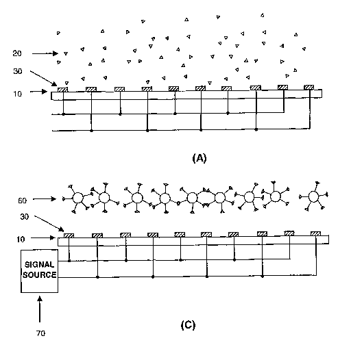
Figure 1 depicts schematic drawing for illustrating the method of binding
partner, e.g., micro-particle, based on-chip manipulation (levitation) of
moieties to be
manipulated, e.g., molecules:
(A) Molecules are suspended in a solution placed on a biochip;
(B) Molecules are coupled onto microparticle surfaces;
(C) Under applied electrical signals to the linear, parallel electrode
elements on the biochip, molecule-microparticle complexes are levitated (or
manipulated) onto certain heights above the chip surface.
Figure 2 depicts schematic representation of a fluidic chamber for moiety,
e.g.,
molecule, manipulation that includes a biochip on the bottom, a spacer and a
top
plate. The molecule manipulation utilizes dielectrophoresis forces.
Figure 3 depicts exemplary electrode structures that may be used for
dielectrophoretic manipulation of binding partners and moieties complexes,
e.g.,
molecules and molecule-particle complexes.
12
CA 02417341 2003-O1-23
WO 02/12896 PCT/US00/25381
Figure 4 depicts schematic representation of a fluidic chamber for acoustic
manipulation of moieties, e.g., molecules. The chamber includes a
piezoelectric
transducer element on the bottom, a spacer, and a top reflective plate.
Figure 5 depicts exemplary electrode structures that may be used for
transportation of moieties, e.g., molecules, through traveling-wave-
dielectrophoresis
of binding partner -moiety complexes, e.g., molecules-particle complexes.
Linear,
parallel electrode array is used:
(A) Schematic drawing of the top view of the electrode array with
molecule-microparticle complexes introduced on the electrodes;
(B) Schematic drawing of the cross-sectional view of the electrode array
and molecules-microparticle complexes are subj ected o a traveling-wave-
dielectrophoresis force; and
(C) Schematic drawing of the cross sectional view showing that molecules-
microparticle complexes are transported to the end of the electrode array.
Figure 6 depicts exemplary electrode structures that may be used for focusing,
transporting, isolating and directing moieties, e.g., molecules, through
traveling-wave
dielectrophoresis of complexes of binding partners and moieties, e.g.,
molecule-
particle complexes. Spiral electrode array comprising four parallel, linear
spiral
electrode elements is used.
' Figure 7 depicts exemplary electrode structures that may be used for
transporting moieties, e.g., molecules, through traveling-wave electrophoresis
of
complexes of binding partners and moieties, e.g., molecule-microparticle
complexes.
Microparticles are electrically charged. Linear electrode array is used.
Figure 8 depicts schematic representative example of binding partner, e.g.,
micro-particle, based on-chip manipulation of moieties, e.g., molecules, for
directing
and focusing on to the chip surfaces:
(A) Molecules are suspended in a solution placed on a biochip;
(B) Molecules are coupled onto microparticle surfaces; and
13
CA 02417341 2003-O1-23
WO 02/12896 PCT/US00/25381
(C) Under applied electrical signals to the electrode elements on the
biochip, molecule-microparticle complexes are directed (focused or
manipulated) into
the chip surfaces.
Figure 9 depicts exemplary manipulation of binding partners and moieties
complexes, e.g., molecules and molecule-particle complexes, using
dielectrophoresis
due to a polynomial electrode array:
(A) Molecule-microparticle complexes are manipulated into the center
region between the electrode elements; and
(B) Molecule-microparticle complexes are manipulated onto the electrode
edges.
Figure 10 depicts exemplary manipulation of binding partners and moieties
complexes, e.g., molecules and molecule-particle complexes, using
dielectrophoresis
due to an interdigitated, castellated electrode array:
(A) Molecule-micraparticle complexes are manipulated into and trapped at
the electrode bay regions between the electrode edges; and
(B) Molecule-microparticle complexes are manipulated onto and trapped at
the electrode edges.
Figure 11 depicts exemplary manipulation of mixtures of different types of
moieties, e.g., molecule mixtures:
(A) Molecule mixtures are placed in a chamber comprising a biochip at a
chamber bottom;
(B) Microparticles are used to couple/link/bind target molecules from a
molecule mixture;
(C) Target-molecule-microparticle complexes are attracted onto the
electrode plane and at electrode edge regions;
(D) Other unbound molecules are washed away from the chamber whilst
the molecule-microparticle complexes are trapped on the electrode edges; and
(E) Molecules are uncoupled or disassociated from microparticle surfaces.
14
CA 02417341 2003-O1-23
WO 02/12896 PCT/US00/25381
Figure 12 depicts exemplary manipulation of mixtures of different types of
moieties, e.g., molecule mixtures:
(A) Molecule mixtures are placed in a chamber comprising a biochip at a
chamber bottom;
(B) Two types of microparticles are used to couple/link/bind two types of
target molecules from a molecule mixture;
(C) Molecule-microparticle complexes are attracted onto the electrode
plane and at electrode edge regions;
(D) Other unbound molecules are washed away from the chamber whilst
the molecule-microparticle complexes are trapped on the electrode edges; and
(E) Two types of molecule-microparticle complexes are separated by
addressing the electrodes with different electrical signals.
Figure 13 shows an example of manipulating two types of target molecules
from a molecule mixture simultaneously using a fluidic chamber similar to that
shown
in Figure 2. Figure 13A shows a molecule mixture introduced on an
interdigitated
electrode array. Figure 13B shows that the two types of target molecules are
coupled
to their corresponding binding partners. Figure 13C shows that the two types
of taxget
molecule-binding partner complexes are separated on the electrode chip.
Figure 14 shows an example of manipulating two types of target molecules
from a molecule mixture simultaneously using a fluidic chamber similar to that
shown
in Figure 2. Figure 14A shows a molecule mixture introduced on a spiral
electrode
array. Figure 14B shows that the two types of target molecules are coupled to
their
corresponding binding partners. Figure 14C shows that the two types of target
molecule-binding partner complexes are separated on the electrode chip.
Figure 15 shows an example of manipulating a molecule mixture in an
acoustic fluidic chamber similar to that shown in Figure 4. Figure 15A shows
the
cross-sectional view of an acoustic chamber, in which two types of target
molecules
are coupled onto their corresponding binding partners. Figure 15B shows that
the two
CA 02417341 2003-O1-23
WO 02/12896 PCT/US00/25381
types of target molecule-binding partner complexes are positioned to different
heights
in the acoustic chamber. .
Modes of Carrying Out the Invention
A. Definitions
Unless defined otherwise, all technical and scientific terms used herein have
the same meaning as is commonly understood by one of ordinary skill in the art
to
which this invention belongs. All patents, applications, published
applications and
other publications and sequences from GenBank and other data bases referred to
herein are incorporated by reference in their entirety.
As used herein, "microfluidic application" refers to the use of microscale
devices, e.g., the characteristic dimension of basic structural elements is in
the range
between less than 1 micron to cm scale, for fluidic manipulation and process,
typically for performing specific biological, biochemical or chemical
reactions and
procedures. The specific areas include, but are not limited to, biochips,
i.e.,
microchips for biologically related reactions and processes, chemchips, i.e.,
microchips for chemical reactions, or a combination thereof.
As used herein, "moiety" refers to any substance whose manipulation in a chip
format is desirable. Normally, .the dimension of the moiety should not exceed
1 cm.
Preferably, the size of the moiety is too small to be manipulated directly by
physical
force in a chip format. Non-limiting examples of moieties that can be
manipulated
through the present methods include cells, cellular organelles, viruses,
molecules, e.g.,
proteins, DNAs and RNAs, or an aggregate or complex thereof.
As used herein, "plant" refers to any of various photosynthetic, eucaryotic
mufti-cellular organisms of the kingdom Plantae, characteristically producing
embryos, containing chloroplasts, having cellulose cell walls and lacking
locomotion.
As used herein, "animal" refers to a mufti-cellular organism of the kingdom of
Animalia, characterized by a capacity for locomotion, nonphotosynthetic
metabolism,
pronounced response to stimuli, restricted growth and fixed bodily structure.
Non-
limiting examples of animals include birds such as chickens, vertebrates such
fish and
16
CA 02417341 2003-O1-23
WO 02/12896 PCT/US00/25381
mammals such as mice, rats, rabbits, cats, dogs, pigs, cows, ox, sheep, goats,
horses,
monkeys and other non-human primates.
As used herein, "bacteria" refers to small prokaryotic organisms (linear
dimensions of around 1 Vim) with non-compartmentalized circular DNA and
ribosomes of about 705. Bacteria protein synthesis differs from that of
eukaryotes.
Many anti-bacterial antibiotics interfere with bacteria proteins synthesis but
do not
affect the infected host.
As used herein, "eubacteria" refers to a major subdivision of the bacteria
except the archaebacteria. Most Gram-positive bacteria, cyanobacteria,
mycoplasmas,
enterobacteria, pseudomonas and chloroplasts are eubacteria. The cytoplasmic
membrane of eubacteria contains ester-linked lipids; there is peptidoglycan in
the cell
wall (if present); and no introns have been discovered in eubacteria.
As used herein, "archaebacteria" refers to a major subdivision of the bacteria
except the eubacteria. There are three main orders of archaebacteria: extreme
halophiles, methanogens and sulphur-dependent extreme thermophiles.
Archaebacteria differs from eubacteria in ribosomal structure, the possession
(in some
case) of introns, and other features including membrane composition.
As used herein, "virus" refers to an obligate intracellular parasite of living
but
non-cellular nature, consisting of DNA or RNA and a protein coat. Viruses
range in
diameter from about 20 to about 300 nm. Class I viruses (Baltimore
classification)
have a double-stranded DNA as their genome; Class II viruses have a single-
stranded
DNA as their genome; Class III viruses have a double-stranded RNA as their
genome;
Class IV viruses have a positive single-stranded RNA as their genome, the
genome
itself acting as mRNA; Class V viruses have a negative single-stranded RNA as
their
genome used as a template for mRNA synthesis; and Class VI viruses have a
positive
single-stranded RNA genome but with a DNA intermediate not only in replication
but
also in mRNA synthesis. The majority of viruses are recognized by the diseases
they
cause in plants, animals and prokaryotes. Viruses of prokaryotes are known as
bacteriophages.
As used herein, "fungus" refers to a division of eucaryotic organisms that
grow
in irregular masses, without roots, stems, or leaves, and are devoid of
chlorophyll or
other pigments capable of photosynthesis. Each organism (thallus) is
unicellular to
17
CA 02417341 2003-O1-23
WO 02/12896 PCT/US00/25381
filamentous, and possesses branched somatic structures (hyphae) surrounded by
cell
walls containing glucan or chitin or both, and containing true nuclei.
As used herein, "binding partners" refers to any substances that both bind to
the moieties with desired affinity or specificity and are manipulatable with
the desired
physical force(s). Non-limiting examples of the binding partners include
cells,
cellular organelles, viruses, microparticles or an aggregate or complex
thereof, or an
aggregate or complex of molecules.
As used herein, "microparticles" refers to particles of any shape, any
composition, any complex structures that are manipulatable by desired physical
forces) in microfluidic settings or applications. The microparticles used in
the
methods could have a dimension from about 0.01 micron to about ten
centimeters.
Preferably, the microparticles used in the methods have a dimension from about
0.1
micron to about several thousand microns. Examples of the microparticles
include,
but are not limited to, plastic particles, polystyrene microbeads, glass
beads, magnetic
beads, hollow glass spheres, metal particles, particles of complex
compositions,
microfabricated free-standing microstructures, etc.
As used herein, "manipulation" refers to moving or processing of the moieties,
which results in one-, two- or three-dimensional movement of the moiety, in a
chip
format, whether within a single chip or between or among multiple chips. Non-
limiting examples of the manipulations include transportation, focusing,
enrichment,
concentration, aggregation, trapping,. repulsion, levitation, separation,
isolation or
linear or other directed motion of the moieties. For effective manipulation,
the
binding partner and the physical force used in the method must be compatible.
For
example, binding partners with magnetic properties must be used with magnetic
force.
Similarly, binding partners with certain dielectric properties, e.g., plastic
particles,
polystyrene microbeads, must be used with dielectrophoretic force. And binding
partners with electrostatic charges) must be used with electrostatic force.
As used herein, "the moiety is not directly manipulatable" by a particular
physical force means that no observable movement of the moiety can be detected
when the moiety itself not coupled to a binding partner is acted upon by the
particular
physical force.
As used herein, "chip" refers to a solid substrate with a single or a
plurality of
one-, two- or three-dimensional micro structures on which certain processes,
such as
18
CA 02417341 2003-O1-23
WO 02/12896 PCT/US00/25381
physical, chemical, biological, biophysical or biochemical processes; etc.,
can be
carried out. The size of the chips useable in the present methods can vary
considerably, e.g., from about 1 rmn2 to about 0.25 m2. Preferably, the size
of the
chips useable in the present methods is from about 4 mm2 to about 25 cm2 with
a
characteristic dimension from about 1 mm to about 5 cm. The shape of the chips
useable in the present methods can also vary considerably, from regular shapes
such
as square, rectangle or circle, to other irregular shapes. Examples of the
chip include,
but are not limited to the dielectrophoresis electrode array on a glass
substrate (e.g.,
Dielectrophoretic Manipulation of Particles by Wang et al., in IEEE
Transaction on
Industry Applications, Vol. 33, No. 3, May/June, 1997, pages 660-669"),
individually
addressable electrode array on a microfabricated bioelectronic chip (e.g.,
Preparation
and Hybridization Analysis of DNA/RNA from E. coli on Microfabricated
Bioelectronic Chips by Cheng et al., Nature Biotechnology, Vol. 16, 1998,
pages 541-
546), capillary electrophoresis chip (e.g., Combination of Sample-
Preconcentration .
and Capillary Electrophoresis On-Chip by Lichtenberg, et al., in Micro Total
Analysis
Systems 2000 edited by A. van den Berg et al., pages 307-310), electromagnetic
chip
disclosed in the co-pending U.S. Patent Application Serial No. 09/399,299,
filed
September 17, 1999, and PCT/US99/21417, filed September 17, 1999, the
disclosure
of which is incorporated by reference in their entireties.
As used herein, "physical force" refers to any force that moves the binding
partners of the moieties without chemically or biologically reacting with the
binding
partners and the moieties, or with minimal chemical or biological reactions
with the
binding partners and the moieties so that the biological/chemical
functions/properties
of the binding partners and the moieties are not altered as a result of such
reactions.
As used herein, "the moiety to be manipulated is substantially coupled onto
surface of the binding partner" means that a certain percentage, and
preferably a
majority, of the moiety to be manipulated is coupled onto surface of the
binding
partner and can be manipulated by a suitable physical force via manipulation
of the
binding partner. Ordinarily, at least 5% of the moiety to be manipulated is
coupled
onto surface of the binding partner. Preferably, at least 10%, 20%, 30%, 40%,
50%,
60%, 70%, 80% or 90% of the moiety to be manipulated is coupled onto surface
of
the binding partner. The percentage of the coupled moiety includes the
percentage of
the moiety coupled onto surface of a particular type of binding partner or a
plurality
19
CA 02417341 2003-O1-23
WO 02/12896 PCT/US00/25381
of binding partners. When a plurality of binding partners is used, the moiety
can be
coupled onto surface of the plurality of binding partners simultaneously or
sequentially.
As used herein, "the moiety to be manipulated is completely coupled onto
surface of the binding partner" means that at least 90% of the moiety to be
manipulated is coupled onto surface of the binding partner. Preferably, at
least 91 %,
92%, 93%, 94%, 95%, 96%, 97%, 98%, 99% or 100% of the moiety to be
manipulated is coupled onto surface of the binding partner. The percentage of
the
coupled moiety includes the percentage of the moiety coupled onto surface of a
particular type of binding partner or a plurality of binding partners. When a
plurality
of binding partners is used, the moiety can be coupled onto surface of the
plurality of
binding partners simultaneously or sequentially.
As used herein, "intracellular moiety" refers to any moiety that resides or is
otherwise located within a celh i.e., located in the cytoplasm or matrix of
cellular
organelle, attached to any intracellular membrane, resides or is otherwise
located
within periplasma, if there is one, or resides or is otherwise located on cell
surface,
i. e., attached on the outer surface of cytoplasm membrane or cell wall, if
there is one.
For clarity of disclosure, and not by way of limitation, the detailed
description
of the invention is divided into the subsections that follow.
B. Moieties
The present methods can be used for manipulating any types of moieties when
the moieties are involved in certain processes, such as physical, chemical,
biological,
biophysical or biochemical processes, etc., in a chip format. Moieties to be
manipulated can be cells, cellular organelles, viruses, molecules or an
aggregate or
complex thereof. Moieties to be manipulated can be pure substances or can
exist in a
mixture of substances wherein the target moiety is only one of the substances
in the
mixture. For example, cancer cells in the blood from leukemia patients, cancer
cells
in the solid tissues from patients with solid tumors and fetal cells in
maternal blood
from pregnant women can be the moieties to be manipulated. Similarly, various
blood cells such as red and white blood cells in the blood can be the moieties
to be
CA 02417341 2003-O1-23
WO 02/12896 PCT/US00/25381
manipulated. DNA molecules, mRNA molecules, certain types of protein
molecules,
or all protein molecules from a cell lysate can be moieties to be manipulated.
Non-limiting examples of manipulatable cells include animal cells, plant
cells,
fungi, bacteria, recombinant cells or cultured cells. Animal, plant cells,
fungus,
bacterium cells to be manipulated can be derived from any genus or subgenus of
the
Animalia, Plantae, fungus or bacterium kingdom. Cells derived from any genus
or
subgenus of ciliates, cellular slime molds, flagellates and microsporidia can
also be
manipulated. Cells derived from birds such as chickens, vertebrates such fish
and
mammals such as mice, rats, rabbits, cats, dogs, pigs, cows, ox, sheep, goats,
horses,
monkeys and other non-human primates, and humans can be manipulated by the
present methods.
For animal cells, cells derived from a particular tissue to organ can be
manipulated. For example, connective, epithelium, muscle or nerve tissue cells
can
be manipulated. Similarly, cells derived from an accessory organ of the eye,
annulospiral organ, auditory organ, Chievitz organ, circumventricular organ,
Corti
organ, critical organ, enamel organ, end organ, external female genital organ,
external
male genital organ, floating organ, flower-spray organ of Ruffini, genital
organ, Golgi
tendon organ, gustatory organ, organ of hearing, internal female genital
organ,
internal male genital organ, intromittent organ, Jacobson organ, neurohemal
organ,
neurotendinous organ, olfactory organ, otolithic organ, ptotic organ, organ of
Rosenmiiller, sense organ, organ of smell, spiral organ, subcommissural organ,
subfornical organ, supernumerary organ, tactile organ, target organ, organ of
taste,
organ of touch, urinary organ, vascular organ of lamina terminalis, vestibular
organ,
vestibulocochlear organ, vestigial organ, organ of vision, visual organ,
vomeronasal
organ, wandering organ, Weber organ and organ of Zuckerkandl can be
manipulated.
Preferably, cells derived from an internal animal organ such as bxain, lung,
liver,
spleen, bone marrow, thymus, heart, lymph, blood, bone, cartilage, pancreas,
kidney,
gall bladder, stomach, intestine, testis, ovary, uterus, rectum, nervous
system, gland,
internal blood vessels, etc can be manipulated. Further, cells derived from
any plants,
fungi such as yeasts, bacteria such as eubacteria or archaebacteria can be
manipulated.
Recombinant cells derived from any eucaryotic or prokaryotic sources such as
animal,
plant, fungus or bacterium cells can also be manipulated. Cells from various
types of
2I
CA 02417341 2003-O1-23
WO 02/12896 PCT/US00/25381
body fluid such as blood, urine, saliva, bone marrow, sperm or other ascitic
fluids,
and subfractions thereof, e.g., serum or plasma, can also be manipulated.
Manipulatable cellular organelles include nucleus, mitochondria, chloroplasts,
ribosomes, ERs, Golgi apparatuses, lysosomes, proteasomes, secretory vesicles,
vacuoles or microsomes. Manipulatable viruses, whether intact viruses or any
viral
structures, e.g., viral particles, in the virus life cycle can be derived from
viruses such
as Class I viruses, Class II viruses, Class III viruses, Class IV viruses,
Class V viruses
or Class VI viruses.
Manipulatable molecules can be inorganic molecules such as ions, organic
molecules or a complex thereof. Non-limiting examples of manipulatable ions
include sodium, potassium, magnesium, calcium, chlorine, iron, copper, zinc,
manganese, cobalt, iodine, molybdenum, vanadium, nickel, chromium, fluorine,
silicon, tin, boron or arsenic ions. Non-limiting examples of manipulatable
organic
molecules include amino acids, peptides, proteins, nucleosides, nucleotides,
oligonucleotides, nucleic acids, vitamins, monosaccharides, oligosaccharides,
carbohydrates, lipids or a complex thereof.
Any amino acids can be manipulated by the present methods. For example, a
D- and a L-amino-acid can be manipulated. In addition, any building blocks of
naturally occurring peptides and proteins including Ala (A), Arg (R), Asn (N),
Asp
(D), Cys (C), Gln (Q), Glu (E), Gly (G), His (H), Ile (I), Leu (L), Lys (K),
Met (M),
Phe (F), Pro (P) Ser (S), Thr (T), Trp (W), Tyr (Y) and Val (V) can be
manipulated.
Any proteins or peptides can be manipulated by the present methods. For
example, membrane proteins such as receptor proteins on cell membranes,
enzymes,
transport proteins such as ion channels and pumps, nutrient or storage
proteins,
contractile or motile proteins such as actins and myosins, structural
proteins, defense
protein or regulatory proteins such as antibodies, hormones and growth factors
can be
manipulated. Proteineous or peptidic antigens can also be manipulated.
Any nucleic acids, including single-, double and triple-stranded nucleic
acids,
can be manipulated by the present methods. Examples of such nucleic acids
include
DNA, such as A-, B- or Z-form DNA, and RNA such as mRNA, tRNA and rRNA.
Any nucleosides can be manipulated by the present methods. Examples of
such nucleosides include adenosine, guanosine, cytidine, thymidine and
uridine. Any
nucleotides can be manipulated by the present methods. Examples of such
22
CA 02417341 2003-O1-23
WO 02/12896 PCT/US00/25381
nucleotides include AMP, GMP, CMP, UMP, ADP, GDP, CDP, UDP, ATP,.GTP,
CTP, UTP, dAMP, dGMP, dCMP, dTMP, dADP, dGDP, dCDP, dTDP, dATP, dGTP,
dCTP and dTTP.
Any vitamins can be manipulated by the present methods. For example,
water-soluble vitamins such as thiamine, riboflavin, nicotinic acid,
pantothenic acid,
pyridoxine, biotin, folate, vitamin B12 and ascorbic acid can be manipulated.
Similarly, fat-soluble vitamins such as vitamin A, vitamin D, vitamin E, and
vitamin
I~ can be manipulated.
Any monosaccharides, whether D- or L-monosaccharides and whether aldoses
or ketoses, can be manipulated by the present methods. Examples of
monosaccharides include triose such as glyceraldehyde, tetroses such as
erythrose
and threose, pentoses such as ribose, arabinose, xylose, lyxose and ribulose,
hexoses
such as allose, altrose, glucose, mannose, gulose, idose, galactose, talose
and fructose
and heptose such as sedoheptulose. ,
Any lipids can be manipulated by the present methods. Examples of lipids
include triacylglycerols such as tristearin, tripalmitin and triolein, waxes,
phosphoglycerides such as phosphatidylethanolamine, phosphatidylcholine,
phosphatidylserine, phosphatidylinositol and cardiolipin, sphingolipids such
as
sphingomyelin, cerebrosides and gangliosides, sterols such as cholesterol and
stigmasterol and sterol fatty acid esters. The fatty acids can be saturated
fatty acids
such as lauric acid, myristic acid, palmitic acid, stearic acid, arachidic
acid and
lignoceric acid, or can be unsaturated fatty acids such as palmitoleic acid,
oleic acid,
linoleic acid, linolenic acid and arachidonic acid.
C. Binding partners
Any binding partners that both bind to the moieties with desired affinity or
specificity and are manipulatable with the compatible physical forces) can be
used in
the present methods. The binding partners can be cells such as animal, plant,
fungus
or bacterium cells; cellular organelles such as nucleus, mitochondria,
chloroplasts,
ribosomes, ERs, Golgi apparatuses, lysosomes, proteasomes, secretory vesicles,
vacuoles or microsomes; viruses, microparticles or an aggregate or complex
thereof.
23
CA 02417341 2003-O1-23
WO 02/12896 PCT/US00/25381
The cells, cellular organelles and viruses described in Section B can also be
used as
binding partners.
Preferably, the microparticles used in the methods have a dimension from
about 0.01 micron to about several thousand microns. Non-limiting examples of
the
microparticles used in the methods include plastic particles, polystyrene
microbeads,
glass beads, magnetic beads, hollow glass spheres, metal particles, particles
of
complex compositions, microfabricated free-standing microstructures (e.g.,
Design of
asynchronous dielectric micromotors by Hagedorn et al., in Journal of
Electrostatics,
1994, Volume: 33, Pages 159-185). Particles of complex compositions refer to
the
particles that comprise or consists of multiple compositional elements, for
example, a
metallic sphere covered with a thin layer of non-conducting polymer film.
In choosing binding partners, the type, material, composition, structure and
size of the binding partners have be comparable with the manipulation format
in the
specific applications. For example, magnetic beads should be used as binding
partners if the means for manipulating moiety-binding-partner are magnetic
field-
based. Beads having appropriate dielectric properties should be used if
dielectrophoretic field is used for manipulating moiety-binding-partner. The
choice
of the beads is further related with specific manipulation details. For
example, for
separating target moiety from a mixture of molecules and particles by
dielectrophoresis manipulation, binding partner's dielectric properties should
be
significantly different from those of molecules and particles so that when
binding
partners are coupled with the target moiety, the moiety-binding-partner
complexes
may be selectively manipulated by dielectrophoresis. In an example of
separating
target cancer cells from a mixture of normal cells, the cancer cells have
similar
dielectric properties to those of normal cells and all the cells behave
similarly in their
dielectrophoretic responses, e.g., negative dielectrophoresis. In this case,
the binding
partners preferably should be more dielectrically-polarizable than their
suspending
medium and will exhibit positive dielectrophoresis. Thus, such binding
partners-
cancer-cell complexes can be selectively manipulated through positive
dielectrophoresis forces while other cells experience negative
dielectrophoresis
forces.
The separation can be achieved by collecting and trapping the positive
dielectrophoresis exhibiting cancer-cell-binding-partner complexes on
electrode edges
24
CA 02417341 2003-O1-23
WO 02/12896 PCT/US00/25381
while removing other cells with forces such as fluidic forces. Similar methods
may
be applied for the case of using negative dielectrophoresis-exhibiting
particles for
selective separation of target cells from cell mixtures where most or many
cell types
exhibit positive dielectrophoresis. Those who are skilled in dielectrophoresis
theory
and application for manipulating cells and microbeads can readily determine
what
properties the binding partners should posses in terms of size, composition
and
geometry in order for them to exhibit positive and/or negative
dielectrophoresis under
specific field conditions and can readily choose appropriate dielectrophoresis-
manipulation methods.
In the case of manipulating multiple types of moieties (e.g. certain mRNAs
and protein molecules), numerous types of binding partners that have specific
physical properties to allow them to be selectively manipulated may be used.
An
example is the use of microbeads that have unique dielectric properties to
separate
two types of molecules from a molecule mixture. The requirements for these two
types of microbeads may be as follows. The surface of each particle type is
modified
so that each particle type allows for specific binding of one type of target
molecules.
If the target molecules are mRNA molecules and a type of protein, the surfaces
of
particles may be modified with poly-T (T-T-T-T...) molecules and antibodies
against
the target protein for the two types of particles used for manipulation of
mRNA and
protein respectively. The dielectric properties of the two particle types may
be chosen
so that under one particular applied field frequency fl, both types exhibit
positive
dielectrophoresis and under the field of another frequency f2, one particle
type exhibit
positive and another type exhibit negative dielectrophoresis. Thus, in
operation, both
types of particles are introduced into the molecule mixture and are allowed
for mRNA
molecules and target protein from the mixture to bind to the particle
surfaces. The
separation of the mRNA-particle complexes and protein-protein complexes from
the
molecule mixture may be achieved by collecting and trapping the positive
dielectrophoresis exhibiting mRNA-particle complexes and protein-particle
complexes on electrode edges under the first field frequency fl in a chip
comprising
dielectrophoresis electrodes while removing other molecules in the mixture
with
additional forces such as fluidic forces (e.g., see example shown in Figure
11). After
removing the other unwanted molecules from the mixture and obtaining the
target
mRNA-particle complexes and protein-particle complexes on the chip, the
additional
CA 02417341 2003-O1-23
WO 02/12896 PCT/US00/25381
forces that have removed the unwanted molecules are stopped and electrical
field is
changed to the second field frequency f2. Under this field condition, only one
type of
molecule-particle complexes (e.g., protein-particle complexes) exhibit
positive
dielectrophoresis, and the other type of molecule-particle complexes (e.g.,
mRNA-
particle complexes) exhibit negative dielectrophoresis. The additional force
may be
applied again to remove the molecule-particle complexes (e.g. mRNA-particle
complex) that exhibit negative dielectrophoresis. This leaves behind on the
chip the
positive-dielectrophoresis exhibiting molecule-particle complexes (e.g.,
protein-
particle complexes). Those who are skilled in dielectrophoresis theory and
application for manipulating cells and microbeads can readily determine what
properties the particles should posses in terms of size, composition and
geometry in
order for them to exhibit positive and/or negative dielectrophoresis under
different
field conditions and can readily choose appropriate dielectrophoresis-
manipulation
methods.
15~
D. Coupling and decoupling of the moieties to the surface of the binding
partners
The moiety to be manipulated can be coupled to the surface of the binding
partner with any methods known in the art. For example, the moiety can be
coupled
to the surface of the binding partner directly or via a linker, preferably, a
cleavable
linker. The moiety can also be coupled to the surface of the binding partner
via a
covalent or a non-covalent linkage. Additionally, the moiety can be coupled to
the
surface of the binding partner via a specific or a non-specific binding.
Preferably, the
linkage between the moiety and the surface of the binding partner is a
cleavable
linkage, e.g., a linkage that is cleavable by a chemical, physical or an
enzymatic
treatment.
Linkers can be any moiety suitable to associate the moiety and the binding
partner. Such linkers and linkages include, but are not limited to, amino acid
or
peptidic linkages, typically containing between about one and about 100 amino
acids,
more generally between about 10 and about 60 amino acids, even more generally
between about 10 and about 30 amino acids. Chemical linkers, such as
heterobifunctional cleavable cross-linkers, include but axe not limited to, N-
succinimidyl (4-iodoacetyl)-aminobenzoate, sulfosuccinimydil (4-iodoacetyl)-
amino-
26
CA 02417341 2003-O1-23
WO 02/12896 PCT/US00/25381
benzoate, 4-succinimidyl-oxycarbonyl-a- (2-pyridyldithio)toluene,
sulfosuccinimidyl-
6- [a-methyl-a-(pyridyldithiol)-toluamido] hexanoate, N-succinimidyl-3-(-2-
pyridyldithio) - proprionate, succinimidyl 6[3(-(-2-pyridyldithio)-
proprionamido]
hexanoate, sulfosuccinimidyl 6[3(-(-2-pyridyldithio)-propionamido] hexanoate,
3-(2-
pyridyldithio)-propionyl hydrazide, Ellman's reagent, dichlorotriazinic acid,
and S-(2-
thiopyridyl)-L-cysteine. Other linkers include, but are not limited to
peptides and
other moieties that reduce stearic hindrance between the moiety and the
binding
partner, photocleavable linkers and acid cleavable linkers.
Other exemplary linkers and linkages that are suitable for chemically linking
the moiety and the binding partner include, but are not limited to, disulfide
bonds,
thioether bonds, hindered disulfide bonds, and covalent bonds between free
reactive
groups, such as amine and thiol groups. These bonds are produced using
heterobifunctional reagents to produce reactive thiol groups on one or both of
the
polypeptides and then reacting the thiol groups on one polypeptide with
reactive thiol
groups or amine groups to which reactive maleimido groups or thiol groups can
be
attached on the other. Other linkers include, acid cleavable linkers, such as
bismaleimideothoxy propane, acid labile-transferrin conjugates and adipic acid
dihydrazide, that would be cleaved in more acidic intracellular compartments;
cross
linkers that are cleaved upon.exposure to LTV or visible light and linkers,
such as the
various domains, such as CHl, CH2, and CH3, from the constant region of human
IgGI
(Batra et al., Molecular Immuuol., 30:379-386 ((1993)). In some embodiments,
several linkers may be included in order to take advantage of desired
properties of
each linker.
Acid cleavable linkers, photocleavable and heat sensitive linkers may also be
used, particularly where it may be necessary to cleave the moiety from the
surface of
the binding partner after manipulation. Acid cleavable linkers include, but
are not
limited to, bismaleimideothoxy propane, adipic acid dihydrazide linkers
(Fattom et
al., Infection & Immun., 60:584-589 (1992)) and acid labile transferrin
conjugates that
contain a sufficient portion of transferrin to permit entry into the
intracellular
transferrin cycling pathway (Welhoner et al., J. Biol. Chem., 266:4309-4314
(1991)).
Photocleavable linkers are linkers that are cleaved upon exposure to light
(see,
e.g., Goldmacher et al., Bioconj. Chem., 3:104-107 (1992)), thereby releasing
the
moiety upon exposure to light. Examples of such photocleavable linkers include
a
27
CA 02417341 2003-O1-23
WO 02/12896 PCT/US00/25381
nitrobenzyl group as a photocleavable protective group for cysteine (Hazum et
al., in
Pept., P~oc. Eu~. Pept. Synzp., 16th, Brunfeldt, K (Ed), pp. 105-110 (1981)),
water
soluble photocleavable copolymers, including hydroxypropylmethacrylamide
copolymer, glycine copolymer, fluorescein copolymer and methylrhodamine
copolymer (Yen et al., Makromol. Chem, 190:69-82 ((1989)), a cross-linker and
reagent that undergoes photolytic degradation upon exposure to near UV light
(350
nm) (Goldmacher et al., Bioconj. Chezzz., 3:104-107 ((1992)) and
nitrobenzyloxycarbonyl chloride cross linking reagents that produce
photocleavable
linkages (Senter et al., Photochem. Photobiol, 42:231-237 (1985)).
Other linkers, include trityl linkers, particularly, derivatized trityl groups
to
generate a genus of conjugates that provide for release of the moiety at
various
degrees of acidity or alkalinity (U.S. Patent No. 5,612,474). Additional
linking
moieties are described, for example, in Huston et al., P>~oc. Natl. Acad. Sci.
U.S.A.,
85:5879-5883 (1988), Whitlow, et al., Protein Engineering, 6:989-995 (1993),
Newton et al., Biochemistry, 35:545-553 (1996), Cumber et al., Bioconj.
Chenz.,
3:397-401 (1992), Ladurner et al., J. Mol. Biol., 273:330-337 (1997) and in
U.S.
Patent. No. 4,894,443. In some cases, several linkers may. be included in
order to take
advantage of desired properties of each linker.
The preferred linkage used in the.present methods are those effected through
biotin-straptoavidin interaction, antigen-antibody interaction, ligand-
receptor
interaction, or nucleic complementary sequence hybridization.
Chemical linkers and peptide linkers may be inserted by covalently coupling
the linker to the moiety and the binding partner. Peptide linkers may also be
linked to
a peptide moiety by expressing DNA encoding the linker and the peptide moiety
as a
fusion protein. Peptide linkers may also be linked to a peptide binding
partner by
expressing DNA encoding the linker and the peptide binding partner as a fusion
protein.
The following description illustrates how molecules, as the moieties to be
manipulated, can be coupled onto surfaces of microparticles, which act as the
binding
partners. In one example, molecules may be passively absorbed on microparticle
surface, depending on the nature of the molecules and the particle surface
compositions. Such absorption may be specific as for the type of the
molecules, e.g.,
protein vs. nucleic acids, and non-specific as for the specific molecule
composition
28
CA 02417341 2003-O1-23
WO 02/12896 PCT/US00/25381
and structures. Protein molecules may be passively absorbed onto surfaces of
polystyrene microbeads. Such passively absorbed proteins are generally stable.
DNA
molecules may be bound to glass bead surfaces under a high-salt condition. The
physical forces such as hydrophobic interactions and ionic electrolyte-related
electrostatic interactions may be involved in passive absorption.
In another example, molecules may be specifically bound to microparticle
surfaces. The specific binding or coupling may involve a covalent or non-
covalent
reaction between the molecules to be manipulated and the molecules on
microparticle
surfaces. For example, protein molecules may be covalently attached to the
surface of
polystyrene microbeads by carbodiimide for carboxylate functional beads or
glutaraldehyde for amino beads. Another example is concerned with
straptoavidin-
coated microbeads. Such microparticles may be coupled with biotinylated
molecules
through biotin-straptoavidin interaction.
In still another example, specific linking molecules may be used to couple the
molecules to be manipulated on microparticle surfaces. The high affinity
binding
between straptoavidin and biotin molecules may be used. One embodiment of this
linkage may be used follows. Straptoavidin molecules axe first deposited or
linked to
microparticle surfaces so that all the microparticles are pre-covered with
straptoavidin
molecules. The molecules to be manipulated are linked to biotin molecules. The
step
of coupling the molecules onto microparticle surfaces may involve the reaction
between biotin (that is linked with molecules to be manipulated) and
straptoavidin
(that is linked with microparticles to be manipulated) molecules. Furthermore,
it is
preferable to use cleavable linking molecules for such an application. So, if
required,
the linking molecules may be cleaved after manipulation so that the molecules
may be
de-coupled from microparticle surfaces.
The following description illustrates the coupling of three classes of bio-
molecules, i. e., DNA, mRNA and protein molecules, to the surface of
microparticles.
DNA molecules can be bound onto particle surfaces in a specific or nonspecific
manner. For non-specific binding, porous bead, such as glass particles, or
particles
having siloxy groups, can be used. DNA can be absorbed onto the beads under
appropriate buffer conditions, such as high salt. The binding of DNA molecules
on
the beads is easily reversible by putting the bead in a low salt or no salt
buffer. So
DNA can be released for further analysis by simply reducing buffer salt
29
CA 02417341 2003-O1-23
WO 02/12896 PCT/US00/25381
concentration. Specific DNA binding to the beads can be realized through
sequence
specific hybridization, such as single strand DNA hybridization capture, DNA
triplex
formation and anti-DNA antibody binding.
For capturing mRNA molecules, microparticle surfaces are modified to attach
oligo-dT poly-nucleotides. Under appropriate conditions, poly-A tails of mRNA
molecules in a sample will specifically bind to poly-T at particle surfaces.
By
changing particle suspension temperature, mRNA molecules can be easily
released
from the micro-particles and be available for further bioanalysis. For
specific mRNA
isolation, complementary oligo-nucleotides or cDNA can be linked to the micro-
particles and used to hybridize against target mRNA molecules. The release of
mRNA from the micro-particles can be realized by denaturation.
Proteins can be bound to microparticles specif cally or nonspecifically. For
nonspecific protein binding, microparticle surfaces can be chemically modified
by
detergent molecules, such as SDS, ince it is well known that protein molecules
non-
specifically bind to SDS. Thus, coupling the SDS on particle surface will then
allow
protein molecules to bind to particle surfaces. For specific protein capture,
antibodies
can be coupled onto the micro-particles.
In some cases, after manipulating the moiety-binding partner, e.g., molecule-
microparticle, complexes to desired locations, microparticles do not interfere
with
reactions the molecules involve in. Thus, it may not be necessary to decouple
molecules from microparticle surfaces. However, in other cases, it may be
desirable
or necessary after the manipulating step. The nature of the decoupling step
depends
on the nature of the moiety, the binding partner, the surface modification of
the
partner and the manipulation step. Generally, the condition of the decoupling
step is
the opposite of the conditions that favor the binding between the moiety and
the
binding partner. For example, if a moiety binds to the binding partner at a
high salt
concentration, the moiety can be decoupled from the binding partner at a low
salt
concentration. Similarly, if a moiety binds to the binding partner through a
specific
linkage or a linker, the moiety can be decoupled from the binding paxtner by
subjecting the linkage to a condition or agent that specifically cleaves the
linkage.
The following description illustrates the decoupling of several molecules from
microparticle surfaces. If the molecules are specifically or non-specifically
absorbed
on microparticle surfaces, they may come off particle surfaces under proper
physic-
CA 02417341 2003-O1-23
WO 02/12896 PCT/US00/25381
chemical conditions. For example, the DNA molecules absorbed onto glass
surface
under high-salt condition in solution may be re-dissolved in solutions if the
salt
(electrolyte) concentration is reduced. Certain covalent or non-covalent
bindings
between molecules and microparticle surfaces may be disrupted under proper
conditions. For example, antibody-antigen binding occurs within certain pH
values of
the binding solution and electrolyte concentration and the antibody-antigen
binding
can be disrupted by changing the pH or electrolyte concentration to non-
binding
values or concentrations. For the case where linking molecules are used to
couple
molecules onto microparticle surfaces, it is preferable to use cleavable
linking
molecules. Thus, after manipulating molecule-microparticle complexes, linking
molecules may be cleaved so that the molecules are de-coupled from
microparticle
surfaces.
E. Physical forces
Any .physical forces can be used in the present methods. For instances, a
dielectrophoresis force, a traveling-wave dielectrophoresis force, a magnetic
force
such as one effected via a magnetic field generated by a ferromagnetic
material or one
effected via a microelectromagnetic unit, an acoustic force such as one
effected via a
standing-wave acoustic field or a traveling-wave acoustic field, an
electrostatic force
such as one effected via a DC electric field, a mechanical force such as
fluidic flow
force, or an optical radiation force such as one effected via a optical
intensity field
generated by laser tweezers, can be used.
Dielectrophoresis refers to the movement of polarized particles in a non-
uniform AC electrical field. When a particle is placed in an electrical field,
if the
dielectric properties of the particle and its surrounding medium are
different,
dielectric polarization will occur to the particle. Thus, the electrical
charges are
induced at the particle/medium interface. If the applied field is non-uniform,
then the
interaction between the non-uniform field and the induced polarization charges
will
produce net force acting on the particle to cause particle motion towards the
region of
strong or weak field intensity. The net force acting on the particle is called
dielectrophoretic force and the particle motion is dielectrophoresis.
Dielectrophoretic
31
CA 02417341 2003-O1-23
WO 02/12896 PCT/US00/25381
force depends on the dielectric properties of the particles, particle
surrounding
medium, the frequency of the applied electrical field and the field
distribution.
Traveling-wave dielectrophoresis is similar to dielectrophoresis in which the
traveling-electric field interacts with the field-induced polarization and
generates
electrical forces acting on the particles. Particles are caused to move either
with or
against the direction of the traveling field. Traveling-wave dielectrophoretic
forces
depend on the dielectric properties of the particles and their suspending
medium, the
frequency and the magnitude of the traveling-field. The theory for
dielectrophoresis
and traveling-wave dielectrophoresis and the use of dielectrophoresis for
manipulation and processing of microparticles may be found in various
literatures
(e.g., "Non-uniform Spatial Distributions of Both the Magnitude and Phase of
AC
Electric Fields determine Dielectrophoretic Forces by Wang et al., in Biochim
Biophys Acta Vol. 1243, 1995, pages 185-194", "Dielectrophoretic Manipulation
of
Particles by Wang et al, in IEEE Transaction on Industry Applications, Vol.
33, No.
3, May/June, 1997, pages 660-669", "Electrokinetic behavior of colloidal
particles in
traveling electric fields: studies using yeast cells by Huang et al, in J.
Phys. D: Appl.
Phys., Vol. 26, pages 1528-1535", "Positioning and manipulation of cells and
microparticles using miniaturized electric field traps and traveling waves. By
Fuhr et
al., in Sensors and Materials. Vol. 7: pages 131-146", "Dielectrophoretic
manipulation of cells using spiral electrodes by Wang, X-B. et al., in
Biophys. J.
Volume 72, pages 1887-1899, 1997", "Separation of human breast cancer cells
from
blood by differential dielectric affinity by Becker et al, in Proc. Natl.
Acad. Sci., Vol.,
92, January 1995, pages 860-864"). The manipulation of microparticles with
dielectrophoresis and traveling wave dielectrophoresis include
concentration/aggregation, trapping, repulsion, linear or other directed
motion,
levitation, separation of particles. Particles may be focused, enriched and
trapped in
specific regions of the electrode reaction chamber. Particles may be separated
into
different subpopulations over a microscopic scale. Particles may be
transported over
certain distances. The electrical field distribution necessary for specific
particle
manipulation depends on the dimension and geometry of microelectrode
structures
and may be designed using dielectrophoresis theory and electrical field
simulation
methods.
32
CA 02417341 2003-O1-23
WO 02/12896 PCT/US00/25381
The dielectrophoretic force FDEPz acting on a particle of radius ~ subjected
to
a non-uniform electrical field can be given by
_ 3 2
FDEP z '- 27L~",7" ,~DEP Germs ~ az
where Ern,s is the RMS value of the field strength, ~", is the dielectric
permitivity of
the medium. xDEP is the particle dielectric polarization factor or
dielectrophoresis
polarization factor, given by
*_
~p ~m
!L DEP - Re * * ,
~p + 2~n,
"Re" refers to the real part of the "complex number". The symbol ~X = ~x - j
~~, is
the complex permitivity (of the particle x=p, and the medium x=m). The
parameters
~p and a-p are the effective permitivity and conductivity of the particle,
respectively.
These parameters may be frequency dependent. For example, a typical biological
cell
will have frequency dependent, effective conductivity and permitivity, at
least,
because of cytoplasm membrane polarization.
The above equation for the dielectrophoretic force can also be written as
3 2
FDEP z = 2?~~", i" ~,'DEP V p(z) az
where p(z) is the square-field distribution for a unit-voltage excitation (V =
1 V) on
the electrodes, V is the applied voltage.
There are generally two types of dielectrophoresis, positive dielectrophoresis
and negative dielectrophoresis. In positive dielectrophoresis, particles are
moved by
dielectrophoresis forces towards the strong field regions. In negative
dielectrophoresis, particles are moved by dielectrophoresis forces towards
weak field
regions. Whether particles exhibit positive and negative dielectrophoresis
depends on
whether particles are more or less polarizable than the surrounding medium.
Traveling-wave DEP force refers to the force that is generated on particles or
molecules due to a traveling-wave electric field. A traveling-wave electric
field is
characterized by the non-uniform distribution of the phase values of AC
electric field
components.
Here we analyze the traveling-wave DEP force for an ideal traveling-wave
field. The dielectrophoretic force FDEP acting on a particle of radius ~
subjected to a
33
CA 02417341 2003-O1-23
WO 02/12896 PCT/US00/25381
traveling-wave electrical field E~~D = E cos(2~( ft - z l ~, o)~CZx (i.e., a x-
direction field
is traveling along the z-direction) is given by
__ 3 2
FTIVD - 2?G~n~l" .~.('TWDE ~ az
where E is the magnitude of the field strength, ~", is the dielectric
permittivity of the
' medium. ~T~~D 15 the particle polarization factor, given by
~p ~m
~TIVD = Im ~~ ,~ 2~",
"Im" refers to the imaginary part of the "complex number". The symbol
~X = ~x - j ~~f is the complex permittivity (of the particle x=p, and the
medium
x=m). The parameters ~p and 6p are the effective permittivity and conductivity
of
the particle, respectively. These parameters may be frequency dependent.
Particles such as biological cells having different dielectric property (as
defined by permittivity and conductivity) will experience different
dielectrophoretic
forces. For traveling-wave DEP manipulation of particles (including biological
cells),
traveling-wave DEP forces acting on a particle of 10 micron in diameter can
vary
somewhere between 0.01 and 10000 pN.
A traveling wave electric field can be established by applying appropriate AC
signals to the microelectrodes appropriately arranged on a chip. For
generating a
traveling-wave-electric field, it is necessary to apply at least three types
of electrical
signals each having a different phase value. An example to produce a traveling
wave
electric field is to use four phase-quardrature signals (0, 90, 180 and 270
degrees) to
energize four linear, parallel electrodes patterned on the chip surfaces. Such
four
electrodes form a basic, repeating unit. Depending on the applications, there
may be
more than two such units that are located next to each other. This will
produce a
traveling-electric field in the spaces above or near the electrodes. As long
as
electrode elements are arranged following certain spatially sequential orders,
applying
phase-sequenced signals will result in establishing traveling electrical
fields in the
region close to the electrodes.
Both dielectrophoresis and traveling-wave dielectrophoresis forces acting on
particles depend on not only the field distributions (e.g., the magnitude,
frequency and
34
CA 02417341 2003-O1-23
WO 02/12896 PCT/US00/25381
phase distribution of electrical field components; the modulation of the field
for
magnitude and/or frequency) but also the dielectric properties of the
particles and the
medium in which particles are suspended or placed. For dielectrophoresis, if
particles
are more polaxizable than the medium (e.g., having larger conductivities
and/or
permittivities depending on the applied frequency), particles will experience
positive
dielectrophoresis forces and are directed towards the strong field regions.
The
particles that are less polarizable than the surrounding medium will
experience
negative dielectrophoresis forces and are directed towards the weak field
regions. For
traveling wave dielectrophoresis, particles may experience dielectrophoresis
forces
that drive them in the same direction as the field traveling direction or
against it,
dependent on the polarization factor ~T«,D . The following papers provide
basic
theories and practices for dielectrophoresis and traveling-wave-
dielectrophoresis:
Huang, et al., J. Phys. D: Appl. Phys. 26:1528-1535 (1993);Wang, et al.,
Biochim.
Biophys. Acta. 1243:185-194 (1995); Wang, et al., IEEE Trahs. Ind. Appl.
33:660-669
(1997).
Microparticles may be manipulated with magnetic forces. Magnetic forces
refer to the forces acting on a particle due to the application of a magnetic
field. In
general, particles have to be magnetic or paramagnetic when sufficient
magnetic
forces are needed to manipulate particles. We consider a typical magnetic
particle
made of super-paramagnetic material. When the particle is subjected to a
magnetic
field B , a magnetic dipole ;u is induced in the particle
B
a =vP(xP -xm)~
m
~P (s~P xm )Hm
where TAP is the particle volume, xP and xn, are the volume susceptibility of
the
particle and its surrounding medium, ,u", is the magnetic permeability of
medium,
Hn, is the magnetic field strength. The magnetic force Fm p~,~t;~ acting on
the particle
is determined by the magnetic dipole moment and the magnetic field gradient:
Fmagnetfc = -0~5 VP (xP - xnr )Hm ~ vBm ~
where the symbols "~ " and "~" refer to dot-product and gradient operations,
respectively. Clearly, whether there is magnetic force acting on a particle
depends on
CA 02417341 2003-O1-23
WO 02/12896 PCT/US00/25381
the difference in the volume susceptibility between the particle and its
surrounding
medium. Typically, particles are suspended in a liquid, non-magnetic medium
(the
volume susceptibility is close to zero) thus it is necessary to utilize
magnetic particles
(its volume susceptibility is much larger than zero). The particle velocity
Vparticle
under the balance between magnetic force and viscous drag is given by:
_ Fmagnetlc
V particle
m
where ~ is the particle radius and r~,n is the viscosity of the surrounding
medium. Thus
to achieve sufficiently large magnetic manipulation force, the following
factors
should be considered: (1) the volume susceptibility of the magnetic particles
should
be maximized; (2) magnetic field strength should be maximized; and (3)
Magnetic
field strength gradient should be maximized.
Paramagnetic particles are preferred whose magnetic dipoles are induced by
externally applied magnetic fields and return to zero when external field is
turned off.
For such applications, commercially available paramagnetic or other magnetic
particles may be used. Many of these particles are between below micron (e.g.,
50
nm - 0.5 micron) and tens of microns. They may have different structures and
compositions. One type of magnetic particles has ferromagnetic materials
encapsulated in thin latex, e.g., polystyrene, shells. Another type of
magnetic
particles has ferromagnetic nanoparticles diffused in and mixed with latex
e.g.,
polystyrene, surroundings. The surfaces of both these particle types are
polystyrene
in nature and may be modified to link to various types of molecules.
The manipulation of magnetic particles requires the magnetic field
distribution
generated over microscopic scales. One approach for generating such magnetic
fields
is the use of microelectromagnetic units. Such units can induce or produce
magnetic
field when an electrical current is applied. The switching on/off status and
the
magnitudes of the electrical current applied to these units will determine the
magnetic
field distribution. The structure and dimension of the microelectromagnetic
units may
be designed according to the requirement of the magnetic field distribution.
Manipulation of magnetic particles includes the directed movement, focusing
and
trapping of magnetic particles. The motion of magnetic particles in a magnetic
field
is termed "magnetophoresis". Theories and practice of magnetophoresis for cell
separation and other applications may be found in various literatures (e.g.,
Magnetic
36
CA 02417341 2003-O1-23
WO 02/12896 PCT/US00/25381
Microspheres in Cell Separation, by Kronick, P. L. in Methods of Cell
Separation,
Volume 3, edited by N. Catsimpoolas, 1980, pages 115-139; Use of magnetic
techniques for the isolation of cells, by Safarik I. And Safarikova M., in J.
of
Chromatography, 1999, Volume 722(B), pages 33-53; A fully integrated
micromachined magnetic particle separator, by Ahn C. H. et al., in J. of
Microelectromechanical systems, 1996, Volume 5, pages 151-157).
Microparticles may be manipulated using acoustic forces, i.e., using acoustic
fields. In one case, standing-wave acoustic field is generated by the
superimposition
of an acoustic wave generated from an acoustic wave source and its reflective
wave.
Particles in standing-wave acoustic fields experience the so-called acoustic
radiation
force that depends on the acoustic impedance of the particles and their
surrounding
medium. The acoustic impedance is the product of the density of the material
and the
velocity of acoustic-wave in the material. Particles with higher acoustic
impedance
than its surrounding medium are directed towards the pressure nodes of the
standing
wave acoustic field. Particles experience different acoustic forces in
different
acoustic field distributions.
One method to generate the acoustic wave source is to use piezoelectric
material. These materials, upon applying electrical fields at appropriate
frequencies,
can generate mechanical vibrations that are transmitted into the medium
surrounding
the materials. One type of piezoelectric materials is piezoelectric ceramics.
Microelectrodes may be deposited on such ceramics to activate the
piezoelectric
ceramic and thus to produce appropriate acoustic wave fields. Various geometry
and
dimensions of microelectrodes may be used according to the requirement of
different
applications. The reflective walls are needed to generate standing-wave
acoustic
field. Acoustic wave fields of various frequencies may be applied, i.e., the
fields at
frequencies between kHz and hundred megahertz. In another case, one could use
non-standing wave acoustic field, e.g., traveling-wave acoustic field.
Traveling-wave
acoustic field may impose forces on particles (see e.g., see, "Acoustic
radiation
pressure on a compressible sphere, by K. Yoshioka and Y. Kawashima in
Acustica,
1955, Vol. 5, pages 167-173"). Particles not only experience forces from
acoustic
fields directly but also experience forces due to surrounding fluid because
the fluid
may be induced to move under traveling-wave acoustic field. Using acoustic
fields,
particles may be focussed, concentrated, trapped, levitated and transported in
a
37
CA 02417341 2003-O1-23
WO 02/12896 PCT/US00/25381
microfluidic environment. Another mechanism for producing forces on particles
in an
acoustic field is through the acoustic-induced fluid convection. An acoustic
field
produced in a liquid may induced liquid convection. Such convection is
dependent on
the acoustic field distribution, properties of the liquid, the volume and
structure of the
chamber in which the liquid is placed. Such liquid convection will impose
forces on
particles placed in the liquid and the forces may be used for manipulating
particles.
One example of such manipulating forces may be exploited for enhancing mixing
of
liquid or mixing of particles into a liquid. For the present invention, such
convection
may be used to enhance the mixing of the binding partners with moiety in a
suspension and to promote the interaction between the moiety and the binding
partners.
A standing plane wave of ultrasound can be established by applying AC
signals to the piezoelectric transducers. For example, the standing wave
spatially
varying along the z axis in a fluid can be expressed as:
~p(e) = pa sin(kz) cos(~t)
where ~p is acoustic pressure at z, po is the acoustic pressure amplitude, k
is the
wave number ( 2~z / ~, , where ~, is the wavelength), z is the distance from
the pressure
node, e~ is the angular frequency, and t is the time. According to the theory
developed
by Yoshioka and Kawashima (see, "Acoustic radiation pressure on a compressible
sphere, by K. Yoshioka and Y. Kawashima in Acustica, 1955, Vol. 5, pages 167-
173"), the radiation force Fa~o"Sr,~ acting on a spherical particle in the
stationary
standing wave field is given by (see "Studies on particle separation by
acoustic
radiation force and electrostatic force by Yasuda K. et al. in Jpn. J. Appl.
Physics,
1996, Volume 35, pages 3295-3299")
Fp~ouSr;~ _ - 43 r3k EQ~ouSr,~ A sin(2kz)
where r is the particle radius, Ea~OtfSIIC is the average acoustic energy
density, A is a
constant given by
A=SPp-2Pn~ -Yp
2Pp + Pm Ym
38
CA 02417341 2003-O1-23
WO 02/12896 PCT/US00/25381
where p", and p p are the density of the particle and the medium, yn, and yP
are the
compressibility of the particle and medium, respectively. A is termed herein
as the
acoustic-polarization-factor.
When A>0, the particle moves towards the pressure node (z=0) of the standing
wave.
When A<0, the particle moves away from the pressure node.
Clearly, particles having different density and compressibility will
experience
different acoustic-radiation-forces when they are placed into the same
standing
acoustic wave field. For example, the acoustic radiation force acting on a
particle of
10 micron in diameter can vary somewhere between 0.01 and 1000 pN, depending
on
the established acoustic energy density distribution.
The piezoelectric transducers are made from "piezoelectric materials" that
produce an electric field when exposed to a change in dimension caused by an
imposed mechanical force (piezoelectric or generator effect). Conversely, an
applied
electric field will produce a mechanical stress (electrostrictive or motor
effect) in the
materials. They transform energy from mechanical to electrical and vice-versa.
The
piezoelectric effect was discovered by Pierre Curie and his brother Jacques in
1880.
It is explained by the displacement of ions, causing the electric polarization
of the
materials' structural units. When an electric field is applied, the ions are
displaced by
electrostatic forces, resulting in the mechanical deformation of the whole
material.
Microparticles may be manipulated using DC electric fields. DC electric field
can exert an electrostatic force on charged particles. The force depends on
the charge
magnitude and polarity on the particles and depends on the field magnitude and
direction. The particles with positive and negative charges may be directed to
electrodes with negative and positive potentials, respectively. By designing
microelectrode array in a microfluidic device, electric field distribution may
be
appropriately structured and realized. With DC electric fields, microparticles
may be
concentrated (enriched), focussed and moved (transported) in a microfluidic
device.
Proper dielectric coating may be applied on to DC electrodes to prevent and
reduce
undesired surface electrochemistry and to protect electrode surfaces.
The electrostatic force F~ on a particle in an applied electrical field E,aZ
can
be given by
39
CA 02417341 2003-O1-23
WO 02/12896 PCT/US00/25381
where Qp is the effective electric charge on the particle. The direction of
the
electrostatic force on the charged particle depends on the polarity of the
particle
charge as well as the applied-field direction.
Thermal convection forces refer to the forces acting on a moiety, e.g., a
particle, due to the fluid-convection (liquid-convection) that is induced by a
thermal
gradient in the fluid. The thermal diffusion occurs in the fluid that drives
the fluid
towards a thermal equilibrium. This will cause a fluid convection. In
addition, for an
aqueous solution, the solution at a higher temperature tends to have a lower
density
than that at a lower temperature. Such a density difference is also not stable
within
the fluid so that convection will be setup: The use of thermal convection may
facilitate liquid mixing. Certain directed thermal convection may act as an
active
force to bring down molecules from further distances.
Thermal gradient distributions may be established within a chip-based
chamber where heating and/or cooling elements may be incorporated into the
chip
structures. A heating element may be a simple joule-heating resistor coil.
Such coil
could be microfabricated onto the chip. Take a coil having a resistance of 10
ohm as
an example. Applying 0.2 A through the coil would result in 0.4 W joule
heating-
power. When the coil is located in an area < 100 micron2, this is an effective
way of
heat generation. Similarly, a cooling element may be a Pettier element that
could
draw heat upon applying electric potentials.
As an exemplary embodiment, the chip may incorporate an array of
individually addressable heating elements. These elements are positioned or
structurally arranged in certain order so that when each of or some of or all
of
elements are activated, thermal gradient distributions can be established to
produce
thermal convection. For example, if one heating element is activated,
temperature
increases in the liquid in the neighborhood of this element will induce fluid
convection. In another exemplary embodiment, the chip may comprise multiple,
interconnected heating units so that these units can be turned on or off in a
synchronized order. Yet, in another example, the chip may comprise only one
heating
element that can be energized to produce heat and induce thermal convection in
the
liquid fluid.
CA 02417341 2003-O1-23
WO 02/12896 PCT/US00/25381
Other physical forces may be applied. For example, mechanical forces, e.g.,
fluidic flow forces, may be used to transport microparticles. Optical
radiation forces
as exploited in "laser tweezers" may be used to focus, trap, levitate and
manipulate
microparticles. The optical radiation forces are the so-called gradient-forces
when a
material (e.g., a microparticle) with a refractive index different from that
of the
surrounding medium is placed in a light gradient. As light passes through
polarizable
material, it induces fluctuating dipoles. These dipoles interact with the
electromagnetic field gradient, resulting in a force directed towards the
brighter region
of the light if the material has a refractive index larger than that of the
surrounding
medium. Conversely, an object with a refractive index lower than the
surrounding
medium experiences a force drawing it towards the darker region. The theory
and
practice of "laser tweezers" for various biological application are described
in various
literatures (e.g., "Making light work with optical tweezers, by Block S. M.,
in Nature,
1992, Volume 360, pages 493-496"; "Forces of a single-beam gradient laser trap
on a
dielectric sphere in the ray optics regime, by Ashkin, A., in Biophys. J.,
1992, Volume
61, pages 569-582"; "Laser trapping in cell biology, by Wright et al., in IEEE
J. of
Quantum Electronics, 1990, Volume 26, pages 2148-2157"; "Laser manipulation of
atoms and particles, by Chu S. in Science, 1991, Volume 253, pages 861-866").
The
light field distribution and/or light intensity distribution may be produced
with the
built-in optical elements and arrays on a chip and the external optical signal
sources,
or may be produced with built-in the electro-optical elements and arrays on a
chip and
the external structures are electrical signal sources. In the former case,
when the light
produced by the optical signal sources passes through the built-in optical
elements and
arrays, light is processed by these elements/arrays through, e.g., reflection,
focusing,
interference, etc. Optical field distributions are generated in the regions
around the
chip. In the latter case, when the electrical signals from the external
electrical signal
sources are applied to the built-in electro-optical elements and arrays, light
is
produced from these elements and arrays and optical fields are generated in
the
regions around the chip.
41
CA 02417341 2003-O1-23
WO 02/12896 PCT/US00/25381
F. Chips and structures internal and external to the chips
The present methods can be used in any chip format. For example, the
methods can be used on silicon, silicon dioxide, silicon nitride, plastic,
glass, ceramic,
photoresist or rubber chips. In addition, the methods can be used on a
chemchip, i.e.,
on which chemical reactions are carried out, a biochip, t. e., on which
biological
reactions are carried out, or a combination of a biochemchip.
The physical forces used in the present methods are effected through a
combination of the structure that is external to the chip and the structure
that is built-
in in the chip. The external structures are energy sources that can be
connected to the
built-in structures for energizing the built-in structures to generate a
physical force
such as dielectrophoresis force, magnetic force, acoustic force, electrostatic
force,
mechanical force or optical radiation force. The built-in structures comprise
a single
unit or a plurality of units, each unit is, when energized and in combination
of the
external structure, capable of effecting the physical force on the binding
partner. In
the case of a plurality of units, the built-in structure may further comprise
the means
for selectively energizing any one of the plurality of units.
In one example, when magnetic force is used to manipulate a complex of a
moiety (e.g., DNA molecules) and its binding partner (e.g., surface modified
magnetic
beads that allows for binding of DNA molecules), the electromagnetic chip
disclosed
in the co-pending U.S. Patent Application Serial No. 09/399, 299, filed
September 16,
1999, which is incorporated by reference in its entirety, can be used in the
methods.
Typically, such electromagnetic chips with individually addressable micro-
electromagnetic units comprise: a substrate; a plurality of micro-
electromagnetic units
on the substrate, each unit capable of inducing magnetic field upon applying
electric
current; means for selectively energizing any one of a plurality of units to
induce a
magnetic field therein. Preferably, the electromagnetic chips further comprise
a
functional layer coated on the surface of the chips for immobilizing certain
types of
molecules. In this example of magnetic manipulation of moiety-binding partner
complexes, microelectromagnetic units are the built-in structures internal to
the chip
and the electrical current source that is connected to the
microelectromagnetic units is
the structures external to the chip. When the electric current from the
external current
source is applied to the microelectromagnetic units, magnetic fields will be
generated
in the regions around the microelectromagnetic units and magnetic forces will
be
42
CA 02417341 2003-O1-23
WO 02/12896 PCT/US00/25381
produced on magnetic particles that are present in the region around the
microelectromagnetic units. Typically, for the case of manipulation force
being
magnetic force, the built-in structures axe electromagnetic units that are
incorporated
on a chip and the external structures are the electrical signal sources (e.g.,
current
sources). When the appropriately designed and fabricated electromagnetic units
are
energized by the electrical signal sources, magnetic fields are generated in
the regions
around the chip. When the binding partner or binding partner-moiety complexes
are
subjected to such magnetic fields, magnetic forces are produced on them, and
such
forces are dependent on the magnetic field distribution, the magnetic
properties of the
binding partner or binding partner-moiety complexes and the magnetic
properties of
the medium that surrounds the binding partner or binding partner-moiety
complexes.
In another example, when dielectrophoresis force and traveling-wave
dielectrophoresis force are used to manipulate a complex of a moiety (e.g.,
protein
molecules) and its binding partner (e.g., surface modified polystyrene beads
that
allows for binding of protein molecules), the spiral electrode array on a
glass chip,
together with a phase-quadrature AC electrical signal source, can be used in
the
methods (see "Dielectrophoretic manipulation of cells using spiral electrodes
by
Wang, X-B. et al., in Biophys. .I. Volume 72, pages 1887-1899, 1997"). In this
example of dielectrophoretic manipulation of moiety-binding partner complexes,
spiral electrode array is the built-in structures internal to the chip and the
AC
electrical signal source that is connected to the spiral electrodes is the
structures
external to the chip. When the AC electrical signals of appropriate phases
from the
external signal source are applied to the spiral electrode array, electrical
fields will be
generated in the regions around the spiral electrode array. Dielectrophoretic
and
traveling-wave dielectrophoretic forces will be produced on moiety-binding
partner
complexes that are present in the region around the spiral electrode array.
Typically,
for the case of manipulation force being dielectrophoresis and/or
dielectrophoresis
force, the built-in structures are the electrode elements and electrode arrays
that are
incorporated on a chip and the external structures are electrical signal
sources. When
the appropriately designed electrode elements and electrode arrays are
energized by
the electrical signal sources, non-uniform electrical fields are generated in
the regions
around the chip. When the binding partner or binding partner-moiety complexes
are
subjected to such non-uniform electrical fields, dielectrophoresis andlor
traveling-
43
CA 02417341 2003-O1-23
WO 02/12896 PCT/US00/25381
wave dielectrophoresis forces acting on the binding partners or binding
partner-
moiety complexes are produced. Such forces are dependent on the interaction
between the electrical field distributions and field induced dielectric
polarization.
In still another example, when acoustic force is used to manipulate a complex
of a moiety (e.g., cells) and its binding partner (e.g., surface modified
polystyrene
beads that allows for binding of cells), the phased array of piezoelectric
transducers
described in US patent 6,029,518 by Oeftering, R. can be used in the methods.
In this
example of acoustic manipulation of moiety-binding partner complexes, the
phased
array of piezoelectric transducers is the built-in structures internal to the
chip and the
AC electrical signal source that is connected to the phased array is the
structures
external to the chip. When the AC electrical signals from the external signal
source
are applied to the phased array of piezoelectric transducers, acoustic wave
will be
generated from the piezoelectric transducers and transmitted into the regions
around
the piezoelectric transducer. Depending on the chamber structure comprising
such a
piezoelectric transducer, when moieties and moiety-binding partner complexes
in a
liquid suspension are introduced into the chamber, acoustic radiation forces
will be
produced on moieties and moiety-binding partner complexes. Typically, for the
case
of manipulation force being acoustic forces, the built-in structures are the
piezoelectric elements or structures that are incorporated on a chip and the
external
structures are electrical signal sources. When the appropriately designed
piezoelectric
elements or structures are energized by the electrical signal sources,
acoustic waves
are generated from piezoelectric elements or structures and acoustic-wave
fields are
produced in the regions around the chip. When the binding partner or binding
partner-moiety complexes are subjected to such acoustic fields, acoustic
forces are
produced on the binding partners or binding partner-moiety complexes and such
forces are dependent on acoustic-wave field distribution, acoustic properties
of the
binding partners or binding partner-moiety complexes and acoustic properties
of the
medium that surrounds the binding partners or binding partner-moiety
complexes.
For the case of manipulation force being electrostatic force, the built-in
structures are the electrode elements and electrode arrays that are
incorporated on a
chip and the external structures are electrical signal sources (e.g., a DC
current
source). When the appropriately designed electrode elements and electrode
arrays are
energized by the electrical signal sources, electrical fields are generated in
the regions
44
CA 02417341 2003-O1-23
WO 02/12896 PCT/US00/25381
around the chip. When the binding partner or binding partner-moiety complexes
are
subjected to electrical fields, electrostatic forces acting on the binding
partners or
binding partner-moiety complexes are produced. Such forces depend on the
electrical
field distributions and charge properties of the binding partners or binding
partner-
s moiety complexes.
For the case of manipulation force being optical radiation force, one example
of the built-in structures is the optical elements and arrays that are
incorporated on a
chip and the external structures is optical signal sources (e.g., a laser
source). When
the light produced by the optical signal sources passes through the built-in
optical
elements and arrays, optical fields are generated in the regions around the
chip and the
optical field distribution is dependent on the geometrical structures, sizes
and
compositions of the built-in optical elements and arrays. When the binding
partner or
binding partner-moiety complexes are subjected to optical fields, optical
radiation
forces acting on the binding partners or binding partner-moiety complexes are
produced. Such forces depend on the optical field distributions and optical
properties
of the binding partners or binding partner-moiety complexes. In other
examples, the
built-in structures are the electro-optical elements and arrays that are
incorporated on
a chip and the external structures are electrical signal sources (e.g., a DC
current
source). When the electrical signals from the external electrical signal
sources are
applied to the built-in electro-optical elements and arrays, light is produced
from these
elements and arrays and optical fields are generated in the regions around the
chip.
When the binding partner or binding partner-moiety complexes are subjected to
optical fields, optical radiation forces acting on the binding partners or
binding
partner-moiety complexes are produced. Such forces depend on the optical field
distributions and optical properties of the binding partners or binding
partner-moiety
complexes.
For the case of manipulation force being mechanical force, the built-in
structures may be the electro-mechanical elements/devices that are
incorporated on a
chip and the external structures are electrical signal sources (e.g., a DC
current
source). The electromechanical devices may be a microfabricated pump that can
generate pressures to pump fluids. When the appropriately designed electro-
mechanical elements/devices are energized by the electrical signal sources,
mechanical forces exerting on the fluid that is introduced to the spaces
around the
CA 02417341 2003-O1-23
WO 02/12896 PCT/US00/25381
chip (e.g., on the chip) are generated. Thus, the binding partner or binding
partner-
moiety complexes in the fluid will experience mechanical forces. The forces on
binding partner or binding partner-moiety complexes depend on the mechanical
forces
on the fluid and depend on the size, composition and geometry of the binding
partners
or binding partner-moiety complexes.
G. Exemplary uses of the present methods
The present methods are generally applicable to microfluidic devices and
systems, i.e., the use of microscale devices, e.g., the characteristic
dimension of basic
structural elements is in the range between less than 1 micron to cm scale,
for fluidic
manipulation and process, typically for performing specific biological,
biochemical or
chemical reactions and procedures. The specific areas include, but not limited
to,
biochips, i.e., microchips for biologically related reactions and processes,
chemchips,
i.e., microchips for chemical reactions, or a combination thereof. In
microfluidic
devices and systems, manipulation and transportation of the moieties, e.g.,
molecules,
is often a basic requirement. For example, an ideal biochip-based analytical
apparatus
may involve steps such as blood cell processing and isolation, target cell
lysis and
mRNA extraction, mRNA transportation, reverse transcription, PCR amplification
and finally target DNA molecule detection. The apparatus may include a number
of
biochip-based, interconnected reaction, chambers. The molecules processed over
one
chip may need to be sent over to a second chip, and the handling, processing,
manipulation and directed movement of target molecules is a basic step for
such
applications. By coupling molecules onto the binding partners, the present
methods
can be used to perform multiple bioprocessing steps in such multiple, biochip-
based,
interconnected reaction chambers. For example, one type of beads may be used
as
binding partners for isolate taxget cells from blood under appropriate
physical forces
(e.g., dielectrophoresis force). After taxget cell-binding partner complexes
are
isolated from the blood cell mixture, the cells are lysed. Then, the binding
partner
beads for binding the cells are removed, and a second type of binding partners
(a
different type of beads) is introduced for mRNA molecules in the cell lysate
to
specifically bind to the surfaces of the binding partners to form mRNA-binding
partner complexes. The mRNA-binding partner complexes are then manipulated and
46
CA 02417341 2003-O1-23
WO 02/12896 PCT/US00/25381
transported to a different chamber where reverse transcription reactions may
be
performed.
The present methods can be used for any type of manipulations. Non-limiting
examples of the manipulations include transportation, focusing, enrichment,
concentration, aggregation, trapping, repulsion, levitation, separation,
isolation or
linear or other directed motion of the moieties. The following description
illustrates
the exemplary uses of the present methods. The first example relates to
"separation of
target molecules" over a biochip. The steps may include the following: 1) a
molecule
mixture that contains two or more-than-two types of molecules is introduced
into a
biochip-based reaction chamber and of all the molecule types in the mixture,
there is a
target, molecule type; 2) add microbeads (binding partners), onto which the
target
molecules can bind, into the reaction chamber; 3) incubate the microbeads with
the
molecule mixture so that the target molecules bind specifically to microbeads,
and if
required, appropriate temperature is maintained and mixing mechanism may be
applied to mix the microbeads with the molecules; 4) apply certain types of
physical
forces to harvest microbeads, and if the microbeads are paramagnetic, magnetic
fields
may be applied to these microbeads by turning on microelectromagnetic array
that is
fabricated into biochip and the microbeads are attracted and trapped to the
microelectromagnets; 5) an external fluid flow force may be applied to the
fluid in the
chamber to flush out the buffer while simultaneously the microelectromagnets
retain
and hold microbeads; and 6) microelectromagnets may be turned off to release
microbeads from their holding locations, and optionally, the target molecules
may
then be released from microparticle surfaces and are separated for further
uses.
The second example relates to "transportation of target molecules" over
certain distance on a biochip. The steps involved are somewhat similar to the
first
example, except that during the manipulation step (4), physical forces are
applied to
transport microparticles. The examples of physical forces for such
transportation may
be traveling-wave dielectrophoresis, electrophoresis and dielectrophoresis.
Furthermore, there is no need for steps (5) and (6) in this example. The third
example
relates to "focusing of target molecules" onto certain regions on a biochip.
The steps
involved axe similar to the second example, except that during the
manipulation step
(4), physical forces are applied to direct and focus microparticles on
specific regions.
The examples of physical forces for such transportation may be
dielectrophoresis,
47
CA 02417341 2003-O1-23
WO 02/12896 PCT/US00/25381
magnetophoresis, and traveling-wave dielectrophoresis. After microparticles
are
focused onto such regions, the molecules linked on the microparticles may be
detached and further processed for participating in certain biochemical
reactions.
Various manipulations, such as levitation, trapping, transportation,
circulation
and linear motion can be achieved using the present methods with a suitable
force for
example, dielectrophoresis (DEP) force (Wang, et al., Biochim. Biophys. Acta.
1243:185-194 (1995)). Several electrode configurations designed to produce
electric
fields capable of inducing DEP and traveling wave DEP forces for the purpose
of
manipulating particles can be used (see e.g., Wang, et al., IEEE Ti~ans. Ind.
Appl.
33:660-669 (1997)). The types of manipulations disclosed in the following
references
can also be achieved using the present methods: Wang, et al., Biophys. J.
72:1887-
1899 (1997) (concentration, isolation and separation using spiral electrodes);
Wang,
et al., Biophys. J. 74:2689-2701 (1998), Huang, et al., Biophys. J. 73:1118-
1129
(1997) and Yang, et al., Ahal. Chem. 71(5):911-918 (1999) (levitation,
repulsion from
electrodes and separation by dielectrophoretic field-flow-fractionation);
Gascoyne,
et al., IEEE Trans. Ind. Apps. 33(3):670-678 (1997), Becker, et al., Proc.
Natl. Acad.
Sci. USA 92:860-864 (1995) and Becker, et al., J. Phys. D: Appl. Phys. 27:2659-
2662
(1994) (trapping, repulsion, redistribution and separation, separation by
dielectrophoretic migration, separation by dielectrophoresis retention);
Huang, et al.,
J. Phys. D: Appl. Phys. 26:1528-1535 (1993) (transportation and trapping by
traveling-wave-dielectrophoresis); and Wang, et al., J. Phys. D: Appl. Phys.
26:1278-1285 (1993) (trapping, separation and repulsion, separation by
dielectrophoretic migration). The target objects for manipulation in these
references
are bioparticles such as cells and surface-coated beads. The manipulation
steps and
devices can also be applied for manipulating binding partners, moiety-binding
partner
complexes as described in this invention.
Other examples of manipulation that are reported in the literature and may be
adapted for manipulating moieties using the present methods with a suitable
force,
preferably dielectrophoresis (DEP) force, include: separation of bacteria from
blood
cells, and of different types of microorganisms (Hawkes, et al., Micf~obios.
73:81-86
(1993); and Cheng, et al., Nat. Biotech. 16:547-546 (1998)); enriching CD34+
stem
cells from blood (Stephens, et al., Bone Marrow Tra~csplahtation 18:777-782
(1996));
DEP collection of viral particles, sub-micron beads, biomolecules (Washizu, et
al.,
48
CA 02417341 2003-O1-23
WO 02/12896 PCT/US00/25381
IEEE Ti~ahs. Ind. Appl. 30:835-843 (1994); Green and Morgan, J. Phys. D: Appl.
Phys. 30:L41-L44 (1997); Hughes, et al., Biochim. Biophys. Acta. 1425:119-126
(1998); and Morgan, et al., Biophys J. 77:516-525 (1999)); DEP levitation for
cell
characterization (Fuhr, et al., BiochinZ. Biophys. Acta. 1108:215-233 (1992));
single-
particle homogeneous manipulation (Washizu, et al., IEEE Trans. Ind. Appl.
26:352-358 (1990); Fiedler, et al., Anal. Chem. 70:1909-1915 (1998); and
Miiller,
et al., Biosenso~s and Bioelectronics 14:247-256 (1999)); dielectrophoretic
field
cages (Schnelle, et al., Biochim. Biophys. Acta. 1157:127-140 (1993); Fiedler,
et al.
(1995); Fuhr, et al. (1995a); Fiedler, et al. (1998); Miiller, et al. (1999));
traveling-
wave DEP manipulation of cells with linear electrode arrays (Hagedorn, et al.,
Electrophoresis 13:49-54 (1992); Fuhr, et al., Sensors and Actuators A: 41:230-
239
(1994); and Morgan, et al., J. Micromech. MicYOeng. 7:65-70 (1997)).
In addition to the examples of microparticle or molecule manipulation
described above, many other on-chip methods or approaches may be used for
manipulating microparticles. For example, the dielectrophoretic field cages
constructed using three-dimensional electrode elements may be used to trap,
position,
and handle and manipulate molecules and molecule-microparticle complexes.
Indeed,
the electrode structures and the processes for manipulating microparticles
described in
the following articles may all be used for manipulating molecule-microparticle
complexes: "Three-dimensional electric field traps for manipulation of cells -
calculation and experimental verification by Sclmelle T., et al., in Biochim.
Biophys.
Acta. Volume 11.57, 1993, pages 127-140", "A 3-D microelectrode system for
handling and caging single cells and particles, by Miiller, T., et al., in
Biosenso~s and
Bioelectronics, Volume 14, pages 247-256, 1999"; "Dielectrophoretic field
cages:
technique for cell, virus and macromolecule handling, by Fuhr, G., et al., in
Cellular
Engineering. Autumn: pages 47-57, 1995"; "Electrocasting - formation and
structuring of suspended microbodies using A.C. generated field cages, by
Fiedler S.
et al., in . Microsystem Technologies. Volume 2: pages 1-?, 1995";
"Dielectrophoretic
sorting of particles and cells in a microsystem, by Fiedler, S., et al., in
Anal. Chem.
Volume 70: pages 1909-1915, 1998".
The following further examples relate to the manipulation of nucleic acid
molecules and blood cells:
49
CA 02417341 2003-O1-23
WO 02/12896 PCT/US00/25381
1. Isolation of mRNA molecules
A fluidic chamber comprising a chip on the bottom surface is used. A
microelectromagnetic array is fabricated on the chip. The units within the
microelectromagnetic array can be turned on or off through switching methods
between the chip and external electrical signal sources. The magnetic fields
can be
further increased or decreased by varying magnitudes of external electrical
signals.
Paramagnetic microparticles, e.g., 0.5 - 5 micron, are used. The polyT (T-T-T-
T...)
molecules are covalently linked to the surfaces of the magnetic particles.
When the
particles are incubated with a solution containing mRNA molecules, e.g., cell
lysate,
or tissue lysate, poly A residues at the 3' end of most mature mRNAs and the
polyT
molecules on the paramagnetic microparticles will be bound by base-pairing
mechanism. The incubation solutions are introduced into the microfluidic
chamber by
introducing mRNA and beads into the chamber through different inlets and the
incubation process occurs in the chamber. By applying electrical current
sources to
microelectromagnets on the chip surfaces, magnetic fields are turned on at
certain
locations of the chip. Magnetic particles may be concentrated or directed or
focused
towards these locations regions on the chip, i. e., concentrating or
transporting
magnetic particles. Thus, mRNAs are concentrated to these regions. With the
magnetic fields on, washing buffer may be introduced so that only magnetic
particles
and their associated mRNAs are retained on the chip. Other molecules in the
solution
will be washed away. mRNAs may then be eluted from the microparticles in DEPC-
treated water (High pH) or by raising temperature and can be used in further
reactions
such as RT-PCR, in vitro transcription, etc.
2. Isolation of DNA molecules
This example is similar to the above example (1). Here, the surfaces of the
magnetic particles may be carboxyl-terminated, or siliconized. The surfaces of
the
magnetic particles may be modified in other ways so that DNA molecules may
bind to
the particles. During the incubation process, DNA molecules from the solution
non-
specifically bind to paramagnetic particles under high concentration of salt,
e.g., 2-3
M guanidine HCI. Once bound, the DNA is stable and may be easily eluted from
the
paramagnetic particles in various aqueous, low-salt, buffers, such as Tris.
Similar
process to the above example is used for directing, concentrating and focusing
CA 02417341 2003-O1-23
WO 02/12896 PCT/US00/25381
magnetic particles on target regions by applying electrical current to the
microelectromagnetic units on the chip surface.
3. Transportation of mRNA or DNA molecules
The fluidic chamber similar to the above examples is constructed. The chip on
the chamber bottom contains a electrode array that can transport particles by
applying
phase-sequenced signals to the electrode array. A traveling-wave electrical
field is
generated in the chamber and, when particles are introduced into the chamber,
traveling-wave dielectrophoresis forces are generated on the particles to move
and
transport them. Thus, after mRNA (or DNA) molecules are bound to
microparticles, molecule-microparticle complexes are transported along certain
paths
to specified locations on biochip surfaces. Thus, mRNA or DNA molecules are
transported.
The above examples employ microparticles that are manipulatable by
traveling-wave dielectrophoresis because of their dielectric properties. Other
particles may be used if acoustic forces or magnetic forces are exploited for
similar
manipulations.
4. Separation of white blood cells from a whole human blood
4.1. Linking or coupling target white blood cells to magnetic bead surfaces
We performed experiments to demonstrate the separation of white blood cells
from a whole human blood using the methods in this invention. The paramagnetic
beads supplied from Dynal (4.5 micron M-450 beads) were used. These beads were
coated with either CD15 or CD45 antibodies and were used to bind CD15 positive
and CD45 positive human leukocytes. First, these two types of the paramagnetic
beads were mixed together by transferring 12.5 microliter bead suspension
(having 5
x 106 beads) of each of the two type of beads supplied from Dynal. The bead
mixtures were then washed three times in a PBS solution (phosphate-buffered-
saline).
The beads were then harvested and mixed with 100 microliter whole human blood
in
an Eppendorf tube. The mixture was incubated at 4°C on an apparatus
that allows
gentle tilting and rotation for ten minutes. This caused that white blood
cells were
bound to the paramagnetic beads. Typically, one white blood cell was bound to
a
magnetic bead or a couple of magnetic beads.
51
CA 02417341 2003-O1-23
WO 02/12896 PCT/US00/25381
4.2. Introducing the mixture of magnetic beads and blood into a chamber
comprising an electromagnetic chip on the bottom
A circular, plastic disc spacer that had been cut in the center was glued to
an
electromagnetic chip. The center-cut hole was round in the shape and formed
the
chamber volume. The electromagnetic chip had microfabricated electromagnetic
units that comprised magnetic cores wrapped with electrical wire coils. When
an
electrical current up to four hundred microamperes was applied to an
electrical coil,
the magnetic field was induced in the vicinities of the magnetic unit. The
white blood
cell/paramagnetic bead complexes were then attracted to the regions of maximum
magnetic field strength. Several minutes after electrical current was applied,
alI the
magnetic beads and magnetic bead-coupled white-blood-cell complexes were
attracted at the poles of the magnetic units. A fluid flow was then introduced
in the
chamber to wash off the red blood cells that were not attached to magnetic
beads.
Thus, this process left behind white-blood-cell/magnetic bead complexes in the
chamber. Depending on the applications, various methods may then be applied to
detach white blood cells from magnetic beads.
5. Isolation and transportation of protein molecules
A fluidic chamber comprising a chip on the bottom surface is used. A
traveling-wave dielectrophoresis array as shown in Figure 5 is fabricated on
the chip
The electrode array can be energized to produce traveling-wave electric filed
to
induce traveling wave dielectrophoresis forces on particles in the vicinity of
array.
Polystyrene beads, e.g., 2 - 20 micron, are used. The antibodies that are
specific
against target protein molecules are linked to the surfaces of the beads. The
bead
suspension and a molecule mixture containing target protein molecules will be
introduced to the chamber through different inlets. The incubation process
occurs in
the chamber to allow target proteins to bind to the bead surfaces. By applying
appropriate electrical signals to the electrode array, protein-bead complexes
may be
directed and trapped on the electrode array. With the electric field on,
washing buffer
may be introduced so that only protein-bead complexes are retained on the
chip.
Other molecules in the solution will be washed away. A different electrical
signal
may then be applied to transport the protein-bead complexes by using traveling
wave
52
CA 02417341 2003-O1-23
WO 02/12896 PCT/US00/25381
dielectrophoresis forces. The proteins may then be detected on the bead, or
released
from the bead for further analysis.
H. Variations of the manipulation methods, kits and uses thereof
The present manipulation methods can have infinite variations and can be used
for many suitable purposes such as isolation, preparation, detection,
diagnosis,
prognosis, monitoring and screening, etc.
In one specific embodiment, the moiety to be manipulated is a cell and the
cell
specifically binds to the surfaces of a binding partner (e.g. magnetic beads)
that is
modified to contain specific antibodies against the cells. In this way, any
target cells
can be manipulated using binding partners with requisite specific
antibody(ies).
In another specific embodiment, the moiety to be manipulated is substantially
coupled onto surface of the binding partner to increase the manipulation
efficiency.
Preferably, the moiety to be manipulated is completely coupled onto surface of
the
binding partner. For example, if mRNA is the moiety to be manipulated, the
mRNA
molecules should substantially bind to their binding partners, e.g.,
microparticles.
Depending on the specific applications, the percentage of mRNA molecules that
should be coupled to the microparticles may be different. For example, in some
applications, "the mRNA molecules substantially binding to their binding
partners"
means that about 5% of mRNA molecules should be coupled to the binding
partners
when 5% of mRNA molecules is a sufficient quantity for the follow-up
manipulations
and assays. In other applications, "substantially binding to their binding
partners"
means that about 80% of mRNA molecules should be coupled to the binding
partners.
If the binding partners are microparticles, the mRNA molecules that
"substantially
bind to the binding partners" may bind to one single microparticle, or may
bind to
multiple or many microparticles. Preferably, the mRNA molecules are completely
bind to such microparticles, although not necessarily to a single or single
kind of
microparticles.
Although the present method can be used to manipulate a single moiety at a
time, the present method is preferably used to manipulate a plurality of
moieties,
whether sequentially or simultaneously, because the present method is easily
amenable to automation. The plurality of moieties can be manipulated via a
single
53
CA 02417341 2003-O1-23
WO 02/12896 PCT/US00/25381
binding partner or a plurality of binding partners. Preferably, the plurality
of moieties
is manipulated via a plurality of corresponding binding partners.
When a plurality of moieties is manipulated simultaneously, the present
method can be used in large-scale detecting, monitoring or screening
procedures, e.g.,
screening for drug or other desirable bioactive substances. For example, the
method
can be used in detecting or monitoring target cells' response, in terms of
gene
expression pattern and protein expression and/or localization pattern, to the
treatment
of drug candidates in drug screening or development procedures. In these
procedures,
the taxget cells can be first manipulated or isolated using the present method
with a
first type of binding partner (e.g., magnetic beads that specifically
recognize and bind
to the target cells). Then, mRNAs and/or proteins can be manipulated and/or
isolated
from the isolated target cells using the present methods. Here certain
treatment of the
target cells may first be performed to obtain the mRNAs and proteins from the
target
cells. The target cells may be lysed so the cell lysate solutions contain many
biomolecules from the cells, e.g., proteins, RNAs, DNAs, lipids, etc. Then a
second
type of binding partner for the target proteins and a third type of binding
partner for
the mRNAs would be used to selectively manipulate proteins and mRNAs. For
example, both types of binding partners may be dielectric microparticles but
possess
different dielectric properties so that one type may exhibit positive
dielectrophoresis
and the other type under same conditions experience negative
dielectrophoresis.
These types of binding partners may be separated and selectively manipulated
using
certain dielectrophoretic manipulation method (e.g., the methods described in
section
G) after they have the proteins and mRNA molecules bound to them. The
selectively
manipulated mRNAs and proteins may then be further analyzed and assayed to
obtain
various information such as their quantities and activities. The mRNA and/or
protein
expression patterns thus obtained in the presence of the drug candidate
treatment can
be compared to that in the absence of the same treatment to assess the
efficacy of the
drug candidate.
The invention is also directed to a method for isolating an intracellular
moiety
from a target cell, which method comprises: a) coupling a target cell to be
isolated
from a biosample onto surface of a first binding partner of said target cell
to form a
target cell-binding partner complex; b) isolating said target cell-binding
partner
complex with a physical force in a chip format, wherein said isolation is
effected
54
CA 02417341 2003-O1-23
WO 02/12896 PCT/US00/25381
through a combination of a structure that is external to said chip and a
structure that is
built-in in said chip, c) obtaining an intracellular moiety from said isolated
target cell;
d) coupling said obtained intracellular moiety onto surface of a second
binding partner
of said intracellular moiety to form an intracellular moiety-binding partner
complex;
and e) isolating said intracellular moiety-binding partner complex with a
physical
force in a chip format, wherein said isolation is effected through a
combination of a
structure that is external to said chip and a structure that is built-in in
said chip. The
isolated intracellular moiety may be further detected, analyzed or assayed.
The intracellular moiety can be isolated from any target cell(s). Preferable,
the
intracellular moiety can be isolated from any target cells) in a biosample.
Non-
limiting examples of target cells include animal, plant, fungi, bacteria,
recombinant or
cultured cells, or cells derived from any particular tissues or organs.
Preferably, the
biosample is a body fluid, e.g., blood, urine, saliva, bone marrow, sperm or
other
ascitic fluids, and subfractions thereof, e.g., serum or plasma. Other non-
fluidic
biosamples, such as samples derived from solid tissues or organs, can be used
in the
present method. Preferably, the method is used in prognosis, diagnosis, drug
screening or development, and the target cells are physiologically normal
cells,
physiologically abnormal cells, e.g., derived from patients with certain
diseases,
disorders or infections, or cells treated with drug candidate.
Any desired intracellular moiety can be isolated from the target cell(s). For
example, cellular organelles, molecules or an aggregate or complex thereof can
be
isolated. Non-limiting examples of such cellular organelles include nucleus,
mitochondria, chloroplasts, ribosomes, ERs, Golgi apparatuses, lysosomes,
proteasomes, secretory vesicles, vacuoles or microsomes. Molecules can be
inorganic
molecules such as ions, organic molecules or a complex thereof. Non-limiting
examples of ions include sodium, potassium, magnesium, calcium, chlorine,
iron,
copper, zinc, manganese, cobalt, iodine, molybdenum, vanadium, nickel,
chromium,
fluorine, silicon, tin, boron or arsenic ions. Non-limiting examples of
organic
molecules include amino acids, peptides, proteins, nucleosides, nucleotides,
oligonucleotides, nucleic acids, vitamins, monosaccharides, oligosaccharides,
carbohydrates, lipids, enzymes, e.g., kinases, hormones, receptors, antigens,
antibodies, molecules involved in signal transduction, or a complex thereof.
CA 02417341 2003-O1-23
WO 02/12896 PCT/US00/25381
The intracellular moiety can be obtained from the target cell-binding complex
by any methods known in the art. In some cases, the target cells may be lysed
to
obtain the intracellular moiety. However, in other cases, target cells can be
made
sufficiently permeable so that the intracellular moiety to be obtained can
move across
the cell membrane and/or wall, and complete cell lysis is not necessary. For
example,
if the intracellular moiety to be obtained resides in the periplasm of plant
or bacterium
cells, such intracellular moiety can be obtained by removing the cell walls
while
maintaining the plasma membrane intact. Similarly, if the intracellular moiety
to be
obtained resides in the cytoplasm, such intracellular moiety can be obtained
by
breaking the plasma membrane while maintaining other cellular organelles or
structures intact. Other suitable variations are possible and are apparent to
skilled
artisans.
The method can comprise additional steps such as decoupling, transporting
and/or detecting steps. In a specific embodiment, the method can further
comprise a
step of decoupling the first binding partner from the target cell-binding
partner
complex before obtaining the intracellular moiety from the isolated target
cell.
In one specific embodiment, the method can further comprise a step of
transporting the obtained intracellular moiety to a new location for coupling
the
obtained intracellular moiety onto surface of a second binding partner, or a
step of
transporting the intracellular moiety-binding partner complex to a new
location for
isolating the intracellular moiety-binding partner complex.
In another specific embodiment, the method can further comprise a step of
detecting the isolated intracellular moiety-binding partner complex, or a step
of
transporting the isolated intracellular moiety-binding partner complex to a
new
location for detecting the intracellular moiety-binding partner complex, e.g.,
for
detecting, monitoring, diagnosis, prognosis or other suitable purposes, and
these
analysis can be qualitative or quantitative. Depending on. the types of the
intracellular
moiety, the analyses can be performed through many different means on a
biochip or
off a biochip. The detection method, the quantification method or the analysis
method for the activity of the intracellular moieties are well known to those
skilled in
the art, e.g., in the field of cell biology, molecular biology, immunology and
clinical
chemistry. For example, reverse transcription of mRNAs to cDNAs followed by a
cDNA amplification and hybridization detection may be used if the interested
56
CA 02417341 2003-O1-23
WO 02/12896 PCT/US00/25381
intracellular moiety is mRNAs. Various enzyme assay methods may be used for
the
enzymatic activity if the interested intracellular moiety is an enzyme
molecule(s).
In still another specific embodiment, the method can further comprise a step
of
decoupling the intracellular moiety from the isolated intracellular moiety-
binding
partner complex and detecting the decoupled intracellular moiety, or a step of
transporting the decoupled intracellular moiety to a new location for
detecting the
intracellular moiety, e.g., for detecting, monitoring, diagnosis, prognosis or
other
suitable purposes, and these analysis can be qualitative or quantitative.
The methods contemplated herein generally have two steps, isolating target
cells and processing the isolated target cells for other purpose(s). Either of
the two
steps may be realized using the present invention. The target cells may be
isolated by
using the binding partners and manipulation of taxget cell-binding partner
complexes
in a chip format. The further processing of the isolated target cells may also
involve
the use of the binding partners and the manipulation of species in a chip
format.
Alternatively, both these two steps may be realized using the present
invention. In
some embodiments, the isolated target cells themselves can be analyzed, e.g.,
for
detecting, monitoring or screening purposes. The analysis of the cells may be
performed off chip using the common methods used in cell biology, for example,
the
fluorescent-activated-cell-sorting analysis after labeling cells with certain
fluorescent
antibodies. The analysis of the cells may also'be performed on a~biochip that
may be
part of the biochip for cell isolation or may be a different chip that may be
integrated
with the cell isolation chip. The biochip analysis of the cells may be through
various
characterization approaches, for example, the dielectric characterization
method of
electrorotation may be used to measure cell dielectric properties. Or the
electrochemical detection sensors or electrical impedance sensors may be used
to
analyzed the cell properties. Or a fluorescent analysis and detection may be
used after
labeling cells with certain fluorescent antibodies. Those skilled in the art
of
electrorotation, electrochemical detection and dielectric impedance detection
may
readily design appropriate chip structures and methods for these cell
analyses.
In other embodiments, certain intracellular moieties can be isolated from the
isolated target cells for further analysis. For example, DNA can be isolated
for further
hybridization, sequencing, mutant or polymorphism, e.g., single nucleotide
polymorphism (SNP), analysis. mRNA can be isolated to assess gene expression.
57
CA 02417341 2003-O1-23
WO 02/12896 PCT/US00/25381
The isolation of DNA or mRNA in these examples may employ the method described
in the present invention (e:g., see the examples described in Section G). The
further
analysis on isolated DNA molecules (e.g., by hybridization, sequencing, mutant
or
polymorphism, e.g., single nucleotide polymorphism (SNP), analysis) or on
isolated
mRNA molecules (e.g., by hybridization, reverse-transcription to cDNAs
followed by
amplification and detection/quantification) may be performed in a biochip
format or
off a-biochip. Common molecular biology techniques employed in the lab for
analyses of DNA and mRNA molecules may be used for such off a-biochip
analysis.
Those skilled in molecular biology may choose appropriate protocols for such
analyses. Various biochip-based methods may be used for the detection and
analysis
of DNA and RNA, for example, capillary-electrophoresis and electroosmosis
driven
separation of molecules, electronically-driven hybridization, and
hybridization on a
DNA array.
Proteins, e.g., kinases, enzymes, can be isolated for proteomics studies,
e.g.,
assessing the level, post-translational modification, cellular location or
function of the
isolated proteins. The isolation of protein molecules may employ the method
described in the present invention (e.g.., see the examples described in
Section G).
Like the cases for isolated DNAs and mRNAs, the isolated protein .molecules
may be
further analyzed either in a biochip-format or off a-biochip using molecular
biology,
immunology, and protein-assay methods. Other small biomolecules (e.g., hormone
and polysaccharides) can also be isolated and analyzed. Again, the isolation
of small
biomolecules may employ the method described in the present invention through
the
use of the binding partners and manipulation forces produced by a biochip. The
isolated biomolecules may then be further analyzed either in a biochip-format
or off
a-biochip format using molecular biology, protein assay and other biochemical
assay
methods.
The manipulation, isolation or analysis of the isolated target cells or
intracellular moieties can be qualitative as well as quantitative. Although'
single target
cell or intracellular moiety can be manipulated, isolated or analyzed, it is
preferable
that a plurality of target cells or intracellular moieties is manipulated,
isolated or
analyzed. For example, a plurality of target cells or intracellular moieties
that are
structurally connected, e.g., isolated from the same tissue or organ,
functionally
connected, e.g., involved in the same biological pathway, or both, e.g.,
involved in the
58
CA 02417341 2003-O1-23
WO 02/12896 PCT/US00/25381
same developmental stage, can be manipulated, isolated or analyzed by the
present
method. In the case of manipulating a plurality of intracellular moieties from
the
isolated target cells, a plurality of the binding partners may be used, each
of which
will be used for binding a single type of intracellular moiety. For example,
magnetic
beads may be used as a binding partner for binding mRNAs, simultaneously,
surface
coated polystyrene beads, glass beads, certain metallic beads may be used as
binding
partners for binding DNAs, proteins, and small biomolecules, respectively.
These
different binding partners may be selectively manipulated in a chip format, so
that
mRNAs, DNAs, proteins and small biomolecules may be separately manipulated and
analyzed.
In one specific example, the invention is directed to a method for generating
a
cDNA library in a microfluidic application, which method comprises: a)
coupling a
target cell to be isolated onto surface of a first binding partner of said
target cell to
form a-target cell-binding partner complex; b) isolating said target cell-
binding
partner complex with a physical force in a chip format, wherein said isolation
is
effected through a combination of a structure that is external to said chip
and a
structure that is built-in in said chip, c) lysing said isolated target cell;
d) decoupling
and removing said first binding partner from said lysed target cell; e)
coupling mRNA
to be isolated from said lysed target cell onto surface of a second binding
partner of
said mRNA to form a mRNA-binding partner complex; f) isolating said mRNA-
binding partner complex with a physical force in a chip format, wherein said
isolation
is effected through a combination of a structure that is external to said chip
and a
structure that is built-in in said chip, and g) transporting said isolated
mRNA-binding
partner complex to a different chamber and reverse transcribing said
transported
mRNA into a cDNA library. The target cell may be from many different sources,
e.g., from a blood sample, or from other body fluids, a cultured cell sample.
In another specific example, the invention is directed to a method for
studying
expressions of certain genes in target cells in a microfluidic application,
which
method comprises: a) coupling a target cell to be isolated onto surface of a
first
binding partner of said target cell to form a target cell-binding partner
complex; b)
isolating said target cell-binding partner complex with a physical force in a
chip
format, wherein said isolation is effected through a combination of a
structure that is
external to said chip and a structure that is built-in in said chip, c) lysing
said isolated
59
CA 02417341 2003-O1-23
WO 02/12896 PCT/US00/25381
target cell; d) decoupling and removing said first binding partner from said
lysed
target cell; e) coupling target mRNA molecules to be isolated from said lysed
target
cell onto surface of a second binding partner of said mRNA to form a mRNA-
binding
partner complex; f) isolating said mRNA-binding partner complex with a
physical
force in a chip format, wherein said isolation is effected through a
combination of a
structure that is external to said chip and a structure that is built-in in
said chip; and g)
quantifying the levels of isolated target mRNA molecules. The quantification
of
mRNA levels may be performed via various molecular biology methods. For
example, mRNA may be first reverse-transcribed to cDNA, the cDNA may then be
hybridized onto a DNA array on which the single stranded DNA that are
complementary to the cDNA to be analyzed are immobilized. The target cells may
be
derived from various sources, e.g., from cells that have been exposed to drug
molecules or candidate drug molecules.
In still another aspect, the invention is directed to a kit for manipulating a
moiety in a microfluidic application, which kit comprises: a) a binding
partner onto
the surface of which a moiety to be manipulated can be coupled to form a
moiety-
binding partner complex; b) means for coupling said moiety onto the surface of
said
binding partner; and c) a chip on which said moiety-binding partner complex
can be
manipulated with a physical force that is effected through a combination of a
structure
that is external to said chip and a structure that is built-in in said chip.
Preferably, the
kit can further comprise instructions) for coupling the moiety onto the
surface of the
binding partner and/or manipulating the moiety-binding partner complex on the
chip.
Other suitable elements, such as means for decoupling said moiety from the
surface of
said binding partner, means for detecting or monitoring said manipulated
moiety,
means for transporting said manipulated moiety to a new location and means for
collecting said manipulated moiety, can also be include in the kit.
I. Detailed description of methods and apparatuses illustrated in drawings
Figure 1 is a schematic drawing for an illustrative example of on-chip
manipulation of molecules based on micro-particles. This is a cross-sectional
view of
a biochip 10 on which a liquid suspension containing molecules 20 to be
manipulated
is placed. The biochip has a parallel electrode array 30 fabricated on its
surface. The
parallel electrode array is an array of linear Iine electrodes that are
parallel to one
CA 02417341 2003-O1-23
WO 02/12896 PCT/US00/25381
another and are connected alternatively. A detailed description of the
electrode
shapes could be found in "Dielectrophoretic Manipulation of Particles by Wang
et al,
in IEEE Transaction on Industry Applications, Vol. 33, No. 3, May/June, 1997,
pages
660-669". The chip could be fabricated from silicon, glass, plastic, ceramics,
or other
solid substrates. The substrate could be made of porous or non-poxous
materials. The
electrode elements could be fabricated using photolithography on the substrate
material and xealized with thin metal films or other conductive layers. An
example of
electrode materials may be a 1000-Angstrom thick gold film over a 70-Angstrom
thick chromium, as described in "Dielectrophoretic Manipulation of Particles
by
Wang et al, in IEEE Transaction on Industry Applications, Vol. 33, No. 3,
May/June,
1997, pages 660-669". Those skilled in the art of microfabrication or
micromachining
could readily determine or choose or develop appropriate fabrication processes
and
materials for fabricating the electrode elements based on the required
geometries and
dimensions.
Figure 1 (A) shows that molecules 20 in a liquid solution are placed on the
biochip (10) surface. Figure 1 (B) shows that molecules 20 are coupled into or
linked
to the surfaces of micro-particles 50 to form molecule-microparticle complexes
60.
The linkage or coupling of molecules onto microparticle surfaces could be
through
various mechanisms. For example, for protein molecules to be manipulated,
antibodies against such proteins could be first coupled to the microparticle
surfaces.
Then the coupling of proteins to the microparticle surfaces may be achieved
through
antibody-protein binding. Figure 1 (C) shows that upon the application of
appropriate
electrical signals from a signal source 70 to the electrode array 30,
dielectrophoretic
forces exerted on the microparticle-molecule complexes due to the non-uniform
electrical fields generated in the spaces above the electrode array levitate
molecule-
microparticles to certain heights above the electrode plane. In this example,
manipulation refers to levitation of molecules to certain heights above the
chamber
bottom surface. The waveform, frequency, magnitude and other properties of
electrical signals may be chosen based on the dielectric and physical
properties of
microparticle-molecule complexes. The related theories in dielectrophoresis
and
dielectrophoretic levitation can be found in "Dielectrophoretic Manipulation
of
Particles by Wang et al, in IEEE Transaction on Industry Applications, Vol.
33, No.
3, May/June, 1997, pages 660-669" and "Introducing dielectrophoresis as a new
force
61
CA 02417341 2003-O1-23
WO 02/12896 PCT/US00/25381
field for field-flow-fractionation by Huang et al, in Biophysical Journal,
Volume 73,
August 1997, page 1118-1129". Those who are skilled in dielectxophoresis and
dielectrophoretic levitation of particles can readily choose or determine
appropriate
electrical signals used for such dielectrophoretic levitation.
To practice the molecule manipulation method shown in Figure 1, a fluidic
chamber may be constructed. Figure 2 shows an example of such chambers. Here,
the chamber comprises a biochip 10 on the bottom, a spacer 80 that is cut in
the
middle to define the chaanber thickness, a top plate 90 that has input fluidic
input port
100 and output port 110 incorporated on the plate 90. These three parts are
bond
together to form a fluidic chamber. For illustration, these three parts are
not drawn
together. The biochip 10 has parallel electrode elements 30 incorporated on
its
surface. For demonstration purpose, these electrode elements are the same as
those in
Figure 1. Typically, for manipulating microparticles, these electrodes have
dimensions for electrode width and gap between 1 micron and 5000 microns, and
preferably, between 10 microns and 200 microns. Note for clarity, the
electrodes are
not drawn to scale. These parallel electrode elements can be used for a number
of
different manipulation applications such as levitation, trapping,
immobilization and
separation. In such cases, dielectrophoretic forces exerted on particles due
to non-
uniform electrical fields are utilized.
In addition to the parallel electrodes depicted in Figures 1 and 2, other
electrode geometries could be used. For example, the
interdigitated/castellated
electrodes and polynomial electrodes described in "Dielectrophoretic
Manipulation of
Particles by Wang et al, in IEEE Transaction on Industry Applications, Vol.
33, No.
3, May/June, 1997, pages 660-669", interdigitated/semicircle-ended electrodes
used in
"Separation of human breast cancer cells from blood by differential dielectric
affinity
by Becker et al, in Proc. Natl. Acad. Sci., Vol., 92, January 1995, pages 860-
864",
and other electrode geometries used in "Selective dielectrophoretic
confinement of
bioparticles in potential energy wells by Wang et al. in J. Phys. D: Appl.
Phys.,
Volume 26, pages 1278-1285" could be used. Figures 3(A) and 3(B) show two
other
examples of interdigitated electrodes with different modified electrode edges,
i.e.,
semicircle edges 120 in Figure 3(A) and triangle edges 130 in Figure 3(B).
Again,
these electrodes could be readily microfabricated on a substrate material
using
photolithography techniques.
62
CA 02417341 2003-O1-23
WO 02/12896 PCT/US00/25381
Figure 4 shows an example of fluidic chambers where acoustic forces are used
to manipulate molecules and molecule-microparticle complexes. The chamber
comprises a piezoelectric transducer element 140 at tl~e chamber bottom, a
spacer 150
that defines the chamber thickness and a top acoustic reflective plate 160. In
operation, the spacer is bond together with the piezoelectric transducer. The
liquid
sample containing the molecules to be manipulated is introduced onto the
chamber
defined by the center cut at the spacer. Upon application of appropriate
electrical
signals 70 to the acoustic transducer 140, the acoustic wave produced on the
transducer 140 will be emitted/transmitted/coupled into the liquid above the
piezoelectric transducer. The acoustic wave travels to the top plate and is
then
partially reflected back into the liquid. The wave then follows similar
"traveling" and
"reflection" path at the bottom transducer surface. These transmitting and
reflective
acoustic waves in the chamber superimpose on each other, leading to a standing
acoustic wave component and a travelling acoustic wave component. Such
acoustic
waves produce forces acting on the particles and molecules. For example,
particles
suspended in a liquid suspension can be subjected to radiation forces that
drive
particles to the pressure node or anti-node of the standing wave, depending on
the
acoustic properties of the particles in respect to those of the particle-
suspending
medium. The acoustic radiation forces exerted on molecules are in general
quite
small, because of the molecules' small dimensions. Thus, molecules that can be
f rst
coupled onto the surfaces of the micro-particles may then subjected to
acoustic
manipulation forces. For example, direct acoustic manipulation of molecules in
a
standing acoustic wave may be difficult. Yet, choosing micro-particles with
appropriate acoustic properties, molecules may then be indirectly transported
or
focused onto the layers in a standing acoustic wave, which correspond to
either the
node or anti-node of the pressure distribution of the standing wave. The
detailed
description of manipulation of microparticles in a standing acoustic wave may
be
found in various literatures including "Ultrasonic manipulation of particles
and cells"
by Coakley et al. Bioseparation. 1994. 4: 73-83", "Particle column formation
in a
stationary ultrasonic field" by Whitworth et al., J. Accost. Soc. Am. 1992.
91: 79-
85", "Manipulation of particles in an acoustic field by Schram, C.J. In
Advances in
Sonochemistry; Mason, T.J., Ed.; JAT Press Ltd., London, 1991; Vol. 2: pp293-
63
CA 02417341 2003-O1-23
WO 02/12896 PCT/US00/25381
322", "Enhanced sedimentation of mammalian cells following acoustic
aggregation
by Kilburn et al., Biotechhol. Bioehg. 1989. 34: pp. 559-562".
Figure 5 shows an example of transpoxting molecule-microparticle complexes
with traveling-wave-dielectrophoresis. Figure 5(A) and 5(B) show the top view
and
the cross-sectional view, respectively, of a linear electrode array. The
linear electrode
elements 170 are connected to a 4-phase signal source 190 through electrode
bus 180
in such a way that every 4-electrode element is connected together. The phase
sequential signals at phase 0, 90, 180 and 270 degrees addressed to the
electrode
elements produce a traveling wave electric field in the regions above the
electrode
elements 170. Molecule-microparticle complexes 60 in such a traveling field
experience a dielectrophoretic force F 200 that is with or against the
traveling
direction of the traveling-wave field. Under a cross-sectional view, Figure
5(C)
shows that molecule-microparticle complexes 60 are transported to the end of
the
electrode array. By using traveling-wave-dielectrophoresis, molecules may be
transported on a biochip in any direction or along any path dependent on the
used
electrode array configuration. Again, the general steps include first coupling
molecules onto microparticle surfaces, then transporting molecule-
microparticle
complexes to desired locations, and then decoupling molecules from
microparticles.
The theories and practices of traveling-wave-dielectrophoresis may be found in
the
literatures, including "Dielectrophoretic Manipulation of Particles by Wang et
al, in
IEEE Transaction on Industry Applications, Vol. 33, No. 3, May/June, 1997,
pages
660-669", "Electrokinetic behavior of colloidal particles in traveling
electric fields:
studies using yeast cells by Huang et al, in J. Phys. D: Appl. Phys., Vol. 26,
pages
1528-1535", "Positioning and manipulation of cells and microparticles using
miniaturized electric field traps and traveling waves. By Fuhr et al., in
Sensors and
Materials. VoI. 7: pages 131-146", "Non-uniform Spatial Distributions of Both
the
Magnitude and Phase of AC Electric Fields determine Dielectrophoretic Forces
by
Wang et al., in Biochim Biophys Acta Vol. 1243, 1995, pages 185-194."
Figure 6 shows an example of focusing, transporting, isolating and directing
molecule-microparticle complexes through traveling-wave dielectrophoresis on a
spiral electrode array 210. In this example, the spiral electrode array
comprises four
parallel, concentric, linear spixal elements. The spiral elements are
energized
sequentially with electrical signals of having phases of 0, 90, I 80 and 270
degrees
64
CA 02417341 2003-O1-23
WO 02/12896 PCT/US00/25381
from an external signal generator 190. Under such signal application, a non-
uniform,
traveling wave electric field is produced in the spaces above the electrode
array.
Molecule-microparticle complexes 60 introduced in such a field may experience
dielectrophoresis forces that has a vertical component in the direction normal
to the
electrode plane and a horizontal component that in the direction parallel to
the
electrode plane. The horizontal force component 220 arises mainly from
traveling-
wave-dielectrophoresis and may direct,the molecule-microparticle complexes 60
either towards or away from the center of the spiral electrode array,
depending on
particle dielectric properties and the phase sequence of the applied
electrical signals.
The operational principle of the spiral electrode array and particle
manipulation
methods using the spiral electrode array may be found in "Dielectrophoretic
manipulation of cells using spiral electrodes by Wang, X-B. et al., in
Biophys. J.
Volume 72, pages 187-1899, 1997". Thus, one application of using the spiral
electrode array is to concentrate or isolate target molecules from a molecule
mixture
I S to the center of the electrode array through binding target molecules on
microparticles, transporting/manipulating microparticles to the center of the
array and
then decoupling the target molecules from microparticles.
Figure 7 shows an example of transporting molecule-microparticle complexes
using traveling-wave electrophoresis induced by a parallel electrode array. In
this
case, microparticles are electrically charged and manipulation of particles is
through
the use of DC electrical fields for generating electrophoretic forces. In
Figure 7,
microparticles are positively charged so that DC electrical field will drive
the particles
towards the electrodes that are negatively biased. Figure 7(A) shows an
intermediate
state of particle transportation in which only one of the electrode elements
is
negatively biased and molecule-microparticle complexes 60 are collected at
this
electrode. All the other electrode elements are positively charged and
microparticles
are repelled from these electrodes. In Figure 7(B), the electrical signal with
the
negative potential is then shifted to the next electrode whilst all other
electrodes are
positively biased. Thus, molecule-rnicroparticle complexes are then directed
and
collected at the current negatively-biased electrode. In Figure 7(C), the
negative
electrical signal shifted further to next electrode element and so did the
molecule-
microparticle complexes. In such a transportation case, the movement of
molecule-
microparticle is synchronized with the application of the negative electrical
signals to
CA 02417341 2003-O1-23
WO 02/12896 PCT/US00/25381
the electrode elements. Because the motion of molecule-microparticles is based
on
electrophoresis and we applied electrical signals in a sequential fashion to
induce an
electrical field that travels, we thus refer this effect as traveling-wave
electrophoresis.
It is obvious to those who are skilled in understanding and practicing
electrophoresis
that various modifications to the present embodiment of traveling-wave
electrophoresis could be realized. For example, if we choose negatively-
charged
micropaxticles, positively-applied electrical signals may be utilized to drive
and
transport particles. Utilizing this basic principle, transportation of
molecules could be
realized on a biochip by designing appropriate electrode arrays and applying
suitable
electric signals for specific types of molecules and microparticles.
Figures 8(A) - 8(C) show an example of directing and transporting molecules
to the surfaces of biochip 10 through dielectrophoresis. The biochip has a
parallel
electrode array 30 incorporated on the chip surface. Figure 8(A) shows that
' molecules are suspended in a liquid solution that is introduced onto
biochip. Figure
8(B) shows that molecules are bound/ linked onto the surfaces of
microparticles to
form the molecule-microparticle complexes 60. Figure 8(C) shows that upon
applying electrical signals at appropriate frequencies and magnitudes from
signal
source 70, molecule-microparticle complexes are focused or manipulated or
brought
down to the chip surface. The molecules may then be further disassociated from
the
~ microparticle surfaces and used for further biochemical reactions, e.g.,
reacting with
molecules that are pre-immobilized on he chip surface. The fluidic chamber
employed for manipulating molecules in this example is similar to that shown
in
Figure 2. Details in using parallel electrode array for directing/manipulating
microparticles to a biochip surface may be found in the article
"Dielectrophoretic
Manipulation of Particles by Wang et al, in IEEE Transaction on Industry
Applications, Vol. 33, No. 3, May/June, 1997, pages 660-669."
Figure 9 shows the use of polynomial electrode array 240 for manipulating
molecule-microparticle complexes. The detailed description for the geometry
and
operational principle of polynomial electrodes may be found in the article
"Electrode
design for negative dielectrophoresis, by Huang and Pethig, in Meas. Sci.
Technol.
Volume 2, 1991, pages 1142-1146." Figure 9(A) shows that molecule-
microparticle
complexes 60 are concentrated into the central regions between the four
electrode
elements 240 up on applying appropriate electrical signals from signal source
70.
66
CA 02417341 2003-O1-23
WO 02/12896 PCT/US00/25381
Figure 9(B) shows that molecule-microparticle complexes 60 are
directed/manipulated to the edges of polynomial electrodes. The polynomial
electrodes may be further employed for separating different types of
microparticles or
molecule-microparticle complexes. The examples of using polynomial electrodes
for
such separation may be found in the article "Selective dielectrophoretic
confinement
of bioparticles in potential energy wells, by Wang et al., in J. Phys D: Appl
Phys.
Volume 26, 1993, pages 1278-1285."
Figure 10 shows the use of interdigitated, castelled electrode array 250 for
manipulating molecule-microparticle complexes. Figure 10(A) shows that
molecule-
microparticle complexes. 60 are directed into and trapped at the edges of the
electrode
elements 250 when molecule-microparticles experience positive
dielectrophoresis
under appropriate electrical signals from signal source 70. Figure 10(B) shows
that
molecule-microparticle complexes are directed and aggregated into the bay
regions
between adjacent electrode tips when they experience negative
dielectrophoresis.
This electrode array in Figure 10 is similar to an interdigitated electrode
array
described in "Positive and negative dielectrophoretic collection of colloidal
particles
using interdigitated castellated microelectrodes by Pethig et al., in J. Phys.
D: Appl
Phys.; -Volume 25, 1992, pages 881-888". Thus further application of the
interdigitated electrode array in Figure 10 for manipulation and separation of
molecules or molecule-microparticle complexes or microparticles may be found
in the
article "Positive and negative dielectrophoretic collection of colloidal
particles using
interdigitated castellated microelectrodes by Pethig et al., in J. Phys. D:
Appl Phys.,
Volume 25, 1992, pages 881-888", and "Selective dielectrophoretic confinement
of
bioparticles in potential energy wells, by Wang et al., in J. Phys D: Appl
Phys.
Volume 26, 1993, pages 1278-1285". Furthermore, electrode arrays depicted in
Figures 3(A) and 3(B) may also employed for similar types of manipulations.
Figure 11 shows an example of manipulation and separation of target
molecules from a molecule mixture using a biochip that has incorporated a
parallel
microelectrode array 30 on its surface. The electrode geometry and the fluidic
chamber for such manipulation are similar to those described in Figures 1 and
2.
Figure 11 (A) shows that molecule mixtures including target molecules 20 are
placed
in a chamber comprising a biochip 10 at a chamber bottom. -Figure 11 (B) shows
that
microparticles 50 are used to couple/link/bind target molecules 20 from a
molecule
67
CA 02417341 2003-O1-23
WO 02/12896 PCT/US00/25381
mixture to form molecule-microparticle complexes 60. Figure 11 (C) shows that
appropriate electrical signals from a signal source 70 are applied to the
electrode
elements 30 to attract molecule-microparticle complexes 60 towards the
electrode
plane and trap them there. After the molecule-micropaxticle complexes are
trapped
onto the electrode plane under dielectrophoretic forces exerting on the
molecule-
microparticle complexes, additional forces such as fluid flow forces are
applied so the
molecules other than target molecules are removed from the chamber. Figure 11
(D)
shows that molecule-microparticle complexes remain on the electrode edges
after the
unwanted molecules are washed away. Figure 11 (E) shows that target molecules
are
disassociated from or removed from the microparticles. Through this process,
only
target molecules are kept in the chamber whilst other molecules are removed.
Dependent on the application, microparticles may then be removed or
manipulated
away from the chamber. The target molecules may then be further used for
biochemical reactions.
Figure 12 shows an example of manipulation and separation of two types of
target molecules (e.g., mRNA molecules and certain protein molecules) from a
molecule mixture using a biochip that has incorporated a parallel
microelectrode array
30 on its surface. The electrode geometry and the fluidic chamber for such
manipulation are similar to those described in Figures 5 and 2. The electrode
structures used here may generate dielectrophoresis forces as well as
traveling wave
dielectrophoresis forces on particles subjected to the induced electrical
field. Figure
12(A) shows that molecule mixtures including target molecules 20 and 25 are
placed
in a chamber comprising a biochip 10 at a chamber bottom. Figure 12(B) shows
that
two types of microparticles are used to couple/link/bind target molecules 20
from a
molecule mixture to form molecule-microparticle complexes 60 and 65. Figure
12(C)
shows that an appropriate electrical signals from a signal source 70 are
applied to the
electrode elements 30 to attract molecule-microparticle complexes 60 and 65
towards
the electrode plane and trap them there. After the molecule-microparticle
complexes
are trapped onto the electrode plane under dielectrophoretic forces exerting
on the
molecule-microparticle complexes, additional forces such as fluid flow forces
are
applied so the molecules other than target molecules are removed from the
chamber.
Figure 12(D) shows that molecule-microparticle complexes remain on the
electrode
edges after the unwanted molecules are washed away and after the additional
forces
68
CA 02417341 2003-O1-23
WO 02/12896 PCT/US00/25381
that have removed the molecules other than the target molecules have stopped.
Figure
12(E) shows that the two types of target molecule-microparticle complexes are
separated by traveling-wave-dielectrophoresis forces that drive the two types
of
complexes to different directions under applied field of a different
condition. This
different condition may include a different field frequency, a different
magnitude and
a different signal excitation mode that allows for the generation of a
traveling wave
electrical field. Through this process, only the two types of target molecules
are kept
in the chamber whilst other molecules are removed, and furthermore, the two
types of
molecules are separated on electrode structures. Dependent on the application,
microparticles may then be removed or manipulated away from the chamber. The
target molecules may then be ftnther used for biochemical reactions. For the
example
shown in Figure 12 to work, the dielectric properties of two types of
microparticles
should be chosen appropriately so that under the first applied field condition
both
particles exhibit positive dielectrophoresis as shown in Figure 12C and under
the
second field condition the two types of particles exhibit traveling-wave-
dielectrophoresis that drive them in opposite directions. Those who are
skilled in
dielectrophoresis and traveling-wave dielectrophoresis may readily determine
what
properties the particles should possess in terms of size, composition and
geometry in
order for them to behave properly in this example. Furthermore, those skilled
in
dielectrophoresis and traveling-wave dielectrophoresis may use a different
dielectraphoresis manipulation method to achieve similar effects to those
shown in
Figure 12 - isolating two types of target molecules from a molecule mixture.
Figures 13A-13C show an example of manipulating two types of target
molecules from a molecule mixture simultaneously using a fluidic chamber
similar to
that shown in Figure 2. The chamber consists of an interdigitated electrode
array 250
on the chamber bottom. Figure 13A illustrates the top view of the electrode
system
250 for the situation after a molecule mixture is introduced. The molecule
mixture
comprises two types of target molecules 300 and 310, other molecules 320, and
two
types of binding partners 330 and 340. The binding partners in this case are
microparticles that can be manipulated by dielectrophoresis forces. The
molecule
mixture may be a cell lysate and the target molecules may be mRNA molecules
and
certain protein molecules. Figure 13 B shows that the target molecules have
bound to
their corresponding binding partners to from molecule-binding partner
complexes 350
69
CA 02417341 2003-O1-23
WO 02/12896 PCT/US00/25381
and 360. Figure 13 C shows that under appropriately applied electrical signals
from
signal source 70, the molecule-binding partner complexes have been selectively
manipulated and separated onto strong and weak field regions of the electrode
system.
In this case, the binding partners 330 and 340 should be chosen to ensure that
they
have appropriate dielectric properties. At the applied field frequency, the
binding
partner 340 is more electrically polarizable (large conductivity and/or
permittivity)
than the surrounding medium and exhibits positive dielectrophoresis. The
binding
partner 330 is less electrically-polarizable (small electrical conductivity
and/or
permittivity) than the surrounding medium, and exhibits negative
dielectrophoresis.
Those who are skilled in the area of dielectrophoresis manipulation and
dielectric
characterization of materials may readily choose appropriate binding partners
in terms
of their size, shape, structure and composition. Such a manipulation step can
be used
to detect the target molecules, and is particular useful for the situations
where the
concentration of the target molecules is low and difficult to measure or
quantify. By
coupling the target molecules onto the surfaces of the binding partners and
concentrating the molecule-binding partner complexes on certain locations
within the
chamber, the identification and quantification of the target molecules are
made easier.
For example, if the target molecules are pre-labeled with fluorescent
molecules,
fluorescent detection may be used in the regions to which the molecule-binding
partner complexes have been manipulated. Furthermore, the example in Figure 13
shows that two types of target molecules may be manipulated and analyzed
simultaneously.
Figure 14 shows an example of manipulating two types of target molecules
from a molecule mixture simultaneously using a fluidic chamber similar to that
shown
in Figure 2. The chamber consists of a spiral electrode array 210 on the
chamber
bottom. Figure 14A illustrates the top view of the electrode system for the
situation
after a molecule mixture is introduced. The molecule mixture comprises two
types of
target molecules 330 and 310, other molecules 320, and two types of binding
partners
330 and 340. The binding partners in this case are microparticles that can be
manipulated by dielectrophoresis and traveling-wave dielectrophoresis forces.
The
molecule mixture may be a cell lysate and the target molecules may be DNA
molecules and certain protein molecules. Figure 14 B shows that the target
molecules
have bound to their corresponding binding partners to from molecule-binding
partner
CA 02417341 2003-O1-23
WO 02/12896 PCT/US00/25381
complexes 350 and 360. Figure 14 C shows that under appropriately applied
electrical field conditions, traveling-wave dielectric field is produced in
the chamber
and under the influence of the field, one type of the molecule-binding partner
complexes 350 has been moved towards the center of the spiral electrode array
and
the other type 360 has been moved towards the peripheral region of the
electrode
array. In this case, the binding partners 330 and 340 should be chosen to
ensure that
they have appropriate dielectric properties. Those who are skilled in the area
of
dielectrophoresis and traveling-wave dielectrophoresis manipulation and
dielectric
characterization of materials may readily choose appropriate binding partners
in terms
~ of their size, shape, structure and composition. The governing equation for
such a
choice is the traveling-wave force equation and the factor
~~~o = Im(sP - sm ~(sp + 2~;, )) described in Section F. Similar to the
example in
Figure 13, such a manipulation step can be used to detect the target
molecules, and is
particular useful for the situations where the concentration of the target
molecules is
low and difficult to measure or quantify.
Figures 1 SA-1 SB show an example of manipulating a molecule mixture in an
acoustic fluidic chamber similar to that shown in Figure 4. The chamber
comprises a
piezoelectric element 140 on the chamber bottom, a spacer and a top plate 160
(see
Figure 4). Figure 1 SA shows the cross-sectional view of the acoustic chamber
for the
situation after a molecule mixture is introduced. Here, the two types of the
target
molecules 300 and 310 have been coupled onto the surfaces of their
corresponding
binding partners to form molecule-binding partner complexes 350 and 360.
Figure
15B shows that when electrical signals from signal source 70 are applied to
the
piezoelectric elements 140 on the chamber bottom, acoustic wave is generated
on the
element and transmitted into the fluid chamber. A standing wave will be
generated
inside the chamber after the acoustic wave is reflected from the top plate.
Under such
a standing wave, binding partners experience acoustic radiation forces so that
the
molecule-binding partner complexes move to certain locations within the
standing
wave. The two types of molecule-binding partner complexes 350 and 360 are
moved
to different heights within the chamber. The positions to which the molecule-
binding
partner complexes settle correspond to the locations where the acoustic
radiation force
and the gravitational force acting on the complexes balance to each other. The
acoustic radiation force depends on the acoustic properties of the binding
partners (see
71
CA 02417341 2003-O1-23
WO 02/12896 PCT/US00/25381
the acoustic force equation in Section F). The gravitation forces depend on
the size
and relative specific density of the binding partner with respect to the
surrounding
medium. Thus, by choosing the binding partners with different properties,
e.g.,
specific density, acoustic impedance, size), their corresponding molecules may
be
selectively manipulated in the acoustic chamber.
The above examples are included for illustrative purposes only and is not
intended to limit the scope of the invention. Since modifications will be
apparent to
those of skill in this art, it is intended that this invention be limited only
by the scope
of the appended claims.
72