Note : Les descriptions sont présentées dans la langue officielle dans laquelle elles ont été soumises.
CA 02417716 2003-O1-29
WO 02/23209 PCT/GBO1/04085
Method
The present invention relates to methods of magnetic
resonance imaging (MRI), in particular for use in MR
angiography (MRA) and in fluid dynamic investigations of
the vascular system and to the use therein of novel
hyperpolarised contrast agents.
Magnetic resonance imaging is a diagnostic technique
that has becom,C~ particularly attractive to physicians as
it is non-invasive and does not involve exposing the
..
patient under study to potentially harmful radiation
such as X-ray.
MR signal strength is dependent upon the population
difference between the nuclear spin states of the
imaging nuclei. In order to achieve effective contrast
between MR images of different tissue types, it has long
been known to administer to the subject MR contrast
agents (e. g. paramagnetic metal species) which affect
relaxation times in the zones in which they are
administered or at which they congregate.
Contrast enhanced MRA is nowadays based on the injection
of a paramagnetic contrast agent that shortens the
relaxation times of the hydrogen atoms present in the
blood vessels. By using imaging pulse sequences with
short repetition times (TR) the background is
suppressed. However the short TZ relaxation leads to
short acquisition time, high sampling rate and a reduced
signal to noise ratio (SNR).
Angiography may also be performed using the "in-flow"
technique without any contrast agent. This method also
depends on the use of sequences utilizing short
repetition times to suppress stationary spin present in
CA 02417716 2003-O1-29
WO 02/23209 PCT/GBO1/04085
- 2 -
the imaged volume. Consequently, it will result in a
high sampling rate and a reduction of the SNR.
Both contrast enhanced MRA and the "in-flow" method may
use the maximum intensity projection (MIP) software
technique in order to generate angiograms. This methods
makes it possible to generate projection images which
mimic the x-ray way of creating angiograms. However,
the quality of images generated using this method
requires a high contrast to noise ratio (CNR) which may
be difficult t,b achieve without disturbing artifacts due
to insufficient suppression of the surrounding tissues.
The present invention thus relates in one aspect to a
MRA method whereby the above-mentioned drawbacks are
addressed. MRA measuring methods may thus be improved
by using ex vitro nuclear spin polarisation and
administration of nuclear spin polarised MR contrast
agents. These agents comprise in their structure nuclei
capable of emitting MR signals in a uniform magnetic
field (e.g. 1H, 13C, 15N' lsF, Z9gi and 31P nuclei) and
capable of exhibiting a long T1 relaxation time, and
preferably additionally a long TZ relaxation time.
,' Ex vivo methods have the advantage that it is possible
to avoid administering the whole of, or substantially
the whole of, the polarising agent to the sample under
investigation, whilst still achieving the desired
nuclear spin polarisation in the MR imaging agent. Thus
such methods are less constrained by physiological
factors such as the constraints imposed by the
administrability, biodegradability and toxicity of
agents in in vivo techniques.
When a hyperpolarised MR contrast agent is used, the
background signal may, if the detection nucleus is not
hydrogen, be totally absent. Thus it may be possible to
CA 02417716 2003-O1-29
WO 02/23209 PCT/GBO1/04085
- 3 -
use not only pulse sequences with short TR when an
angiogram is collected. Instead sequences that make
more efficient use of the available polarization, such
as mufti-echo sequences (e. g. RARE, EPI, GREASE), fully-
balanced gradient sequences (e. g. true FISP), steady
state gradient sequences, and line scanning methods, may
be utilized. An advantage of the present invention is
that the extraction of micro-flow information is
simplified.
Some of the advantages with the present invention for
magnetic resonance angiography (MRA) using
hyperpolarised contrast agents are as follows:
- images can be obtained without any background
signal,
- there is no need for pulse sequence techniques to
suppress stationary spins,
- projection images showing the blood vessels in a
arbitrary direction,
- High SNR to allow for coronary angiography, and
- due to long T1 relaxation values, vessels far from
the injection point may be enhanced.
' Techniques have been developed which involve ex vivo
nuclear spin polarisation of contrast agents containing
non-zero nuclear spin nuclei (e.g. 3He), prior to
administration and MR signal measurement.
It has also been demonstrated that it is possible to
hyperpolarise compounds comprising e.g. 13C and 15N ex
vivo, in order to produce injectable polarised contrast
agents e.g. by polarisation transfer from a noble gas,
by "brute force", by the dynamic nuclear polarisation
(DNP) or the para-hydrogen methods (see, for example,
the present Applicant's publications WO-99/35508 and WO
99/24080, the disclosures of which are hereby
CA 02417716 2003-O1-29
WO 02/23209 PCT/GBO1/04085
- 4 -
incorporated by reference). Some of these techniques
involve the use of polarising transfer agents which are
defined as any agents suitable for producing the ex vivo
polarisation of an MR contrast agent.
In all aspects of the present invention, any suitable
way of hyperpolarisation may be used. In effect, it is
not dependent on the hyperpolarisation method used.
However, in many situations hyperpolarisation methods
using para-hydrogen and DNP are preferred.
After the ex vivo hyperpolarisation step i_s performed,
any polarising transfer agent is preferably separated
from the composition comprising the polarised MR
contrast agent. The polarised MR contrast agent is then
administered to the body using any suitable delivery
system and injected into the patient for an angiographic
and/or fluid dynamic investigation of the vascular
system.
The present invention thus relates in one aspect to a
method of contrast enhanced magnetic resonance imaging
of a sample, preferably~a human or non-human animal
body, said method comprising:
o' a) administering, e.g. by injection, a hyperpolarised
MR contrast agent comprising non-zero nuclear spin
nuclei into said sample for angiographic investigations,
b) exposing said sample or part of said sample to
radiation of a frequency selected to excite nuclear spin
transitions in said non-zero nuclear spin nuclei,
c) detecting MR signals from said sample using any
suitable manipulation method including pulse sequences,
d) optionally ensuring that the execution of the pulse
sequence and/or the administration of the contrast agent
are gated against heart rhythm and/or the respiration
rhythm of the body,
e) optionally, generating an image, spectroscopic
CA 02417716 2003-O1-29
WO 02/23209 PCT/GBO1/04085
- 5 -
data, dynamic flow data or physiological data from said
detected signals.
In some investigations and according to a preferred
aspect of the present invention with zero background
signal, angiograms may be generated by using projection
in the desired direction of the vessels in question.
The lack of a background signal reduces the risk of
"back folding" artifacts. This may be particularly
useful when coronary angiography is performed which is
another preferred aspect of the invention. An image of
a slice of the~same thickness as the heart, in any given
direction, may be used to generate a projection of the
complete heart. This approach mimics the way X-ray
angiography is performed.
In conventional fluid dynamic methods for investigations
of the vascular system used today e.g. for micro-flow
(perfusion), methods are based on recording signal drop
during the passage of a contrast bolus or by using
tagging methods. The tagging methods use the inflow of
blood, from the tagged region, to the imaged region and
measure the change in signal intensity as the base for
calculation of a perfusion map. Such methods generate
perfusion maps and regional cerebral blood volume (rCBV)
maps with only limited SNR.
In the case of conventional velocity measurements, the
methods are based on signal phase data and the signal
medium is either blood or blood comprising a
paramagnetic contrast medium e.g. a Gd-based contrast
agent. However, such velocity measurement using phase
methods are sensitive to phase error due to the
surrounding tissues.
When a hyperpolarised MR contrast agent is used in a
method as provided by the present invention, the --- .-.--
CA 02417716 2003-O1-29
WO 02/23209 PCT/GBO1/04085
- 6 -
background signal may, if the detection nucleus is not
hydrogen, be totally absent. Thus it may be possible to
use pulse sequences other than those with short TR.
Instead sequences that more efficient make use of the
available polarization, such as multi echo sequences
(RARE, EPI, GREASE), fully-balanced gradient sequences
(e.g. true FISP), steady state gradient sequences, and
line scanning methods, may be utilized. An advantage
with the present invention is that the extraction of
micro-flow information is simplified.
Thus viewed from another aspect, the invention provides
a fluid dynamic investigation of the vascular system
whereby the above-mentioned drawbacks are addressed.
Methods for obtaining flow and micro-flow measurements
and/or quantifying data are preferred. Especially
preferred are methods for obtaining perfusion, flow
velocity, flow profile, tissue perfusion maps and
regional blood volume including regional cerebral blood
volume (rCBV) data.
The present invention thus relates in another aspect to
a method of contrast enhanced magnetic resonance imaging
of a sample, preferably a human or non-human animal
body, said method comprising:
a) administering, e.g. by injection, a hyperpolarised
MR contrast agent comprising non-zero nuclear spin
nuclei into said sample for fluid dynamic investigations
of the vasculature,
b) exposing said sample or part of said sample to
radiation of a frequency selected to excite nuclear spin
transitions in said non-zero nuclear spin nuclei,
c) detecting MR signals from said sample using any
suitable manipulation method including pulse sequences,
d) optionally ensuring that the execution of the pulse
sequence and/or the administration of the contrast agent
are gated against heart rhythm and/or the respiration
CA 02417716 2003-O1-29
WO 02/23209 PCT/GBO1/04085
rhythm of the body,
e) optionally, generating an image, spectroscopic
data, dynamic flow data, perfusion data, blood volume
data and/or any other suitable physiological data from
said detected signals.
According to a preferred embodiment of the present
invention, the specific pulse sequence used will depend
on the flow velocity in the vessel type to be imaged.
In some situations, fast, single shot sequences (e. g.
EPI, RARE, GRE~.SE, BURST, QUEST) are preferred for
imaging of the coronary arteries.
Any diffusion of the hyperpolarised contrast agent
molecule may be measured using the method suggested by
Stajskal et al and referred to as the Stajskal-Tanner
(ST) method in standard NMR and MRI literature. The ST
sequence works by the dephasing and subsequent rephasing
of protons using two equally-sized gradient pulses
separated by a 180° pulse. This gradient/rf pulse
sequence may be incorporated as a pre-phase before the
actual data collection part of a pulse sequence.
Several different pulse sequences (e. g. spin echo, EPI,
STEAM, RARE) have been modified in order to incorporate
the ST-method. During the application of the ST part of
a diffusion sequence the protons NMR-signal is
attenuated due to TZrelaxation. The effective TE (echo
time) may often reach values of 60 ms or longer. The
influence from relaxation may thus be strong. This
relaxation will result in signal attenuation and a
reduced SNR. When a hyperpolarised contrast medium with
a long T1/TZ is used, the signal attenuation will be less
due to relaxation, when utilising a pulse sequence with
a long TE.
The lack of background signal also simplifies the
calculation of micro-flow data as perfusion maps and
CA 02417716 2003-O1-29
WO 02/23209 PCT/GBO1/04085
_ g -
regional cerebral blood volume (rCBV) maps. This method
is thus a preferred aspect of the invention.
Due to the long T1 relaxation time of the hyperpolarised
contrast agent, vessels far from the injection point may
be visualized, including vessels in the brain and in the
lungs, and this is another preferred aspect of the
invention.
As mentioned earlier as an optional step (step d) of
this invention; and in order to optimize the image
window used for angiography or for fluid dynamic
investigations of the vasculature, the execution of the
pulse sequence and/or the administration, e.g.
injection, of the hyperpolarised contrast agent may need
to be gated against the heart and/or respiration of the
patient. The gating may also be used to ensure that the
organ/imaged volume is in the same position during the
collection of the series of images. The gating step may
be performed in order to image the volume/organ in
question before and during the passage of a contrast
medium bolus.
In all aspects of the invention, it is preferred to use
,' a tagging or saturation technique. This technique may
be used to show only those hyperpolarised spins in the
final image that have entered the imaged region through
specific vessels or from a given flow direction. It may
also be used to remove the signal from hyperpolarised
spins in a given part of an imaged volume, e.g. within
the heart when the coronary arteries are to be
visualized.
Tagging and saturation techniques may preferably be used
when micro-flow/perfusion data is collected. This
technique may be performed by destroying all of the
hyperpolarisation, using a saturation pulse from the
CA 02417716 2003-O1-29
WO 02/23209 PCT/GBO1/04085
- 9 -
volume to be investigated and by observing the inflow
due to micro-flow. The observation is then performed
using a volume selective image pulse sequence. Any
inflow into a small volume element (voxel) may also be
measured using a point scanning method. The performed
measurements may include collection of spectroscopic
and/or physiological information in order to distinguish
between different tissue types or/and flow velocities.
In another preferred aspect of the invention, a "native
image" of the ,body (i.e. one obtained prior to
administration of the hyperpolarised MR contrast agent
or one obtained for the administered MR contrast agent
without prior polarisation as in a conventional MR
experiment) may be generated to provide structural (e. g.
anatomical) information upon which the image obtained in
the method according to the invention may be
superimposed. A "native image" is generally not
available where 13C or 1sN is the imaging nucleus because
of the low abundance of 13C and 1sN in the body. In this
case, a proton MR image may be taken to provide the
anatomical information upon which the 13C or 1sN image may
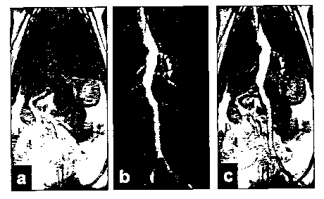
be superimposed, see e.g. fig lc of the accompanying
drawings.
Using standard phase contrast techniques and/or extra
gradient/rf pulse to perform encoding, of spatial or
movement information, flow velocity may be measured.
Also the flow velocity profile may be measured using
through-plane sequences.
By "angiography", we mean any investigation regarding
any angiographic vessel, i.e. the arteries and the
capillary system. In some situations, measurements of
veins may also be covered by the present invention. A
preferred aspect of the invention provides MRA imaging
of the arteries.
CA 02417716 2003-O1-29
WO 02/23209 PCT/GBO1/04085
- 10 -
By the "vascular system", we mean any system of blood-
containing vessels, i.e. arteries, veins and
capillaries.
By "hyperpolarised", we mean polarised to a level over
that found at room temperature and 1T, preferably
polarised to a polarisation degree in excess of 0.1o,
more preferably in excess of lo, even more preferably in
excess of 100.
The hyperpolar,,ised contrast agent should preferably
exhibit a long Tz relaxation time, preferably greater
than 0.5 secs, more preferably greater than 1 sec, even
more preferably than 5 sets.
Suitable MR imaging agents according to the invention,
may contain nuclei such as e.g. 3Li, 13C, lsN, 19F, ~9Si or
31P, as well as 1H, preferably 1H, 13C, lsN, 19F and 31P
nuclei, with 1H, 13C, zsN and 31P nuclei being particularly
preferred. Most especially preferred are 13C nuclei.
As noted above, 1H, 13C, 15N and 31P are the nuclei most
suited to use in a method of the present invention with
13C being most especially preferred. 1H nuclei have the
advantages of being present in high concentration in
natural abundance and having the highest sensitivity of
all nuclei. 13C nuclei are advantageous as the
background signal from hyperpolarised 13C nuclei is very
low and much less than from, for example, 1H nuclei. 19F
nuclei have the advantage of high sensitivity.
Hyperpolarisation of contrast agents comprising 31P
nuclei allows endogenous substances to be used.
Where the MR imaging nucleus is other than a proton
(e.g. 13C or 1sN) , there will be essentially no
interference from background signals (the natural
abundance of 13C and lsN, for instance, being negligible)
CA 02417716 2003-O1-29
WO 02/23209 PCT/GBO1/04085
- 11 -
and the image contrast will be advantageously high.
This is especially true where the MR contrast agent
itself is enriched above natural abundance in the MR
imaging nucleus. Thus the method according to the
invention has the benefit of being able to provide
significant spatial weighting to a generated image.
The MR contrast agent should preferably be artificially
enriched with nuclei (e.g. 15N and/or 13C nuclei) having a
long T1 relaxation time.
The long T1 relaxation time of certain 13C and 15N nuclei
is particularly advantageous and certain MR contrast
agents containing 13C or 15N are therefore preferred for
use in the present method. Preferably the polarised MR
contrast agent has an effective nuclei 13C polarisation
of more than 0.1%, more preferably more than 1.0o, even
more preferably more than 10%, particularly preferably
more than 250, especially particularly preferably more
than 50a and finally most preferably more than 950.
The MR contrast agent is more preferably 13C enriched at
carbonyl or quaternary carbon positions, given that a 13C
nucleus in a carbonyl group or in certain quaternary
carbons may have a Tz relaxation time typically of more
than 2s, preferably more than 5s, especially preferably
more than 30s. Preferably the 13C enriched compound
should be deuterium labelled, especially adjacent the 13C
nucleus. Preferred 13C enriched compounds are those in
which the 13C nuclei are surrounded by one or more non-MR
active nuclei such as O, S, C or a double or triple
bond.
MR contrast agents for use in methods of the present
invention are of the formula (I):
CXq (I)
CA 02417716 2003-O1-29
WO 02/23209 PCT/GBO1/04085
- 12 -
wherein each X is independently D, CD3, CD~OR', S03H,
SOZH, SO~NH2, CONR' 2, COZH and OCHO,
wherein R1 is independently H or Me,
or two of the X groups and the C atom they are attached
to form either the 3-membered ring
~ CD2
i
~CD2
or the 4-membered ring
C , CDY
CD2-Z
wherein Y is D or CD~ORl
and Z i s CDZ , CD ( CD20R1 ) or O .
Shown below as compounds 1-17 are particular examples of
agents suitable for use in the present invention. Such
agents are water soluble, non-toxic, easy to synthesise
and have relatively long T1-values in water, for example
in excess of 60 sets.
For instance, compounds 1 and 2 are found to have T1
values of 95 sets and 133 sets, respectively.
With the exception of compounds 1-3 shown below which
are known from the applicant's own published application
no. WO-A-99/35508, these agents are themselves novel and
form a further aspect of the present invention.
Examples are shown below as compounds 4-17. The agents
can be 13C enriched.
ZTiewed from a further aspect the invention provides a
physiologically tolerable MR imaging agent composition
comprising an MR imaging agent together with one or more
physiological tolerable carriers or excipients, said
CA 02417716 2003-O1-29
WO 02/23209 PCT/GBO1/04085
- 13 -
imaging agent being chosen from one of the compounds in
general formula (I) above, preferably compounds numbered
1-l7 as below, for example compounds numbered 4-17 as
below.
Viewed from a still further aspect the invention
provides the use of a compound from general formula (I)
above, preferably a compound numbered 1-17 as below, for
example a compound 4-17 as below, in a method of the
present invention.
Viewed from a yet still further aspect the invention
provides the use of a compound from general formula (I)
above, preferably a compound numbered 1-17 as below, for
example a compound 4-Z7 as below, for the manufacture of
an MR imaging agent for use in a method of diagnosis
involving the generation of an MR image by MR imaging of
a human or non-human being.
HO'CDz CD3 DZC
DzC--f -NOz DzC--~-CD3 D C~ \CDz
HHO,CDz HHO'CDz HO ~z
HO
1 Z 3
X~CDz X'CDz X'CDz
DZC--~--S03N DZC--~-SOzH D2C-~-SOZNHz
H HO'CDZ H HO~CDz H HO'CDz
4 5 6
X'CDz ~ X'CDz X'C X~CD
D z
DZC-~-CONHz D2C-~--CONHMe
DZC~--CONMezZC~ --COON
H H
HO'CDz NO'CDz H H H Dz
~CDz H
'C
O O
90
X'CDzOCH3 X'CD X 'CD
DZC--~--CDzDzC-~--S03H
O DZC-f --OCHO
~Cp HO GOON HO
H ~CDz
HO
z
11 1~ O
13
D
OH ~H H C HOv
C.OH p C DzC / . OH CD
~
z , OD-CD DzC
Dz CD-CDz D CD
~
Cpz
~
pz DZC-CD ,OH D O-CDz
~pz OH
C
14 pz 16 17
15 X=D or
OH
CA 02417716 2003-O1-29
WO 02/23209 PCT/GBO1/04085
- 14 -
The MR contrast agent should of course be
physiologically tolerable or be capable of being
provided in a physiologically tolerable, administrable
form with conventional pharmaceutical or veterinary
carriers or excipients. Preferred MR contrast agents
are soluble in aqueous media (e.g. water) and are of
course non-toxic.
The formulation, which preferably will be substantially
isotonic, may conveniently be administered at a
concentration .sufficient to yield a 1 micromolar to lOM
concentration of the MR contrast agent in _the imaging
zone; however the precise concentration and dosage will
of course depend upon a range of factors such as
toxicity and the administration route.
Parenterally administrable forms should of course be
sterile and free from physiologically unacceptable
agents, and should have low osmolality to minimize
irritation or other adverse effects upon administration
and thus the formulation should preferably be isotonic
or slightly hypertonic.
It may be convenient to inject simultaneously at a
series of administration sites such that a greater
proportion of the vascular tree may be visualized before
the polarization is lost through relaxation.
The dosages of the MR contrast agent used according to
the method of the present invention will vary according
to the precise nature of the MR contrast agents used and
of the measuring apparatus. Preferably the dosage
should be kept as low as possible while still achieving
a detectable contrast effect. In general, the maximum
dosage will depend on toxicity constraints.
After the polarisation, the hyperpolarised MR contrast
CA 02417716 2003-O1-29
WO 02/23209 PCT/GBO1/04085
- 15 -
agent may be stored at low temperature e.g. in frozen
form. Generally speaking, at low temperature the
polarisation is retained longer and thus polarised
contrast agents may conveniently be stored e.g. in
liquid nitrogen. Prior to administration, the MR
contrast agent may be rapidly warmed to physiological
temperatures using conventional techniques such as
infrared or microwave radiation.
The contents of all publications referred to herein are
incorporated by reference.
..
Embodiments of the invention are described further with
reference to the following non-limiting Examples and the
accompanying drawings.
Example 1
The method of para-hydrogen polarisation transfer as
described in WO 99/24080 (to Nycomed Imaging AS) using a
(PPh3)RhCl catalyst was performed using a malefic acid
dimethyl ester 13C labelled in the carbonyl group, (see
fig 2 of the accompanying drawings). After
. polarisation, the polarised compound was injected as a
- contrast medium into the tail vein of a rat.
The concentration and the polarization of 13C nuclei in
the bolus that was injected into the rat was 150 mM and
approximately 0.30, respectively, and the imaging was
performed, see fig l of the accompanying drawings.
The images shown in fig. 1 were generated using a BioMed
animal scanner operating at 2.4 Tesla. The image shown
in fig 1a is a proton image and has been generated using
a standard spin echo pulse sequence and without the use
of any contrast medium. Pulse sequence parameters were
TR/TE/a = 3.3 ms/1.4 ms/5° and a total scan time of 4:23
CA 02417716 2003-O1-29
WO 02/23209 PCT/GBO1/04085
- 16 -
min. A dose of the hyperpolarised contrast medium was
then generated. The resonance frequency was changed to
the one needed to perform 13C-imaging and a single shot
RARE sequence was executed. The to total scan time was
0.9 sec., the used inter-echo time was 28 ms and the
matrix size was 128 x 32. The resulting image is shown
in fig 1b. The total lack of background signal is
clearly demonstrated. This image was generated as a
projection right through the complete animal
demonstrating the possibility of generating an angiogram
in the same way that when x-rays are used. In fig 1c
the 13C image has been superimposed on the hydrogen
image.