Note : Les descriptions sont présentées dans la langue officielle dans laquelle elles ont été soumises.
29-10-2001 ES000030~
CA 02419589 2003-O1-31
ORTHOTOPIC TOTAL ARTIFICIAL HEART
FIELD OF THE INVENTION
This invention pertains to medical prostheses, and in particular, to a total
artificial heart. It has been conceived to satisfy the current need for
creating a new
and original design to achieve a Total Artificial Heart in order to replace a
native
sick heart in its terminal stage or as a bridge to cardiac transplantation or
to be
used after a heart transplantation failure.
BACKGROUND OF THE INVENTION
1o At present, when there is a patient with a serious heart disease, which for
different reasons is nonreversible, cardiac transplantation is considered as
the
solution, provided that the patient gets a donor. However, in the United
States, for
example, there are about 60,000 patients per year in this situation and only
about
6% to 10% get a transplantation due to the current difficulties to find an
adequate
heart donor.
A Total Artificial Heart (TAH) is recognized as progress in cases of such
extreme cardiac failure. The present generation of this kind of devices
includes
the use of different models. In addition, there are partial circulatory
assistance
devices in use, generally called Left Ventricular Assistance Systems (LVAS).
2o Under extreme haemodynamic failure circumstances, these devices are
used at present as a bridge to transplantation. They allow the patient to be
kept
alive while the patient awaits for the appropriate donor, preventing a serious
systemic damage caused by the progressive deterioration of the haemodynamia,
which can later compromise the viability of other organs if the patient gets a
transplantation.
However, the present Total Artificial Heart generation has had problems.
Even though these devices have kept patients alive under extreme
circumstances,
they have not been able to provide them with an acceptable quality of life.
Most important, due to the difference between the sizes of the current
3o generation devices and the space inside the mediasfinum available for the
current
AMENDED SHEET
29-10-2001 ES000030a
CA 02419589 2003-O1-31
-2-
models, i.e. the so called lack of anatomical fit, many of these devices do
not place
the artificial ventricular and other elements necessary for their operation in
an
orthotopic position. Several elements are placed outside the body and coupling
of
parts located both inside and outside of the human body is made through the
skin.
Several pathological phenomena occur, such as local infections that are later
transformed into more serious infections, ascendent infections, skin
ulcerations
and countless problems for the patient and his/her quality of life. An example
of
their limitations is the need for the patient to be connected to a pneumatic
console.
In addition, by placing these parts outside the chest, risks and problems are
increased during operation; it also causes surgical complications and problems
during the postoperative period such as bleeding, hematomas, infection, and
compressions.
Furthermore, due to the reduced space available within the chest, some of
the devices of the present generation do not have an adequate size to produce
a
~5 good final diastolic volume. Hence, often times, in order to obtain an
adequate
blood flow rate, these devices resort to a significant increase of the heart
frequency which causes additional turbulence as a result of and increase in
the
blood flow linear velocity. This situation can be the cause of more serious
haematological complications, such as haemolysis and bleeding and cause a
faster
deterioration of the materials that form these devices. On these grounds, a
better
utilization of the space available inside the mediastinum to achieve a
significant
increase on the diastolic volume would be highly desirable.
Another type of haematological complications associated with devices of
the present generation is thrombosis and embolisms. In some of these devices
the
internal walls of the cavities through which blood circulates have areas with
stasis,
corners or boundaries between the different materials of their surfaces and
with
stitches between them, all of which created a very high embolism risk.
The aforementioned artificial devices present haemostatic complications
such as bleeding, occurnng because the blood has to go through long circuits
of
3o rigid prosthetic tubes with many stitches at each end. These artificial
prosthetic
AMENDED SHEET
29-10-2001 ES000030~
CA 02419589 2003-O1-31
- -3-
tubes do not respond to a need to increase the blood flow rate like native
vessels
do in reflex mode, i.e. by greatly increasing their diameter. This deficiency
causes
a larger increase in blood presswe which fiuther stresses the above mentioned
stitches and causes the present generation Total Artificial Hearts to operate
under
more stringent conditions.
Another important problem of these devices is their limitation to
compensate the different blood volumes physiologically handled by the
pulmonary
circuit and the systemic circuit. To alleviate this situation, the swgeon has
to
create a communication between the two circuits during the implantation
swgical
1o procedwe, usually making an interauricular communication. However, the size
of
the swgical opening, in particular and the efficacy of this procedwe in
general, is
often questioned because of frequently occurring systemic or pulmonary
hemodynamic congestions.
With reference to the prior art. USPTO No. 4.863,461, titled Artificial
Ventricle, describes an artificial heart known as Jarvik-7, to be implanted in
an
orthotopic position. This device has two artificial ventricles which have
their
ventricle-artery connections in the same position as in the native heart. It
has a
pneumatic mechanism, with connections to an exterior control panel. USPTO
Document No 5,674,281 titled Artificial Heart Braking System describes a
device
2o with two blood chambers and in between such an electrical actuator to
compress
the chambers. Document WO 98/41247 discloses two ventricular chambers which
are arranged so as to form an upside-down V, between which are placed two
actuators and the space for the actuating fluid.
Accordingly, there is still a need for an artificial heart without the
attendant disadvantages of conventionally available artificial hearts.
SUMMARY OF THE INVENTION
In general, the present invention comprises an artificial heart that may be
implanted in orthotopic position in a circulatory system of a living being,
e.g.,
AMENDED SHEET
29-10-2001 tSUUUU;iO~
CA 02419589 2003-O1-31
..4-
mammals, that preferrably and anatomically fits within a mediastinum space
created by removing the two native ventricles.
Said artificial heart comprising:
-one right blood chamber, said right blood chamber having an elongated
flow essentially directed up and back, said right blood chamber having one
right
inlet port for blood to enter, said right inlet port having means for
attachment to
the right atrium,
-one posterior outlet port for blood to exit said right blood chamber, said
posterior outlet port being located above and behind the right inlet port,
said
1o posterior outlet port having means for attachment to the main pulmonary
artery,
said posterior outlet port either including or being adjacent to the valve for
the
main pulmonary artery,
-one left blood chamber, said left blood chamber having an elongated flow
essentially directed up and to the right, said left blood chamber having one
left
inlet port for blood to enter, said left inlet port having means for
attachment to the
left atrium,
-one anterior outlet port for blood to exit said left blood chamber, said
anterior outlet port being located above and to the right of the left inlet
port,
approximately at the same height as and in front of said posterior outlet
port, said
2o anterior outlet port having means for attachment to the aorta artery, said
anterior
outlet port either including or being adjacent to the valve for the aorta
artery,
the spatial arrangement between said blood chambers being such that,
when they are simultaneously fully expanded, a part of the right blood chamber
(i.e., which projects onto the anterior thoracic wall and coincides with the
projection onto said anterior thoracic wall of a corresponding part of the
left blood
chamber) is posterior to said corresponding part of the left blood chamber.
In particular, the artificial heart of the instant invention comprises an
assembly of two artificial ventricles or blood chambers, each having an inlet
and
an outlet. The incoming blood from the right auricle enters the right blood
3o chamber through the right inlet port and exits it through the posterior
outlet port.
AMENDED SHEET
29-1 U-2001 tSUUUU;iU~
CA 02419589 2003-O1-31
-
The incoming blood from the left auricle enters the left blood chamber through
the
left inlet port and exits it through the anterior outlet port.
The unique spatial arrangement of both blood chambers, inlet ports and
outlet ports give the instant invention a radically better utilization of the
space
available inside the mediastinum after having surgically removed both native
ventricles and having surgically liberated both great vessels, main pulmonary
artery and aorta artery. An important advantage of the instant invention
consists
on the location of the posterior outlet port, which is placed posterior and
above to
the right inlet port. This specific placement enables the utilization of the
space
to available above both auricles for blood pumping purposes, which otherwise
would
be unutilized. This arrangement places the posterior outlet port in the space
normally occupied by the initial sector of the aorta artery. From that native
posterior position, the aorta artery travels upwards and forward to the right
to exit
the anterior mediastinum. The anterior outlet port is placed approximately at
the
same height to and in front of the posterior outlet port. Hence, the lower
sectors
of the aorta artery and main pulinonary artery are surgically liberated and
transposed with respect to their antero-posterior position so as to connect
them to
their corresponding outlet ports. If additional space is desired, the initial
sector of
both great vessels will be removed and both outlet ports will be placed at a
higher
2o position, close to a plane located at the level of the right pulmonary
artery and the
mid sector of the ascending aorta artery.
Another important advantage of the instant invention consists on the flow
and location of both blood chambers which enables a significantly better
utilization of the space available in the mediastinum. In their fully expanded
position both blood chambers reach the anterior thoracic wall. The right blood
chamber has an elongated flow, essentially directed up and back. The left
blood
chamber has an elongated flow, essentially directed up and to the right. The
aorta
artery, in its upward path, occupies an anterior position at its crossing of
the right
pulmonary artery. Therefore, the space available inside the mediastinum is
3o significantly better utilized in the instant invention by keeping the
pathway of the
AMENDED SHEET
29-10-2001 t=SUUUU;iUa
CA 02419589 2003-O1-31
- -6-
blood coming from the right auricle into the main pulmonary artery into an
posterior position with respect to the pathway of the blood coming from the
left
auricle into the aorta artery. Systemic and pulmonary pathways do not comply
with this requirement in the native ventricles and the previous art in the
field of
Total Artificial Hearts has not changed it either. The instant invention
changes
this native disposition, placing the right blood chamber always behind the
left
blood chamber, when their projections onto an anterior thoracic wall coincide.
In
doing so the instant invention is able to utilize the space available for
pumping
blood and not merely transporting it through artificial tubes to reach its
intended
1 o destination.
The volume available for pumping in the instant invention is fiu~ther
increased by placing the outlet ports close to the valves leading to the great
vessels. Therefore, because of the described arrangement of the different
components comprising the instant invention, the pumping volume provided by
the blood chambers actually reach higher in the mediastinum than in the
previous
art.
The better use of the space available in the mediastinum enables the Total
Artificial Heart of the instant invention to have a higher final diastolic
volume of
the blood chambers, obtaining in this way ejected volumes large enough to
2o achieve an acceptable blood flow rate without a significant increase of the
heart
frequency, and thereby reducing both hemolysis and mechanical wear of movable
parts.
Furthermore, in the preferred embodiment of the instant invention and in
some variant, this Orthotopic Total Artificial Heart can be completely placed
inside the mediastinum, i.e. the blood chambers, the driving mechanism, for
example the compressing mechanism, and power source. In this manner, the
instant invention can be an integrated, "one-piece" system.
The significantly better space utilization of the instant invention is used
for
at least one of two purposes: a) To place the driving mechanism for the Total
3o Artificial Heart inside the mediastinum; b) To increase the diastolic
volume of
AMENDED SHEET
29-10-2001 ES000030~
CA 02419589 2003-O1-31
each blood chamber. This new design realizes a fundamental need expressed by
the medical community for the necessary anatomical fit of the Total Artificial
Heart within the available and restricted space of the mediastinum.
Due to the different structural layout of the Total Artificial Heart of the
instant invention, the following important improvements are made:
1- Keeping the pathway of the blood coming from the right auricle into the
main pulmonary artery in a posterior position with respect to the pathway of
the
blood coming from the left auricle into the aorta artery
2- Outflow tract paths of the artificial ventricles are placed closer to the
1o circulatory system that is going to be irrigated, therefore not needing
prosthetic
' tubes to reach the corresponding arteries.
3- The artificial ventricles are placed in a higher position inside the
mediastinum.
4- In the preferred embodiment and other variants of the instant invention,
both the outer compressing chamber and the power source are also placed inside
the mediastinum.
Furthermore, if necessary, additional space can be conveniently created by
resecting the initial sector of the large arterial trunks.
An important hemodynamic and hematological advantage of the instant
2o invention is that, by placing the blood chambers' outflow near the systemic
and
pulinonary vascular regions, it no longer requires the use of prosthetic tubes
at the
outflow of these blood chambers. This characteristic provides the instant
invention with the great advantage of being directly connected with the
vascular
systems through native vessels which respond to increased blood flow with the
vasodilatation autonomous reflex response. Hence, no increased pressures are
needed to get a higher blood flow, thereby reducing the pressure on the walls
of
the blood chambers and the turbulence and associated liquid shear stress, all
of
which greatly reduces the subsequent damage that this causes to blood cells
and to
the life of the Total Artificial Heart itself.
AMENDED SHEET
29-10-2001 ES0000302
CA 02419589 2003-O1-31
The artificial ventricles of the instant invention are one-piece blood
chambers that have two non-thrombogenetic characteristics, their morphology
and
their surfaces in contact with the blood. The blood chambers have an elongated
and upward flow, having neither stasis areas nor corners or boundaries between
dissimilar materials; neither they have stitches among them.
Another advantage of the instant invention is the non-thrombogenic walls
of the blood chambers. These inner walls are made with biological surfaces,
soft
and flexible, which protect blood cells and red corpuscles against cellular
traumatism, therefore avoiding hemolysis. In addition, the cellular damage is
reduced in the instant invention because blood is pumped by the action of
forces
homogeneously distributed and approximately concentric and also because the
blood chambers are made of a single material without comers, borders or
stitches,
and without prosthetic materials or tubes of a more or less fixed diameter at
the
outflow of the blood chambers.
Yet another advantage of the instant invention also provides for the
independent variation of the discharging volumes of each blood chamber. Such
independent handling of the volumetric flow rates for each blood chamber
enables
the compensation of the imbalance in the blood flow circulating through the
pulinonary circuit and the systemic circuit. Physiological differences and
shunts
2o between these circulatory circuits shall be compensated in such a way that
there
shall be no need for creating surgical shunts.
Additional objects and attendant advantages of the present invention will
be set forth, in part, in the description that follows, or may be learned from
practicing or using the present invention. The objects and advantages may be
realized and attained by means of the instrumentalities, features and/or
combinations particularly pointed out in the appended claims. It is to be
understood that the foregoing general description and the following detailed
description are exemplary and explanatory only and are not to be viewed as
being
restrictive of the invention, as claimed.
AMENDED SHEET
29-10-2001 E50UUU~0
CA 02419589 2003-O1-31
-9-
BRIEF DESCRIPTION OF THE DRAWINGS
The accompanying drawings, which are incorporated in, and constitute a
part of the specification, illustrate embodiments of the present invention
and,
together with the description, serve to explain the principles of the present
invention.
Fig. 1 Schematic representation of the systemic-pulmonary circulatory
system of a human being.
Fig. 2 General schematic representation of the preferred embodiment of
the electro-hydraulic variant of the instant invention.
Fig. 3 Anterior view of the preferred embodiment of the electro-hydraulic
variant of the instant invention, implanted in the systemic-pulmonary
circulatory
system of a human being, in which the initial sector of the large vessels is
removed.
Fig. 3.A Clarification of Fig. 3, showing an anterior view of the preferred
embodiment of the instant invention, in a diastolic or filling position of the
blood
chambers, with the driving mechanism implanted in the systemic-pulmonary
circulatory system of a human being, in which the initial sector of the large
vessels
is removed.
Fig. 3.B Clarification of Fig. 3, an anterior view of the preferred
2o embodiment of the instant invention, in which an ejection or emptying
position of
the blood chamber, is shown.
Fig. 3.C Representation of an electro-hydraulic variant of the instant
invention, in which the compressing effect is produced by two lateral moving
surfaces, implanted in the systemic-pulmonary circulatory system of a human
being, where the initial sector of the large vessels is removed.
Fig. 3.D Representation of a pneumatic variant of the instant invention,
where the compressing effect is produced by the introduction of a compressing
fluid inside the outer compressing chamber, which has senu-rigid walls.
Fig. 4. Inner and anterior view of the two assembled blood chambers of the
instant invention seen in an ejection or emptying position.
AMENDED SHEET
29-10-2001 ~~uuuu,~u~
CA 02419589 2003-O1-31
- -10-
Fig. 5. Inner and anterior view of the two assembled blood chambers of the
instant invention seen in a diastolic or filling position.
Fig. 5.A Inner and right side view of the two assembled blood chambers
of the instant invention shown in a diastolic or filling position.
s Fig. 6 Upper, rear and left side view of the outer compressing chamber of
the electro-hydraulic variant of the instant invention.
Fig. 6.A Clarification of Fig. 6, showing an upper, rear and left side view
of the outer compressing chamber.
Fig. 6.B Upper, rear and left side view of the outer compressing chamber
to of the electro-hydraulic variant of the instant invention, showing
displacement of a
moving surface.
Fig. 7 Right side view of the outer compressing chamber of the electro-
hydraulic variant of instant invention.
Fig. 7.A Clarification of Fig. 7 showing the right side view of the outer
~5 compressing chamber of the electro-hydraulic variant of the instant
invention.
Fig. 7.B Right side view of the outer compressing chamber of the electro-
hydraulic variant of the instant invention, showing displacement of a moving
surface.
Fig. 8 Schematic comparison between the aorta artery and the main
2o pulmonary artery before and after the surgical removal of their initial
sectors.
Fig. 8.A Schematic comparison of the spaces created after having
surgically removed both ventricles, before and after the surgical removal of
the
initial sectors of the aorta artery and the main pulmonary artery.
Fig. 8.B View of the free spaces created by removal of the initial sector of
25 the large vessels.
Fig. 9. View of the moving surface of the compressing mechanism of the
preferred embodiment of the instant invention.
Fig. 10. Anterior view of an electro-mechanic variant of the instant
invention, showing the compressing mechanism directly acting on the two
3o assembled blood chambers.
AMENDED SHEET
29-10-2001 ES000030~
CA 02419589 2003-O1-31
-11-
Fig. 11. View of the upper part of the cross section A-Al in Fig 10, where
a set of two lateral moving surfaces is outlined.
Fig. 12. Anterior view of another electro-mechanic variant of the instant
invention, showing an independent double-compressing mechanism directly acting
on each blood chamber.
Fig. 13. View of the upper part of the cross section B-B1 in Fig 12, where
two sets of two lateral moving surfaces is outlined.
DETAILED DESCRIPTION OF THE INVENTION
1 o This invention is herein described in detail, as a non-limiting model and
as
. the preferred way to develop it at present. It is also illustrated in the
pictures
attached hereto.
At present, the specific and preferred way to build the Orthotopic Total
Artificial Heart, according to this invention, is the one illustrated as a
model in the
pictures attached hereto. Notwithstanding, the present invention may be
subject to
different shape and size modifications and the present specifications are not
intended to limit the invention to the particular shapes and/or sizes herein
described. On the contrary, the intention is to cover all modifications and
alternative executions that are within the subject matter and the purpose of
the
2o invention in accordance with the claims attached hereto.
Moreover, as there shall be several modifications and changes that shall be
analyzed by the technicians in this field, we do not wish to limit the
invention to
the exact construction or operation described herein. Therefore, any and all
equivalent modifications shall be considered as included within the scope of
the
instant invention.
T7~e novelu: of the present invention is the transposition of the outlet
pathvju~-s and the. outlet ports of the t~~lo assembled blood chambers in tire
design
of a or-rhotopic total artificial heart. It is placed into the chest of a
lioirtg being bv°
surgery. irr the anatomical space called unter~ior rnediastirrum. The,
rrrediastinum is
irr tire chest, between both lrrrrgs. Its anterior central =one is culled the
arrterior
AMENDED SHEET
29-10-2001 ES0000302
CA 02419589 2003-O1-31
-12-
mediastirzum and belongs to the pericurdic cmjit~:, in ~:hich the heart unrl
the low
sector of the great vessels, Inrown as tire aorta nrten~ and the nrain
pulmorrun~
arter3=, are. located. Tlre native heart of a living beirrg, as it is seen in
the scheme
of the fcg. l, presents hvo native ventricles. The right ventricle 101 elects
the
blood to the lungs 103 throzrglr the nrairr pulnronar~: nrten° 12, un~l
the left
ventricle 102 that ejects the blood to the bodl: 104 tlzrorsgh the aorta
amen° 11. In
the nornral anatomy of the native heart, the outlet pahtNJuy acrd the. outlet
of the
pulmonary valve of the right ventricle, have an anterior position with regard
to
the outlet pathway and the outlet of the aorta value of tlae left ventricle.
The
present Inl'erJtlOlr TS designed to be placed in the snare space us the native
heart.
therefore in un ortlrotopic position, after the remooal of the tlvo ncrtive
ventricles
and the lower sector- of the uoruu arten.~ and rrruin pulrnonar~- urter~~ haoe
been
surgically liberated, and their trarzsposed or irrver~ted, o-ith respect to
their native
untero Posterior position.
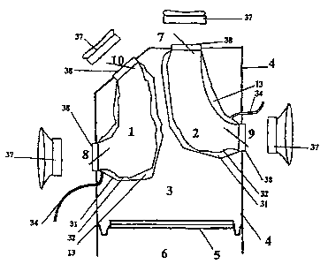
The electro-hydraulic preferred embodiment of the Orthotopic Total
Artificial Heart, as shown in the schematic representation of Fig. 2,
comprises an
outer compressing chamber 4, a compressing fluid 3, two assembled blood
chambers, right 1 and left 2, and a mechanism for independently varying their
discharging volumes.
2o The outside wall of the instant invention is an outer compressing chamber
4, the external shape of which has an anatomy which agrees with the
rnediastinal
space that it shall occupy, as shown in Figs. 3, 3.A, 3.B, 6, 6.A, 6.B, 7, 7.A
and
7.B, with an oval or kidney or pyramidal shape, with a upper vertex and a
lower
and left base. It will occupy the space called anterior mediastinum.
2s The outer compressing chamber 4 of the preferred embodiment of instant
invention is constituted by different sectors, as observed in Fig. 3 and 3.A:
a mid
sector 14, which is placed in front of the right auricle 17 and left auricle
18; an
upper sector or vertex 15, which is raised up to a horizontal plane crossing
at the
right pulmonary artery's lower border level 12, and is extended in the front
up to
3o the breastbone 24 (see Fig. 7.A and 7.B); a lower sector or base 16 which
is
AMENDED SHEET
29-10-2001 ES000030~
CA 02419589 2003-O1-31
-13-
extended up to the diaphragm 21 and the area of the native heart end,
occupying
the supradiaphragmatic free space 33 (see Fig. 8.B) created by removing the 2
native ventricles. In all these figures of the preferred embodiment, Figs. 3,
3A,
3B, 4, S, SA, the initial sector of both great vessels has been surgically
removed,
as seen in Fig. 8B.
The outer compressing chamber 4, as shown in a frontal view in Figs. 3,
3.A and 3.B, has its left edge 22 moving from left to right as it ascends
distancing
itself from the diaphragm 21 and reaches close to the left edge of the aorta
artery
at its upper part. The right edge 23 of said outer compressing chamber 4
travels
up more or less vertically from the diaphragm Level 21.
The outer compressing chamber 4's depth is extended from the right 8 and
left 9 auricular-ventricular inlet ports to the breastbone 24, as shown in
Figs. 7,
7.A and 7.B.
The rear side, in its upper and right sector, above the right inlet port 8 of
i 5 the right blood chamber 1, as shown in Figs. 6, 6.A, 6.B, 7, 7.A and 7.B,
presents
a geometric structure, a cone trunk 25, which creates a growing and oblique
protuberance, to the upper and to the rear parts. In the upper edge of this
cone
trunk 25, the posterior outlet port 10 of the right blood chamber 1 is located
and
connected directly to the pulmonary circulatory circuit.
2o The outer compressing chamber 4, as shown in the Fig. 2 outline, has four
holes, two inlets and two outlets, for the connection of two assembled blood
chambers to the patient's circulatory systems,. The inlet ports 8 and 9 are
placed
on the rear side of the present invention, as shown in Figs. 5.A, 6, 6.A, 6.B,
7, 7.A
and 7.B. The right inlet port 8 of the right blood chamber 1 receives blood
from
25 the right auricle 17. The left inlet port 9 of the left blood chamber 2,
receives
blood from the left auricle 18, as shown in Fig. 3.A and SB.
The outlet ports 7 and 10 are located in the upper side of outer
compressing chamber 4, as shown in Figs. 3, 3.A, 3.B, 6, 6.A, 6.B, 7, 7.A and
7.B.
The anterior outlet port 7 of the left blood chamber 2, connects through the
3o neoentrance 26 (see Figs. 8, 8.A and 8.b) to the systemic circulatory
circuit. The
AMENDED SHEET
29-10-2001 ESUUUU;iU~
CA 02419589 2003-O1-31
-14-
posterior outlet port 10 of the right blood chamber 1, is located in the upper
part
vertex of the cone trunk structure 25, which creates a protuberance in the
rear side
of the outer compressing chamber 4, and is connected through the neoentrance
27
with the pulmonary circulatory circuit, as shown in Fig. 8, S.A and 8.B.
After removing the initial sector of the great vessels, neoentrance 26 and
neoentrance 27 are in a higher position than that of the aortic valve 28 and
the
pulmonary valve 29, as shown in Fig. 8 and 8.A.
As shown in Figs. 3, 3.A, 3.B, 4, 5 and S.A, posterior outlet port 10 of the
instant invention is behind the anterior outlet port 7, that is they are in an
inverted
position compared to the outlet valves of the native ventricles.
Inside the outer compressing chamber 4 of the preferred embodiment of
the instant invention, as shown in the outline represented in Fig. 2, there
are the
compressing fluid 3, and two structures with the shape, size, walls and
connections of the instant invention, the right blood chamber 1 and the left
blood
~ 5 chamber 2. They occupy the whole inner volume of this outer compressing
chamber 4, which is sealed.
The compressing fluid 3 (for example glycerin), as shown in the schematic
representation of Fig. 2, occupies the volume defined by the inner side of
outer
compressing chamber 4, the moving surface 5, and the external walls 31 of both
2o assembled blood chambers. This compressing fluid 3 is used to transfer the
driving force of the moving surface 5 to the external walls 31 of both blood
chambers. The compressing fluid contained inside the outer compressing chamber
4, acts in such a way that when the moving surface 5 is in a filling or
diastolic
position, as shown in figures 3, 3.A, 5 and S.A, it allows the right 1 and the
left 2
25 blood chambers to reach each of them a volume of 90 cc, when the blood
enters
through the right inlet port 8 of the right blood chamber 1 and through the
left
inlet port 9 of the left blood chamber 2. Both assembled blood chambers shall
have their respective outlet ports 10 and 7 closed.
While the moving surface 5 moves 3 centimeters forward inside the outer
3o compressing chamber 4, reaching its maximum blood ejection or systolic
position,
AMENDED SHEET
29-10-2001 tSUUUUau~
CA 02419589 2003-O1-31
-15-
as shown in Figs. 3.B and 4, it transfers the forces received from the driving
mechanism to the compressing fluid 3, which shall compress the external wall
31
of the right 1 and left 2 blood chambers producing the emptying effect of
their
inner volume, obtaining in such a way the expulsion or ejection of the blood
contained inside them, through the posterior outlet port 10 of the right blood
chamber 1, and the anterior outlet port 7 of the left blood chamber 2. Both
two
assembled blood chambers shall have their respective inlet ports closed.
The right blood chamber 1 is a soft and flexible sack created to pump the
blood, and is placed in front and above the position of the right auricle, and
l0 located to the right in the outer compressing chamber 4, as shown in
figures 3, 3
A, 3 B, 4, 5 and 5 A. It is composed of two soft and flexible walls, its inner
cavity
has no comers, stitches or boundaries between the different materials, as its
inner
biological membrane 32 shall be totally constituted by a single-piece pig
pericardium for example. Its external wall 31 is a synthetic one, made of
Pebax
3533, for example. This right blood chamber 1 is connected through its right
inlet
port 8 to the right auricle 17 and through its posterior outlet port 10 to the
pulmonary circulatory system. Said right blood chamber 1, when fully expanded,
reaches the anterior thoracic .wall and has an elongated flow essentially
directed up
and back. As shown in Figs. 3, 3 A, 3 B, 4, S and 5 A, this right blood
chamber 1
2o shall have a significant difference to the anatomic structure of the native
right
ventricle. Its blood flow pathway goes up and back from the right inlet port
8,
almost in a straight line as shown in Figs. 3, 3.A, 3.B, 4, S and 5 A, until
reaching
and connecting directly to the neoentrance 27 of the pulmonary circulatory
system,
which occupies a posterior position inside the mediastinum, as shown in the
schematic comparison of Figs. 8 and 8.A.
Therefore, this right blood chamber 1 avoids the loop originated in the
development of the embryonic circulatory tube, or downward path that the blood
makes inside the native right ventricle, by entering through the tricuspid
valve and
descending to the diaphragm 21. Thereafter the blood flow from the native
right
3o ventricle 101 takes an upward and leftward path, crossing in front of the
blood
AMENDED SHEET
29-10-2001 ES000030~
CA 02419589 2003-O1-31
-16-
flow from the outlet of the left native ventricle 102, in which position the
outlet
pathway of the right native ventricle 101 connects to the pulmonary artery 12
via
the pulmonary valve, which is located in front of the valve of the aorta
artery 11.
The pulmonary artery 12 thereafter takes a backward direction.
s The supradiaphragmatic space 33 of the anterior mediastinum shown in
Fig. 8.B, is reserved to place the driving mechanism 6 located in the base
sector 16
of the preferred embodiment of the instant invention, as shown in Figs. 3, 3.A
and
3.B.
The left blood chamber 2 is also a soft and flexible sack created to pump
1o blood, and is placed in front and above the position of the left auricle
located to
the left in the outer compressing chamber 4. As shown in Fig. 3, 3 A, 3B, 4, 5
and
A, it is composed of two soft and flexible walls, its inner cavity has no
corners,
stitches or boundaries between different materials since the inner biological
membrane 32 is totally composed by a single-piece pig pericardium for example.
~ s Its external wall 31 is a synthetic one, made of Pebax 3533, for example.
From its
left inlet port 9 or mitral valve, which connects it to the left auricle 18 as
shown in
Figs. 3, 3.A, 3.B, 4, 5 and S.A. Said left blood chamber 2, when fully
expanded
reaches the anterior thoracic wall and has an elongated flow essentially
directed up
and to the right in the anterior mediastinum, having its outflow pathway in
front of
2o the right outflow pathway. The position of the anterior outlet port 7 is
shown in
Figs. 3, 3.A, 4, 5 and S.A and is placed in front of the posterior outlet port
10. In
this way, the left blood chamber 2 allows the blood flow to be almost straight
and
in an anterior, upward and right direction, to the systemic circulatory
system.
These two assembled blood chambers of the instant invention, right 1 and
25 left 2, soft and flexible, have a double membrane wall, as shown in the
schematic
representation of Fig. 2, and have an inner cavity the volume of 90cc each.
However, the discharging volume of each blood chamber can be independently
varied. To decrease or increase the final diastolic volume of each blood
chamber
independently, the preferred embodiment of the instant invention has a
mechanism
3o for independently varying discharging volumes. In the interstitial space
between
AMENDED SHEET
29-10-2001 ES0000302
CA 02419589 2003-O1-31
-17-
the inner walls 32 and the external ones 31 of each blood chamber a fluid,
called
interstitial fluid 13, is introduced through a catheter 34 of the mechanism
for
independently varying discharging volumes, as shown in the outline of Fig. 2.
When the space between the inner wall 32 and the external wall 31 is filled
with
the interstitial fluid 13, the inner volume of each blood chamber is reduced.
When
the intersticial liquid 13 is removed by means of the catheter 34 of the
mechanism
for independently varying discharging volumes, the final diastolic volume of
each
blood chamber is increased independently. The interstitial fluid 13 may be,
for
example, glycerin. This mechanism for independently varying discharging
1 o volumes is handled through the catheter 34 as shown in the schematic
representation of Fig. 2, and inserted, for example, via a central vein. This
catheter 34 is introduced into the outer compressing chamber 4, next to the
inlets 8
and 9 of the blood chamber, from the neck veins and it is connected to the
external
wall 31. In this way, during the implantation period and the postoperative
period,
the physician can vary the interstitial volume of each two assembled blood
chambers, being able to independently vary their final diastolic volume, to
achieve
a blood flow in the systemic circuit and in the pulmonary circuit, according
to the
physiological needs of each patient and the specific operation of each device.
An electro-hydraulic variant in the design of the outer compressing
2o chamber 4 of the instant invention, having two lateral moving surfaces 39
to
produce the compressing effect on the two assembled blood chambers, is shown
in
Fig. 3C. In this outer compressing chamber 4, the lateral moving surfaces 39
are
shown in a diastolic position. These two lateral moving surfaces 39 when
displaced to the center of the outer compressing chamber 4 increase the
pressure
of compressing fluid 3, which effects the compressing action of the two
assembled
blood chambers, right 1 and left 2.
Another variation in the design of the outer compressing chamber 4 of the
instant invention is shown in Fig. 3D. To produce the compressing effect on
both
two assembled blood chambers, a variation of the volume of the compressing
fluid
40A inside the outer compressing chamber 4 is produced. A variation on the
AMENDED SHEET
29-10-2001 ES000030~
CA 02419589 2003-O1-31
-18-
volume of the compressing fluid 40A is produced, for example, by gas injection
and extraction within the outer compressing chamber 4; this outer compressing
chamber 4 is characterized by its semi-rigid wall, with low volume change upon
changes in the internal pressure upon gas injection and extraction. Fig. 3D
shows a
schematic representation of the outer compressing chamber 4 with the same
layout
for the right 1 and the left 2 blood chambers and with one connection for a
tube 40
which connects to a source that introduces and extracts gas. Fig. 3D shows the
compressing fluid 40 A in the outer compressing chamber 4 which, in this case,
is
a gas.
to An electro-mechanic variation of the compresing mechanism of the instant
invention is that in which the blood pumping function is effected by a
different
driving mechanism as shown in Figs. 10, 11, 12 and 13. The right blood chamber
1, is connected on the back to its respective right inlet port 8 through which
it
receives the blood from the right auricle. The left blood chamber 2 is
connected
~5 on the back to its left inlet port 9 through which it receives the blood
from the left
auricle. In the front of inlet ports 8 and 9, as shown in Figs. 11 and 13,
both
assembled blood chambers are extended up to the breastbone 24, and this part
of
both blood chambers are placed parallel, in a somewhat oblique direction to
the
left.
2o In Fig. 10, we can see the simultaneous, joint and direct compressing
action produced by two lateral moving surfaces 41, on the right lateral wall
of the
right blood chamber 1, and on the left lateral wall of the left blood chamber
2,
moved by the driving mechanism 6. Fig. 11 is a cross-sectional view A-A1 of
Fig
10, which is at the level of inlet ports 8 and 9 of the right 1 and left 2
blood
25 chambers. Here we can see that their front sectors, close to the breastbone
24, are
placed with their inner lateral sides in a parallel position and together, and
are
supported by each other to receive the joint lateral compressing effect of the
two
lateral moving surfaces 41.
An electro-mechanic variation of said compressing mechanism is shown in
30 Fig. 12 and Fig. 13, in which the direct compressing action is produced by
one
AMENDED SHEET
29-10-2001 ESUUUU3U~
CA 02419589 2003-O1-31
-19-
pair of lateral moving surfaces 42 for each blood chamber, each pair acting
independently on the lateral walls of said blood chambers. Said moving
surfaces
42 are moved by the driving mechanism 6.
Yet another variation of the instant invention consists on an arrangement
comprising two outer compressing chambers, each one enclosing its respective
blood chamber, said outer compressing chambers having one or more moving
surfaces. Said outer compressing chambers can be separate from each other or
share a common wall, said common wall becoming then a septum dividing the
inner spaces of each outer compressing chamber. Each said outer compressing
to chamber has at least two openings; one of the openings coincides with the
inlet
port and another opening coincides with the outlet port through which blood
comes in an out respectively. The space enclosed between each outer
compressing
chamber and its respective blood chamber is filled with a compressing fluid.
Said
compressing fluid's function is to transmit the forces exerted on the movable
~5 surfaces of the outer compressing chamber into the blood chamber which is
soft
and flexible. Hence, a reduction in the volume effected on the outer
compressing
chamber by the compressing mechanism results in a concomitant reduction in the
inner volume of the blood chamber. Said compressing mechanism is driven by at
least one power source, also located inside the mediastinum. Said reduction in
the
2o inner volume of each blood chamber ejects the blood contained by it. This
assembly enables the independent management of systemic and pulinonary flaw
rates with all the advantages outlined above.
Still another variation of the instant invention consists on an arrangement
comprising two outer compressing chambers. Said outer compressing chambers
25 can be separate from each other or share a common wall, said common wall
becoming then a septum dividing the inner spaces of each outer compressing
chamber. Each said outer compressing chamber encloses its respective blood
chamber, said outer compressing chambers being characterized by their semi-
rigid
wall, with low volume change upon changes in internal pressure occurring
during
3o use. Each said outer compressing chamber has at least three openings; one
of the
AMENDED SHEET
29-10-2001 t5000030~
CA 02419589 2003-O1-31
' -20-
openings coincides with the inlet port and another opening coincides with the
outlet port through which blood comes in an out respectively. The space
enclosed
between each outer compressing chamber and its respective blood chamber is
filled with a compressing fluid. Each said outer compressing chamber has at
least
one opening through which compressing fluid is added or withdrawn into each
outer compressing chamber. The cyclic addition and withdrawal of compressing
fluid in and out of each outer compressing chamber effects the compression and
expansion of the enclosed two assembled blood chambers, which are soft and
flexible. The preferred compressing fluid is a gas, more preferably an inert
gas.
1o The reduction in the inner volume of each blood chamber ejects the blood
contained by it. This assembly enables the independent management of systemic
and pulinonary flow rates with all the advantages outlined above.
Another variation of the instant invention refers to a variation of the
independently varying discharging volumes mechanism. This mechanism has
been designed in order to be able to vary independently the volume ejected by
each blood chamber. This variation consists on two assembled blood chambers
with different volumes. For example, the right blood chamber 1 has an inner
volume of 85 cc, and the left blood chamber 2 has an inner volume of 95cc. The
right blood chamber 1 ejects blood to the pulinonary circuit, which pumps
against
an average pressure of 50 to 25 mm of Hg. This pressure is lower than the
pressure at which the left blood chamber 2 ejects to the systemic circuit,
which has
an average arterial pressure of 120 to 80 mm Hg. Due to the different
pressures at
which each of the blood chambers ejects, being the pressure of the right blood
chamber 1 lower, when the variable displacement of the moving surfaces
displaces
a volume lower than 170cc, for example 160cc, the right blood chamber 1 is
totally emptied and ejects 85cc and the left blood chamber 2 ejects only 75cc.
When there is a compressing displacement of 170cc, both blood chambers eject
SScc each. When there is a compressing displacement of 180cc, the left blood
chamber 2 ejects lOcc more than the right blood chamber.
3o Summarizing:
AMENDED SHEET
' 29-10-2001 E50000302
CA 02419589 2003-O1-31
-2i-
Moving surface Displacement IZBC Ejection LBC Ejection
160cc. 85cc. 75cc.
170cc. 85cc. 85cc.
180cc. 85cc. 95cc.
This improvement of the independently varying discharging volumes is
also applied to the variant of the instant invention effecting the blood
pumping
using a direct compressing action of the two assembled blood chambers as shown
in Fig. 10 and Fig. 11 where the pressure is produced jointly by the lateral
moving
surfaces 41. It first empties the right blood chamber 1, which ejects against
a
lower pulmonary circuit pressure and the lateral moving surfaces displacement
is
regulated to vary the blood flow in each circuit according to the
physiological
needs.
In the variant in which each blood chamber has two separate lateral
moving surfaces 42, the displacement of each pair of them is adjusted in order
to
independently handle the volumes ejected.
Those skilled in the art will recognize, or be able to ascertain using no
more than routine experimentation, many equivalents to the specific
embodiments
of the invention specifically described herein. Such equivalents are intended
to be
encompassed in the scope of the following claims.
AMENDED SHEET