Note : Les descriptions sont présentées dans la langue officielle dans laquelle elles ont été soumises.
CA 02424261 2003-03-28
WO 02/28323 PCT/US01/30405
METHOD AND APPARATUS FOR STABILIZING ADJACENT BONES
Technical Field
The present invention is directed to a method and
apparatus for stabilizing adjacent bones, and is
particularly directed to a method and apparatus for
attaching and stabilizing adjacent vertebral bodies
while the vertebral bodies fuse together.
Background of the Invention
Each adjacent pair of vertebrae in the human
spinal column are separated by an intervertebral disc
that makes relative movement of the vertebrae possible.
Problems, however, can develop with one or more of the
discs, causing severe back pain. In some cases, it is
necessary to remove a problematic disc and to fuse the
adjacent vertebrae together in order to relieve pain.
One known method for fusing an adjacent pair of
vertebrae following removal of a disc is to implant a
device, commonly referred to as a fusion cage, into the
interbody space where the disc was removed. The fusion
CA 02424261 2003-03-28
WO 02/28323 PCT/US01/30405
-2-
cage facilitates fusion of the vertebrae. Typically,
procedures such as reaming and/or tapping of adjacent
vertebrae are required to prepare the adjacent
vertebrae to receive the fusion cage. Such procedures
normally involve substantial cutting of the hard
cortical bone of the end plates of the adjacent
vertebrae, which can weaken the end plates and lead to
collapse of the vertebrae. The fusion cage is then
positioned in the interbody space and into engagement
with the adjacent vertebrae. At least one known fusion
cage has relatively movable parts that enable the
fusi.on cage to be expanded after the fusion cage is
positioned in the interbody space between adjacent
vertebrae. The design of this expandable fusion cage
is, however, relatively complex.
Typically, a fusion cage includes an internal
cavity that is filled with bone graft material. The
fusion cage and the bone graft material promote bone
growth that slowly unites the adjacent vertebrae. The
typical fusion cage, while in engagement with the
adjacent vertebrae, does not attach to the vertebrae
and thus does not resist relative movement of the
vertebrae, through bending or rotation, along any one
of the three planes of motion (sagittal, coronal, or
CA 02424261 2003-03-28
WO 02/28323 PCT/US01/30405
-3-
horizontal). Rather, the typical fusion page relies on
the viscoelasticity of the surrounding ligaments to
stabilize the adjacent vertebrae.
It is desirable to provide an apparatus for
implantation into an adjacent pair of vertebral bodies
that attaches to and thus fastens the vertebral bodies
while they fuse together despite the forces on the
apparatus from human body movement and muscle memory.
It is further desirable to provide an apparatus which
has a simple one-piece construction and which may be
implanted into an adjacent pair of vertebrae without
having to prepare the adjacent vertebrae to accept the
apparatus by substantial cutting of the cortical bone.
Summary of the Invention
The present invention is an apparatus for
implantation into an adjacent pair of vertebral bodies
'having first and second surfaces that oppose each
other. The apparatus, when implanted, is attached to
the adjacent pair of vertebral bodies and stabilizes
the vertebral bodies while the vertebral bodies fuse
together. The apparatus comprises a platform having a
third surface extending transverse to the first and
second surfaces. The apparatus further comprises at
least one helical spike for embedding into each of the
CA 02424261 2003-03-28
WO 02/28323 PCT/US01/30405
-4-
adjacent pair of vertebral bodies upon rotation of the
platform to attach the at least one helical spike to
each of the vertebral bodies and thus fasten (pin) the
vertebral bodies together. The at least one helical
spike projects from the platform and extends around a
longitudinal axis. The at least one helical spike has
a tip portion at a distal end for penetrating the first
and second surfaces and for screwing into the adjacent
pair of vertebral bodies as the platform is rotated.
The at least one helical spike at least partially
defines an internal cavity for receiving material that
promotes fusion of the vertebral bodies.
In accordance with one embodiment of the present
invention, the apparatus comprises a pair of helical
spikes. The proximal ends of the pair of helical
spikes are spaced 180 apart.
In accordance with another embodiment of the
present invention, the apparatus comprises three
helical spikes extending around the longitudinal axis.
The proximal ends of the three helical spikes are
spaced 120 apart.
The present invention also provides a method for
attaching and stabilizing an adjacent pair of vertebral
bodies while the vertebral bodies fuse together, the
CA 02424261 2003-03-28
WO 02/28323 PCT/US01/30405
-5-
vertebral bodies having first and second surfaces that
oppose each other. The method comprises the step of
removing disc material disposed between the vertebral
bodies to create an interbody space and the step of
providing an interbody stabilizer for insertion into
the interbody space by implanting the interbody
stabilizer into both of the adjacent pair of vertebral
bodies. The interbody stabilizer comprises a platform
and at least one helical spike. The platform has a
third surface extending transverse to the first and
second surfaces of the vertebral bodies. The at least
one helical spike projects from the platform and
extends around a longitudinal axis. The at least one
helical spike at least partially defines an internal
cavity for receiving material that promotes fusion of
the vertebral bodies. The method further comprises the
step of embedding the interbody stabilizer into each of
the adjacent pair of vertebral bodies by rotating the
platform of the interbody stabilizer. Rotation of the
platform causes the at least one helical spike to
penetrate into and subsequently out of each of the
vertebral bodies in an alternating manner to attach the
interbody stabilizer to each of the vertebral bodies
and thus fasten (pin) the vertebral bodies together.
CA 02424261 2003-03-28
WO 02/28323 PCT/US01/30405
-6-
Material that promotes fusion of the vertebral bodies
is placed into the internal cavity in the interbody
stabilizer.
Brief Description of the Drawings
The foregoing and other features of the present
invention will become apparent to those skilled in the
art to which the present invention relates upon reading
the following description with reference to the
accompanying drawings, in which:
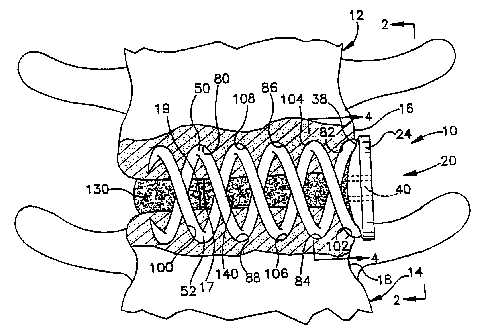
Fig. 1 is a schematic anterior view of an
apparatus implanted in an adjacent pair of vertebral
bodies in accordance with the present invention;
Fig. 2 is a side view taken along line 2-2 in
Fig. 1;
Fig. 3 is a perspective view of the apparatus of
Fig. 1;
Fig. 4 is a sectional view taken along 4-4 in
Fig. 1;
Fig. 5 illustrates an alternate configuration for
an end portion of the apparatus of Fig. 1;
Fig. 6 is a schematic anterior view illustrating a
-second embodiment of the present invention;
CA 02424261 2003-03-28
WO 02/28323 PCT/US01/30405
-7-
Fig. 7 is an exploded perspective view of the
apparatus of Fig. 6, and includes a driver for rotating
the apparatus;
Fig. 8 is a side view illustrating a third
embodiment of the present invention;
Fig. 9 is a side view illustrating a fourth
embodiment of the present invention; and
Fig. 10 is a sectional view taken along line 10-10
in Fig. 9.
Description of Preferred Embodiments
The present invention is directed to a method and
apparatus for stabilizing adjacent bones, and is
particularly directed to a method and apparatus for
attaching and stabilizing adjacent vertebral bodies
while the vertebral bodies fuse together. As
representative of the present invention, Fig. 1
illustrates an apparatus 10 implanted into an adjacent
pair of lumbar vertebrae 12 and 14 in a vertebral
column (not shown). It should be understood that the
apparatus 10 could be implanted into any adjacent pair
of vertebrae. The vertebrae 12 has a side surface 16
and a lower surface (or end plate) 17 (Fig. 2). The
vertebrae 14 has a side surface 18 and an upper
surface (or end plate) 19.
CA 02424261 2003-03-28
WO 02/28323 PCT/US01/30405
-8-
The apparatus 10 comprises an interbody
stabilizer 20 made from a biocompatible material, such
as titanium or stainless steel. It is contemplated
that the biocompatible material used to make the
interbody stabilizer 20 could also be biodegradable.
The interbody stabilizer 20 is centered about a
longitudinal axis 22 (Fig. 3). The interbody
stabilizer 20 includes a platform 24 having a generally
cylindrical outer surface 26 extending between
oppositely disposed first and second ends 28 and 30.
The second end 30 of the platform 24 includes an end
surface 38 that extends transverse to the side
surfaces 16 and 18 of the adjacent vertebrae 12 and 14,
respectively. The end surface 3,8 of the platform 24
has a shape that is complimentary to the side
surfaces 16 and 18 of the vertebrae 12 and 14,
respectively.
The platform 24 of the interbody stabilizer 20
further includes an axial passage 40 that extends from
the first end 28 to the end surface 38. The passage 40
has a hexagonal configuration for receiving a rotatable
driver (not shown).
First and second helical spikes 50 and 52 project
from the end surface 38 of the platform 24. The
CA 02424261 2003-03-28
WO 02/28323 PCT/US01/30405
-9-
helical spikes 50 and 52 resemble a pair of inter-
twined corkscrews. According to the embodiment
illustrated in Figs. 1-4, the first and second helical
spikes 50 and 52 extend around the axis 22. The
spikes 50 and 52 extend in a helical pattern about the
axis 22 at the same, constant radius R1. It is
contemplated, however, that the first and second
helical spikes 50 and 52 could extend about the axis 22
at different radiuses. Further, it is contemplated
that the radius of one or both of the first and second
helical spikes 50 and 52 could increase or decrease as
the helical spikes extend away from the platform 24.
In order for the interbody stabilizer 20 to be
implanted endoscopically through a typical cannula (not
shown), it is preferred that the platform 24 and the
helical spikes 50 and 52 are less than 20mm in overall
diameter. It should be understood that the interbody
stabilizer 20 could have an overall diameter that is
greater than 20mm for certain applications, and that
the interbody stabilizer could also be implanted in an
open surgical procedure. However, for structural
stability reasons, the overall diameter of the helical
spikes 50 and 52 should remain less than or equal to
the diameter of the platform 24.
CA 02424261 2003-03-28
WO 02/28323 PCT/US01/30405
-10-
In the illustrated embodiment of Figs. 1-4, the
first and second helical spikes 50 and 52 have the same
axial length, and also have the same circular
cross-sectional shape. It is contemplated, however,
that the first and second helical spikes 50 and 52
could have different axial lengths. Further, it is
contemplated that the helical spikes 50 and 52 could
have a different cross-sectional shape, such as an oval
shape. It also contemplated that the first and second
helical spikes 50 and 52 could have different cross-
sectional shapes and/or areas (i.e., one spike being
thicker than the other spike). Finally, it is
contemplated that the helical spikes 50 and 52 should
have the same pitch, and that the pitch of the helical
spikes would be selected based on the specific surgical
application and quality of the bone in which the
interbody stabilizer 20 is to be implanted.
Each of the first and second helical spikes 50
and 52 can be divided into three portions: a connecting
portion 54, an intermediate portion 56, and a tip
portion 58. The connecting portion 54 of each of the
helical spikes 50 and 52 is located at a proximal
end 60 that adjoins the end surface 38 of the
platform 24. The connecting portion 54 may include
CA 02424261 2003-03-28
WO 02/28323 PCT/US01/30405
-11-
barbs (not shown) for resisting pull-out of the helical
spikes 50 and 52 from the vertebrae 12 and 14.
According to one method for manufacturing the interbody
stabilizer 20, the connecting portion 54 of each of the
helical spikes 50 and 52 is fixedly attached to the
platform 24 by inserting, in a tangential direction,
the proximal ends 60 of the helical spikes into
openings (not shown) in the end surface 38 and welding
the connecting portions 54 to the platform. The
inserted proximal'ends 60 of the helical spikes 50
and 52 help to reduce tensile bending stresses on the
helical spikes under a tensile load.
Alternatively, the helical spikes 50 and 52 may be
formed integrally with the platform 24, such as by
casting the interbody stabilizer 20. If the interbody
stabilizer 20 is cast, it is contemplated that a fillet
(not shown) may be added at the junction of the helical
spikes 50 and 52 and the platform 24 to strengthen the
junction and minimize stress concentrations at the
connecting portions 54. The fillet at the junction of
the helical spikes 50 and 52 and the platform 24 also
helps to reduce bending stresses in the connecting
portions 54 of the helical spikes under a tensile load.
CA 02424261 2003-03-28
WO 02/28323 PCT/US01/30405
-12-
As best seen in Fig. 4, the connecting portions 54
at the proximal ends 60 of the first and second helical
spikes 50 and 52 are spaced 1800 apart about the
axis 22 to balance the interbody stabilizer 20 and
evenly distribute loads on the helical spikes. The
connecting portion 54 of each of the helical spikes 50
and 52 has a first cross-sectional diameter D1
(Fig. 3).
The tip portion 58 of each of the helical
spikes 50 and 52 is located at a distal end 62 of the
helical spikes. The intermediate portion 56 of each of
the helical spikes 50 and 52 extends between the tip
portion 58 and the connecting portion 54. The
intermediate portion 56 and the tip portion 58 of each
of the helical spikes 50 and 52 has a second
cross-sectional diameter D2 that is less than or equal
to the first cross-sectional diameter Dl of the
connecting portions 54. If the second cross-sectional
diameter D2 is less than the first cross-section
diameter D1, the increased thickness of the connecting
portions 54 of the helical spikes 50 and 52 will help
to provide the interbody stabilizer 20 with increased
tensile strength at the junction of the helical spikes
and the platform 24.
CA 02424261 2006-09-13
-13-
The tip portion 58 of each of the helical
spikes 50 and 52 is self-penetrating and provides the
helical spikes with the ability to penetrate into a
respective one of the vertebrae 12 and 14 as the
platform 24 of the interbody stabilizer 20 is rotated
in a clockwise direction. The tip portions 58
illustrated in Figs. 1-4 have an elongated conical
shape with a sharp pointed tip 68. Fig. 5 illustrates
an alternative, self-tapping configuration for the tip
portions 58 which includes a planar surface 66 for
driving into the vertebrae 12 and 14, in the same
manner that a wood chisel turned upside-down drives
.into wood, as the platform 24 is rotated. It is
contemplated that the tip portions 58 could also have a
pyramid shape, similar to the tip of a nail.
Figs. 1 and 2 illustrate the interbody
stabilizer 20 implanted in the adjacent lumbar
vertebrae 12 and 14 to stabilize the vertebrae.
First, disk material that normally separates the*
vertebrae 12 and 14 is removed by the surgeon. Removal
of the disk material leaves an interbody space 61
(Fig. 2) between the vertebrae 12 and 14. A tool (not
shown) is then used to punch a hole (not shown) in the
cortical bone (not shown) of each of the vertebrae 12
CA 02424261 2003-03-28
WO 02/28323 PCT/US01/30405
-14-
and 14. The hole in the vertebrae 12 may be punched in
either the side surface 16 or the lower surface 17.
The hole in the vertebrae 14 may be punched in either
the side surface 18 or the upper surface 19. The holes
in the vertebrae 12 and 14 are punched in locations
that correspond to the spacing of the tip portions 58
of the helical spikes 50 and 52 of the interbody
stabilizer 20. The holes in the vertebrae 12 and 14
are intended to make the initial rotation of the
stabilizer 20 easier. It should be noted that one or
both of the configurations of the tip portions 58
illustrated in Figs. 1-5 may be able to punch through
the cortical bone upon rotation of the interbody
stabilizer 20, thus eliminating the need for the
aforementioned tool to punch holes in the cortical
bone.
The tip portions 58 of the interbody stabilizer 20
are placed in the holes in the vertebrae 12 and 14 and
a rotatable driver (not shown) is inserted into the
passage 40 in the platform 24. The driver is then
rotated, causing the interbody stabilizer 20 to rotate
as well. It is contemplated that a cylindrical sleeve
(not shown) may be placed around the intermediate
portions 56 and the connecting portions 54 of the
CA 02424261 2006-09-13
-15-
helical spikes 50 and 52 to prevent the helical spikes
from deforming radially outward during the initial
rotation of the interbody stabilizer 20.
Rotation of the interbody stabilizer 20 screws the
helical spikes 50 and 52 into the vertebrae 12 and 14,
respectively. The tangentially-oriented connection
between the connection portions 54 of the helical
spikes 50 and 52 and the platform 24 minimizes bending
loads on the connecting portions during rotation of the
interbody stabilizer 20. Further, the tangentially-
oriented connection ensures that the force vector
resulting from axial force torque and applied by the
driver (not shown) to the platform 24 is transmitted
along the helical centerline (not shown) of each of
the helical spikes 50 and 52.
As the interbody stabilizer 20 is rotated, the tip
portion 58 of the first helical spike 50 penetrates the
cancellous bone in the vertebrae 12 and cuts a first
helical segment 82 of a first tunnel 80 (Fig. 1) in the
vertebrae 12. Simultaneously, the tip portion 58 of
the second helical spike"52 penetrates the cancellous
bone of the vertebrae 14 and cuts a first helical
segment 102 of a second tunnel 100 in the vertebrae 14.
CA 02424261 2006-09-13
-16-
At some point between 90 and 180 of rotation of
the interbody stabilizer 20, the tip portions 58 of the
helical spikes 50 and 52 penetrate back out of the
vertebrae 12 and 14, respectively and into the
interbody space 61. More specifically, the tip
portiori 58 of the first helical spike 50 projects
through the lower surface 17 of the vertebrae 12 and
into the interbody space 61. Simultaneously, the tip
portion 58 of the second helical spike 52 projects
through the upper surface 19 of the vertebrae 14 and
into.the interbody space 61.
As the interbody stabilizer 20 is rotated
beyond 180 , the tip portions 58 of the helical
spikes 50 and 52 move through the interbody space 60
and engage the vertebrae 14 and 12, respectively. The
tip portion 58 of the first helical spike 50 penetrates
into the upper surface 19 of the vertebrae 14, while
'the tip portion 58 of the second helical spike 52
projects through the lower surface 17 of the
vertebrae 12. Continued rotation of the interbody
stabilizer 20 causes the tip portion 58 of the first
helical spike 50 to cut a second helical segment 84 of
the first tunnel 80 in the vertebrae 14. Similarly,
the continued rotation causes the tip portion 58 of the
CA 02424261 2006-09-13
-17-
second helical spike 52 to cut a second helical
segment 104 of the second tunnel 100 in the
vertebrae 12.
After another 90 to 180 of rotation of the
interbody stabilizer 20, the tip portions 58 of the
helical spikes 50 and 52 penetrate back out of the
vertebrae 14 and 12, respectively, and into the
interbody space 61. More specifically, the tip
portion 58 of the first helical spike 50 projects
through the upper surface 19 of the vertebrae 14 and
the tip portion 58 of the second helical spike 52
projects through the lower surface 17 of the
vertebrae 12.
As the interbody stabilizer 20 is rotated further,
the tip portions 58 of the helical spikes 50 and 52
move through the interbody space 61 and re-engage the
vertebrae 12 and 14, respectively. The tip portion 58
of the first helical spike 50 penetrates the lower
surface 17 of the vertebrae 12 and cuts a third helical
segment 86 of the first tunnel 80 in the vertebrae 12.
Simultaneously, the tip portion 58 of the second
helical spike 52 penetrates the upper surface 19 of the
vertebrae 14 and cuts a third helical segment 106 of
the second tunnel 100 in the vertebrae 14.
CA 02424261 2006-09-13
-18-
After further rotation of the interbody
stabilizer 20, the tip portions 58 of the helical
spikes 50 and 52 again penetrate back out of the
vertebrae 12 and 14, respectively and into the,
interbody space 61. The tip portion 58 of the first
helical spike 50 projects through the lower surface 17
of the vertebrae 12, while the tip portion 58 of the
second helical spike 52 projects through the upper
surface 19 of the vertebrae 14. The interbody
stabilizer 20 is then rotated so that the tip
portions 58 of the helical spikes 50 and 52 move
through the interbody space 61 and re-engage the
vertebrae 14 and 12, respectively. The tip portion 58
of the first helical spike 50 again penetrates into the
upper surface 19 of the vertebrae 14, causing the tip
portion 58 of the first helical spike 50 to cut a
fourth helical segment 88 of the first tunnel 80 in the
vertebrae 14. Similarly, the tip portion 58 of the
second helical spike 52 again penetrates through the
lower surface 17 of the vertebrae 12, causing the tip
portion 58 of the second helical spike 52 to'cut a
fourth helical segment 108 of the second tunnel 100 in
the vertebrae 12.
CA 02424261 2006-09-13
-19-
This pattern of screwing the helical spikes 50
and 52 of the interbody stabilizer 20 into and out of
each of the vertebrae 12 and 14 in an alternating
manner continues with each revolution of the
platform 24 by the driver. The continued rotation of
the platform 24 embeds the helical spikes 50 and 52 of
the interbody stabilizer 20 into the vertebrae 12
and 14 and attaches the interbody stabilizer to each of
the vertebrae. With each rotation of the interbody
stabilizer 20, the connection between the interbody
stabilizer and each of the vertebrae 12 and 14 gets
stronger. The attachment of the interbody
stabilizer 20 to each of the vertebrae 12 and 14 thus
fastens, or pins, the vertebrae together, yet spaced
apart. Rotation of the platform 24 is terminated when
the end surface 38 of the platform seats against one or
both of the side surfaces 16 and 18 of the vertebrae 12
and 14, respectively.
Once the interbody stabilizer 20 is implanted,
bone graft material 130 (shown schematically in Figs. 1
and 2) for permanently fusing the vertebrae 12 and 14
is placed into the interbody space 61. More
specifically, the bone graft material 130 is placed
into a cavity 140 defined by the helical spikes 50
CA 02424261 2006-09-13
_20_
and 52, the lower surface 17 of the vertebrae 12, and
the upper surface 19 of the vertebrae 14. The bone
graft material 130, which may comprise bone chips
and/or synthetic bone material, is placed into the
cavity 140 through the axial passage 40 in the
platform 24 of the interbody stabilizer 20. A
sufficient amount of the bone graft material 130 is
placed into the cavity 140 to fill not only the cavity,
but also the entire interbody space 61.
When implanted, the interbody stabilizer 20 is
attached to both of the vertebrae 12 and 14 and
securely fastens the vertebrae together. Because each
of the helical spikes 50 and 52 penetrates into=and
subsequently out of each of the vertebrae 12 and 14,
the helical spikes provide multiple fixation locations
between the interbody stabilizer 20 and the vertebrae
that pin the vertebrae together. The interbody
stabilizer 20 is therefore able to resist relative
movement of the vertebrae 12 and 14 toward or away from
each other, and does not rely on surrounding ligaments
to stabilize the vertebrae. More specifically, the
interbody stabilizer 20 resists relative movement of
the vertebrae 12 and 14, through bending or rotation,
along any one of the three planes of motion (sagittal,
CA 02424261 2003-03-28
WO 02/28323 PCT/US01/30405
-21-
coronal, or horizontal). Thus, the interbody
stabilizer 20 is able to maintain proper intervertebral
spacing and provide effective temporary stabilization
of the adjacent vertebrae 12 and 14, despite -
substantial forces on the interbody stabilizer caused
by human body movement and muscle memory, while the
bone graft material 130 fuses the vertebrae together.
Advantageously, the interbody stabilizer 20 has a
simple one-piece construct and does not require
substantial cutting of cortical bone (i.e., a reaming
or tapping procedure) to prepare the vertebrae 12
and 14 to accept the interbody stabilizer. Thus, the
interbody stabilizer 20 is'not only a simplified
construct, but also simplifies the steps required for
implantation into adjacent vertebrae.
Figs. 6 and 7 illustrate an apparatus 210
constructed in accordance with a second embodiment of
the present invention. In the second embodiment of
Figs. 6 and 7, reference numbers that are the same as
those used in the first embodiment of Figs. 1-4
designate parts that are the same as parts in the first
embodiment.
According to the second embodiment, the
apparatus 210 comprises an interbody stabilizer 220
CA 02424261 2003-03-28
WO 02/28323 PCT/US01/30405
-22-
having a platform 224. The platform 224 includes a
generally rectangular slot 232 that extends axially
from a first end 228 toward a second end 230 of the
platform. Adjacent the first end 228, the platform 224
includes first and second segments of external
threads 234 and 236 that are separated by the slot 232.
The slot 232 and the threads 234 and 236 provide
structure for connecting spinal fixation
instrumentation to the platform 24. The first and
second helical spikes 50 and 52 project from the end
surface 38 at the second end 230 of the platform 224.
Fig. 6 illustrates how the interbody
stabilizer 220 may be used for segmental spinal
fixation. Lumbar vertebrae L3 and L4, indicated by
reference numbers 290 and 292, respectively, are shown
in Fig. 6. The interbody stabilizer 220 according to
the second embodiment of the present invention is
implanted in the interbody space between the
vertebrae 290 and 292. The interbody stabilizer 220 is
implanted into the vertebrae 290 and 292 in much the
same manner as described above regarding the first
embodiment. A rotatable driver 270 (Fig. 7) fits into
the slot 232 in the interbody stabilizer 220 and is
used to rotate the interbody stabilizer.
CA 02424261 2003-03-28
WO 02/28323 PCT/US01/30405
-23-
Once the interbody stabilizer 220 is implanted,
spinal fixation instrumentation such as a beam 280
which has been bent into a desired shape by the
surgeon, is placed into the slot 232 in the interbody
stabilizer. A nut 282 is then screwed onto the
threads 234 and 236 on the platform 24 and tightened to
secure the beam 280 to the interbody stabilizer 220.
As in the first embodiment, the interbody
stabilizer 220 fastens the vertebrae 290 and 292
together and stabilizes the vertebrae until the bone
graft material 130 placed in the cavity 140 defined
inside each of the interbody stabilizers fuses the
vertebrae. The beam 280 helps to further support the
vertebrae 290 and 292 until the vertebrae fuse
together.
Fig. 8 illustrates an apparatus 310 constructed in
accordance with a third embodiment of the present
invention. In the third embodiment of Fig. 8,
reference numbers that are the same as those used in
the first embodiment of Figs. 1-4 designate parts that
are the same as parts in the first embodiment.
According to the third embodiment, the interbody
stabilizer 20 is implanted into two cervical
vertebrae 312 and 314 in the same manner as described
CA 02424261 2003-03-28
WO 02/28323 PCT/US01/30405
-24-
above regarding the first embodiment. The end
surface 38 of the interbody stabilizer 20 seats against
anterior surfaces 316 and 318 of the vertebrae 312
and 314, respectively. As in the first embodiment, the
interbody stabilizer 20 fastens the vertebrae 312
and 314 and stabilizes the vertebrae until the bone
graft material 130 placed in the cavity 140 in the
interbody stabilizer fuses the vertebrae.
Figs. 9 and 10 illustrate an apparatus 410
constructed in accordance with a fourth embodiment of
the present invention. In the fourth embodiment of
Figs. 9 and 10, reference numbers that are the same as
those used in the first embodiment of Figs. 1-4
designate parts that are the same as parts in the first
embodiment.
According to the fourth embodiment, the
apparatus 410 comprises an interbody stabilizer 420
having three helical spikes 430, 431, and 432
projecting tangentially from the end surface 38 of the
platform 24. The spikes 430-432 are centered about the
axis 22. As shown in Fig. 10, the connecting
portions 54 at the proximal ends 60 of the helical
spikes 430-432 are spaced 120 apart about the axis 22,
which balances the interbody stabilizer 420 and evenly
CA 02424261 2003-03-28
WO 02/28323 PCT/US01/30405
-25-
distributes loads on the helical spikes. As in the
first embodiment of Figs. 1-4, in the fourth embodiment
of Figs. 9 and 10, the cross-sectional diameter of the
connection portions 54 of the helical spikes 430-432 is
greater than or equal to the cross-sectional diameter
of the intermediate portions 56 and the tip portions 58
of the helical spikes.
Each of the three helical spikes 430-432 extend in
a helical pattern about the axis 22 at the same,
constant radius R1. It is contemplated, however, that
one or more of the helical spikes 430-432 could extend
about the axis 22 at different radiuses. Further, it
is contemplated that the radius of one or more helical
spikes 430-432 could increase or decrease as the
helical spikes extend away from the platform 24.
As shown in Fig. 9, the three helical
spikes 430-432 have the same axial length and also have
the same circular cross-sectional shape. It is
contemplated, however, that one or more of the helical
spikes 430-432 could have different axial lengths.
Further, it is contemplated that one or more of the
helical spikes 430-432 could have a different
cross-sectional shape, such as an oval shape. It also
contemplated that the one or more of the helical
CA 02424261 2003-03-28
WO 02/28323 PCT/US01/30405
-26-
spikes 430-432 could have different cross-sectional
shapes and/or areas (i.e., one spike being thicker or
thinner than the other two spikes). Finally, it is
contemplated that the helical spikes 430-432 should
have the same pitch, and that the pitch of the helical
spikes would be selected based on the specific surgical
application and quality of the bone in which the
interbody stabilizer 20 is to be implanted.
The tip portion 58 of each of the helical
spikes 430-432 illustrated in Fig. 8 has an elongated
conical shape for penetrating into a vertebrae as the
platform 24 of the interbody stabilizer 420 is rotated
in the clockwise direction. It should be understood
that the tip portions 58 of the helical spikes 430-432
of the interbody stabilizer 420 could alternatively be
configured like the tip portions illustrated in Fig. 5.
The interbody stabilizer 420 according to the
fourth embodiment of Figs. 9 and 10 is implanted into
an adjacent pair of vertebrae in the same manner as the
interbody stabilizer 20 according to the first
embodiment. Further, the interbody stabilizer 420
according to the fourth embodiment may also be used to
mount spinal fixation instrumentation as shown in the
second embodiment of Figs. 6 and 7. When implanted,
CA 02424261 2003-03-28
WO 02/28323 PCT/US01/30405
-27-
the interbody stabilizer 420 is attached to both of the
adjacent vertebrae and fastens the vertebrae together.
Further, the interbody stabilizer 420 maintains proper
intervertebral spacing and provides effective temporary
stabilization of the adjacent vertebrae while the bone
graft material placed in the cavity in the interbody
stabilizer fuses the vertebrae together.
Advantageously, the interbody stabilizer 420 is a
simple one-piece construct does not require substantial
cutting of cortical bone (i.e., a reaming or tapping
procedure) to prepare the adjacent vertebrae to accept
the interbody stabilizer.
It should be noted that the interbody stabilizers
according to the present invention can be used not only
to stabilize a degenerative disc, but can also be used
to correct spinal deformity suc.h as scoliosis,
kyphosis, lordosis, and spondylosisthesis.
From the above description of the invention, those
skilled in the art will perceive improvements, changes
and modifications. It should be understood that the
method and apparatus according to the present invention
could be used to attach and stabilize other adjacent
bones, not just bones in the spine or pelvis. Further,
it is contemplated that the present invention could
CA 02424261 2003-03-28
WO 02/28323 PCT/US01/30405
-28-
comprise a single helical spike, or more than three
spikes. Such improvements, changes and modifications
within the skill of the art are intended to be covered
by the appended claims.