Note : Les descriptions sont présentées dans la langue officielle dans laquelle elles ont été soumises.
CA 02424306 2003-03-28
WO 02/32496 PCT/USO1/32184
OVER-THE-WIRE INTERLOCK ATTACHMENT/DETACHMENT MECHANISM
This application claims the benefit of provisional patent application Serial
No.
60/241,005 filed October 18, 2000.
Background of the Invention
[0001] In recent years, a number of medical devices have been designed which
are
adapted for compression into a small s~~.e to facilitate introduction into the
heart or a vascular
passageway and which are subsequently expandable. These devices, among others,
include
septal occluders, stems and free standing filters which expand and are held in
position by
engagement with the wall of an organ or vessel. It has 'been found to be
advantageous to
form such devices of a shape memory material having a first, relatively
pliable low
temperature condition and a second, relatively rigid high-temperature
condition. By forming
such devices of temperature responsive material, the device in a flexible and
reduced stress
state may be compressed to fit within the bore of a delivery catheter when
exposed to a
temperature below a predetermined transition temperature, but at temperatures
at or above the
transition temperature, the device expands and becomes relatively rigid.
[0002] Originally, these implantable medical devices were intended to
permanently
remain in place, but recently it has become advantageous to retrieve the
previously implanted
device.
[0003] The development of removable implantable medical devices such as septal
occluders, stems and filters which expand and are held in position by
engagement with the
wall of an organ or vessel has led to the development of infra vascular snares
to retrieve these
foreign bodies, usually from the peripheral vessels of the cardiovascular
system. Single loop
snares, such as those shown by U.S. Patent Nos. 3,828,790 to Curtiss et al.
and 5,171,233 to
Amplatz et al. are commonly used snares. The Arnplatz snare consists of a
super-elastic
nitinol cable with a single-formed loop. Because of the snare's super elastic
construction, the
loop can be introduced through small lumen catheters without risk of
deformation. The loop
is formed at approximately 90° to a cable, and this allows for the user
to advance the loop
over a foreign body and ensnare it by closing the loop with a small catheter.
The foreign
CA 02424306 2003-03-28
WO 02/32496 PCT/USO1/32184
body is removed from the vasculature by withdrawing the device into a guiding
catheter or
vascular sheath.
[0004] In an attempt to provide a snare with improved cross sectional vessel
coverage,
multi-loop snares such as those shown by U.S. Patent Nos. 5,098,440 to
Hillstead and
6,099,534 to Bates have been developed. These snares include loops which are
joined only at
their proximal ends to a shaft, and otherwise are not joined at any point
between the shaft and
the distal ends of the loops. This provides the advantage over single loop
snares of enhanced
cross sectional vessel coverage, and the free distal ends of the loops can be
brought together
to engage multiple surfaces of an intravascular medical device to be removed.
[0005] The problem with known snare recovery devices is that they are
difficult to
advance over a medical implant device and require skilled manipulation to
retrieve an
implanted device. Once the medical implant device is engaged by a recovery
snare, there is
no assurance that the device will not slip out of the snare during the
recovery process.
[0006] It is particularly difficult to remove medical implants from the heart,
such as
septal occluders, with known snare recovery devices. Such snare recovery
devices normally
require appropriate sizing to the vasculature in order to facilitate
successful ensnarement, and
the geometry of mufti loop snares is difficult to maintain during delivery.
The relative
position of the loops can change, both within a catheter or delivery tube and
within a vessel,
and the loops can actually become displaced or entangled during delivery.
Summary of the Invention
[0007] A primary object of the present invention is to provide a novel and
improved over-
the-wire interlock attachment/detachment mechanism adapted to engage and
positively lock
on to an implanted medical device.
[0008] Another object of the present invention is to provide a novel and
improved over-
the-wire interlock attachment/detachment mechanism which automatically aligns
to form an
interlock attachment with an implanted medical device.
[0009] A further obj ect to the present invention is to provide a novel and
improved over-
the-wire interlock attachment/detachment mechanism well adapted for use with
over-the-wire
implanted medical devices.
CA 02424306 2003-03-28
WO 02/32496 PCT/USO1/32184
[0010] Yet another object of the present invention is to provide a novel and
improved
over-the-wire interlock attachment/detachment mechanism which includes a
cylindrical
locking section for engagement with a cylindrical lock receiving section
connected to the
medical implant.
[0011] A further object of the present invention is to provide a novel and
improved over-
the-wire interlock attachment/detachment mechanism which includes no
overlapping
components and which maintains a low profile configuration during passage
through a vessel
and/or catheter.
[0012] These and other objects of the present invention are achieved by
providing a
cylindrical lock receiving section of a small diameter attached to an
irnplantable medical
device such as a blood clot filter, a stmt, or a septal occluder. This
cylindrical lock receiving
section has a plurality of spaced, curved cutouts to receive both the guide
fingers and
contoured locking fingers formed on a cylindrical locking section. The locking
fingers are
angled outwardly from the cylindrical body of the cylindrical locking section,
and are moved
inwardly into engagement with the curved cutouts of the cylindrical lock
receiving section by
a sheath which slides over the cylindrical locking section, or by another
suitable operator
which can be activated to move the fingers inwaxdly.
Brief Description of the Drawings
[0013] Figure 1 is a perspective view of the over-the-wire interlock
attachment/detachment mechanism of the present invention with the control
sheath shown in
section;
[0014] Figure 2 is a perspective view of an over-the-wire free standing filter
with the
cylindrical lock receiving section for the over-the-wire interlock
attachment/detachment
mechanism of Figure 1;
[0015] Figure 3 is a perspective view of the partially engaged locking and
lock receiving
sections for the over-the-wire interlock attachment/detachment mechanism of
Figure 1;
[0016] Figure 4 is a perspective view of the engaged locking and lock
receiving sections
for the over-the-wire interlock attachment/detachment mechanism of Figure 1;
CA 02424306 2003-03-28
WO 02/32496 PCT/USO1/32184
[0017] Figure 5 is a second embodiment of a locking section for the over-the-
wire
interlock attachment/detachment mechanism of the present invention; and
[0018] Figure 6 is a third embodiment of a locking section for the over-the-
wire interlock
attachment/detachment mechanism of the present invention.
Description of the Preferred Embodiments
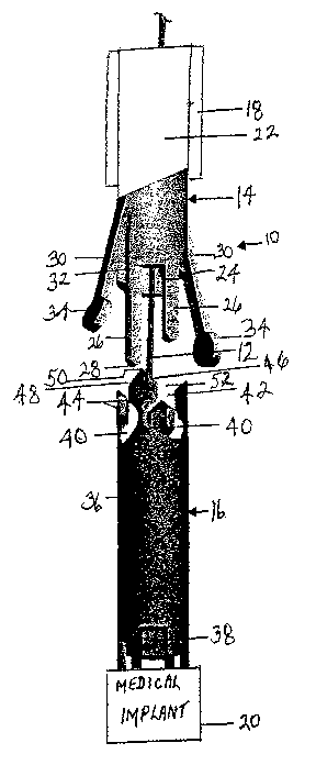
[0019] Referring to Figure l, the over-the-wire interlock
attachment/detachment
mechanism of the present invention indicated generally at 10 is adapted for
movement along
a conventional guidewire 12 such as a .014" guidewire. The over-the-wire
interlock
attachment/detachment mechanism includes a male locking section 14, a female
lock
receiving section 16, and a tubular sheath 18 dimensioned to slide over the
male and female
sections. Preferably, the female section 16 is secured to an implantable
medical device 20
such as a septal occluder, a filter or stmt to be released in the heart or a
blood vessel or other
vessel of the human body or to be retrieved or repositioned within the heart
or vessel.
[0020] The male locking section 14 includes a tubular body 22 which defines an
open
ended central chamber 24 through which the guidewire 12 passes. Projecting
outwardly from
the forward end of the tubular body 22 are one or more elongate guide fingers
26. ~ These
guide fingers are straight, elongate pins with arcutely shaped ends 28, and
two such guide
fingers are shown in Figure 1 although more than two can be provided. The
outer surface of
each guide finger is preferably coextensive with the outer surface of the
tubular body 22.
[0021] Also projecting outwardly from the forward end of the tubular body 22
are one or
more flexible, elongate locking anus 30 which are substantially equal in width
to the width of
the guide fingers 26. Underlying each of the locking arms is a slot 32 formed
in the tubular
body to receive the locking arm. When unconfined, each locking arm is formed
to angle
outwardly beyond the outer surface of the tubular body 22.
[0022] A shaped locking member 34 is formed at the end of each locking arm.
Preferably, this locking member, which extends laterally from at least one
side of the locking
arm, is circular in shape, but other shapes Which extend laterally from the
locking arm
including but not limited to an ellipse, a "T", a rectangle, a square, a hook,
a triangle or an
"L" can be used. A circular locking member facilitates engagement with the
lock receiving
CA 02424306 2003-03-28
WO 02/32496 PCT/USO1/32184
section 16. The guide forgers and locking arms are equally spaced around the
tubular body
22. They are preferably equal in number, and although two of each are shown,
more can be
used.
[0023] The female lock receiving section 16 includes a tubular body 36 which
defines an
open ended central chamber 38 for receiving the guidewire 12. The tubular body
36 is
substantially equal in diameter to the tubular body 22 so that the two are
coextensive when
the male locking section is engaged with the female lock receiving section
[0024] The female lock receiving section includes a plurality of shaped
locking cutouts
40 which are shaped to conform to and receive the shaped locking members 34.
The number
of shaped locking cutouts 40 is equal to the number of guide fingers 26 and
locking arms 30.
Extending into each of the shaped locking cutouts 40 is a straight, open
ended, cutout entry
section 42 which is formed to receive either a guide finger 26 or a locking
arm 30.
[0025] The shaped locking cutouts 40 and open ended entry cutout sections 42
are
equally spaced around the tubular body 36 to conform to the spacing of the
guide fingers 26
and locking arms 30. Outwardly projecting spacer sections 44 extend outwardly
between
adjacent shaped cutouts and open ended entry cutout sections and each
terminate in inclined
outer end surfaces 46 and 48 which form an apex S0. Each inclined outer
surface angles
downwardly toward an open ended entry cutout section 42 and the inclined outer
end surface
46 of a spacer section 44 forms with the inclined outer end surface 48 of an
adjacent spacer
section an enlarged outwardly tapered opening S2 for each open ended cutout
section.
[0026] The female lock receiving section 16 is secured to one end of a medical
implant
20, which can be an over the wire device such as a septal occluder. For
purpose of
illustration, the female lock receiving section is shown with the over-the-
wire free standing
filter S4. The free standing filter S4 has a filter body with an elongate
guidewire receiving
member S6 extending centrally therethrough to define an open ended channel
configured to
receive a plurality of different sized guidewires. An expandable and
contractible frame 58
surrounds the elongate guidewire receiving member and is connected at a
proximal end to the
elongate guidewire receiving member. A porous embolic capturing unit 60 has an
open end
62 connected to the frame and a closed end 64 connected to the elongate
guidewire receiving
member which extends through the porous embolic capturing unit.
CA 02424306 2003-03-28
WO 02/32496 PCT/USO1/32184
[0027] Figures 1, 3 and 4 disclose the manner in which the over-the-wire
interlock
attachment/detachment mechanism 10 is operable to positively engage and remove
a medical
implant 20 from a body organ or vessel. The male locking section 14 is
enclosed within the
sheath 18 so that the locking arms 30 are forced into the slots 32 and do not
project outwardly
beyond the periphery of the male locking section. In this configuration, the
male locking
section is passed along the wire 12 until it is positioned in close proximity
to the female lock
receiving section 16. At this point, the sheath 18 is drawn back to permit the
locking arms 30
to angle outwardly from the male locking section 14. The male locking section
is then
moved toward the female lock receiving section 16 until the guide fingers 26
engage the
outer end surface 46 or 48 of a spacer section 44. As the male locking section
continues to
move toward the female lock receiving section, each guide finger will be
guided by an
inclined outer end surface 46 or 48 into an open ended cutout entry section 42
which then
guides the guide finger into the associated shaped cutout 40. The over-the-
wire interlock
attachment/detachment mechanism is now in the configuration illustrated in
Figure 3. It will
be noted that when the guide fingers move into the open ended cutout entry
sections 42, they
position the locking arms 30 and the locking members 34 above and in aligmnent
with open
ended cutout sections 42 and their associated shaped cutouts 40. Now, shown in
Figure 4, the
tubular sheath 18 is moved forwardly over the tubular bodies 22 and 36 to
force the locking
members 34 into the shaped cutouts 40 and positively engage the male locking
section I4
with the female lock receiving section I6.
[0028] Once a positive engagement has been established between the male
locking
section and female lock receiving section, the over-the-wire interlock
attachment/detachment
mechanism can be drawn back over the wire 12 to remove the medical implant 20.
Because
of the positive locking engagement, forces present on the medical implant as
it is withdrawn
will not result in detachment from the over-the-wire interlock
attachment/detachment
mechanism. This is very important for medical implants such as the removable
filter 54
where hooks 58 must be withdrawn from the wall of the vessel.
[0029] It is often difficult to accurately position a medical implant within a
vessel without
disconnecting or misaligning the implant relative to the positioning device.
This problem is
rectified by the over-the-wire interlock attachment/detachment mechanism 10.
The medical
implant 20 with an attached female lock receiving section 16 is positively
locked to the male
locking section 14 in the manner shown by Figure 4 before it is moved over the
wire 12 into
CA 02424306 2003-03-28
WO 02/32496 PCT/USO1/32184
position within a body vessel. The positive locking action between the male
locking section
and female lock receiving section facilitates accurate positioning of the
medical implant
within a vessel without misorientation or the likelihood of a disconnect. Once
the implant
device is positioned, the sheath 18 can be moved back as shown in Figure 3
allowing the
locking arms 30 to spring outwardly to disengage the locking members 34 from
the shaped
cutouts 40. Now the male locking section 14 can be drawn back over the wire 12
away from
the female lock receiving section 16.
[0030] The sheath 18 may be replaced by other operating mechanisms capable of
moving
the locking arms 30 into the slots 32. For example, elongate tethers attached
to the ends of
the locking arms which extend back through the central chamber 24 might
perform this
function.
[0031] The male locking section 14 can be modified as shown in Figures 5 and 6
to
provide a flexible end section 68 adj acent to the elongate guide fingers 26
and elongate
locking arms 30. By providing a flexible section 68 in the body 22 proximal to
the guide
fingers and locking arms, it becomes easier to align the guide fingers,
locking arms and
locking members 34 with the cutouts in the female lock receiving section 16.
The flexible
section 68 can be formed in a variety of ways. For example, a spring section
can be welded
or bonded to the body 22 between the main portion of the body and the guide
fingers and
locking arms to form the flexible section 68. Ideally, as shown in Figure 5,
the body 22 is
formed with a unitary spring section 68 by cutting the body in a spiral to
create a helical
spring 70. This can be done with a laser which can also be used to shape the
guide fingers,
locking arms and locking members in the tubular body 22.
[0032] Alternatively, as shown in Figure 6, a flexible, tubular polymer
section 72 can be
formed between the main portion of the body 22 and the guide fingers and
locking arms to
provide the flexible section 68.