Note : Les descriptions sont présentées dans la langue officielle dans laquelle elles ont été soumises.
CA 02427229 2006-12-01
2
Title Of Invention
METHOD AND APPARATUS FOR
PROTECTION FROM HIGH INTENSITY LIGHT
Field Of The Invention
[0001] This invention generally relates to a method and apparatus
for automatically protecting personnel from direct exposure to the output of a
high intensity light source. More specifically, this invention relates to a
method and apparatus for protecting the eyes from direct exposure to high
intensity light used in medical devices such as endoscopes and the like.
Additionally, the risk of inadvertent ignition of combustible material, such
as
paper surgical drapes, caused by close proximity to high intensity light
source
outputs, is avoided.
Background Of The Invention
[0002] The imaging of body surfaces through an endoscope is well
known within the medical and veterinarian fields. Typically, this involves
inserting an endoscope into a body cavity and directing an intense light
source output through the endoscope to illuminate body tissue. Light reflected
by the body tissue then is guided along an optical path to an image sensor to
generate a video image of the tissue. One such approach is described in U.S.
Patent 5,162,913 to Chatenever, et al, and provides a technique for an
automatic adjustment of the exposure of video images detected with a CCD
(charge coupled device) image sensor.
[0003] The use of high intensity light sources involves potential
hazards to medical personnel and patients. For example, when a light guide
cable, used to convey the intense light source output, is momentarily
disconnected from the endoscope and placed on a sterile drape used to
CA 02427229 2003-04-29
-3-
protect the patient, the light output intensity can be sufficient to ignite
the
drape and pose a fire hazard; or, the user can inadvertently hold the
disconnected light guide cable in such a way as to temporarily blind another
person in the room. In some instances, when the endoscope is pulled out of a
patient, there can be a risk of these same hazards. When the light source is
used with an endoscopic video camera, which has an automatic exposure
system, the light source may be turned up to an intensity level higher than
required for the camera to produce well-exposed images. This increased light
intensity level can desiccate body tissue and cause serious injury to the
patient. Typically, endoscopic video camera automatic exposure systems can
produce well-exposed images with an electronic shutter setting of
approximately 1/125th to 1/500'h of a second. If an endoscope distal end is
placed within close proximity to tissue being imaged, typically, a relatively
low
light intensity level will stili enable an endoscopic video camera to produce
well-exposed images. An undesirable, and potentially dangerous, scenario
can occur if the light source output is set to a high level, and the endoscope
distal end is placed within close proximity to tissue being imaged. Typically,
in
such a case, camera automatic exposure systems will adjust electronic
shutter settings to approximately 1/10,000th of a second (or faster) to
compensate for the high illumination reflections from the tissue. In such a
situation, the risk of desiccating delicate tissue is greatly increased.
[0004) A technique for automatically controlling the light intensity
from a light source, on the basis of an image signal from an imaging unit
associated with an endoscope, is described in Japanese Unexamined Patent
Publication No. 62-155689 as mentioned at column 2, lines 1-21 in U.S.
Patent 5,957,834 to A. Mochida. As recognized in the Mochida patent, when
light intensity control is made dependent upon a signal derived from the
image, then upon removal of the endoscope from the body, the control is
likely to increase the intensity level from the light source, when instead it
should decrease it to protect the operator's eyes from accidental exposure
and prevent ignition of combustible material. In the Mochida patent a switch
CA 02427229 2003-04-29
-4-
is added to manually adjust and control the output of the light source when
the
endoscope is removed from a body.
[oaos] As further described in the Mochida patent the intensity level
of the light source is controlled by regulating the position of a diaphragm
with
respect to the light source. The control signal for doing this is derived from
an
image sensor in the endoscope.
[ooos] In U.S. Patent 4,527,552 a photoelectric element generates
a signal indicative of the intensity of light reflected from an object
illuminated
by a light source associated with the endoscope to control the light source
output level. In U.S. Patent 5,131,381 a light source associated with an
endoscope is controlled by a signal that represents the density value of each
line of a camera video image derived through the endoscope. Other patents
relevant to light intensity level controls for endoscopes are U.S. Patents
5,159,380; 3,670,722; 5,134,469; 4,963,960; and 4,561,429.
[00071 Techniques have been proposed to reduce the risks
associated with high intensity light sources. One involves a special light
guide
cable with wires in it that are shorted together when the cable is attached to
an endoscope. The short is detected at the iight source and light intensity is
reduced when the cable is disconnected and the short is subsequently
removed. A retractable mechanical shroud, which covers the light guide when
not connected to an endoscope, has also been suggested.
[ooos] These safety solutions are not necessarily effective against
all potential hazardous conditions that may arise; such as when the
endoscope with the light guide cable still attached is pulled out of a patient
and inadvertently directed at a person or surgical drape, or when the light
guide or source initially is directed to treat openly accessible tissue and
inadvertently misdirected during or after a procedure, or when a video camera
CA 02427229 2006-03-28
head, attached to the endoscope light guide cable combination, is
disconnected from its corresponding control unit.
Summary Of The Invention
With a method and apparatus, in accordance with the
invention, the output from a high intensity light source is controlled so that
whenever the output is not directed at tissue (meaning that the endoscope /
video camera / light source combination is not currently being used to image
body tissue), the light source output intensity is automatically reduced to a
safe level. This is achieved by monitoring the reflected light from tissue and
when this reflection indicates that the light source is not directed at
tissue, the
light intensity is turned down to a safe level.
According to the present invention, there is provided a method for
protecting personnel from an intense light source output used for the
illumination
of a surface observed through an endoscope, comprising the steps of:
generating a modulation signal;
modulating the intensity of the light source output with the modulation
signal;
monitoring light received along a light path in the endoscope from the
surface illuminated by the light source output and detecting the modulation in
the
received light;
reducing the intensity of the light source output to a selected level when
the detected modulation is below a reference level; and
adjusting the intensity of the light source output when the detected
modulation is above the reference level;
said adjusting of the intensity of the light source output based upon the
exposure setting of an endoscopic video camera.
According to the present invention, there is also provided an
apparatus for protecting personnel from direct illumination by an intense
light
CA 02427229 2006-03-28
6
source output used for the illumination of a surface observed through an
endoscope, comprising:
an endoscope having an imaging path through which the surface at a
distal end can be observed;
a light source for illumination of the surface;
a camera head including an image sensor aligned to detect light reflected
from the surface and passed along the endoscope imaging path and for
generating image signals;
a camera control unit for processing the image signals received from the
camera head;
a modulator producing modulation signals utilized in varying the light
source output intensity with a selected modulation;
a correlator receiving the image signals for determining the presence of
the selected modulation within the image signals; and
a communication bus coupled to a plurality of bus interfaces for
communication between the light source, camera control unit, modulator, and
correlator.
According to the present invention, there is also provided an
apparatus for protecting personnel from an intense light source output used
for
the illumination of a surface observed through an endoscope, comprising:
a light source having a light output with a selected modulation;
an image sensor associated with the endoscope for detecting light
passed therethrough and reflected from the illuminated surface;
a correlator responsive to the output from the image sensor to produce a
safety signal indicative of the modulation level of the light source output;
and
a controller responsive to the safety signal for reducing the intensity of the
light source output to a selected level when the safety signal is indicative
of the
light source output modulation level being below a selected reference level.
According to the present invention, there is also provided a method
for protecting personnel from an intense light source output directed at a
surface, comprising the steps of:
CA 02427229 2006-03-28
7
generating a modulation signal;
modulating the output intensity of the light source with the modulation
signal;
monitoring light reflected by the surface;
detecting the modulation in the monitored light; and
reducing the intensity of the light source intensity when the detected
modulation is below a reference level.
As described herein for one preferred form of the invention, the
light source is provided with a characteristic signal. The absence of this
characteristic signal from reflected light becomes indicative that the light
source is not directed at tissue and the light intensity needs to be reduced
to
avoid inadvertent iight related injury. This characteristic signal can be a
frequency or wavelength modulation, but preferably is an amplitude or
intensity modulation at a distinctive frequency so that the modulation can be
detected in reflected light.
Preferably, in one embodiment, in accordance with the invention, a
modulation signal is generated and is superimposed on the high intensity light
source output. The light source output thus includes a modulation signal that
is
also present in reflected light, which can be detected by a video camera
imager.
The lack of detection of the modulation signal can then be used to indicate
when
the light source output is not directed at tissue, thus initiating a dramatic
reduction in light intensity.
Preferably, the technique of this invention can be particularly
effective in assuring protective control over a high intensity light source
used as
part of an endoscopic video system utilizing a communication bus to
interconnect various devices. In such case, the light source moduiation is
extracted from the pixel signals at the output of an endoscopic image sensor
used to detect light reflected from tissue observed through an endoscope. As
long as this modulation signal is detected, the high intensity light source
output
remains at a level adequate to produce well-exposed video images. However,
once the modulation signal either disappears or drops below a specific
reference
CA 02427229 2006-03-28
7a
level, it is assumed that the light guide cable is no longer attached to the
endoscope, or that the endoscope itself is removed from the patient, and a
protective reaction is initiated. The loss or reduction of the modulation
signal is
thus converted to a control signal that is sent by means of the bus to the
light
source, causing the output intensity to be turned down to a safe level.
Additionally, as described herein, a method is provided, by the
invention, to determine when the light source output exceeds a level
necessary for an endoscopic video camera to produce well-exposed images.
It is, therefore, an object of the invention to provide a method
and apparatus with which automatic protection of personnel against
accidental exposure to high intensity light from a light guide cable, used
with
or without an endoscope attached, is obtained.
It is a further object of the invention to provide a video
camera/light source control for an enhanced safety of the use of an
endoscope using the high intensity light source for the illumination of
tissue.
It is still further an object of the invention to provide a control
over the overall light output from a high intensity light source used to
illuminate an object observed through an endoscope.
These and other objects of the invention can be understood
from the following detailed description of a preferred embodiment of the
invention in conjunction with the drawings.
Brief Description Of The Drawings
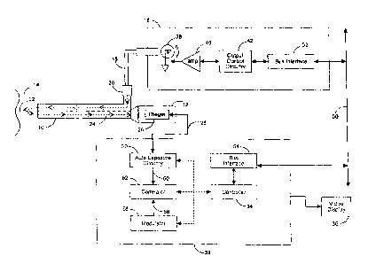
Figure 1 is a schematic block diagram view of an apparatus for
controlling a light source in a safe manner in accordance with the invention.
Figure 2 is a flow diagram for controlling a high intensity light
CA 02427229 2006-03-28
7b
source to reduce risk of injury to personnel; and for optimizing the overall
output level of a high intensity light source, when used to illuminate target
tissue with an endoscope and endoscopic video camera system.
Figure 3 is a schematic block diagram view of another apparatus
for controlling a light source in a safe manner in accordance with the
invention.
Detailed Description Of The Drawings
With reference to Figure 1, a typical endoscope 10 is illustrated
having a camera head 12 mounted thereto at the proximal end to produce
video images in a manner for example as described in the aforementioned
'913 patent. The distal end of endoscope 10 is directed at tissue 14 to
inspect
the tissue with light from a high intensity light source 16 and passed to the
distal end through a light guide cable 18. Typically, light guide cable 18 can
be disconnected from endoscope 10 at connector 20, thus, posing a safety
hazard as previously described.
The light from light guide cable 18 is directed to illuminate tissue
14 as suggested with path 22 and light reflected by tissue 14 is passed along
optical path 24 to imager 26 within camera head 12. Imager 26 detects light
CA 02427229 2003-04-29
-8-
reflected off tissue 14 by means of optical path 24. Imager 26 may be any
type commonly used within the art, such as but not limited to CCD, CID or
CMOS imagers. Camera head 12 produces image signals 28, which are
received by auto exposure circuitry 30, within camera control unit (CCU) 32.
Auto exposure circuitry 30 may consist of various types of methods for
controlling the electronic shutter of imager 26, as well as adjusting
amplification gain in response to illumination levels received by imager 26.
Typically, within the field of video endoscopy, auto exposure circuitry has
high-speed and wide dynamic range capabilities. Various methods may be
utilized, that are well known within the art. Video display 36, receives
signals
from CCU 32, where an image of tissue 14 is presented.
[0023] In the embodiment of Figure 1, iight source 16 is controlled by
CCU 32 controller 34, by means of CCU bus interface 54, digital
communication bus 50, and light source bus interface 52. Controller 34, may
be any type of device designed to receive and execute software programs, or
which is designed to be modified in functionality by software programs, and
preferably is from the group consisting of digital signal processors,
microcontrollers, and microprocessors, or the group consisting of field
programmable gate arrays, and computer programmable logic devices.
[0024] Typically, high intensity light sources utilize an incandescent
bulb 38 (being a xenon bulb, or other type), driven by an amplifier 40, which
in
turn is controlled by output control circuitry 42, to set the light intensity
level of
the light source 16. Other types of light source intensity output control are
known within the art; such as mechanical diaphragm or iris, liquid crystal
shutter, rotary reed or slot devices, and the like. These various types of
light
source output controi may be utilized within the scope of the present
invention. In the present embodiment, output control circuitry 42 varies the
intensity of bulb 38 in accordance with a"slow" time varying signal 56 output
from modulator 58 (within CCU 32) by means of CCU bus interface 54, bus
50, and light source bus interface 52, under the control of controller 34.
What
CA 02427229 2003-04-29
_g_
is meant by "'slow' time varying signal" is that, preferably, signal 56, is of
the
order of approximately two to four seconds per cycle (well below the response
time of auto exposure circuitry 30). Also, "slow" time varying signal 56 is of
an
amplitude level that produces about a 5% to 10% modulation (change in
intensity) of the maximum output intensity of light source 16. Therefore, CCU
32 can control the overall light output level of light source 16, and CCU 32
can
also vary the set overall light output level in accordance with "slow" time
varying signal 56, both in rate and intensity amplitude.
[0025] Light reflected from tissue 14 contains the amplitude (intensity)
modulation, which is detected by imager 26, and is present in video data on
line 28. Auto exposure circuitry 30 not only adjusts the camera exposure to
produce an optimized video image, but also, as part of this exposure
adjustment, compensates for the intensity modulation (driven by modulator 58
"slow" time varying signal 56). Due to the modulation's "slow" rate and "low"
amplitude, auto exposure circuitry 30 easily compensates for the varying light
level, and thus the change in light amplitude is not perceived by viewer's of
video monitor 36.
[0026] Auto exposure circuitry 30 outputs detected modulation signal
60, which retains data corresponding to the intensity modulation. Detected
modulation signal 60 is received by correlator 62. Correlator 62 also receives
"slow" time varying signal 56 from modulator 58.
[0027] Figure 2 is a flow diagram for a method of controlling a high
intensity light source to reduce risk of injury to personnel; and for
optimizing
the overall output level of a high intensity light source, when used to
illuminate
target tissue with an endoscope and endoscopic video camera system. The
light source output is varied 200 in accordance with "slow" time varying
signal
56 output by modulator 58, as previously described. Detected modulation
signal 60 and "slow" time varying signal 56 are compared for correlation 202.
If the two modulation signals (60, 56) do not correlate 204 (i.e. the
modulation
CA 02427229 2003-04-29
-10-
is not detected by auto exposure circuitry 30), controller 34, by means of CCU
bus interface 54, bus 50, light source bus interface 52, and output control
circuitry 42, reduces the light source intensity level to a minimum safe level
206. What is meant by "minimum safe level" is that the light source output is
reduced to a level where the modulation signal can be detected when a
condition causing non-correlation is corrected (i.e. the light intensity is at
its
lowest level in which the modulation signal can still be detected, within the
light source output, by auto exposure circuitry 30). Some conditions which
cause non-correlation 204 are: the light guide cable 18 being disconnect from
light source 16 or endoscope 10, the camera head 12 being disengaged from
endoscope 10, or the light guide cable I camera head I endoscope
combination being removed from a patient (thus, light is not reflected off
body
tissue).
[0028] If the two modulation signals (60, 56) do correlate 208 (i.e, the
modulation is detected by auto exposure circuitry 30), to provide a control
loop which sets the overall light source output 22 to an optimum level, the
current CCU electronic shutter setting is checked. What is meant by
"optimum level" is that the light source intensity is kept at the lowest
possible
level which produces a well-exposed video image from CCU 32. Typically,
light source outputs are set manually by medical personnel, and CCU auto
exposure circuitry adjusts imager electronic shutter and/or gain amplification
levels to produce an acceptable image, with the existing manually set light
intensity level.
[0029] As previously described, the light intensity level may be
manually set much higher than is required for the CCU to produce well-
exposed video images. After correlation has been established 208, in order to
maintain the light source output intensity at a safe level, if the CCU
electronic
shutter is less than or equal to 1/500 of a second 210, the light intensity
level
is decreased by a certain percentage 212 (of the total light source output
CA 02427229 2003-04-29
-11 -
capability). This control loop, 202, 208, 210, and 212 is repeated until the
CCU electronic shutter is greater than 1/500th of a second 214.
[003o] To ensure adequate light is present for the CCU to produce well-
exposed video images, if the CCU electronic shutter is greater than 1/500fh of
a second 214, and if the CCU electronic shutter is greater than or equal to
1/125th of a second 216, the light source intensity level is increased by a
certain percentage 218. This control loop, 202, 208, 214, 216, and 218 is
repeated until the CCU electronic shutter is less than 1/125th of a second
220.
If the CCU electronic shutter is greater than 1/5001h of a second 214, and
less
than 1/125th of a second (as depicted by loop 202, 208, 214, and 220) the
light source output level is maintained at its current intensity level.
[0031] The percentage of light intensity increase or decrease will
determine the speed at which the level is adjusted. Preferably, the
percentage is relatively small (approximately 1% to 3% of the total light
source
capability), which will allow the light source output to slowly "creep"
(within
approximately 5 seconds) to an optimal intensity level.
[0032] Figure 3 depicts another embodiment of the present invention, in
which correlator/modulator 300 is connected to bus 50, separate from CCU
310 and light source 16. Correlator/modulator 300 comprises, bus interface
302, correlator 304, and modulator 306. Within CCU 310, controller 34 still
functions as described in the previous embodiment, except that control of
correlator 304 and modulator 306 is accomplished by means of CCU bus
interface 54, bus 50, and bus interface 302.
[0033] The present embodiment is identical to the previous
embodiment with the following exceptions: light source 16 output control
circuitry 42 varies the intensity of bulb 38 in accordance with a "slow" time
varying signal 308 output from modulator 306 by means of commands routed
from controller 34 to CCU bus interface 54, bus 50, bus interface 302, and
CA 02427229 2003-04-29
-12-
light source bus interface 52. Auto exposure circuitry 30 outputs detected
modulation data 312 (corresponding to the light level modulation) to bus 50 by
means of bus interface 54, under control of controller 34. Detected
modulation data 312 is received by correlator 304 via bus interface 302.
Correlator 304 also receives "slow" time varying signal 308 from modulator
306. Steps to control light source 16, to reduce risk of injury to personnel;
and
for adjusting the average light output intensity, are identical as described
for
the previous embodiment, as detailed for Figure 2.
[0034] Having thus described several embodiments for practicing the
invention, its advantages and objects can be understood. Variations from the
drawings and description can be made by one skilled in the art without
departing from the scope of the invention, which is to be determined from the
following claims. One example being that light source 16 and CCU 32 may be
housed within a single housing, thus obviating bus 50, and bus interfaces 52
and 54.