Note : Les descriptions sont présentées dans la langue officielle dans laquelle elles ont été soumises.
CA 02442334 2003-09-25
WO 02/078555 PCT/US02/10065
DISTAL BONE ANCHORS FOR BONE FIXATION
WITH SECONDARY COMPRESSION
Background of the Invention
Field of the Invention
[0001] The present invention relates to internal bone fracture fixation
devices.
In one application, the present invention relates to bone fracture fixation
devices and
methods adapted for fixation, among other fractures, of femoral neck and other
proximal
femoral fractures.
Description of the Related Art
[0002] The femur, otherwise known as the thigh bone, generally comprises an
elongate shaft extending from the hip to the knee. The proximal end of the
shaft includes a
head, a neck, a greater trochanter and a lesser trochanter. The head of the
femur fits into the
acetabular cup of the hip bone to form a ball and socket joint at the hip. The
distal end of
the femur includes a medial condyle and a lateral condyle. The condyles engage
an upper
end of the tibia to form the knee joint. Overall, the femur is the longest and
strongest bone
in the skeleton. However, portions of the femur are extremely susceptible to
fracturing.
[0003] Pertrochanteric fractures among geriatric patients are the most
frequent
in connection with those of the region of the neck of the bone. The advanced
age and the
pathologies which are encountered in these patients make a timely
stabilization of skeletal
injuries necessary in order to reduce to a minimum the bed confinement and the
rehabilitation times. Preferably, devices and procedures are utilized which
minimize
complications brought about by the so-called immobilization syndrome, which
may be
lethal for patients in delicate metabolical circumstances. It is also
preferable to reduce to a
minimum blood losses related to surgical intervention. At the same time, the
syntheses
means utilized must be stable in order to allow the patient to very timely
assume a seated
position and, two or three days following the intervention, to reassume an
erect posture
with progressive bearing of weight.
[0004] Internal fixation of femoral fractures in general is one of the most
common orthopedic surgical procedures. Fractures of the femur occur in both
the proximal
portion of the femur and the distal portion of the femur. Fractures of the
proximal portion
CA 02442334 2003-09-25
WO 02/078555 PCT/US02/10065
of the femur (hip fractures) are generally classified as femoral neck
fractures (capital or
sub-capital), intertrochanteric fractures and subtrochanteric fractures.
Fractures of the distal
portion of the femur (knee fractures) are referred to as supracondylar
fractures.
Supracondylar fractures generally extend vertically between the condyles at
the lower end
of the femur to separate the distal portion of the femur into two main bone
fragments. A
fracture line may be further comminuted to create a plurality of smaller bone
fragments.
Fractures of the femur which extend into the neck of the bone are generally
more difficult
to treat than fractures restricted to the shaft of the femur.
[0005] Operative treatment of the fractures requires that the fractures be
internally fixed and possibly compressed. Fractures of the neck, head or
trochanters of the
femur have been treated with a variety of compression screw assemblies which
include
generally a compression plate having a barrel member, a lag screw and a
compressing
screw. The compression plate is secured to the exterior of the femur and the
barrel member
is inserted into a predrilled hole in the direction of the femoral head. The
lag screw which
has a threaded end and a smooth portion is inserted through the barrel member
so that it
extends across the break and into the femoral head. The threaded portion
engages the
femoral head. The compressing screw connects the lag screw to the plate. By
adjusting the
tension of the compressing screw the compression (reduction) of the fracture
can be
adjusted.
[0006] A variety of elongated implants (nail, screw, pin, etc.) have been
developed, which are adapted to be positioned along the longitudinal axis of
the femoral
neck with a leading (distal) end portion in the femoral head so as to
stabilize a fracture of
the femoral neck. The elongated implant may be implanted by itself or
connected to another
implant such as a side plate or intramedullary rod. The leading end portion of
the implant
typically includes means to positively grip the femoral head bone (external
threads,
expanding arms, etc.), but the inclusion of such gripping means can introduce
several
significant problems. First, implants with sharp edges on the leading end
portion, such as
the externally threaded implants, exhibit a tendency to migrate proximally
towards the hip
joint weight bearing surface after implantation. This can occur when the
proximal cortical
bone has insufficient integrity to resist distal movement of the screw head.
Such proximal
migration under physiological loading, which is also referred to as femoral
head cut-out,
can lead to significant damage to the adjacent hip joint. Also, the externally
threaded
-2-
CA 02442334 2009-01-02
implants can generate large stress concentrations in the bone during
implantation which
can lead to stripping of the threads formed in the bone and thus a weakened
grip. The
movable arms of known expanding arm devices are usually free at one end and
attached at
the other end to the main body of the leading end portion of the implant. As a
result, all
fatigue loading is concentrated at the attached ends of the arms and
undesirably large
bending moments are realized at the points of attachment. In addition,
conventional
threaded implants generally exhibit insufficient holding power under tension,
such that the
threads can be stripped out of the femoral head either by overtightening
during the
implantation procedure or during post operative loading by the patient's
weight.
[0007] Thus, notwithstanding the variety of efforts in the prior art, there
remains a need for an orthopedic fixation device with improved locking force
such as
within the femoral head in a femoral neck application, which resists migration
and rotation,
and which can be easily and rapidly deployed within the bone.
Summary of the Invention
[0008] There is disclosed, a method of securing a first bone fragment to a
second bone fragment. The method comprises the steps of drilling a bore
through the first
bone fragment in the direction of the second bone fragment, and advancing
through the
bore a fixation device comprising a first portion and a second portion that
are coupled to
each other. A distal anchor of the fixation device is rotated to secure the
fixation device to
the second fragment, and the proximal anchor is axially advanced to engage the
first
fragment and provide compression across the fracture.
(0009] In one application of the method, the second bone fragment
comprises the head of a femur. Altematively, the second bone fragment
comprises a tibia, a
fibula, a femur, a humurus, a radius, or an ulna. The first bone fragment may
comprise a
condyle.
[0010] The method may additionally comprise the step of uncoupling the
first portion from the second portion.
[0011] The present invention provides a bone fixation device, comprising:
an elongate body, having a proximal end and a distal end; a helical anchor on
the distal
end; a first retention structure on the body, proximal to the anchor; an anti-
rotational
structure on the body; and a proximal anchor, moveably carried by the body,
wherein the
proximal anchor is movable in the distal direction with respect to the body
and the
-3-
CA 02442334 2009-01-02
retention structure resists proximal movement of the proximal anchor with
respect to the
body, and wherein the anti-rotational structure prevents rotational movement
of the body
with respect to the proximal anchor.
[0011 a] In one embodiment, the helical anchor is wrapped about a central
core or axial lumen. An outer edge of the helical anchor defines an outer
boundary and the
central core or axial lumen defines a minor diameter.
[0012] In accordance with a further aspect of the present invention, there is
provided a bone fixation device, comprising: an elongate body having a
proximal end and a
distal end, the elongate body comprising a first portion and a second portion
being
detachably coupled to each other at a junction; a cancellous bone anchor on
the distal end;
a proximal anchor axially movably carried on the body; and complimentary
surface
structures in between the body and the proximal anchor that permit advancing
the proximal
anchor in the distal direction to tighten the fixation device but that resist
axial proximal
movement of the proximal anchor.
[0012a] In one embodiment, the cancellous bone anchor comprises a
helical flange wrapped about a central core or axial lumen and an outer edge
of the helical
anchor defines an outer boundary and the central core or axial lumen defines a
minor
diameter.
[0013] There is disclosed a method of treating a femoral fracture. The
method comprises the steps of drilling at least one and preferably two or
three bores
distally into the femur in the direction of a fracture, and advancing into
each bore a fixation
device that comprises a body having a first portion that forms a distal bone
anchor and a
second portion that forms a proximal end. A proximal component is rotated to
engage the
distal anchor with the bone distal to the fracture, and a proximal anchor is
advanced
distally along the fixation device to compress the fracture.
[0014] In accordance with another aspect of the invention a bone fracture
fixation device comprises an elongate body having a proximal end and a distal
end. The
body also includes a helical anchor on the distal end. A first retention
structure is on the
body located proximal to the anchor. A proximal anchor is moveably carried by
the body
and has a tubular housing. The tubular housing has at least one barb extending
radially
outwardly from the tubular housing and defining an engagement surface that
lies within a
plane that is transverse to a longitudinal axis of the tubular housing. The
proximal anchor is
-4-
CA 02442334 2009-01-02
movable in the distal direction with respect to the body and the retention
structure resists
proximal movement of the proximal anchor with respect to the body.
[0015] There is also provided a femoral neck fracture fixation device. The
device comprises an elongated body, having a proximal end and a distal end and
comprising a first portion and a second portion detachably coupled to each
other at a
junction. The first portion includes an anti-rotational structure. A helical
distal anchor is
provided on the distal end. A first retention structure is provided on the
body, proximal to
the distal anchor, and a proximal anchor surface is moveably carried by the
body. The
proximal anchor includes a tubular sleeve that in a first position extends
distally past the
junction between the first portion and the second portion. The proximal anchor
surface is
moveable in the distal direction with respect to the body. The retention
structure resists
proximal movement of the proximal anchor surface with respect to the body, and
the anti-
rotational structure prevents rotational movement of the first portion with
respect to the
proximal anchor.
[0016] In one embodiment, the first retention structure comprises a series of
ridges or grooves. A second retention structure is preferably provided on the
interior of the
tubular sleeve for cooperating with the first retention structure on the body.
[0017] There is also provided a bone fracture fixation device. The fixation
device comprises an elongate body having a proximal end and a distal end and
comprising
a first portion and a second portion that are detachably coupled to each other
at a junction.
A cancellous bone anchor and/or a cortical bone anchor is carried by the
distal end. A
proximal anchor is axially moveably carried on the body and includes a tubular
portion that
extends distally past the junction. Complementary surface structures are
provided in
between the first portion of the body and the proximal anchor to permit
advancing the
proximal anchor in the distal direction to tighten the fixation device but
resist axial
proximal movement of the proximal anchor and to prevent rotational movement
between
the first portion and the proximal anchor.
[0018] Preferably, the drilling step comprises drilling the bore along an axis
which extends into the femoral neck and in the direction of the head of the
femur. In one
embodiment, the advancing a proximal anchor step comprises axially advancing
the
proximal anchor without rotating the proximal anchor with respect to the
fixation device.
The femoral fracture may be a femoral neck fracture (e.g., capital or
subcapital), an
intertrochanteric fracture or a subtrochanteric fracture.
-5-
CA 02442334 2003-09-25
WO 02/078555 PCT/US02/10065
[0019] Further features and advantages of the present invention will become
apparent to those of skill in the art in view of the detailed description of
preferred
embodiments which follows, when considered together with the attached drawings
and
claims.
Brief Description of the Drawings
[0020] Figure 1 is a posterior elevational posterior cross section through the
proximal portion of the femur, having two femoral neck fracture fixation
devices positioned
therein.
[0021] Figure 2 is a posterior cross section as in Figure 1, with a modified
fixation device positioned therein.
[0022] Figure 3A is a side elevational cross section of a fixation device
similar
to that of Figure 1.
[0023] Figure 3B is a side elevational cross section of a fixation device
similar
to that of Figure 2.
[0024] Figure 3C is a side elevational view of a double helix distal anchor.
[0025] Figure 3D is a side elevational view of a "V" thread distal anchor.
[0026] Figure 3E is a side elevational view of a buttress thread distal anchor
[0027] Figure 3F is a side elevational view of a triple helix distal anchor.
[0028] Figure 3G is a side elevational view of a split triple helix distal
anchor.
[0029] Figure 3H is a side elevational view of a tapered transition thread
distal
anchor.
[0030] Figure 31 is a side elevational view of a tapered thread distal anchor.
[0031] Figure 4A is a front elevational perspective view of a modified
fixation
device of the present invention.
[0032] Figure 4B is a front elevational perspective view of a further
modification to the fixation device of the present invention.
[0033] Figure 5 is an axial cross sectional view through a distal end of a
fixation
device of the present invention.
[0034] Figure 6 is a posterior cross section as in Figures 1, with a fixation
device and integral proximal plate anchor positioned therein.
-6-
CA 02442334 2003-09-25
WO 02/078555 PCT/US02/10065
[0035] Figure 6A is a cross sectional schematic view of a combination proximal
anchor and plate in accordance with the present invention.
[0036] Figure 7A is a posterior cross section as in Figures 1, with a plate
and
fixation device positioned therein.
[0037] Figure 7B is a cross section through a proximal portion of the femur,
illustrating the use of a fixation device in combination with a plate.
[0038] Figure 7C is a cross section as in Figure 7B, illustrating the use of a
fixation device of the present invention in combination with an intramedullary
nail.
[0039] Figure 8 is a cross sectional view through an angularly adjustable
proximal anchor plate.
[0040] Figure 9 is a front perspective view of the proximal anchor plate of
Figure 8.
[0041] Figure 10 is an anterior view of the distal tibia and fibula, with
fixation
devices across lateral and medial malleolar fractures.
[0042] Figure 11 is a side perspective view of another embodiment of a
fixation
device having certain features and advantages according to the present
invention..
[0043] Figure 12 is a side elevational view of the fixation device of Figure
11.
[0044] Figure 13 is a cross-sectional view taken through line 13-13 of Figure
12.
[0045] Figure 13A is an enlarged view of portion 13A of Figure 13.
[0046] Figure 13B is an enlarged view of portion 13B of Figure 13 with the
fixation device in a first position.
[0047] Figure 13C is an enlarged view of portion 13C of Figure 13 with the
fixation device in a second position.
[0048] Figure 14 is a cross-sectional view taken through line 14-14 of Figure
12.
[0049] Figures 15A-C illustrate a procedure for using of the fixation device
of
Figure 11 to secure a femoral neck fracture.
Detailed Description of the Preferred Embodiment
[0050] Although the fixation devices of the present invention will be
disclosed
primarily in the context of fractures of the proximal femur, the methods and
structures
-7-
CA 02442334 2003-09-25
WO 02/078555 PCT/US02/10065
disclosed herein are intended for application in any of a wide variety of
bones and fractures,
as will be apparent to those of skill in the art in view of the disclosure
herein. For example,
the bone fixation device of the present invention is applicable in a wide
variety of fractures
and osteotomies in the hand, such as interphalangeal and metacarpophalangeal
arthrodesis,
transverse phalangeal and metacarpal fracture fixation, spiral phalangeal and
metacarpal
fracture fixation, oblique phalangeal and metacarpal fracture fixation,
intercondylar
phalangeal and metacarpal fracture fixation, phalangeal and metacarpal
osteotomy fixation
as well as others known in the art. A wide variety of phalangeal and
metatarsal osteotomies
and fractures of the foot may also be stabilized using the bone fixation
device of the present
invention. These include, among others, distal metaphyseal osteotomies such as
those
described by Austin and Reverdin-Laird, base wedge osteotomies, oblique
diaphyseal,
digital arthrodesis as well as a wide variety of others that will be known to
those of skill in
the art. The bone fixation device may be used with or without plate(s) or
washer(s), all of
which can be either permanent, absorbable, or combinations.
[0051] Fractures of the fibular and tibial malleoli, pilon fractures and other
fractures of the bones of the leg may be fixated and stabilized with the
present invention
with or without the use of plates, both absorbable or non-absorbing types, and
with
alternate embodiments of the current invention. Fractures and osteotomies of
the mid and
hind foot, tarsal arthrodesis and osteotomy, or others as are known to those
with skill in the
art. One example is the fixation of the medial malleolar avulsion fragment
fixation.
[0052] The fixation device of the present invention may also be used to attach
tissue or structure to the bone, such as in ligament reattachment and other
soft tissue
attachment procedures. Plates and washers, with or without tissue spikes for
soft tissue
attachment, and other implants may also be attached to bone, using either
resorbable or
nonresorbable fixation devices depending upon the implant and procedure. The
fixation
device may also be used to attach sutures to the bone, such as in any of a
variety of tissue
suspension procedures.
[00531 For example, peripheral applications for the fixation devices include
utilization of the device for fastening soft tissue such as capsule, tendon or
ligament to
bone. It may also be used to attach a synthetic material such as marlex mesh,
to bone or
allograft material such as tensor fascia lata, to bone. In the process of
doing so, retention of
-8-
CA 02442334 2003-09-25
WO 02/078555 PCT/US02/10065
the material to bone may be accomplished with the collar as shown, or the pin
and or collar
may be modified to accept a suture or other material for facilitation of this
attachment.
[0054] Specific examples include attachment of the posterior tibial tendon to
the
navicular bone in the Kidner operation. This application may be accomplished
using an
appropriately sized implant of the present invention along with a washer with
distally
extending soft tissue spikes. Navicular-cuneiform arthrodesis may be performed
utilizing
the device and concurrent attachment of the tendon may be accomplished.
Attachment of
the tendon may be accomplished in the absence of arthrodesis by altering the
placement of
the implant in the adjacent bone.
[0055] Ligament or capsule reattachment after rupture, avulsion or detachment,
such as in the ankle, shoulder or knee can also be accomplished using the
devices disclosed
herein.
[0056] The fixation devices can also be used to aid bone fusion between
adjacent bones, bone fragments or any of a variety of articulating joints,
such as, for
example, a first and a second adjacent vertebral bodies of the spine.
[0057] The fixation devices may be used in combination with semi tubular, one-
third tubular and dynamic compression plates, both of metallic and absorbable
composition,
if the collar is modified to match the opening on the plate.
[0058] The cannulated design disclosed below can be fashioned to accept an
antibiotic impregnated rod for the slow adsorption of medication locally. This
may be
beneficial for prophylaxis, especially in open wounds, or when osteomyelitis
is present and
stabilization of fracture fragments is indicated.
[0059] A kit may be assembled for field use by military or sport medical or
paramedical personnel. This kit contains an implanting tool, and a variety of
implant
device size and types. The kit may include additional components such as
sterilization or
disinfectant materials, a skin stapler, bandages, gloves, and basic tools for
emergent wound
and fracture treatment. Antibiotic rods may be included for wound prophylaxis
during
transport.
[0060] Referring to Figure 1, there is illustrated a posterior side
elevational view
of the proximal portion of a femur 10, having a two fixation devices 12
positioned therein.
The proximal end of the femur 10 comprises a head 14 connected by way of a
neck 16 to
the long body or shaft 17 of the femur 10. As illustrated in F'igure 1, the
neck 16 is smaller
-9-
CA 02442334 2003-09-25
WO 02/078555 PCT/US02/10065
in diameter than the head 14. The neck 16 and head 14 also lie on an axis
which, on
average in humans, crosses the longitudinal axis of the body 17 of the femur
10 at an angle
of about 126 . The risk of fracture at the neck 16 is thus elevated, among
other things, by
the angular departure of the neck 16 from the longitudinal axis of the body 17
of femur 10
and also the reduced diameter of the neck 16 with respect to the head 14.
[0061] The greater trochanter 18 extends outwardly above the junction of the
neck 16 and the body 17 of the femur 10. On the medial side of the greater
trochanter 18 is
the trochanteric fossa 20. This depression accommodates the insertion of the
obturator
externus muscle. The lesser trochanter 21 is located posteromedially at the
junction of the
neck 16 and the body 17 of the femur 10. Both the greater trochanter 18 and
the lesser
trochanter 21 serve for the attachment of muscles. On the posterior surface of
the femur 10
at about the same axial level as the lesser trochanter 21 is the gluteal
tuberosity 22, for the
insertion of the gluteus maximus muscle. Additional details of the femur are
well
understood in the art and not discussed in further detail herein.
[0062] Figure 1 illustrates a subcapital femoral neck fracture 24. Fractures
of
the proximal portion of the femur 10 are generally classified as capital or
subcapital femoral
neck fractures, intertrochanteric fractures and subtrochanteric fractures. All
of these
fractures will be deemed femoral neck fractures for the purpose of describing
the present
invention.
[0063] Referring to Figures 1-4, the fixation device 12 comprises a body 28
extending between a proximal end 30 and a distal end 32. The length, diameter
and
construction materials of the body 28 can be varied, depending upon the
intended clinical
application. In embodiments optimized for various fractures in an adult human
population,
the body 28 will generally be within the range of from about 6 mm to about 150
mm in
length after sizing, and within the range of from about 2 mm to about 12 mm in
maximum
diameter. The major diameter of the helical anchor, discussed below, may be
within the
range of from about 2.0 mm to about 15 mm. In general, the appropriate
dimensions of the
body 28 will vary, depending upon the specific fracture. In rough terms, for a
malleolar
fracture, shaft diameters in the range of from about 3 mm to about 4.5 mm may
be used,
and lengths within the range of from about 20 mm to about 70 mm. For condylar
fractures,
shaft diameters within the range of from about 3.5 mm to about 8.0 mm may be
used with
lengths within the range of from about 25 mm to about 70 mm. For colles
fractures (distal
-10-
CA 02442334 2003-09-25
WO 02/078555 PCT/US02/10065
radius and ulna), diameters within the range of from about 2.0 mm to about 4.5
mm may be
used with any of a variety of lengths within the range of from about 6 mm to
about 70 mm.
[0064] In one embodiment, the body 28 comprises titanium. However, as will
be described in more detail below, other metals or bioabsorbable or
nonabsorbable
polymeric materials may be utilized, depending upon the dimensions and desired
structural
integrity of the finished fixation device 12.
[0065] The distal end 32 of the body 28 is provided with a cancellous bone
anchor or distal cortical bone anchor 34. Additional details of the distal
bone anchor are
described below. In general, in a femoral neck application, distal bone anchor
34 is adapted
to be rotationally inserted into the cancellous bone within the head 14 of the
femur 10, to
retain the fixation device 12 within the femoral head.
[0066] The proximal end 30 of the fixation device is provided with a proximal
anchor 36. Proximal anchor 36 is axially distally moveable along the body 28,
to permit
compression of the fracture 24 as will be apparent from Figure 1 and the
description below.
As will be explained below, complementary locking structures such as threads
or ratchet
like structures between the proximal anchor 36 and the body 28 resist proximal
movement
of the anchor 36 with respect to the body 28 under normal use conditions. The
proximal
anchor 36 can be axially advanced along the body 28 either with or without
rotation,
depending upon the complementary locking structures as will be apparent from
the
disclosure herein.
[0067] In the illustrated embodiment, proximal anchor 36 comprises a housing
38 such as a tubular body, for coaxial movement along the body 28. The housing
38 is
provided with one or more surface structures 40 such as radially inwardly
projecting teeth
or flanges, for cooperating with complementary surface structures 42 on the
body 28. The
surface structures 40 and complementary surface structures 42 permit distal
axial travel of
the proximal anchor 36 with respect to the body 28, but'resist proximal travel
of the
proximal anchor 36 with respect to the body 28. Any of a variety of
complementary surface
structures which permit one way ratchet like movement may be utilized, such as
a plurality
of annular rings or helical threads, ramped ratchet structures and the like
for cooperating
with an opposing ramped structure or pawl.
[0068] Retention structures 42 are spaced axially apartalong the body 28,
between a proximal limit 54 and a distal limit 56. The axial distance between
proximal
-11-
CA 02442334 2003-09-25
WO 02/078555 PCT/US02/10065
limit 54 and distal limit 56 is related to the desired axial working range of
travel of the
proximal anchor 36, and thus the range of functional sizes of the fixation
device 12. In one
embodiment of the fixation device 12, the retention structure 42 comprise a
plurality of
threads, adapted to cooperate with the retention structures 40 on the proximal
anchor 36,
which may be a complementary plurality of threads. In this embodiment, the
proximal
anchor 36 may be distally advanced along the body 28 by rotation of the
proximal anchor
36 with respect to the body 28. Proximal anchor 36 may be advantageously
removed from
the body 28 by reverse rotation, such as to permit removal of the body 28 from
the patient.
In this embodiment, a flange 44 is preferably provided with a gripping
structure to permit a
removal tool to rotate the flange 44 with respect to the body 28. Any of a
variety of
gripping structures may be provided, such as one or more slots, flats, bores
or the like. In
one embodiment, the flange 44 is provided with a polygonal, and, in
particular, a
pentagonal or hexagonal circumference. See, e.g. Figure 4A.
[0069] Figures 4A and 4B additionally illustrate a profile modification that
can
be made on any of the embodiments discussed herein. Referring to Figure 4A,
the retention
structures 42 are positioned on a reduced diameter segment 31. The reduced
diameter
segment 31 is separated from the remainder of the body 28 by an annular
shoulder 29. This
construction allows the outside diameter of the tubular housing 38 to be
approximately the
same as the outside diameter of the distal portion of body 28. In this manner,
a single
diameter bore hole may be formed in the proximal bone segment, to receive both
the body
28 and tubular housing 38 with minimal extra tolerance. Alternatively, as
illustrated in
Figure 4B, the body 28 may have the same diameter throughout its axial length
with the
retention structures 42 formed thereon. In this embodiment, the outside
diameter of
proximal housing 38 will be larger than the outside diameter throughout the
body 28.
[0070] Thus, the present invention provides a bone fixation device which can
provide compression across a fracture throughout a range of motion following
the
placement of the distal anchor. The distal anchor may be positioned within the
cancellous
and/or distal cortical bone, and the proximal anchor may be distally advanced
throughout a
range to provide compression across the fracture without needing to relocate
the distal
anchor and without needing to initially locate the distal anchor in a precise
position with
respect to the proximal side of the bone. Providing a working range throughout
which
tensioning of the proximal anchor is independent from setting the distal
anchor allows a
-12-
CA 02442334 2003-09-25
WO 02/078555 PCT/US02/10065
single device to be useful for a wide variety of fractures, as well as
eliminates the need for
accurate device measurement and accurate placement of the distal anchor. In
many
applications, the working range is at least about 10% of the overall length of
the device, and
may be as much as 20% or 30% or more of the overall device length. In the
context of a
femoral application, working ranges of up to about 10 mm may be provided,
since
estimates within that range can normally be readily accomplished within the
clinical setting.
In other applications, such as a metatarsal fracture, a working range in the
area of from
about 1 mm to about 2 mm may be all that is necessary. The embodiments
disclosed herein
can be scaled to have a greater or a lesser working range, as will be apparent
to those of
skill in the art in view of the disclosure herein.
[0071] The proximal anchor 36 includes a flange 44 that seats against the
outer
surface of the femur or tissue adjacent the femur. The flange 44 is preferably
an annular
flange, to optimize the footprint or contact surface area between the flange
44 and the
femur. Circular or polygonal shaped flanges for use in femoral head fixation
will generally
have a diameter of at least about 4 mm greater than the adjacent body 28 and
often within
the range of from about 4 mm to about 20 mm or more greater than the adjacent
body 28.
In a modified embodiment, the flange 44 can be curved to match the curved
shape of the
femur and further optimize the footprint or contact surface area between the
flange 44 and
the femur.
[0072] In the illustrated embodiment, the bone contacting surface 46 of the
flange 44 resides in or approximately on a plane which is inclined with
respect to the
longitudinal axis of the body 28. Any of a variety of angular relationships
between the
bone contacting surface 46 of the flange 44 and the longitudinal axis of the
body 28 and
housing 38 may be utilized, depending upon the anticipated entrance angle of
the body 28
and associated entrance point surface of the femur 10. In general, the
longitudinal axis
extending through the head 14 and neck 16 of the human femur is inclined at an
angle of
approximately 126 from the longitudinal axis of the long body 17 of the femur
10. Angles
between the longitudinal axis of body 28 and tissue contacting surface 46
within the range
of from about 90 to about 140 will generally be utilized, often within the
range of from
about 100 to about 120 , for fixed angle fixation devices. Perpendicular
flanges (i.e., 90 )
are illustrated in Figures 3A and 3B.
-13-
CA 02442334 2003-09-25
WO 02/078555 PCT/US02/10065
[0073] The clinician can be provided an array of proximal anchors 36 of
varying
angular relationships between the bone contacting surface 46 and the
longitudinal axis of
the body 28 and housing 38 (e.g., 90 , 100 , 110 , 120 , and 130 ). A single
body 28 can
be associated with the array such as in a single sterile package. The
clinician upon
identifying the entrance angle of the body 28 and the associated entrance
point surface
orientation of the femur 10 can choose the anchor 36 from the array with the
best fit angular
relationship, for use with the body 28.
[0074] In accordance with an optional feature, illustrated in Figures 8 and 9,
the
flange 44 is angularly adjustable with respect to the longitudinal axis of the
body 28. More
specifically, in this embodiment, the tubular housing 38 is a separate
component from the
flange 44. The housing 38 and the flange 44 preferably include corresponding
semi-
spherical or radiused surfaces 45a, and 45b. The surface 45b surrounds an
aperture 49 in
the flange 44. This arrangement allows the housing 38 to extend through and
pivot with
respect to the flange 44. As such, the angular relationship between the bone
contacting
surface 46 of the flange 44 and the longitudinal axis of the body 28 can vary
in response to
the entrance angle.
[0075] As an independent feature in Figures 8 and 9, the flange 44 is enlarged
and includes one or two or more openings 47 for receiving one or two or more
femoral
shaft screws (not shown). The flange 44 may be elongated anatomically distally
parallel to
the axis of the femur, so that it functions simultaneously as a plate, as will
be discussed in
connection with Figure 6.
[0076] With reference back to Figures 3a and 3b, the proximal end 30 of the
body 28 is preferably additionally provided with rotational coupling 48, for
allowing the
body 28 to be rotationally coupled to a driving device. Any of a variety of
driving devices
may be utilized, such as electric drills or hand tools which allow the
clinician to manually
rotate the cancellous bone anchor 34 into the head of the femur. Thus, the
rotational
coupling 48 may have any of a variety of cross sectional configurations, such
as one or
more flats or splines.
[0077] In one embodiment, the rotational coupling 48 comprises a proximal
projection of the body 28 having a polygonal cross section, such as a
hexagonal cross
section. The rotational coupling 48 is illustrated as a male component,
machined or milled
or attached to the proximal end 30 of the body 28. However, the rotational
coupling may
-14-
CA 02442334 2003-09-25
WO 02/078555 PCT/US02/10065
also be in the form of a female element, such as a hexagonal or other
noncircular cross
sectioned lumen extending throughout a proximal portion or the entire length
of the body
28. Although illustrated as solid throughout, the body 28 may be cannulated to
accommodate installation over a placement wire as is understood in the art.
The cross
section of the central cannulation can be made non circular, e.g., hexagonal,
to
accommodate a corresponding male tool for installation or removal of the
device regardless
of the location of the proximal break point, as will be discussed.
[0078] The body 28 may be provided with at least one and preferably two or
three or more break points 50 spaced axially apart along the proximal portion
of the body
28. Break points 50 comprise a weakened transverse plane through the body 28,
which
facilitate severing of the proximal portion of the body 28 following proper
tensioning of the
proximal anchor 36. Break point 50 may be constructed in any of a variety of
ways, such as
by machining or milling an annular recess into the exterior wall of the body
28, or created
one or more transverse perforations through the body 28 such as by mechanical,
laser, or
EDM drilling.
[0079] The body 28 may also be provided with at least one and preferably two
or three or more graduation markings axially spaced along the proximal portion
of the body
28. Such graduation markings can be used to indicate how far the body 28 has
been
inserted into the bone. Such graduation markings may include indicia
indicating the
distance (e.g., in millimeters or inches) from the proximal surface of the
bone to the distal
tip of the distal bone anchor 34.
[0080] In all of the embodiments illustrated herein, the distal anchor 34
comprises a helical locking structure 60 for engaging cancellous and/or distal
cortical bone.
In the illustrated embodiment, the locking structure 60 comprises a flange
that is be
wrapped around a central core 62 or an axial lumen, as discussed below. The
central core
62 or axial lumen defines a minor diameter of the helical locking structure
60. In a similar
manner, the outer edge of the helical flange 60 defines a major diameter or
outer boundary
of the helical locking structure 60. The flange extends through at least one
and generally
from about two to about 50 or more full revolutions depending upon the axial
length of the
distal anchor and intended application. For most femoral neck fixation
devices, the flange
will generally complete from about 2 to about 20 revolutions. The helical
flange 60 is
-15-
CA 02442334 2003-09-25
WO 02/078555 PCT/US02/10065
preferably provided with a pitch and an axial spacing to optimize the
retention force within
cancellous bone, to optimize compression of the fracture.
[0081] The helical flange 60 of the embodiment illustrated in Figure 1 is
shaped
generally like a flat blade or radially extended screw thread. However, it
should be
appreciated that the helical flange 60 can have any of a variety of cross
sectional shapes,
such as rectangular, triangular or other as deemed desirable for a particular
application
through routine experimentation in view of the disclosure herein. The ratio of
the major
diameter to the minor diameter can be optimized with respect to the desired
retention force
within the cancellous bone and giving due consideration to the structural
integrity and
strength of the distal anchor 34. Another aspect of the distal anchor 34 that
can be
optimized is the shape of the major and minor diameters, which in the
illustrated
embodiment are generally cylindrical with a tapered distal end 32.
[0082] The distal end 32 and/or the outer edges of the helical flange 60 may
be
atraumatic (e.g., blunt or soft). This inhibits the tendency of the fixation
device 12 to
migrate anatomically proximally towards the hip joint bearing surface after
implantation
(i.e., femoral head cut-out). Distal migration is also inhibited by the
dimensions and
presence of the proximal anchor 36, which has a larger footprint than
conventional screws.
[0083] Referring to Figures 2 and 3B, a variation of the distal anchor 34 is
illustrated. The distal anchor 34 comprises an elongated helical locking
structure 60 that is
spirally wrapped about an axial lumen through at least one and preferably from
about two
to about 20 or more full revolutions. The axial lumen defines a minor diameter
that is
generally cylindrical. As with the previous embodiment, the elongated body 60
is provided
with a pitch and an axial spacing to optimize the retention force within
cancellous bone,
which optimizes compression of the fracture. The tip 72 of the elongated body
60 may be
pointed. Although not illustrated, this variation is particularly suited for a
canulated
fixation device 12. That is, a design wherein a central lumen extends through
the body 28
and the distal anchor 34.
[0084] Figure 5 is an axial cross sectional view through a distal anchor of
the
type illustrated in Figures 2 and 3B. Figure 5 also illustrates the cross-
section of the helical
flange which forms the spiral locking structure. The cross-section has a width
w, and a
height h. Through routine experimentation, the shape, the width w and height h
of the
elongated body can be varied to optimize the retention force within cancellous
bone. When
-16-
CA 02442334 2003-09-25
WO 02/078555 PCT/US02/10065
w is approximately equal to h, the cross section can be circular, square or
faceted. In
general, w and h are within the range of from about 1 mm to about 8 mm for use
in the
femoral neck application.
[0085] With reference to Figure 3C, another variation of the distal anchor 34
is
illustrated. In this arrangement, the distal anchor 34 forms a double helix
comprising two
elongated structures 360, 362 spirally wrapped around an axial lumen through
at least one
and preferably from about 2 to about 20 or more full revolutions. As with the
previous
embodiments, the shape, the width w and height h of the elongated bodies 360,
362 along
with pitch and an axial spacing can be optimized through routine
experimentation to
optimize the retention force within cancellous bone, which optimizes
compression of the
fracture. The diameter of the axial lumen can also be optimized. The tip 364
of helical
flanges 360, 362 may be tapered or pointed to permit easier insertion through
self-tapping
and self-drilling. The double helix design may be incorporated into any of the
designs
disclosed elsewhere herein. In one embodiment for use in the femoral neck, the
elongated
structures 360, 362 have a generally rectangular cross sectional shape with a
height and
width within the range of about 1.0 - 4.0 millimeters. In such an embodiment,
the major
diameter is in the range of about 4.0 -15 millimeters, the minor diameter is
in the range of
about 2.0 - 8.0 millimeters, and the pitch is in the range of from about 3 to
about 12 threads
per inch.
[0086] With reference to Figure 3D, yet another variation of the distal anchor
34
is illustrated. In this embodiment, the anchor 34 comprises a helical flange
370 having a
generally "V" shaped cross-section. The illustrated flange 370 has sides
angled at about 60-
degrees, forming two load bearing surfaces 372, 374 and a blunted outer edge
376. The
proximally facing surface 372 carries the axial load to resist pullout. In
this embodiment of
the helical flange 370, the minor diameter is approximately equal to zero.
Such an
arrangement advantageously leaves more bone in place when the distal anchor 34
is
inserted into the distal bone fragment such as a portion of the femur 10.
However, it should
be appreciated that in a modified arrangement the minor diameter can be
increased giving
due consideration to the balance between the desired retention force within
the cancellous
bone and the structural integrity and strength of the distal anchor 34. The
angle between
the two surfaces 372, 374 along with the pitch and axial spacing of the
helical flange 370
-17-
CA 02442334 2003-09-25
WO 02/078555 PCT/US02/10065
are selected to optimize the retention force within cancellous bone, to
optimize
compression of the fracture.
[00871 Still yet another variation of the distal anchor 34 is illustrated in
Figure
3E. In this variation, the distal anchor 34 comprises a helical flange 380
having a buttress
thread design. That is, the flange 380 has a generally rectangular cross-
section, and extends
radially outwardly and in some embodiments is inclined proximally to form a
proximally
concave spiral. This arrangement enhances the pullout strength of the distal
anchor 34
because the bearing surfaces 382, 384 of the flange 380 lie generally
perpendicular to the
load direction. As with the previous arrangement, the helical flange 380 has a
minor
diameter that is approximately equal to zero. However, it should be
appreciated that in a
modified arrangement the minor diameter can be increased minor diameter can
increased
giving due consideration to the balance between the desired retention force
within the
cancellous bone and the structural integrity and strength of the distal anchor
34. As with
the previous embodiments, the pitch and axial spacing can also be optimized to
enhance the
retention force within cancellous bone and to optimize compression across the
fracture.
[0088] Referring to Figures 3F and 3G, additional variations of distal anchor
34
are illustrated. With initial reference to Figure 3F, the distal anchor 34
comprises at least
three helical threads or flanges 390, 392, 394 spirally wrapped around a
generally
cylindrical central core 395, which in the illustrated arrangement also
defines the wall of an
axial lumen 397 that can extend through the body 28. The major diameter of the
distal
anchor 34 is generally cylindrical. The leading tips 396 of the helical
flanges 390, 392, 394
may be sharpened so as to aid the screw in being self tapping and/or self
drilling. In this
arrangement, the helical flanges 390, 392, 394 can be provided with a lower
pitch as
compared to the arrangement described above. Moreover, as compared to the
previous
arrangements, this arrangement requires less turns to insert the distal anchor
34 any given
axial distance.
[0089] For example, in an embodiment for use in the femoral neck, the pitch of
the helical flanges 390, 392, 394 may be within the range of from about 2to
about 12
threads per inch. The distal anchor 34 therefore requires fewer turns during
insertion to
achieve the same axial travel as a single helix thread having a greater pitch.
In addition,
this arrangement leaves more of the bone intact. In a modified arrangement,
the distal
anchor can include two or four helical flanges such as flanges 390, 392, 394.
The number,
-18-
CA 02442334 2003-09-25
WO 02/078555 PCT/US02/10065
pitch and axial spacing of the helical flanges can be optimized through
routine
experimentation in light of the disclosure herein. In one dual helical flange
embodiment,
the minor diameter is about 4.5 millimeters, the major diameter is about 7.0
millimeters and
the pitch is about 5.5 threads per inch.
[0090] In Figure 3G, the distal anchor 34 comprises split triple helix distal
anchor design that is similar to the arrangement described above. However, in
this
arrangement, one of the helical flanges is cut through to the axial lumen 397
that is defined
by the central core 395. As such, three flanges 400, 402, 403 remain wrapped
around the
central core 395. As compared to the previous arrangement, this arrangement
leaves more
bone intact. As with the previous embodiments, the pitch and axial spacing can
be
optimized through routine experimentation. A split double helix, with two
flanges or
threads may also be provided.
[0091] Figures 3H and 31 illustrate more variations of the distal anchor 34.
In
Figure 3H, the distal anchor 34 comprises a generally V-shaped flange 410 that
is wrapped
around a central core 412 that also defines a central lumen 413, which can
extend through
the body 28. The major diameter of the V-shaped flange 410 is generally
cylindrical. In
contrast, the minor diameter of the central core tapers in the distal
direction. As such, in the
illustrated arrangement, the central core disappears into the generally
cylindrical central
lumen 413 at a point in between the proximal and distal ends of the threads,
and, in the
illustrated embodiment, at approximately the longitudinal center 414 of the
distal anchor
34. This arrangement strengthens the proximal portion 416 of the distal anchor
34, where
stretching and fatigue may be most likely to occur on pullout. It is
anticipated that the
shape of the flange 410 along with the pitch, axial spacing and the taper of
the central core
can be optimized through routine experimentation given the disclosure herein.
[0092] In Figure 31, the distal anchor 34 also comprises a V-shaped helical
flange 420 that is wrapped around an axial lumen. In this arrangement, both
the major and
minor diameters taper from the proximal end 422 of the anchor 34 to the distal
end 424. At
the distal end 424, the minor diameter is approximately equal to zero. In this
arrangement,
the distal end 424 of tapered distal anchor 34 can provide for self tapping
while the
proximal end 422 of the anchor 34 provides for self drilling. As with the
previous
embodiments, the shape, pitch, axial spacing of the helical flange 430 and the
taper of the
major and minor diameters can be further optimized through routine
experimentation. In a
-19-
CA 02442334 2003-09-25
WO 02/078555 PCT/US02/10065
modified arrangement, the helical flange 430 can be wrapped around a central
core that
tapers from the proximal end 422 to the distal end 424.
[0093] In any of the embodiments herein, an anti-rotation lock may be provided
between the distal anchor and the proximal collar or plate, such as a spline
or other interfit
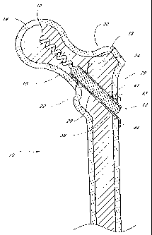
structure to prevent relative rotation of the proximal and distal ends of the
device following
implantation.
[0094] In use, the clinician first identifies a patient having a fracture to
be
treated, such as a femoral neck fracture, which is fixable by an internal
fixation device. The
clinician accesses the proximal femur, reduces the fracture if necessary and
selects a bone
drill and drills a hole 80 in accordance with conventional techniques. In the
example of a
femoral neck fracture, three holes and fixation devices will often be used as
has been
discussed. Preferably, the hole 80 has a diameter within the range from about
3 mm to
about 8 mm. This diameter may be slightly larger than the diameter of the
distal anchor 34.
The hole 80 preferably extends up to or slightly beyond the fracture 24.
[0095] A fixation device 12 having an axial length and outside diameter
suitable
for the hole 80 is selected. The distal end 32 of the fixation device 12 is
advanced distally
into the hole 80 until the distal anchor 34 reaches the distal end of the hole
80. The
proximal anchor 36 may be carried by the fixation device 12 prior to advancing
the body 28
into the hole 80, or may be attached following placement of the body 28 within
the hole 80.
Once the body 28 is in place, the clinician may use any of a variety of
driving devices, such
as electric drills or hand tools to rotate the cancellous bone anchor 34 into
the head of the
femur.
[0096] While proximal traction is applied to the proximal end 30 of body 28,
such as by conventional hemostats, pliers or a calibrated loading device, the
proximal
anchor 36 is advanced distally until the anchor 36 fits snugly against the
outer surface of the
femur or tissue adjacent the femur. Appropriate compression of the fixation
device 12
across the fracture is accomplished by tactile feedback or through the use of
a calibration
device for applying a predetermined load on the implantation device. One
advantage of the
structure of the present invention is the ability to adjust compression
independently of the
setting of the distal anchor 34.
[0097] Following appropriate tensioning of the proximal anchor 36, the
proximal extension 30 of the body 28 is preferably cut off, snapped off,
unscrewed or
-20-
CA 02442334 2003-09-25
WO 02/078555 PCT/US02/10065
otherwise removed. Body 28 may be cut using conventional saws, cutters or bone
forceps
which are routinely available in the clinical setting. Alternatively, the
fixation device can
be selected such that it is sized to length upon tensioning, so that no
proximal projection
remains.
[0098] Following removal of the proximal end 30 of body 28, the access site
may be closed and dressed in accordance with conventional wound closure
techniques.
[0099] With reference to Figure 2, in one arrangement, the proximal anchor 36
can include one or more barbs 41 extending radially outwardly from the tubular
housing 28.
The barbs 41 may be radially symmetrically distributed about the longitudinal
axis of the
tubular housing 38. Each barb 41 is provided with a transverse engagement
surface 43, for
anchoring the proximal anchor 36 in the bone. The transverse engagement
surface 43 may
lie on a plane which is transverse to the longitudinal axis of the tubular
housing 38 or may
be inclined with respect to the longitudinal axis of the tubular housing 38.
In either
arrangement, the transverse engagement surface 43 generally faces the bone
contacting
surface 46 of the flange 44. As such, the transverse engagement surface 43
inhibits
proximal movement of the proximal anchor 36 with respect to the bone.
[0100] The barbs 41 allow the bone fixation device to capture "secondary
compression" of the fracture. As explained above, the bone fixation device can
be used to
provide an initial compression across the fracture when the proximal anchor 36
is
appropriately tensioned. However, as the patient applies weight or stress to
the bone post
procedure, the fracture typically undergoes secondary compression, which
further
compresses the fracture. During such secondary compression, the barbs 41
prevent
proximal movement of the proximal anchor 36 with respect to the bone. The
ratchet-type
structures 40, 42 of the proximal anchor 36 and the body 28 allow the proximal
anchor 36
to move distally along the body 28. Thus, any slack caused by the secondary
compression
is taken up by the proximal anchor 36 as the retention structures 40, 42
prevent proximal
movement of the proximal anchor 36 with respect to the body 29. This device is
therefore
self tightening after it has been implanted in the patient.
[0101] Preferably, the clinician will have access to an array of fixation
devices
12, having, for example, different diameters, axial lengths and angular
relationships. These
may be packaged one per package in sterile envelopes or peelable pouches, or
in dispensing
cartridges which may each hold a plurality of devices 12. Upon encountering a
fracture for
-21-
CA 02442334 2003-09-25
WO 02/078555 PCT/US02/10065
which the use of a fixation device is deemed appropriate, the clinician will
assess the
dimensions and load requirements, and select a fixation device from the array
which meets
the desired specifications.
[0102] In some types of fractures such as a femoral neck fracture, a clinician
may want to introduce two or three or more fixation devices 12 into the
femoral head 14 to
secure the fracture 24. This may be desirable if the clinician determines
that, based upon
the nature of the fracture 24, there is a possibility that the head 14 of the
femur 10 could
rotate about a single fixation device 12. Even minor rotation can inhibit the
healing of the
fracture. Significant rotation can result in failure of the fixation device or
necrosis of the
femoral head. Two fixation devices 12 may also be desirable where the
direction of the
fracture is generally parallel to the axis of implantation as is understood in
the art.
[0103] Referring to Figure 6, there is disclosed a variation of the proximal
anchor 36 in which the proximal anchor 36 is integrally formed with or
attached to a plate.
The fixation device 12 in Figure 6 may otherwise be identical to the
embodiments
previously discussed. The proximal anchor 90 comprises an elongated flange 92,
which
extends from the housing 93 longitudinally down (anatomically caudad or
distally) the body
17 of the femur 10. The elongated flange 92 preferably includes one or more
openings 94
for receiving one or more femoral shaft screws 96. The flange 92 may or may
not extend
above (anatomically proximal to) the housing 93. Elimination of a proximal
flange may
more easily permit rotational removal of the proximal anchor 36 from the body
28 by
reverse rotation in an inclined flange embodiment.
[0104] Referring to Figure 6A, there is illustrated a cross sectional
schematic
view of an integral proximal anchor 36 and proximal plate. The dimensions and
orientation
of the proximal anchor 36 may be varied widely, depending upon the intended
application.
For example, a longitudinal axis of the housing 93 may be inclined or
perpendicular with
respect to the plane of flange 92. The flange 92 may have any of a variety of
dimensions
and profiles, depending upon the intended application. Lengths of the plate 92
in the
vertical direction as illustrated on Figure 6A, for use in femoral neck
fixation fractures, may
range from at least about 0.5 inches to about 10 inches or more. The plate 92
may be planar
as illustrated, particularly in small plate embodiments, or may be curved or
contoured to
improve seating of the plate 92 against the adjacent bone. Plate 92 may be
provided with
-22-
CA 02442334 2003-09-25
WO 02/078555 PCT/US02/10065
one or more apertures for receiving bone screws or other fixation devices as
illustrated in
Figures 6 and 7A.
[0105] Referring to Figure 7A, the fixation device 12 is schematically
illustrated
in combination with a conventional plate 100. The fixation device 12 in Figure
7A may be
identical to the embodiments described elsewhere herein. The fixation device
12 is used
with an elongated side support or plate 100, which extends longitudinally
above and below
the hole 80. The elongated side plate 100 includes an opening 102 that
preferably has a
diameter that is slightly larger than the diameter of the housing 38. The
elongated side
plate 100 preferably also includes one or more openings 104 for receiving one
or more
femoral shaft screws 106. Advantageously, the elongated side plate 100 spreads
the forces
exerted by the flange 44 across a larger area of the femur 17, and affects the
distribution of
load. In an alternate embodiment, the elongated side plate can 100 include one
or more
openings above the housing 38 for receiving trochanteric anchor screws (not
shown).
[0106] A contoured side plate 100 is illustrated in Figure 7B. The proximal
anchor 36 is also formed with a tapered (e.g. conical or concave outwardly)
bone or plate
contacting surface on flange 44.
[0107] The fixation device 12 of the present invention may also be used in
combination with intramedullary nails or rods 101 as schematically illustrated
in Figure 7C,
as will be understood by those of skill in the art.
[0108] The fixation device 12 of the present invention may be used in any of a
wide variety of anatomical settings beside the proximal femur, as has been
discussed. For
example, lateral and medial malleolar fractures can be readily fixed using the
device of the
present invention. Referring to Figure 10, there is illustrated an anterior
view of the distal
fibula 120 and tibia 122. The fibula 120 terminates distally in the lateral
malleolus 124,
and the tibia 122 terminates distally in the medial malleolus 126.
[0109] A fixation device 12 in accordance with the present invention is
illustrated as extending through the lateral malleolus 124 across the lateral
malleolar
fracture 128 and into the fibula 120. Fixation device 12 includes a distal
anchor 34 for
fixation within the fibula 120, an elongate body 28 and a proximal anchor 36
as has been
discussed.
[0110] Figure 10 also illustrates a fixation device 12 extending through the
medial malleolus 126, across a medial malleolar fracture 130, and into the
tibia 122.
-23-
CA 02442334 2003-09-25
WO 02/078555 PCT/US02/10065
Although Figure 10 illustrates fixation of both a lateral malleolar fracture
128 and medial
malleolar fracture 130, either fracture can occur without the other as is well
understood in
the art. Installation of the fixation devices across malleolar fractures is
accomplished
utilizing the same basic steps discussed above in connection with the fixation
of femoral
neck fractures.
[0111] Figures 11-13 illustrate a modified embodiment of a fixation device 512
having certain features and advantages according to the present invention. As
with the
previous embodiments, the fixation device 512 comprises a body 28 extending
between a
proximal end 30 and a distal end 32. The length, diameter and construction
materials of the
body 28 can be varied, depending upon the intended clinical application. In an
embodiments optimized for various fractures in an adult human population, the
body 28
will generally be within the range of from about 10 mm to about 150 mm in
length after
sizing, and within the range of from about 2 mm to about 8 mm in maximum
diameter.
The major diameter of the helical anchor, discussed below, may be within the
range of from
about 2.7 mm to about 12 mm. In general, the appropriate dimensions of the
body 28 will
vary, depending upon the specific fracture. In rough terms, for a malleolar
fracture, shaft
diameters in the range of from about 3 mm to about 4.5 mm may be used, and
lengths
within the range of from about 25 mm to about 70 mm. For condylar fractures,
shaft
diameters within the range of from about 3.5 mm to about 6.5 mm may be used
with
lengths within the range of from about 25 mm to about 70 mm. For colles
fractures (distal
radius and ulna), diameters within the range of from about 2.0 mm to about 4.5
mm may be
used with any of a variety of lengths within the range of from about 6 mm to
about 70 mm.
[0112J Referring to Figures 12, 13, and 13A, the body 28 comprises a first
portion 536 and a second portion 538 that are coupled together at a junction
540. In the
illustrated embodiment, the first portion 536 carries the distal anchor 34
while the second
portion 538 forms the proximal end 30 of the body 28. The first and second
portions 536,
538 are preferably detachably coupled to each other at the junction 540. In
the illustrated
embodiment, the first and second portions 536, 538 are detachably coupled to
each other
via interlocking threads. Specifically, as best seen in Figure 13A, the body
28 includes an
inner surface 541, which defines a central lumen 542 that preferably extends
from the
proximal end 30 to the distal end 32 throughout the body 28. At the proximal
end of the
first portion 536, the inner surface 541 includes a first threaded portion
544. The first
-24-
CA 02442334 2003-09-25
WO 02/078555 PCT/US02/10065
threaded portion 544 is configured to mate with a second threaded portion 546,
which is
located on the outer surface 545 of the second portion 538. The interlocking
annular
threads of the first and second threaded portions 544, 546 allow the first and
second
portions 536, 538 to be detachably coupled to each other. In one modified
embodiment, the
orientation of the first and second threaded portions 544, 546 can be
reversed. That is, the
first threaded portion 544 can be located on the outer surface of the first
portion 536 and the
second threaded portion 546 can be located on the inner surface 541 at the
distal end of the
second portion 538. Any of a variety of other releasable complementary
engagement
structures may also be used, to allow removal of second portion 538 following
implantation, as is discussed below.
(0113] In a modified arrangement, the second portion 538 can comprise any of a
variety of tensioning elements for permitting proximal tension to be placed on
the distal
anchor 34 while the proximal anchor is advanced distally to compress the
fracture. For
example, any of a variety of tubes or wires can be removably attached to the
first portion
536 and extend proximally to the proximal handpiece. In one such arrangement,
the first
portion 536 can include a releasable connector in the form of a latching
element, such as an
eye or hook. The second portion 538 can include a complementary releasable
connector
(e.g., a complementary hook) for engaging the first portion 536. In this
manner, the second
portion 538 can be detachably coupled to the first portion 536 such proximal
traction can be
applied to the first portion 536 through the second portion as will be
explained below.
Alternatively, the second portion 548 may be provided with an eye or hook, or
transverse
bar, around which or through which a suture or wire may be advanced, both ends
of which
are retained at the proximal end of the device. Following proximal tension on
the
tensioning element during the compression step, one end of the suture or wire
is released,
and the other end may be pulled free of the device. Alternate releasable
proximal
tensioning structures may be devised by those of skill in the art in view of
the disclosure
herein.
[0114] As described above, the proximal end 30 of the fixation device is
provided with a proximal anchor 36 that is distally moveable along the body
28, to permit
compression of the fracture 24 as will be apparent from the description below.
As will be
explained below, complimentary locking structures such as threads or ratchet
like structures
between the proximal anchor 36 and the body 28 resist proximal movement of the
anchor
-25-
CA 02442334 2003-09-25
WO 02/078555 PCT/US02/10065
36 with respect to the body 28 under normal use conditions. The proximal
anchor 36
preferably can be axially advanced along the body 28 without rotation as will
be apparent
from the disclosure herein.
[0115] In the illustrated embodiment, proximal anchor 36 comprises a housing
552 such as a tubular body, for coaxial movement along the body 28. As best
seen in
Figures 11 and 13, in a final position, the housing 552 extends distally past
the junction 540
between the first portion 536 and the second portion 538. The housing 552 is
provided
with one or more surface structures 554 such as a radially inwardly projecting
flange 556
(see Figures 13B and 13C), for cooperating with complementary surface
structures 558 on
the first portion 536 of the body 28. In the illustrated embodiment, the
complimentary
surface structures 558 comprise a series of annular ridges or grooves 560. The
surface
structures 554 and complementary surface structures 558 permit distal axial
travel of the
proximal anchor 36 with respect to the body 28, but resist proximal travel of
the proximal
anchor 36 with respect to the body 28.
[0116] For example, as best seen in Figure 13B, the proximal end of the flange
556 is biased towards the longitudinal axis of the body 28. As such, when the
proximal
anchor 36 is urged proximally with respect to the body 28, the flange 556
engages the
grooves or ridges 560 of the complementary surface structures 558. This
prevents proximal
movement of the proximal anchor 36 with respect to the body 28. In contrast,
as best seen
in Figure 13C, when the proximal anchor 36 is moved distally with respect to
the body 28,
the flange 556 can bend outwardly away from the body 28 and the ridges 560 so
as to allow
the proximal anchor 36 to move distally. Of course, those of skill in the art
will recognize
that there are a variety of other complementary surface structures, which
permit one way
ratchet like movement. For example, a plurality of annular rings or helical
threads, ramped
ratchet structures and the like for cooperating with an opposing ramped
structure or pawl
can also be used. In one embodiment, opposing screw threads are dimensioned to
function
as a ratchet.
[0117] Retention structures 558 are spaced axially apart along the body 28,
between a proximal limit 562 and a distal limit 564. The axial distance
between proximal
limit 562 and distal limit 564 is related to the desired axial working range
of the proximal
anchor 36, and thus the range of functional sizes of the fixation device 512.
Thus, as
-26-
CA 02442334 2003-09-25
WO 02/078555 PCT/US02/10065
described above, the fixation device 512 can provide compression across a
fracture
throughout a range of motion following the placement of the distal anchor.
[0118] As with the previous embodiments, the proximal anchor 36 includes a
flange 44 that seats against the outer surface of the femur or tissue adjacent
the femur. In
the illustrated embodiment, the flange 44 is an annular flange, to optimize
the footprint or
contact surface area between the flange 44 and the femur. Circular or
polygonal shaped
flanges for use in femoral head fixation will generally have a diameter of at
least about 4
mm greater than the adjacent body 28 and often within the range of from about
4 mm to
about 20 mm or more greater than the adjacent body 28. In the illustrated
embodiment, the
bone contacting surface 46 of the flange 44 is tapered and generally faces the
shaft 17 of the
femur 10.
[0119] In a modified embodiment, the housing 552 of the proximal anchor 36
can include one or more one or more barbs that extend radially outwardly from
the tubular
housing 552. As described above, such barbs provide for self tightening after
the device has
been implanted in the patient. The barbs may be radially symmetrically
distributed about
the longitudinal axis of the housing 552. Each barb is provided with a
transverse
engagement surface, for anchoring the proximal anchor 50 in the bone. The
transverse
engagement surface may lie on a plane which is transverse to the longitudinal
axis of the
housing 552 or may be inclined with respect to the longitudinal axis of the
tubular housing
552. In either arrangement, the transverse engagement surface generally faces
the bone
contacting surface 46 of the flange 44. As such, the transverse engagement
surface inhibits
proximal movement of the proximal anchor with respect to the bone.
[0120] As mentioned above, the clinician can be provided an array of proximal
anchors 36 of varying angular relationships between the bone contacting
surface 46 and the
longitudinal axis of the body 28 and housing 552 (e.g., 90 , 100 , 110 , 120 ,
and 130 ). A
single body 28 can be associated with the array such as in a single sterile
package. The
clinician upon identifying the entrance angle of the body 28 and the
associated entrance
point surface orientation of the femur 10 can choose the anchor 36 from the
array with the
best fit angular relationship, for use with the body 28.
[0121] With particular reference to Figures 12 and 13, the proximal end 30 of
the body 28 may be provided with a rotational coupling 570, for allowing the
second
portion 538 of the body 28 to be rotationally coupled to a rotation device.
The proximal
-27-
CA 02442334 2003-09-25
WO 02/078555 PCT/US02/10065
end 30 of the body 28 may be desirably rotated to accomplish one or two
discrete functions.
In one application of the invention, the proximal end 30 is rotated to remove
the second
portion 538 of the body 28 following tensioning of the device across a
fracture or to anchor
an attachment to the bone. Rotation of the rotational coupling 570 may also be
utilized to
rotationally drive the distal anchor into the bone. Any of a variety of
rotation devices may
be utilized, such as electric drills or hand tools, which allow the clinician
to manually rotate
the proximal end 30 of the body. Thus, the rotational coupling 570 may have
any of a
variety of cross sectional configurations, such as one or more flats or
splines.
[0122] In one embodiment, the rotational coupling 570 comprises a proximal
projection of the body 28 having an axial recess with a polygonal cross
section, such as a
hexagonal cross section. The rotational coupling 570 is illustrated as a
female component,
machined or milled or attached to the proximal end 30 of the body 28. However,
the
rotational coupling may also be in the form of a male element, such as a
hexagonal or other
noncircular cross sectioned projection.
[0123] In the illustrated embodiment, the body 28 is cannulated to accommodate
installation over a placement wire as is understood in the art. The cross
section of the
illustrated central cannulation is circular but in other embodiments may be
non circular,
e.g., hexagonal, to accommodate a corresponding male tool for installation or
removal of
the second portion 538 of the body 28 as will be explained below. In other
embodiments,
the body 28 may partially or wholly solid.
[0124] As with the previous embodiments, the distal anchor 34 comprises a
helical locking structure 60 for engaging cancellous and/or distal cortical
bone. In the
illustrated embodiment, the locking structure 60 comprises a flange that is
wrapped around
the axial lumen. The flange extends through at least one and generally from
about two to
about 50 or more full revolutions depending upon the axial length of the
distal anchor and
intended application. For most femoral neck fixation devices, the flange will
generally
complete from about 2 to about 20 revolutions. The helical flange 60 is
preferably
provided with a pitch and an axial spacing to optimize the retention force
within cancellous
bone, to optimize compression of the fracture.
[0125] The helical flange 60 of the illustrated embodiment has a generally
triangular cross-sectional shape. However, it should be appreciated that the
helical flange
72 can have any of a variety of cross sectional shapes, such as rectangular,
oval or other as
-28-
CA 02442334 2003-09-25
WO 02/078555 PCT/US02/10065
deemed desirable for a particular application through routine experimentation
in view of the
disclosure herein. As described above with respect to Figures 3A-31, the outer
edge of the
helical flange 60 defines an outer boundary. The ratio of the diameter of the
outer boundary
to the diameter of the central lumen can be optimized with respect to the
desired retention
force within the cancellous bone and giving due consideration to the
structural integrity and
strength of the distal anchor 34. Another aspect of the distal anchor 34 that
can be
optimized is the shape of the outer boundary and the central core, which in
the illustrated
embodiment are generally cylindrical.
[0126] The distal end 32 and/or the outer edges of the helical flange 60 may
be
atraumatic (e.g., blunt or soft). This inhibits the tendency of the fixation
device 12 to
migrate anatomically proximally towards the hip joint bearing surface after
implantation
(i.e., femoral head cut-out). Distal migration is also inhibited by the
dimensions and
presence of the proximal anchor 36, which has a larger footprint than
conventional screws.
[0127] A variety of other arrangements for the distal anchor 32 can also be
used.
For example, the various distal anchors described above be incorporated into
the fixation
device 512. In particular, the distal anchor may comprise a single helical
thread
surrounding a central core, much as in a conventional screw, which has been
cannulated to
facilitate placement over a wire. Alternatively, a double helical thread may
be utilized,
with the distal end of the first thread rotationally offset from the distal
end of the second
thread. The use of a double helical thread can enable a greater axial travel
for a given
degree of rotation and greater retention force than a corresponding single
helical thread.
Specific distal anchor designs can be optimized for the intended use, taking
into account
desired performance characteristics, the integrity of the distal bone, and
whether the distal
anchor is intended to engage exclusively cancellous bone or will also engage
cortical bone.
[0128] With particular reference to Figures 11 and 14, the fixation device 512
may include an antirotation lock between the first portion 536 of the body 28
and the
proximal collar 36. In the illustrated embodiment, the first portion 536
includes a pair of
flat sides 580, which interact with corresponding flat structures 582 in the
proximal collar
36. One or three or more axially extending flats may also be used. As such,
rotation of the
proximal collar 36 is transmitted to the first portion 536 and distal anchor
34 of the body
28. Of course, those of skill in the art will recognize various other types of
splines or other
-29-
CA 02442334 2003-09-25
WO 02/078555 PCT/US02/10065
interfit structures can be used to prevent relative rotation of the proximal
anchor and the
first portion 536 of the body 28.
[0129] To rotate the proximal collar, the flange 44 is preferably provided
with a
gripping structure to permit an insertion tool to rotate the flange 44. Any of
a variety of
gripping structures may be provided, such as one or more slots, flats, bores
or the like. In
one embodiment, the flange 44 is provided with a polygonal, and, in
particular, a
pentagonal or hexagonal recess 84. See Figure 13.
[0130] In use, the clinician first identifies a patient having a fracture to
be
treated, such as a femoral neck fracture, which is fixable by an internal
fixation device. The
clinician accesses the proximal femur, reduces the fracture if necessary and
selects a bone
drill and drills a hole 80 (see Figure 15A) in accordance with conventional
techniques.
Frequently, the hole 80 has a diameter within the range from about 3 mm to
about 8 mm.
This diameter may be slightly larger than the diameter of the distal anchor
34. The hole 80
preferably extends up to or slightly beyond the fracture 24.
[0131] A fixation device 12 having an axial length and outside diameter
suitable
for the hole 80 is selected. The distal end 32 of the fixation device 12 is
advanced distally
into the hole 80 until the distal anchor 34 reaches the distal end of the hole
80. The
proximal anchor 50 may be carried by the fixation device 12 prior to advancing
the body 28
into the hole 80, or may be attached following placement of the body 28 within
the hole 80.
Once the body 28 and proximal anchor 36 are in place, the clinician may use
any of a
variety of driving devices, such as electric drills or hand tools to rotate
the proximal anchor
36 and thus cancellous bone anchor 34 into the head of the femur.
[0132] Once the anchor 34 is in the desired location, proximal traction is
applied to the proximal end 30 of body 28, such as by conventional hemostats,
pliers or a
calibrated loading device, while distal force is applied to the proximal
anchor 36. In this
manner, the proximal anchor 36 is advanced distally until the anchor 36 fits
snugly against
the outer surface of the femur or tissue adjacent the femur and the fracture
24 is completely
reduced as shown in Figure 15B. Appropriate tensioning of the fixation device
512 is
accomplished by tactile feedback or through the use of a calibration device
for applying a
predetermined load on the implantation device. One advantage of the structure
of the
present invention is the ability to adjust compression independently of the
setting of the
distal anchor 34.
-30-
CA 02442334 2003-09-25
WO 02/078555 PCT/US02/10065
[0133] Following appropriate tensioning of the proximal anchor 36, the second
portion 538 of the body 28 is preferably detached from the first portion 536
and removed.
See Figure 15C. In the illustrated embodiment, this involves rotating the
second portion
538 with respect to the first portion via the coupling 570. In connection with
many of the
fractures identified previously herein, a single fixation device 512 may be
all that is
clinically indicated. However, two or three or more fixation devices 512 may
be utilized to
reduce a single fracture in a similar manner as illustrated in Figure 1,
depending upon the
location and physical requirements of the fractured portion of the bone. For
example, in the
case of proximal femoral fractures of the type illustrated herein, typically
at least two and
preferably three fixation devices 12 will be implanted to span the femoral
neck. The use of
three fixation devices 512 desirably provides sufficient compression across
the fracture, as
well as minimizes the risk of rotation of the head of the femur around the
axis of a single
fixation device 512. The proximal end of the fixation devices may be connected
together
such as through a three-holed plate or rod, or may be independent of each
other.
[0134) Following removal of the second portion 538 of each body 28, the access
site may be closed and dressed in accordance with conventional wound closure
techniques.
[0135] In a modified arrangement, the second portion 538 may form part of the
driving device, which is used to rotate the proximal anchor 36 and thus
cancellous bone
anchor 34 into the head of the femur. The second portion 538 is used to apply
proximal
traction so as to compress the fracture. After appropriate tensioning, the
second portion
538 can be de-coupled from the first portion 536 and removed with the driving
device.
[0136] In the foregoing variation, the second portion 538 may be connected to
a
rotatable control such as a thumb wheel on the deployment device. A container
may be
opened at the clinical site exposing the proximal end of the implant, such
that the distal end
of the second portion 538 may be removably coupled thereto. Proximal
retraction of the
hand tool will pull the implant out of its packaging. The implant may then be
positioned
within the aperture in the bone, rotated to set the distal anchor, and the
hand piece may be
manipulated to place proximal traction on the second portion 538 while
simultaneously
distally advancing the proximal anchor. Following appropriate tensioning
across the
fracture, the second portion 38 may be disengaged from the implant, and
removed from the
patient. In the example of a threaded engagement, the second portion 538 may
be
disengaged from the implant by rotating a thumb wheel or other rotational
control on the
-31-
CA 02442334 2009-01-02
hand piece. In an alternate embodiment, such as where the second portion 538
comprises a
pull wire, following appropriate tensioning across the fracture, a first end
of the pull wire is
released such that the pull wire may be removed from the implant by proximal
retraction of
the second end which may be attached to the hand piece.
[0137] As mentioned above, the fixation device 512 may also be used in
combination with intramedullary nails or rods, as will be understood by those
of skill in the
art. The fixation device 512 may be used in any of a wide variety of
anatomical settings beside
the proximal femur, as has been discussed. For example, lateral and medial
malleolar fractures
can be readily fixed using the device of the present invention. See e.g.,
Figure 10.
[0138] The fixation devices of the present invention may be made from either
conventional bioabsorbable materials or conventional non-absorbable materials,
combinations
thereof and equivalents thereof. In addition, natural materials such as
allografts may be used.
Examples of absorbable materials include homopolymers and copolymers of
lactide,
glycolide, trimethylene carbonate, caprolactone, and p-dioxanone and blends
thereof The
following two blends may be useful: 1) the blend of poly(p-dioxanone) and a
lactide/glycolide
copolymer, as disclosed in U.S. Pat. No. 4,646,741 and (2) the glycolide-rich
blend of two or
more polymers, one polymer being a high lactide content polymer, and the other
being a high
glycolide content disclosed in U.S. Pat. No. 4,889,119.
[0139] A variety of polymers which may be useful for the components of the
present invention are identified below. Many of these polymers have been
reported to be
biodegradable into water-soluble, non-toxic materials which can be eliminated
by the body:
Polycaprolactone
Poly (L-lactide)
Poly (DL-lactide)
Polyglycolide
Poly (L-Lactide-co-D, L-Lactide)
70:30 Poly (1-Lactide-co-D, L-Lactide)
95:5 Poly (DL-lactide-co-glycolide)
90:10 Poly (DL-lactide-co-glycolide)
85:15 Poly (DL-lactide-co-glycolide)
-32-
CA 02442334 2003-09-25
WO 02/078555 PCT/US02/10065
75:25 Poly (DL-lactide-co-glycolide)
50:50 Poly (DL-lactide-co-glycolide)
90:10 Poly (DL-lactide-co-caprolactone)
75:25 Poly (DL-lactide-co-caprolactone)
50:50 Poly (DL-lactide-co-caprolactone)
Polydioxanone
Polyesteramides
Copolyoxalates
Polycarbonates
Poly (glutamic-co-leucine)
[0140] The desirability of any one or a blend of these or other polymers can
be
determined through routine experimentation by one of skill in the art, taking
into account
the mechanical requirements, preferred manufacturing techniques, and desired
reabsorption
time. Optimization can be accomplished through routine experimentation in view
of the
disclosure herein.
[0141] Alternatively, the components can be molded, formed or machined from
biocompatible metals such as Nitinol, stainless steel, titanium, and others
known in the art. In
one embodiment, the components of the bone fixation device are injection-
molded from a
bioabsorbable material, to eliminate the need for a post-healing removal step.
One suitable
bioabsorbable material which appears to exhibit sufficient structural
integrity for the purpose
of the present invention is poly-p-dioxanone, such as that available from the
Ethicon Division
of Johnson & Johnson. Poly (L-lactide, or co-DL-lactide) or blends of the two
may
alternatively be used as mentioned above. As used herein, terms such as
bioabsorbable,
bioresorbable and biodegradable interchangeably refer to materials which will
dissipate in
situ, following a sufficient bone healing period of time, leaving acceptable
byproducts. All or
portions of any of the devices herein, as may be appropriate for the
particular design, may be
made from allograft material, or synthetic bone material as discussed
elsewhere herein.
[0142] The bioabsorbable implants of this invention can be manufactured in
accordance with any of a variety of techniques known in the art, depending
upon the
particular polymers used, as well as acceptable manufacturing cost and
dimensional tolerances
as will be appreciated by those of skill in the art in view of the disclosure
herein. For
example, any of a variety of bioabsorbable polymers, copolymers or polymer
mixtures can be
-33-
CA 02442334 2009-01-02
molded in a single compression molding cycle, or the surface structures can be
machined on
the surface of the pin or sleeve after the molding cycle. It is also possible
to use the techniques
of U.S. Pat. No. 4,743,257, to mold absorbable fibers and binding polymers
together, to create
a fiber- reinforced absorbable device.
[0143] An oriented or self-reinforced structure for the device can also be
created during extrusion or injection molding of absorbable polymeric melts
through a
suitable die or into a suitable mold at high speed and pressure. When cooling
occurs, the flow
orientation of the melt remains in the solid material as an oriented or self-
reinforcing structure.
The mold can have the form of the finished device component, but it is also
possible to
manufacture the components of the invention by machining injection-molded or
extruded
semifinished products. It may be advantageous to make the components from melt-
molded,
solid state drawn or compressed, bioabsorbable polymeric materials, which are
described, e.g.,
in U.S. Pat. Nos. 4,968,317 and 4,898,186.
[0144] Reinforcing fibers suitable for use in the anchor components of the
present invention include ceramic fibers, like bioabsorbable hydroxyapatite or
bioactive glass
fibers. Such bioabsorbable, ceramic fiber reinforced materials are described,
e.g., in published
European Patent Application No. 0146398 and in WO/96/21628.
[0145] As a general feature of the orientation, fiber-reinforcement or self-
reinforcement of the components, many of the reinforcing elements are oriented
in such a way
that they can carry effectively the different external loads (such as tensile,
bending and shear
loads) that are directed to the anchor as used.
[0146] The oriented and/or reinforced anchor materials for many applications
have tensile strengths in the range of about 100-2000 MPa, bending strengths
in the range of
about 100-600 MPa and shear strengths in the range of about 80-400 MPa,
optimized for any
particular design and application. Additionally, they are relatively stiff and
tough. These
mechanical properties may be superior to those of non-reinforced or non-
oriented absorbable
polymers, which often show strengths between about 40 and 100 MPa and are
additionally
may be flexible or brittle. See, e.g., S. Vainionpaa, P. Rokkanen and P.
Tormnld, "Surgical
Applications of Biodegradable Polymers in Human Tissues", Progr. Polym. Sci.,
Vol. 14,
(1989) at 679-716.
[0147] The components of the invention (or a bioabsorbable polymeric coating
layer on part or all of the component surface), may contain one or more
bioactive substances,
such as antibiotics, chemotherapeutic substances, angiogenic growth factors,
substances for
-34-
CA 02442334 2009-01-02
accelerating the healing of the wound, growth hormones, antithrombogenic
agents, bone
growth accelerators or agents, and the like. Such bioactive implants may be
desirable because
they contribute to the healing of the injury in addition to providing
mechanical support.
[0148] In addition, the components may be provided with any of a variety of
structural modifications to accomplish various objectives, such as
osteoincorporation, or more
rapid or uniform absorption into the body. For example, osteoincorporation may
be enhanced
by providing a micropitted or otherwise textured surface on the anchor
components.
Alternatively, capillary pathways may be provided throughout the pin and
collar, such as by
manufacturing the anchor components from an open cell foam material, which
produces
tortuous pathways through the device. This construction increases the surface
area of the
device which is exposed to body fluids, thereby generally increasing the
absorption rate.
Capillary pathways may alternatively be provided by laser drilling or other
technique, which
will be understood by those of skill in the art in view of the disclosure
herein. In general, the
extent to which the anchor can be permeated by capillary pathways or open cell
foam
passageways may be determined by balancing the desired structural integrity of
the device
with the desired reabsorption time, taking into account the particular
strength and absorption
characteristics of the desired polymer.
[0149] One open cell bioabsorbable material is described in U.S. Patent No.
6,005,161 as a poly(hydroxy) acid in the form of an interconnecting, open-cell
meshwork
which duplicates the architecture of human cancellous bone from the iliac
crest and possesses
physical property (strength) values in excess of those demonstrated by human
(mammalian)
iliac crest cancellous bone. The gross structure is said to maintain physical
property values at
least equal to those of human, iliac crest, cancellous bone for a minimum of
90 days following
implantation.
-35-
CA 02442334 2003-09-25
WO 02/078555 PCT/US02/10065
[0150] The fixation devices may also be made from conventional non-
absorbable, biocompatible materials including stainless steel, titanium,
alloys thereof,
polymers, composites and the like and equivalents thereof. In one embodiment,
the distal
anchor comprises a metal helix, while the body and the proximal anchor
comprise a
bioabsorbable material. Alternatively, the distal anchor comprises a
bioabsorbable
material, and the body and proximal anchor comprise either a bioabsorbable
material or a
non-absorbable material. As a further alternative, each of the distal anchor
and the body
comprise a non-absorbable material, connected by an absorbable link. This may
be
accomplished by providing a concentric fit between the distal anchor and the
body, with a
transverse absorbable pin extending therethrough. This embodiment will enable
removal of
the body following dissipation of the pin, while leaving the distal anchor
within the bone.
[0151] The components of the invention (or a bioabsorbable polymeric coating
layer on part or all of the anchor surface), may contain one or more bioactive
substances,
such as antibiotics, chemotherapeutic substances, angiogenic growth factors,
substances for
accelerating the healing of the wound, growth hormones, antithrombogenic
agents, bone
growth accelerators or agents, and the like. Such bioactive implants may be
desirable
because they contribute to the healing of the injury in addition to providing
mechanical
support.
[0152] In addition, the components may be provided with any of a variety of
structural modifications to accomplish various objectives, such as
osteoincorporation, or
more rapid or uniform absorption into the body. For example,
osteoincorporation may be
enhanced by providing a micropitted or otherwise textured surface on the
components.
Alternatively, capillary pathways may be provided throughout the body and
collar, such as
by manufacturing the anchor and body from an open cell foam material, which
produces
tortuous pathways through the device. This construction increases the surface
area of the
device which is exposed to body fluids, thereby generally increasing the
absorption rate.
Capillary pathways may alternatively be provided by laser drilling or other
technique,
which will be understood by those of skill in the art in view of the
disclosure herein. In
general, the extent to which the anchor can be permeated by capillary pathways
or open cell
foam passageways may be determined by balancing the desired structural
integrity of the
device with the desired reabsorption time, taking into account the particular
strength and
absorption characteristics of the desired polymer.
-36-
CA 02442334 2003-09-25
WO 02/078555 PCT/US02/10065
[01531 The components of the present invention may be sterilized by any of the
well known sterilization techniques, depending on the type of material.
Suitable
sterilization techniques include heat sterilization, radiation sterilization,
such as cobalt 60
irradiation or electron beams, ethylene oxide sterilization, and the like.
[01541 The specific dimensions of any of the bone fixation devices of the
present invention can be readily varied depending upon the intended
application, as will be
apparent to those of skill in the art in view of the disclosure herein.
Moreover, although
the present invention has been described in terms of certain preferred
embodiments, other
embodiments of the invention including variations in dimensions, configuration
and
materials will be apparent to those of skill in the art in view of the
disclosure herein. In
addition, all features discussed in connection with any one embodiment herein
can be
readily adapted for use in other embodiments herein. The use of different
terms or
reference numerals for similar features in different embodiments does not
imply differences
other than those which may be expressly set forth. Accordingly, the present
invention is
intended to be described solely by reference to the appended claims, and not
limited to the
preferred embodiments disclosed herein.
-37-