Note : Les descriptions sont présentées dans la langue officielle dans laquelle elles ont été soumises.
CA 02444413 2003-10-09
-1-
VIOLET LASER INDUCED FLUORESCENCE FOR CANCER DIAGNOSIS
This invention provides an economical, portable, device which can produce
images (visible by eye
and recorded) of violet laser induced fluorescence on natural materials more
especially breast cancer.
During surgery, the use of laser fluorescence helps to determine the extent of
the malignancy, while
the recording unit preserves images of the actual operation.
BACKGROUND OF THE INVENTION
Cancer is one of the major diseases of modern societies and, in particular,
breast cancer is the
leading cause of death among women in North America. About 1 in 8 women in the
United States
and Canada will develop breast cancer. In 2003, 211,300 new cases of breast
cancer will be found
and 39,800 men and women will be lost to this disease in the United States
alone. Because surgery
is the first treatment for breast cancer, such surgery is the most common type
of surgical procedure
in Cancer Clinics.
It has been known for some considerable time that cancer cells fluoresce a
different colour than
normal cells of the same type. However, fluorescence has not been effectively
used as real time aid
in surgical procedures. It is also suspected that certain types of pre-
cancerous tissue may possess a
characteristic fluorescent colour. In addition, different types of normal
tissue not known to be
diseased can also have a variable fluorescence signature. Also, the same
disease may present in
different patients with different characteristics. Obviously, the more
information that surgeons and
pathologists and the like have immediately at their disposal, the more rapid
and accurate will be the
diagnosis and the less will be the discomfort for the patient. It is axiomatic
that rapid treatment
increases the survival rate. The present invention is designed to address
these concerns.
PRIOR ART
Other inventions have dealt with similar problems (but generally not
specifically breast cancer) by
using spectrometry, computers, compound optical trains, etc. Existing
inventions have technical
problems common in this field such as high cost (estimated to be in the range
of $100,000 to
$500,000), complexity of design, difficulty of use (combinations of
spectrometry, video imaging)
and a lack of ruggedness and portability. In particular, surgeons and
pathologists may not be
familiar with computerized digital spectroscopy and it may not be appropriate
to place such
CA 02444413 2003-10-09
-2-
equipment in operating theaters. In cases where ultraviolet light is used to
illuminate the sample to
be examined, there is also an inherent safety problem as ultraviolet light is
dangerous to the human
eye. Safety is seldom discussed in any of the patents in this field.
U. S. Pat. 6393315 May 21, 2002 Aprahamian, P. M., et al.
This invention requires two light sources. "Luminous excitation is preferably
constituted by two
wavelengths or two spectral bands, namely one of about 590 nm or centered on
590 nm and adapted
to excite the porphyrins, and the other of about 400 nm or centered on 400 nm
(or if desired about
355 nm), adapted to excite other endogenous chromophores". The method requires
acquiring, for
the same tissues to be analyzed, fluorescence signals in the "spectral bands
centered, respectively, on
about 600 nm (or else about 680 nm) and on about 630 nrn andlor 680-690 nm,
and, as the case may
be, on about 470 nm and/or 510-520 nm, for each of the points to be measured".
None of figures
relate to human patients and there is no provision for real time imaging or
portability. The figured
spectra are different from the spectra collect in this work using Violet Laser
Excitation.
U.S. Pat. 5,467,767 Nov. 29, 1995 Alfano, R. R., et al.
In one embodiment, the method comprises irradiating a human breast tissue
sample with light at a
wavelength of about 310 nm (Ultra Violet) and measuring the time-resolved
fluorescence emitted
therefrom at about 340 nm. The time-resolved fluorescence profile is then
compared to similar
profiles obtained from known malignant and non-malignant human breast tissues.
This invention
has a similar objective to the present invention but used a different method
U.S. Pat. 5381224 Jan. 10, 1995 Dixon, A. E., et al.
This invention relates to the field of Scanning Laser Imaging Systems when
used to image
macroscopic specimens. The diagrams show a quite complicated conoscopic device
which is likely
to be quite expensive to manufacture. There are no photos produced by the
device and fluorescence
is mentioned in passing and apparently requires a *theta laser scan lens.
There is no provision for
portability.
U.S. Pat. 6584342 Jan. 24, 2003Trushin et al.
Tissue is irradiated with low intensity monochrome radiation in the wave
length band of 630 to 645
nm and the fluorescent image is recorded within the band of 650 to 730 nm.
This is a complicated
CA 02444413 2003-10-09
device based on different technology than the present invention and which is
not portable and is
likely to be quite expensive to manufacture.
SUMMARY OF THE INVENTION
The object of this invention is to provide an economical, portable, device
which can provide images
(visible by eye and recorded) of violet laser induced fluorescence on natural
materials. Specimen
include but are not limited to: biological or medical samples which are useful
for diagnosis and study
of various diseases, or more especially breast cancer. The invention provides
real time imaging in a
portable unit and can have a sterile cover for use in operating theaters or
pathology laboratories. The
use of violet laser light provides a safety feature while maximizing the
visible luminescent effect of
the laser as explained in the description below. Violet Laser Induced
Fluorescence of human breast
tissue from known cancer patients yields fluorescent peaks at about 510, 560,
and 590 nm).
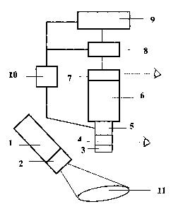
BRIEF DESCRIPTION OF FIGURE 1
FIGURE 1 is a schematic representation of the present invention with the parts
described below:
1 ) Laser, 404 + 2 nm (violet)
2) beam expander/scanner + filters
3) filters (laser blocking, visible transmission)
4) coupler for real time observation by eye
5) coupler to allow analysis of light other than by imaging
6) focusing imaging device, (6 or 7 or 8 can have a method to view the image
before permanently
saving the image)
7) recording medium for imaging device 6
8) device to transport or transmit images or other information
9) computer for analysis and enhancement
10) ancillary equipment such as optical spectrometer and the like
11 ) the specimen (such as a cancer tumour) lies within the elliptical area
illuminated by the Laser
scanner device
CA 02444413 2003-10-09
-4-
DETAILED DESCRIPTION OF THE INVENTION
Fig. 1 is a schematic diagram of the device for certain embodiments of the
present invention. The
laser 1 outputs 404 + 2 nm, more or less, of violet radiation. The output is
then expanded by 2 into a
broad beam sufficient to illuminate the sample 11. Said beam expander 2 can
use lenses, or a
scanning raster, or both, to expand the beam in an adjustable manner to
illuminate the sample area.
The sample 11, which may be observed during and after surgery, is illuminated
by the Laser
Illumination Unit, LIU (1, 2). The laser working distance between LIU and 11
is variable (from as
little as 0.1 m tb as much as several meters) using an attachment system which
is suited to the
environment of observation (for an operating room, all equipment must be
sterile).. The angle
between the axis of laser illumination and the axis of the visualizing unit is
similarly variable in two
dimensions. Thus the geometry of the total device may be adjusted in 3
dimensions to fit the
geometry and safety considerations of the environment in which the
observations are made. For use
in operating theaters and the like, the laser unit LIU is covered by a sterile
covering which can be
disposable. It is understood that in some cases it might be advantageous to
add a filter or filters to
the laser to shape the wavelength output of the laser.
The observations of the fluorescence emitted by the sample are made using the
Visualization and
Recording Unit (VRU), 3,4,5,6,7, 8, 9. Items 3,4,6,7, 8 together, form a
recording macroscope with
real time imaging. The filter unit 3 comprises at least one filter matched to
the laser so as to block
the laser illumination but which passes the rest of the visible light spectrum
produced by
fluorescence in the sample. Other filters in 3 can reduce overall intensity
and/or enhance certain
colored effects by selectively blocking certain bands within the visible light
spectrum. The working
distance between 11 and VRU (3cm to lm, more or less) is adjustable in 3
dimensions (by
attachment not shown in Fig. 1). Said adjustment is made to suit the sample
and the environment in
which the observations are being made. In most cases, the environment must be
darkened in order to
see or record the fluorescence. The visualization unit VRU contains a coupling
units in 4 and 7
which permit light to form an image for the eye (dashed line and eye-symbols
in Fig. 1). The light
can also be outputted via a coupling unit 5 to a device 10 for further
analysis such as by a
spectrometer or the like. Imaging for recording is performed by 6, an imaging
device which can be a
simple as an adjustable lens. The image is recorded on 7 using a recording
medium such as film,
ccd, video tape, video or the like, depending on the application.
CA 02444413 2003-10-09
-5-
The observations may be transported to other equipment for fwrther work by a
transport or
transmitting device 8 which can be as simple as a "memory stick". Further
analysis and/or
enhancement (not generally in real time) can be performed by 9, a computing
device. Equipment 10
for further analytical work, such as a spectrometer and the like, is coupled
optically to 5 and
electronically to 8 and 9. Device 7 can have a method to view the image before
permanently saving
the image for example on high-density digital storage devices (7, and within
9). For use in operating
theaters and the like, the Visualization Recording Unit, VRU (items 3 to 10)
is covered by a sterile
covering which can be disposable.
It is understood that power supplies, lightweight batteries, or the like, are
included in parts 1,6,8,9,
and 14 where needed. As laser diodes are extremely susceptible to adverse
heating effects, it is
taken as given that the laser 1 has a cooling apparatus including thermal
sensing device, and
switching circuit to prevent destruction by overheating. It is further
understood that since the
sample is illuminated by a broad laser beam, therefore, everyone present in
the room (or the
location) in which the fluorescent observations are taking place must wear
safety glasses of a type
matched to block the wavelength of the laser. Said safety glasses are provided
for that use with the
invention.
Violet laser light is essential to this invention for a number of reasons.
Both ultraviolet (UV), and
violet light are dangerous to the eye and both are capable of inducing
fluorescence. However, UV is
invisible to the eye. If the eye is exposed to UV light damage can be done to
the eye without
anything being seen or even felt (as there are no pain receptors in the eye,
it has no feeling of pain).
In contrast, the eye sees any stray violet light, for example, due to specular
reflection or focusing by
drops of fluid, as uncomfortably bright. The eye tends to be naturally averted
from bright points of
laser light. This phenomenon results in a built-in safety feature of the
present invention. Violet laser
light can be seen and cause the eye to look away before the accidental
exposure does any damage.
In addition, experiments done by the inventor have shown that the fluorescent
signature produced by
laser light is sharper and less noisy compared with UV light sources.
Therefore the violet laser is a
superior excitation source for the purposes of this invention. The fluorescent
effect produced by this
invention is different than that produced by UV, or visible light such as blue
(442 nm).
CA 02444413 2003-10-09
-6-
The new laser diodes are small, light; powerful, and have a nominal 10,000
hour lifetime. They do
not require plasma tubes and require about 3 to 5 volts electrical potential.
This new technology
relaxes design criteria when making an embodiment of this invention, resulting
in designs which
would not have been practicable even a few years ago. The equipment can be
battery powered and
completely portable; in some embodiments, its use is not even limited to the
surface of the earth.
In one practice, the device would be used in an operating room by a surgeon
during an operation to
remove a tumor. When the surgeon wishes to examine the texture or structure of
the material
surrounding a tumor he can dim or extinguish the room lights and simultaneous
turn on the Laser
Illumination Unit. The laser unit illuminates the tumor and surrounding tissue
causing auto-
fluorescence. Because the tumor and surrounding normal tissue fluoresce
different colors and tend
to have different textures and structures, the margins of the tumors can be
visually enhanced by
comparing the white-light and fluorescent images. Thus the use of laser
fluorescence helps to
determine the extent of the malignancy, while the recording unit preserves
images of the actual
operation. After surgery, but before fixing the tissue in formaldehyde, the
pathologist can examine
the tumor sample in detail using fluorescence and white light while he is
preparing his report. Also,
the pathology observations can be recorded and compared with the results fram
the surgery.