Note : Les descriptions sont présentées dans la langue officielle dans laquelle elles ont été soumises.
CA 02447619 2003-11-18
f1~
1
DESCRIPTION
A COMPOSITION FOR ACCELERATING BONE FRACTURE HEALING
TECHNICAL FIELD
The present invention relates to a composition for
accelerating bone fracture healing, specifically, to a
pharmaceutical composition for accelerating bone fracture
healing, which comprises as an active ingredient a PDE4
inhibitor, preferably a PDE4 inhibitor together with a
biocompatible and biodegradable polymer, which is
especially in the form of microsphere preparation, more
preferably, microsphere-containing injectable preparation,
and which is able to promote bone fracture healing when
locally administered.
BACKGROUND ART
Bone fracture is a condition where a physiological
continuity of bone tissue is partially or completely broken
off and generally classified on the basis of the outbreak
mechanism into (a) fracture by external force, (b)
pathological fracture, and (c) fatigue fracture. In
addition, the state of bone fracture is classified on the
basis of the fracture line (the line tracing the epiphysis
generated by bone transection), into fissure fracture,
greenstick fracture,' transverse fracture, oblique fracture,
spiral fracture, segmental fracture, comminuted fracture,
avulsion fracture, compression fracture, depression
fracture, and the like (IGAKU-DAIJITEN, 18th ed., pp. 719
720, published by Nanzando).
CA 02447619 2003-11-18
2
Generally, it takes a considerable time until a bone
fracture heals, which can be an obstacle in daily life.
Further, the number of bone fracture of the osteoporosis
patients, which is one of pathological fractures, has
markedly increased with the aging of population. In
particular, the transcervical fracture requires a long-term
hospitalization and often develops internal complication
including dementia due to a long-term hospitalization,
which is becoming a major social and economic issue.
The fracture healing process is mainly classified into
the following three stages ("Kossetsu Chiryougaku (Fracture
Therapeutics)", April, 2000, pp. 29-37, 46-51, Nanko-do),
and it is considered that, the healing progresses in the
reparative phase, an important stage for bone fracture
healing, by a mechanism different from that in the bone
remodeling phase where osteogenesis and osteolysis (bone
resorption) occur repeatedly.
(1) An inflammatory phase: tissue surrounding bone is
damaged, a fracture crevice is occupied with hematoma, and
inflammation arises at the fracture region.
(2) A reparative phase: two processes progress in
parallel; a process in which hematoma in the fracture
crevice is removed yielding granulation tissue, soft callus
is formed and gradually replaced by hard callus via
osteogenic mechanism (endochondral ossification), and a
process in which a new bone is formed by osteogenic cells
present in periost (fibrous/intramembranous ossification).
(3) A re-molding phase: the formed new bone extends for a
long term by repeating the bone resorption and the bone
formation, while the bone deformation is corrected and
CA 02447619 2003-11-18
3
defect region reinforced.
The new bone formed during the re-molding phase has
intensity of certain degree, and one's daily life is less
hampered; however, the reparative phase takes a long term
and restricts patient's daily life greatly. Accordingly,
it is clinically important to shorten the term of
reparative phase.
As substances accelerating bone fracture healing,
there have been disclosed peptide-type physiologically
active substances such as bone morphogenetic protein (BMP)
and transforming growth factor (TGF) (Pros. Natl. Acad.
Sci., USA, vol. 87, pp. 2220-2224 (1989) . Further, it has
been disclosed a pharmaceutical preparation for local
administration containing a compound of the formula below
(JP-04-364179A (1992)) as a bone formation accelerator
after microcapsulation with lactic acid-glycolic acid
copolymer (PLGA) in JP-09-263545A (1997).
O
N
"' O
p;0/~'CH3
I
O
H3C
The possibility of improving the bone mass by
increasing the intracellular cyclic AMP (CAMP) level with
phosphodiesterase (PDE) inhibitor was studied, and it was
reported that the increase of bone mineral density of the
backbone and the femur, and hyperplasia of cortical bone
CA 02447619 2003-11-18
r
4
were observed in a mouse received daily subcutaneous
injection of Pentoxifylline, a general PDE inhibitor, or
Rolipram that is a selective PDE4 inhibitor (Bone, vol. 27,
6th issue, pp. 811-817 (2000)).
However, the researches above are focused on the
pharmacological effect on a normal region, which is an
osteogenic region during re-modeling process, and not a
bone fracture region and are totally silent about bone
fracture healing accerelating activity of PDE4 inhibitor.
DISCLOSURE OF INVENTION
One of purposes of the present invention is to provide
a novel pharmaceutical composition for accelerating bone
fracture healing, which accelerates the healing of a
fracture in the early stage. Another purpose of the
present invention is to provide a novel pharmaceutical
composition for local administration which, when applied to
a fracture region, exerts efficiently the fracture healing
accelerating activity only at an intended site while
avoiding the manifestation of systemic action of an active
ingredient. Yet another purpose of the present invention
is to provide a sustained release depot preparation for
accelerating bone fracture healing, which, when applied
locally, can release an active ingredient gradually and
exert the drug efficacy over a long term by one time dosage.
The present inventors have investigated into
pharmacological actions of various compounds and noticed
that compounds having PDE4 inhibiting activity could affect
the fracture healing process. The inventors have then
found that the compounds having PDE4 inhibiting activity
CA 02447619 2003-11-18
~d i
can accelerate the fracture healing and established the
present invention.
The present invention provides a composition for
accelerating bone fracture healing, which comprises a PDE4
5 inhibitor as an active ingredient. In particular, the
present invention provides a pharmaceutical preparation
suitable for local administration at the fracture region,
specifically, a bone fracture healing accelerating
composition in the form of depot preparation.
BRIEF DESCRIPTION OF DRAWINGS
Figure 1 is a copy of photography showing chondrocyte
calcification (calcium deposits) effect of Compound (1) in
cultured rabbit costicartilage cells.
Figure 2 is a graph showing the reproduction of a
defective radius in a rabbit treated with PDE4 inhibitor
(Compound (2)) microsphere. The relation between the total
bone area (cross sectional) (mm2) or stress-strain index
(SSI: mma), and the dosage of Compound (2) are shown in the
upper and the lower graphs, respectively.
Figure 3 is a graph showing a time-course of cAMP
content in rat fibula fracture region that was treated with
a PDE4 inhibitor (Compound (2)) microsphere.
Figure 4 is a graph showing a time-course of cAMP
content in fibula fracture region of normal and STZ-induced
diabetic rats.
Figure 5 is a graph showing the in vitro elution
characteristics of microspheres obtained in Example 1-(4),
2-(1) and 3-(1) .
Figure 6 is a graph showing the time-course of plasma
' CA 02447619 2003-11-18
r
6
concentration of Compound (1) administered intravenously.
Data are shown by mean f standard deviation (n = 3).
Figure 7 is a graph showing the time-course of plasma
concentration of an active ingredient following the
subcutaneous injection of microsphere dispersion obtained
in Example l-(5), 2-(2) or 3-(2). Data are shown by mean f
standard deviation (n = 5).
Figure 8 is a graph showing the time-course of
Compound (1) remaining in the preparation following the
subcutaneous injection of microsphere dispersion obtained
in Example 2-(2). Data are shown by mean t standard
deviation (n = 5) .
Figure 9 is a graph showing the time-course of
Compound (2) remaining in the preparation following the
subcutaneous injection of microsphere dispersion obtained
in Example 6-(5) or 7-(2). Data are shown by mean t
standard deviation (n = 4).
BEST MODE FOR CARRYING OUT THE INVENTION
The bone fracture healing accelerating composition of
the present invention has a superior effect on the bone
fracture healing process especially in the reparative phase.
The present composition can accelerate the fracture healing
by accelerating the endochondral ossification wherein a
soft callus is formed at the fracture region, which in turn
is replaced by hard callus.
The pharmaceutical composition of the present
invention can be prepared by combining a PDE4 inhibitor as
an active ingredient and a conventional pharmaceutically
acceptable excipient or a diluting agent therefor.
CA 02447619 2003-11-18
7
Preferred pharmaceutical composition is a sustained release
composition for local administration, which contains a PDE4
inhibitors) and a biocompatible and biodegradable
polymer(s). It is further preferred that said composition
for local administration is in the form of microsphere,
which microsphere can be formulated as an injectable
preparation.
Examples of PDE4 inhibitor usable as an active
ingredient of pharmaceutical compositions of the present
invention include all the compounds having PDE4 inhibitory
activity, for example, those described in JP 05-229987A
(1993), JP 09-59255A (1997), JP 10-226685A (1998), EP
158380, WO/94/25437, USP 5,223,504, WO/95/4045, EP 497564,
EP 569414, EP 623607, EP 163965, USP 5,605,914, WO/95/35282,
WO/96/215, USP 5,804,588, U5P 5,552,438, WO/93/9118,
WO/96/31485, EP 459505, WO/97/22585, EP 738715, WO/91/16314,
WO/96/218, WO/97/18208, EP 158380, WO/99/50270, EP 260817,
WO/98/11113, WO/94/22852, EP 432856, USP 4193926,
WO/98/13348, WO/96/6843, JP 2000-503678A (W0/98/14432), JP
2000-502724A (W0/98/9961), JP 2000-510105A (W0/97/40032),
JP 2000-514804A (W0/98/2440), JP 2000-502350A (W0/97/23457),
JP 2000-501741A (W0/97/2585), and the like.
PDE can be classified into PDE1-5 according to the
teaching of "Trends in Pharmacological Sciences, vol. 11,
pp. 150-155", and PDE4 inhibitors suitable for the present
bone fracture healing accelerating composition are
preferably selective to PDE4 with higher inhibitory
activity against PDE4 compared to others (PDE1-3, 5), more
preferably have 10 times or more inhibitory activity on
PDE4 than on the other PDEs. The inhibitory activity of
CA 02447619 2003-11-18
,t
8
such PDE4 inhibitor on PDE4 is particularly preferably 50
times or more, and yet more preferably 100 times or more of
that on the other PDEs.
Preferable PDE4 inhibitors are compounds of which ICso
of PDE4 inhibitory activity is 0.1-1000nM, preferably 0.1-
100nM, more preferably less than 100nM, when determined by
a method described in "Advances in Cvclic Nucleotide
Research", vol. 10, pp. 69-92, 1979, Raven Press.
Specific examples of selective PDE4 inhibitors include
Compounds (1) to (57) represented by the following formulas
or pharmaceutically acceptable salts thereof.
a nu nr~ m m,~ .
H3CH2C0~~ ~ ~CH20H
H3CH
N_ _U
CH2CH20CH3
Compound (1 ) Compound (2)
HO. F
H3C~0 / ~ ~ N H3C~0 / \ N-OH
HsC~O ~ S NHz HsC
O S~NHz
Compound (3) Compound (4)
CA 02447619 2003-11-18
9
CI
O~ N
N
~N
HsC H
O CH3 O
Compound (5) Compound (6)
CI ~ N
/
CH3
CI
/ N O
H3C~0 1N' _N \ CH3 HO
~-- F
H3C~N
Compound (7) Compound (8)
N.O\/NH2 O
O ~ ~O O ~ \ CHs
OH
H C~ \ ~ ~CH3 HO~H ~.S.~O /
O CH3
Compound (9) Compound (10)
i
CA 02447619 2003-11-18
O CI ~ N
O CI / N
O \ N \
O I / H CI O ~ ~ N
i H3C~0 \ F H CI
CH3
Compound (11) Compound (12)
CI
a N ~ /N+
CI
~'N
~J
O'
Compound (13) Compound (14)
5
C
CI
Compound (15) Compound (16)
~NH2
O~'''~
NH
' CA 02447619 2003-11-18
11
H3C~\O HN
HsC~O / I \ I
\ I / I CI O S ~ ~3v
O
N~OH \ N p\N~
~O
CI
Compound (17) Compound (18)
OCHs N CIO
O ~ ~ N_NH O \ H I \ O F
F--C CI ~O~ F
F
Compound (19) Compound (20)
O~CH3
/ ( N~
H3C~0 I \ \ /
O / \
O NH
I ~ N CI
\ ~ \ N
CI
Compound (21 ) Compound (22)
s~
CA 02447619 2003-11-18
12
O~CH3
/ ~ N~
HO ~NH Hs
CI
I I
CI \ N
Compound (23) Compound (24)
CH3
O /
O
H
n
N N' /NH
~O
~' N O
Compound (25) Compound (26)
O
~ N ~b
~N i N
H3C
\ ~ +..O
NI
O-
Compound (27) Compound (28)
CA 02447619 2003-11-18
13
I O
N I JH
O
O~ N N ~ N
H3C
N
O N H
\ N+..O \/CH3
O-
Compound (29) Compound (30)
N~ O
\ NH ~ N ~ NH
~N~N~O O'"N N~NH2
/
Compound (31 ) Compound (32)
F~
H3C-O
/ \ ~ ~p
H3C N-N
O
HN ~O~CH3
~O
Compound (33) Compound (34)
CA 02447619 2003-11-18
14
N
HN~CH3
N HN
N ~CH3
HN
~N 1N CH3 ~ N
N ~ N
O
/ ~ \ N
\ I N N l
O HsC~ ~g~N ~N
CH3 ~CH3
Compound (35) Compound (36)
HN'CH3 HsC / \
N , N ~N ~ /
F ~ ~ \~ F
N N / ,-N
F F / \ \ ~N
Compound (37) Compound (38)
O~CH3
O
\)
N~
H2N ~~ OH
O '~-NH
O
Compound (39) Compound (40)
CA 02447619 2003-11-18
O
CH3 N
.,~~~iNH \
CH3 I ~N H C ~ I ~N I N
N II N 3 v O/
O
~I
Compound (41 ) Compound (42)
H3~1
o
N H ~''~
N N ~O
I ~ N O NH
H3C
O O
H3C
Compound (43) Compound (44)
H3
CH3
/ ~~ N~
H3C~N H \ I / ~ NH
\N_ _N- 'O
N O~~O
N~S /
H ~~ I/
~CH3
Compound (45) Compound (46)
CA 02447619 2003-11-18
.s
16
O
H
H
N N O
O~CH
3
~~CH3H3 O
Compound (47) Compound (48)
CH3
O Hs
HN / O H2N
O~ N
H v
Compound (49) Compound (50)
3
N
\ CH3 \ O
O O N- -N \
i
O N O \
CH3 O~
O N
H2N ~O
Compound (51 ) Compound (52)
' CA 02447619 2003-11-18
a
17
CH3 CH3
~'OH "~/ ~ ~''~ ~OH
N ~ w ~N
CH3 I \ O CH3 I \ O
NON / I NJ~N I
N~ \ N~ /
N
NON
Compound (53) Compound (54)
H3c1
CH3
H
CH3 I \ O
NON
\ ~ /
O~CH3
Compound (56)
Compound (55)
,r
CA 02447619 2003-11-18
18
CH3
The compounds having PDE4 inhibitory activity can be
classified into (A) to (D) below according to the chemical
structure, and a PDE4 inhibitor for the present invention
can be selected from these compounds appropriately; however,
preferred compounds belong to (A) and (B), in particular,
(A) .
(A) Compounds having naphthalene skeleton or a partial
structure analogous thereto [e. g., Compounds (1), (2), (38),
(47) , and (52) to (57) ] ;
(B) Compounds having 3-cyclopentyloxy-4-methoxyphenyl
structure or a partial structure analogous thereto [e. g.,
Compounds (6), (9), (11), (12), (14), (17), (19), (20),
(21) , (24) , (25) , (26) , (27) , (33) , (34) , (35) , (39) , (40) ,
(44), (49), (50) and (51)]~
(C) Compounds having a xanthine skeleton or a partial
structure analogous thereto [e. g., Compounds (5), (7), (28),
(29), (30), (31), (32), (36), (37), (41), (43) and (46)];
and
Compound (57)
CA 02447619 2003-11-18
19
(D) Compounds having a different structure from those
described in (A) to (C) above [e. g., Compounds (3), (4),
(8) , (10) , (13) , (15) , (16) , (18) , (22) , (23) , (42) , (45)
and ( 4 8 ) ] .
Examples of compounds of group (A) include those shown
by the following formulas (I) to (III) and
pharmacologically acceptable salts thereof.
R~
/ \ ~OR4
R2 \ I / OR5
R3
Wherein R1 and R2 are the same or different and each a
hydrogen atom, a hydroxyl group, a cyclo-lower alkyloxy
group, or an optionally substituted lower alkoxy group, or
bind together at the ends to form a lower alkylenedioxy
group;
R3 is an optionally substituted 6-membered nitrogen-
containing heterocyclic group; and
-OR" and -ORS are the same or different and each an
optionally protected hydroxyl group. JP 05-229987A, (1993).
R ~ , Ra,
R2~ \ / R4r
R5,
C J N~R6
N
CA 02447619 2003-11-18
".
Wherein R1'and R2' are the same or different and each a
hydrogen atom or an optionally protected hydroxyl group;
either of R3'and R'' is an optionally protected hydroxy-
5 substituted methyl group and the other is a hydrogen atom,
a lower alkyl group or an optionally protected hydroxy-
substituted methyl group; and
RS' and R6' are the same or different and each a hydrogen
atom, an optionally substituted lower alkyl group, an
10 optionally substituted phenyl group or an optionally
protected amino group, or bind together at the ends and
form in association with the adjacent nitrogen atom an
optionally substituted heterocyclic group. JP-09-59255A,
( 1993 ) .
A
11 N~R5" (IIn
C ~ . R6"
N
Wherein A is a group selected from those shown by the
formulas:
R~"
/ ~ .~ / ~ wN
R2" \ , N R2.. \ / R41
,. / N\ R32 R~" / N,Rss R~" / ~ N
i I i
R2" \ / R42 ~ 2" \ i N , R2" \ ~ N
CA 02447619 2003-11-18
21
wherein Rhand Rz~ are the same or different and each a
hydrogen atom or an optionally protected hydroxyl group;
R31 is an optionally protected hydroxymethyl group; R32 is a
hydrogen atom, a lower alkyl group or an optionally
protected hydroxymethyl group; R33 is an optionally
substituted lower alkyl group; R'1 is an optionally
protected hydroxymethyl group; R'Z is an optionally
protected hydroxymethyl group; the dotted line represents
the presence or absence of a double bond; and
Rs~and R6n are the same or different and each a hydrogen atom
or an optionally protected amino group, or bind together at
the ends and form in association with the adjacent nitrogen
atom an optionally substituted heterocyclic group. JP-10-
226685A, (1998) .
As a PDE4 inhibitor which is an active ingredient of
the present bone fracture healing accelerating composition,
among group (A), compounds having naphthalene or
isoquinoline skeleton and pharmaceutically acceptable salts
thereof are more preferred, and Compounds (1) and (2) and
their pharmaceutically acceptable salts are still more
preferred.
Since PDE4 inhibitors may cause vomiting or gastric
acid secretion depending on dosage when acted systemically
(Cellular Signaling, 9 (3-4), pp. 227-236 (1997)), the bone
fracture healing accelerating composition of the present
invention is preferably applied locally to a vicinity of
fracture region so that the drug concentration in the
systemic blood does not increase but the one at the
fracture region is maintained. To establish this purpose,
it is preferred to formulate the composition into a
CA 02447619 2003-11-18
22
sustained release form, which can advantageously reduce the
frequency of administration and also decrease the burden of
patients.
Examples of preferred embodiments of the present
composition include depot preparations which gradually
release a drug when administered locally (e. g., pellet
preparation, gel preparation, matrix preparation,
microsphere preparation, a sustained release preparation
obtained by adding a drug into an aqueous solution of a
biocompatible and biodegradable polymer, a preparation
which is designed to be a liquid at the time of
administration and to form a gel in a living body after
administration, a preparation embedded in various bases
which are reported to be generally used in the field of
orthopedics, and the like.)
Examples of pellet preparations include a long-term
sustained release preparation obtainable by compressing a
drug and fine particles of lactic acid-glycolic acid
copolymer of which terminal carboxyl group is esterified by
an alcohol, and the like. (JP2001-187749A)
Examples of gel preparations include those obtained by
dissolving into a phosphate buffer a drug and hyaluronic
acid which is chemically bound to polyethylene glycol
(Journal of Controlled Release, 59 (1999) pp. 77-86), and
the like.
Examples of matrix preparations comprising a drug
include those obtained by impregnating a drug into granular
material of collagen or fibrous membrane preparation, or by
adding a drug to a granular material of collagen or a
reaction mixture for preparing a fibrous membrane
CA 02447619 2003-11-18
23
preparation, and the like (JP10-182499A (1998), JP06-305983
(1994) ) .
Examples of a sustained release preparation obtained
by adding a drug into an aqueous solution of a
biocompatible and biodegradable polymer include those
obtained by adding a drug into an aqueous sodium
hyaluronate solution, and the like.
Examples of a preparation designed to be a liquid at
the time of administration and to form a gel in a living
body after administration include those wherein a drug and
a lactic acid-glycolic acid copolymer are dissolved in N-
methyl-2-pyrrolidone (Journal of Controlled Release, 33
(1995) pp. 237-243), or a preparation comprising a drug and
a polymer that exists as an solution at low temperature but
forms a gel at body temperature, such as a block co-polymer
of lactic acid-glycolic acid copolymer and polyethylene
glycol and the like (ibid., 27(1993), 139-147).
Examples of a preparation embedded in various bases
which are reported to be generally used in the field of
orthopaedics include those prepared by mixing a drug and a
base (e. g., water-insoluble biocompatible and biodegradable
polymer, polymethyl methacrylate, hydroxyapatite,
tricalcium phosphate or the like). Biomaterials, vol. 21,
pp. 2405-2412 (2000); and International Journal of
Pharmaceutics, vol. 206, pp. 1-12 (2000).
Preparations for local administration that release an
effective amount of PDE4 inhibitor gradually in affected
fracture region are preferred in the respect that the
administration frequency during the term required for bone
fracture healing can be reduced.
CA 02447619 2003-11-18
24
Among depot preparations, in the case of microspheres
feasible for local administration by injection, the
particle size of such microspheres is preferably in the
range suitable for passing a needle, more preferably 0.01-
150 a m, particularly preferably 0.1-100 a m in the respect
that the irritation at the affection site can be reduced.
Since the present bone fracture healing accelerating
composition containing a PDE4 inhibitor as an active
ingredient is administered locally to a vicinity of
fracture region, it would be preferable to make the dosage
small. Accordingly, the PDE4 inhibitor content in the
composition such as microsphere preparation can be
preferably 0.0001-80~ by weight, more preferably 0.001-50~
by weight, and further more preferably 0.01-50~ by weight.
The dose of a PDE4 inhibitor as an active ingredient
may vary depending on the kind of PDE4 inhibitor to be used,
the weight, age, conditions of the subject or a site to be
applied and is generally determined by a physician; however,
for local administration, the dose can usually be in the
range of from 1 ng to 1 g per affected site.
The bone fracture healing accelerating composition of
the present invention can be prepared in a conventional
manner using a PDE4 inhibitor and a pharmaceutically
acceptable excipient or a carrier therefor. Preferred
composition can be prepared by combining a PDE4 inhibitor
and a biocompatible and biodegradable polymer.
Among them, the water-insoluble biocompatible and
biodegradable polymer is a water-insoluble biocompatible
and biodegradable polymer that requires at least 1000 ml of
water to dissolve 1 g of the polymer at 25°C, and specific
" CA 02447619 2003-11-18
example include hydroxy fatty acid polyesters and
derivatives thereof (for example, poly lactic acid, poly
glycolic acid, poly citric acid, poly malic acid, poly- (3 -
hydroxybutyric acid, ring-opening polymerized F -
5 caprolactones, lactic acid-glycolic acid copolymer, 2-
hydroxybutyric acid-glycolic acid copolymer, block
copolymer of poly lactic acid and polyethylene glycol,
block copolymer of poly glycolic acid and polyethylene
glycol, and block copolymer of lactic acid-glycolic acid
10 copolymer and polyethylene glycol, etc.), polymers of alkyl
a -cyanoacrylates (e. g., polybutyl-2-cyanoacrylate, etc.),
polyalkylene oxalate (e. g., polytrimethylene oxalate,
polytetramethylene oxalate, etc.), polyortho-esters,
polycarbonates (e. g., polyethylene carbonate,
15 polyethylenepropylene carbonate, etc.), polyortho-
carbonates, polyamino acids (e. g., poly- y -L-alanine, poly-
y -benzyl-L-glutamic acid, poly- y -methyl-L-glutamic acid,
etc.), hyaluronic acid esters. One or more of these
polymers can be used. Other biocompatible and
20 biodegradable polymers include sodium hyaluronate,
chondroitin sulfate, collagen, gelatin, fibrin, and the
like.
Among the water insoluble biocompatible and
biodegradable polymers above, hydroxy fatty acid polyesters
25 are particularly preferred. Above all, those of which
average molecular weight ranging in between 2000 and about
800000 are more preferred, those ranging in between 2000
and about 200000 are especially preferred and those ranging
in between 5000 and 50000 are most preferred.
In addition, among the hydroxy fatty acid polyesters
CA 02447619 2003-11-18
26
above, poly lactic acid, lactic acid-glycolic acid
copolymer and 2-hydroxybutyric acid-glycolic acid copolymer
are more preferred. The molar ratio of lactic acid and
glycolic acid in a lactic acid-glycolic acid copolymer is
preferably 90:10 to 30:70, more preferably 80:20 to 40:60,
and the molar ratio of 2-hydroxybutyric acid and glycolic
acid in a 2-hydroxybutyric acid-glycolic acid copolymer is
preferably 90:10 to 30:70, more preferably 80:20 to 40:60.
When formulating a PDE4 inhibitor above into a depot
preparation, it can be carried out appropriately depending
on the intended embodiment, optionally after pulverizing a
PDE4 inhibitor if necessary.
Pulverization of PDE4 inhibitor can be carried out
using any one of conventional methods for producing fine
particles including mechanical pulverization methods such
as jet mill, hammer mill, convolution ball mill, jar ball
mill, beads mill,, shaker mill, rod mill and tube mill
pulverizations, or so-called crystallization method wherein
a drug is first dissolved in a solvent and then
recrystallized by adjusting pH, changing temperature, or
altering the constitution of solvent, and recovering the
particles by centrifugation, filtration, or the like.
When preparing the above-mentioned various types of
formulations of the present pharmaceutical composition, any
appropriate process can be used depending on the selected
PDE4 inhibitor.
For example, microsphere preparation can be prepared
by the following methods. In case that a salt of a PDE4
inhibitor shows low incorporation rate into a microsphere,
it may be converted into corresponding free form using an
CA 02447619 2003-11-18
2T
acid or a base prior to the preparation of microspheres.
(1) In-Water Drying Method
In this method, a drug is added to a solution of
water-insoluble biocompatible and biodegradable polymer in
a water-immiscible organic solvent of which boiling point
is lower than water (water-insoluble polymer solution), and
the resultant organic phase is dispersed into an aqueous
phase to give an 0/W emulsion, which is followed by removal
of the organic solvent. This method can be conducted in a
manner similar to those described in, for example, JP 56-
19324B (1981), JP 63-91325A (1988), JP 08-151321A (1996),
Kajeev Jain et al., "Controlled Drug Delivery by
Biodegradable Poly (Ester) Devices: Different Preparative
Approaches", Drug Development and Industrial Pharmacy, vol.
24(8), pp. 703-727, 1998, JP 60-100516A (1985), JP 62-
201816A (1987), JP 09-221417A (1997) and JP 06-211648A
(1994) .
(2) Phase Separation Method
In this method, into a solution of water-insoluble
biocompatible and biodegradable polymer in an organic
solvent is dissolved or dispersed a drug, or is dispersed
an aqueous solution of the drug. A hardening agent is then
added gradually with stirring to obtain solid
precipitations. This method can be conducted in a manner
similar to those described in, for example, JP 60-67417A
(1985), USP 5503851, USP 5000886, Eur. J. Pharm. Biopharm.
vol. 42 (1), pp.l6-24 (1996) and the forecited Jain et aI.
(ibid.)
(3) Spray Drying Method
In this method, to a solution of water insoluble
CA 02447619 2003-11-18
28
biocompatible and biodegradable polymer in an organic
solvent is dissolved or dispersed a drug, or is dispersed
an aqueous solution of the drug. The resultant solution or
dispersion is then sprayed via a nozzle into a drying
chamber of a spray drier to volatilize the organic solvent
in the fine droplets in a very short time. This method can
be conducted in a manner similar to those described in, for
example, JP Ol-155942A (1989), JP 05-194200A (1993), JP 05-
70363A (1993), JP 08-151321A (1996), JP 09-221417A (1997),
USP 5,922,253, "Spray Drying Handbook" (John Wiley & Sons,
New York 1984), Partick B. Deasy, "Microcapsulation and
Related Drug Processes" (Marcel Dekker, Inc., New York
1984) and the forecited Jain et al. (ibid), and the like.
(4) Solvent Diffusion Method
In this method, a solution of a drug and a water
insoluble biocompatible and biodegradable polymer in a
water miscible organic solvent is added to an aqueous
solution of protective colloid, followed by emulsification
with stirring to yield fine particles. This method can be
conducted in a manner similar to those described in, for
example, JP 05-58882A (1993), JP 09-110678A (1997) and
International Journal of Pharmaceutics, vol. 187, pp. 143-
152 (1999) .
In the aforementioned "In-Water Drying Method",
different preparation processes may be employed depending
on the type of organic phase though they all can be
conducted in a conventional manner. Examples of organic
phase include the followings.
(a) An organic phase wherein a drug is directly dissolved
or dispersed in a solution of a water-insoluble,
CA 02447619 2003-11-18
29
biocompatible and biodegradable polymer. This, when
dispersed in an aqueous phase, gives 0/W emulsion (JP 56
19324B (1981), JP 63-91325A (1988), JP 06-32732A (1994), JP
08-151321A (1996), JP 06-32732A (1994), and the forecited
Jain, etc.)
(b) An organic phase which is W/0 emulsion wherein an
aqueous solution of a drug is dispersed in a solution of a
water-insoluble, biocompatible and biodegradable polymer.
The W/O emulsion, when dispersed in an aqueous phase, gives
(W/0)/W emulsion (JP 60-100516A (1985), JP 62-201816A
(1987), JP 09-221417A (1997), and the forecited Jain, etc.)
(c) An organic phase which is O/0 emulsion, which uses two
or more water-insoluble, biocompatible and biodegradable
polymers, wherein a drug is dissolved or dispersed in a
polymer solution that is dispersed in the other(s). The
O/0 emulsion, when dispersed in an aqueous phase, gives
(0/0)/W emulsion (JP 06-211648A (1994)).
By using any of the organic phases above, the
emulsification can be achieved by a conventional method,
for example, the intermittent shaking method, the method
using a mixer such as a propeller shaker or a turbine
shaker, the colloidal mill method, the homogenizer method
and the ultrasonication method.
Examples of organic solvent usable in these methods
include halogenated hydrocarbons (methylene chloride,
chloroform, carbon tetrachloride, chloroethane,
dichloroethane, trichloroethane, etc.), aliphatic esters
(ethyl acetate, butyl acetate, etc.), aromatic hydrocarbons
(benzene, etc.), aliphatic hydrocarbons (n-hexane, n
pentane, cyclohexane, etc.), ketones (methylethyl ketone,
CA 02447619 2003-11-18
..
etc.), ethers (diethyl ether, diisopropyl ether, methyl
isobutyl ether, etc.)
In preparation of emulsion above, an emulsifier may be
added to an aqueous phase to stabilize emulsion, which
5 emulsifier includes, for example, anionic surfactants
(sodium oleate, sodium stearate, sodium lauryl sulfate,
etc.), nonionic surfactants (polyoxyethylene sorbitan fatty
acid ester [Tween80, Tween 60 (Nikko Chemicals, Co., Ltd.)],
polyethylene castor oil derivatives [HCO-60, HCO-50 (Nikko
10 Chemicals, Co., Ltd.)], polyvinylpyrrolidone, polyvinyl
alcohol, carboxymethyl cellulose, methyl cellulose,
lecithin, gelatin, etc.
Further, when one or more other ingredients are
incorporated in addition to PDE4 inhibitor, the former can
15 be preferably added to the organic phase at the time of
preparation of 0/W emulsion. To obtain a microsphere
preparation with an elevated concentration of medicinal
ingredient, it is necessary to prepare an organic phase
containing an active ingredient at high concentration. For
20 this purpose, an osmoregulatory agent may be included in an
aqueous phase to prevent the outflow of an active
ingredient into an aqueous phase (JP 2608245).
The 0/W emulsion obtained in the above-mentioned
manner is then subjected to in-water-drying to remove
25 organic solvent present in emulsion to give microspheres.
Organic solvent can be removed from emulsion in a
conventional manner such as heating, placing under reduced
pressure, blowing air, or the like, and for example, a
method where a solvent is distilled off in an open system
30 (JP 56-19324B (1981), JP 63-91325A (1988), JP 08-151321A
CA 02447619 2003-11-18
31
(1996), JP 06-211648A (1994)) or in a closed system (JP 09
221418A (1997)) can be employed. In addition, a method
where a solvent is extracted and removed by means of a
large quantity of outside water phase (JP-2582186) can also
be used.
Further, the following methods can be appropriately
used depending on the PDE4 inhibitor.
A method wherein a solution containing a drug, a
biodegradable polymer and a water-miscible good solvent
(Solvent A: acetone, tetrahydrofuran, etc.) for the said
polymer is first added to a homogeneous mixed solution
comprising a poor solvent (Solvent B: water, ethanol, etc.)
for the said polymer, which is miscible with solvent A, and
a poor solvent (Solvent C: glycerin, etc.) for the said
polymer, which is immiscible with solvent A. The mixture,
upon emulsification, give s emulsion wherein the polymer
solution constitutes the dispersed-phase and the
homogeneous mixed solution constitutes the continuous-phase.
The solvent A is then removed from the dispersed phase
(W0/01/80835) .
A method for preparing microspheres from emulsion by
in-water-drying method, in which emulsion an organic phase
containing an organic solvent with a boiling point lower
than water (methylene chloride, ethyl acetate, etc.) and a
water insoluble polymer is emulsified in an aqueous phase,
comprising (1) employing a device equipped with a gas
separation membrane (permeable evaporation membrane, porous
membrane, etc.), (2) providing emulsion to be subjected to
the in-water-drying to one side of the gas separating
membrane, and (3) distilling off the organic solvent in
CA 02447619 2003-11-18
32
emulsion to the other side of the gas separating membrane
(W0/01/83594).
Furthermore, the organic solvent remaining in
microspheres can be removed by heating microspheres in an
aqueous phase at temperature higher than the boiling point
of the organic solvent (JP 2000-239152A) or heating the
microspheres to dry after coating with an additive of high
melting point (JP 09-221417A (1997)).
The resultant microspheres are recovered by
centrifugation, filtration or sieving, washed to remove
substances attached on the surface such as additives in the
water-phase, and subjected to lyophilization optionally
after combining with an aggregation inhibitor to prevent
the agglomeration of microspheres, for example, sugar,
sugar alcohol or inorganic salt, preferably lactose,
mannitol or sorbitol. It is preferred to use a sieve to
obtain microspheres of an intended particle size, and it is
more preferred to use a sieve allowing particles of, for
example, 150 ~ m or below to pass so as to improve the
syringeability when the microsphere preparation is used as
injectable solution.
For preparing microspheres by "Phase Separation
Method", amphiphilic solvents such as acetone, acetonitrile,
tetrahydrofuran and dioxane in addition to the organic
solvents used in the "In-water Drying Method" above can be
used. A PDE4 inhibitor and optionally one or more
additional ingredients, or a solution thereof, are
dissolved or dispersed in an organic solution of water
insoluble polymer in any one of these organic solvents to
form an organic phase. The organic phase is added
CA 02447619 2003-11-18
33
gradually to a solvent (disperse medium) immiscible with
the organic solvent above, for example, silicon oil, liquid
paraffin, sesame oil, soybean oil, corn oil, cotton seed
oil, coconuts oil, linseed oil, with stirring to form 0/0
emulsion. If desired, a surfactant may be added to the
disperse medium. The water insoluble polymer can be
solidified by cooling the emulsion or evaporating the
solvent in the organic phase by heating. Alternatively, a
hardening agent such as hexane, cyclohexane, methyl ethyl
ketone, octamethyl-cyclotetrasiloxane or the like can be
added gently to emulsion with stirring, or versa, to
separate out the water insoluble polymer from emulsion
thereby forming microspheres.
The resultant microspheres are recovered by
centrifugation, filtration or sieving, washed with hexane
or purified water to remove solvents, additives, etc.
attached on its surface, and optionally subjected to air
drying, vacuum-drying, or lyophilization. Alternatively,
it can be lyophilized after adding an aggregation inhibitor
in a manner similar to that used in the above-mentioned in-
water-drying method.
Examples of internal organic phase in the phase
separation method include the following embodiments.
(a) An organic phase wherein a drug is directly dissolved
or dispersed in a solution of a water-insoluble,
biocampatible and biodegradable polymer.
(b) An organic phase which is W/0 emulsion wherein an
aqueous solution of a drug is dispersed in a solution of a
water-insoluble, biocompatible and biodegradable polymer.
(c) An organic phase which is O/O emulsion, which uses two
CA 02447619 2003-11-18
34
or more water-insoluble, biocompatible and biodegradable
polymers, wherein a drug or a solution thereof is dissolved
or dispersed in a polymer solution that is dispersed in the
other(s).
Further, the preparation of microspheres by ~Spray
Drying Method" is conducted using the same organic solvent
as the above-mentioned phase separation method. To an
organic solvent is dissolved a water insoluble
biocompatible and biodegradable polymer, and a PDE4
inhibitor and optionally one or more additional ingredients,
or a solution thereof, are dissolved or dispersed in the
solution, and sprayed via a nozzle into a drying chamber of
a spray drier to volatilize the organic solvent to form
microspheres.
For the present invention, any commercially available
spray dryers, for example, such as Pulvis Mini Spray GS31
(YAMATO Scientific Co., Ltd.), Mini Spray Dryer (Shibata
Scientific Technology, Co., Ltd.), can be used.
The resultant microspheres are then worked-up in a
manner similar to that used in the in-water drying method
to yield the desired microsphere preparation.
Examples of water-miscible organic solvents usable in
the "Solvent Diffusion Method, include acetone, methanol,
ethanol or a mixture thereof, which may further contain a
volatile solvent (methylene chloride, chloroform) in which
a drug can dissolve, if necessary. Examples of colloid
protective agent include polyvinyl alcohol.
When the microsphere preparation of the present
composition for accelerating the bone fracture healing
comprising a PDE4 inhibitor as an active ingredient is
CA 02447619 2003-11-18
administered to a vicinity of fracture region, it can be
preferably applied locally, more preferably, as injection
or implant.
An injectable preparation of microspheres can be
5 prepared by dispersing/suspending microspheres obtained by
the present invention at a concentration of 0.0001-1000
mg/ml, preferably 0.0005-800 mg/ml, more preferably 0.001
500 mg/ml into an aqueous solution containing a dispersant.
Examples of dispersant include nonionic surfactants
10 such as polyoxyethylene sorbitan fatty acid ester (Tween80,
Tween60, Nikko Chemicals Co., Ltd.), polyethylene castor
oil (HCO-60, HCO-50, Nikko Chemicals Co., Ltd.), cellulose
derived dispersants such as carboxymethyl cellulose sodium,
sodium alginate, dextran, sodium hyaluronate, and the like.
15 These dispersants can serve to improve the dispersibility
of microspheres and stabilize the elution of an active
ingredient. A dispersant can generally be added to a
composition at a concentration of 0.01-2 ~ by weight,
preferably 0.05-1 ~ by weight.
20 The injectable preparation above may optionally
contain a preservative (methylparaben, propylparaben,
benzyl alcohol, chlorobutanol, sorbic acid, boric acid,
amino acid, polyethylene glycol, etc.), an isotonizing
agent (sodium chloride, glycerin, sorbitol, glucose,
25 mannitol, etc.), a pH modifier (sodium hydroxide, potassium
hydroxide, hydrochloric acid, phosphoric acid, citric acid,
oxalic acid, carbonic acid, acetic acid, arginine, lysine,
etc.), a buffer (sodium hydrogen phosphate, potassium
hydrogen phosphate, etc.) or the like.
30 If necessary, a steroid antiinflammatory analgesic or
CA 02447619 2003-11-18
36
non-steroidal antiinflammatory analgesic may be dissolved
or dispersed in the injectable preparation. Examples of
steroidal antiinflammatory analgesic include dexamethasone,
triamcinolone, triamcinolone acetonide, halopredone,
paramethasone, hydrocortisone, prednisolone,
methylprednisolone, betamethasone, and the like. Examples
of non-steroidal antiinflammatory analgesic include
ibuprofen, ketoprofen, indomethacin, naproxen, piroxicam,
and the like.
In addition to the above-mentioned suspension, the
microsphere injection containing PDE4 inhibitor can be in
the form of a kit for preparing an injectable preparation
at the time of use, which kit comprises a solid preparation
of an aggregation inhibitor and microspheres, a dispersant
and injectable distilled water.
The solid preparation used in a kit can be prepared by
suspending microspheres in an aqueous solution containing
an aggregation inhibitor, and subjecting the suspension to
lyophilization, vacuum drying, or spray drying, and/or the
like. The lyophilization is especially preferred.
When preparing a solid preparation, a dispersant can
be added to an aqueous solution containing aggregation
inhibitor (mannitol, sorbitol, lactose, glucose, xylitol,
maltose, galactose, sucrose, etc.) in order to improve the
re-dispersibility into injectable distilled water, thereby
yielding a solid preparation of good dispersibility. If
necessary, it can be formulated into a kit for preparing an
injectable preparation, in which a steroidal
antiinflammatory analgesic and/or a non-steroidal
antiinflammatory analgesic as well as a dispersant are
CA 02447619 2003-11-18
37
combined.
The present bone fracture healing accelerating
composition comprising a PDE4 inhibitor as an active
ingredient can be used in treatment of various warm blood
mammals such as human, a domestic animal (a horse, a bull,
a sheep, a pig), a pet (a dog, a cat), and the like.
Examples of disorders to which the present fracture
healing accelerating composition comprising a PDE4
inhibitor as an active ingredient applicable include (a)
fracture by external force, (b) pathological fracture
(fracture associated with osteoporosis, osteomalacia,
malignant tumor, multiple myeloma, osteogenesis imperfecta
congenita, cyctic bone, suppurative myelitis, osteopetrosis
or nutrition disorders), and (c) fatigue fracture. In
addition, the present fracture healing accelerating
composition comprising a PDE4 inhibitor as an active
ingredient can be applied to any of the following fractures,
including fissure fracture, greenstick fracture, transverse
fracture, oblique fracture, spiral fracture, segmental
fracture, comminuted fracture, avulsion fracture,
compression fracture, depression fracture, and the like
[EXAMPLES]
The following Experimental Examples, Examples and Test
Examples are provided to further illustrate the present
invention. Throughout the following examples, a compound
with a given number is the same compound indicated by the
same number in the list above which shows specific examples
of preferred compounds with chemical structure.
CA 02447619 2003-11-18
38
EXPERIMENTAL EXAMPLE 1: Acceleration of Fracture Healing in
Normal Rat
(Acclimation)
CD (SD) IGS rats (Charles River Japan, Inc.; male; 7-
week-old) were housed for seven days at room temperature
(23 ~ 2°C) and 40-70 ~ humidity. During the housing period,
the rats were free to access commercially available food
(from Oriental Bio; CE-2).
(Fracture Healing)
Under ether anesthesia, the left-lower legs of rats
were shaved and sterilized with 70 ~ aqueous ethanol,
fibulas were exposed with scissors and cut with nail
scissors (Natsume Seisakusyo; B17). The cut sections of
the fibula were re-matched with tweezers. For the test
groups (20 rats/group), the drug-containing microspheres
prepared in Example 2-(1), which contains 0.1 or 0.5 mg of
Compound(1), were placed around the cutting site of each
rat using spatula followed by suturing with silk thread.
For the control group (20 rats/group), the same amount of
drug-free microspheres prepared in Control Example 1-(1)
were placed around the cutting site using spatula followed
by suturing with silk thread. Following the suturing,
animals of every group were sterilized with 70 o aqueous
ethanol. At six weeks after the suturing, under ether
anesthesia, 10 rats from each group were sacrificed by
laparotomy with bleeding and the fibulas were excised.
(Experimental Results)
(1) Measurements of Fibula Bone Mineral Density and Bone
Mineral Content
The fibula excised at 6 weeks after suturing was
CA 02447619 2003-11-18
39
subjected to DXA bone densitometer (Aloka; DCS-600) to
determine bone mineral density and bone mineral content at
fracture region (scanning width: 1 mm).
The results of the measurement of bone mineral density
and bone mineral content are shown in Table 1 and 2,
respectively.
TABLE l: Fibula Bone Mineral Density (mg/cmz)
Drug Drug Content Bone Mineral
Density (mg/cm
)
Control 0 33.69 0.80
Compound(1) 0.1 mg 36.77 1.52
Compound(1) 0.5 mg 40.05 1.34
TABLE 2: Fibula Bone Mineral Content (mg)
Drug Drug Content Bone Mineral
Content (mg)
Control 0 11.99 0.50
Compound(1) 0.1 mg 14.27 0.76
Compound(1) 0.5 mg 15.20 0.82
As shown in Tables 1 and 2, it is found that, in the
groups treated with drug-containing microspheres, the bone
mineral density and bone mineral content in the fracture
area have increased in a dose-dependent manner.
(2) Measurements of Fibula Volume
The volume of fibula excised 6 weeks after suturing
was determined using Plethysmometer (Muromachi Kikai Co.,
Ltd.; TK-101).
The results of bone volume measurement are shown in
Table 3.
TABLE 3: Fibula Volume (u1)
CA 02447619 2003-11-18
Drug Drug Content Bone volume (u1)
Control 0 62.7 2.0
Compound(1) O.l mg 68.1 2.9
- -.
~Compound(1j T ~ X7.2 + 2.9
0.5 mg
As shown in Table 3, it is found that, in the groups
treated with drug-containing microspheres, the bone volume
of fibula have increased in a dose-dependent manner.
5 (3) Measurements of Fibula Strength
The fibula excised at 6 weeks after suturing was
subjected to a three-point bending test with bone strength
tester (from Muromachi Kikai Co., Ltd.; TK-252C) to
determine bone strength. Briefly, the fibula was supported
10 by two supports apart from each other by 8 mm and the cut
section was positioned at the middle of these supports,
i.e., 4 mm apart from the respective two supports. Loading
(3 mm/minute) from upper direction was kept on the middle
point (fracture section) of fibula until fibula begins to
15 fracture. The maximum pressure necessary to break the bone
was defined as the breaking force and the total energy
spent to break the bone was defined as the breaking energy.
The results of measurement of breaking energy and
breaking force are shown in Tables 4 and 5, respectively.
TABLE 4: Breaking Energy (mJ)
Drug Drug Content Breaking Energy (mJ)
Control 0 2.27 0.14
Compound(1) 0.1 mg 4.37 0.71
Compound(1) 0.5 mg 6.44 1.06
TABLE 5: Breaking Force (N)
Drug Drug Content Breaking force (N)
Control 0 15.0 ~ 0.94
CA 02447619 2003-11-18
41
Compound(1) 0.1 mg 19.8 ~ 2.14
Compound(1) 0.5 mg 23.0 ~ 1.68
As shown in Tables 4 and 5, it is found that, in the
groups treated with drug-containing microspheres , the bone
strength at fracture region have increased in a dose
s dependent manner.
EXPERIMENTAL EXAMPLE 2: Acceleration of Fracture Healing in
Normal Rats
(Acclimation)
CD (SD) IGS rats (Charles River Japan, Inc. ; male; 7-
week-old) were housed for seven days at room temperature
(23 ~ 2°C) and 50 ~ 20 o humidity. During the housing
period, the rats were free to access commercially available
food (from Oriental Bio; CE-2).
(Fracture Healing)
Under ether anesthesia, the left-lower legs of rats
were shaved and sterilized with 70 % aqueous ethanol;
fibulas were exposed with scissors and cut with nail
scissors (Natsume Seisakusyo; B17). The cut sections of
the fibula were re-matched with tweezers. For the test
groups (15 rats/group), the drug-containing microspheres
prepared in Example 7, which contains 0.004, 0.02, 0.1 or
0.5 mg of Compound(2), were placed around the cutting site
using spatula followed by suturing with silk thread. For
the control group (15 rats/group), the same amount of drug-
free microspheres prepared in Control Example 2 were placed
around the cutting site using spatula followed by suturing
with silk thread. Following the suturing, every animal was
r
CA 02447619 2003-11-18
42
sterilized with 70 ~ aqueous ethanol. Five rats at two
weeks later and 10 rats at four weeks later were taken from
each group and sacrificed by laparotomy with bleeding under
ether anesthesia and the fibulas were excised.
(Experimental Results)
(1)Measurements of Fibula Bone Mineral Density and Bone
Mineral Content
The fibula excised at 2 weeks after suturing was
subjected to DXA bone densitometer (from Aloka; DCS-600) to
determine bone mineral density and bone mineral content at
fracture region (scanning width: 1 mm).
The results of the measurement of bone mineral density
and bone mineral content are shown in Tables 6 and 7,
respectively.
TABLE 6: Fibula Bone Mineral Density (mg/cm2)
Drug Drug Content Bone Mineral Density (mg/cm2)
~~
Control 0 40.48
1.36
Compound(2) 0.004 mg 46.18 3.18
Compound(2) 0.02 mg 50.72 2.68
Compound(2) 0.1 mg 56.06 4.19
Compound(2) 0.5 mg 52.18 4.36
TABLE 7: Fibula Bone Mineral Content (mg)
Drug Drug Content Bone Mineral Content (mg)
Control 0 13.62 0.78
Compound(2) 0.004 mg 15.90 1.88
Compound(2) 0.02 mg 19.86 1.75
Compound(2) 0.1 mg 23.10 2.72
Compound(2) 0.5 mg 23.24 2.97
As shown in Tables 6 and 7, it is found that, in the
groups treated with drug-containing microsphere, the bone
mineral density and bone mineral content in the fracture
CA 02447619 2003-11-18
43
area have increased in a dose-dependent manner.
(2)Measurements of Fibula Volume
The volume of fibula excised at 2 weeks after suturing
was determined using Plethysmometer (Muromachi Kikai Co.,
Ltd.; TK-101). The results of bone volume measurement are
shown in Table 8.
TABLE 8: Fibula Volume (u1)
Drug Drug Content Bone Volume (u1)
Control 0 79.20 4.87
Compound(2) 0.004 mg 85.0 8.14
Compound(2) 0.02 mg 97.6 4.99
Compound(2) 0.1 mg 121.6 13.76
Compound(2) 0.5 mg 132.2 11.72
As shown in Table 8, it is found that, in the groups
treated with drug-containing microspheres, the bone volume
of fibula have increased in a dose-dependent manner.
(3) Measurements of Fibula Strength
The fibula excised at 4 weeks after suturing was
subjected to a three-point bending test with bone strength
tester (from Muromachi Kikai Co., Ltd. TK-252C) to
determine bone strength. Briefly, the fibula was supported
by two supports apart from each other by 8 mm and the cut
section was positioned at the middle of these supports,
i.e., 4 mm apart from the respective two supports. Loading
(3 mm/minute) from upper direction was kept on the middle
point (fracture section) of fibula until fibula begins to
fracture. The breaking force and the total energy spent to
break the bone was defined as the breaking energy.
The results of measurement of breaking energy are
shown in Table 9.
CA 02447619 2003-11-18
~ . r
44
TABLE 9: Breaking Energy (mJ)
Drug Drug Content Breaking Energy (mJ)
Control 0 2.19 0.33
Compound(2) 0.004 mg 2.66 0.49
Compound(2) 0.02 mg 2.59 0.55
Compound(2) 0.1 mg 3.15 0.54
Compound(2) 0.5 mg 3.27 0.61
As shown in Table 9, it is found that, in the groups
treated with drug-containing microsphere, the bone strength
at fracture region have increased in a dose-dependent
manner.
EXPERIMENTAL EXAMPLE 3: in vitro test
(Isolation of Costicartilage Cell)
Costicartilages were isolated from NZ line rabbit
(Kitayama Labes., Co Ltd.; male; 4-week-old) and soaked
into Hank's balanced salt solution (calcium- and magnesium-
free; LifeTech Co., Ltd.; hereinafter, referred to as
~HBSS"). One costicartilage and one Costa were excised
together, the adipose tissue and the muscle tissue were
removed and then, the proliferating chondrocyte layer of
costicartilage was excised. The collected proliferating
chondrocyte layer was cut into sections with a surgical
knife (FEATHER Safety Razor Co., Ltd.) and the all
proliferating chondrocyte layer sections from 4 rabbits
were combined in a centrifuge tube. To the centrifuge tube
was added 40 ml of HBSS (pH 7.2) supplied with 0.1
tetrasodium ethylenediamine tetraacetate to obtain
suspension of the proliferating chondrocyte layer sections,
..
CA 02447619 2003-11-18
which was shaken at 37°C for 20 minutes and centrifuged
(1500 rpm, 10 minutes). The supernatant was aspirated off,
and 40 ml of HBSS (pH 7.2) supplied with 0.2 % trypsin was
added to the tube to suspend the precipitates and shaken at
5 37°C for 1 hour. The tube was centrifuged (1500 rpm, 10
minutes), and the supernatant was aspirated off. The
precipitates were washed twice with HBSS, suspended in 100
ml of HBSS supplied with 0.1 % collagenase (Wako Pure
Chemical Industries, Ltd., 034-10533) and shaken at 37°C
10 for 3 hours. The content of the tube was passed through
the Cell Strainer (pore size 40 um) and the filtrate was
divided into four centrifuge tubes. To each of all four
tubes was added 40 ml of medium (a-MEM, LifeTech Co., Ltd.),
and the tubes were centrifuged (1500 rpm, 10 minutes). The
15 supernatant was aspirated off, and the resultant
precipitates were collected in one tube with Pipetman.
After addition of 40 ml of the same medium, the tube was
re-centrifuged (1500 rpm, 10 minutes). Washing procedure
comprising addition of medium followed by centrifugation
20 was repeated additional three times. The precipitates were
suspended in the same medium to give an about 5 ml
suspension, and the cell number was counted.
(Cultivation of Costicartilage Cells)
The suspension of costicartilage cell described above
25 was added to 24-well plate at 50,000 cells per well (day 1).
Next day, medium was replaced with the same medium. After
the medium replacement at day 5, the medium in the well for
test sample was replaced with a medium (including 0.1 %
dimethylsulfoxide as a vehicle) containing a test compound
30 indicated in Table 10 below. At the same time, the medium
CA 02447619 2003-11-18
46
in the well for control was replaced with the same medium
as the above except that it does not contain a test
compound (vehicle only). At that time, ascorbate-phosphate
ester was added to the medium at 2 mM of final
concentration. In the case of Alcian blue staining, medium
was changed at 5, 7, 9, 12 and 14 days, and the Alcian blue
staining was carried out at day 16 from the initiation of
cultivation.
(Cartilage Matrix Production)
After removing the medium, cells in each well were
fixed by addition of 1 ml of neutral buffer containing 4
paraformaldehyde and incubation for 30 minutes at room
temperature. Cells were then washed twice with 1 ml of
phosphate buffer (pH 7.2), and to each well was added 1 ml
of pre-filtered 0.1 M HC1 containing 0.1 % Alcian blue BGX
(Sigma; A3157), which stains cartilage matrix proteoglycan
selectively. Alcian blue was dissolved by adding 0.5 ml of
aqueous 6 M guanidine hydrochloride solution to each well.
Absorbance at 620 nm was measured, and cartilage matrix
amount (proteoglycan production amount) in each well was
estimated, and proteoglycan production rate(%) of each test
compound for vehicle (100%) was calculated. The results
are shown in Table 10.
TABLE 10
Proteoglycan
Test Compound Concentration (M) production (%)
Vehicle - 100
Compound ( 1 ) 1X10-' 524
Compound ( 2 ) 1X 10⁻⁶ 2 04
Compound ( 2 ) 1X10⁻⁵ 90 6
Compound ( 9 ) 1X10⁻⁶ 132
CA 02447619 2003-11-18
47
Compound ( 9 ) 1X 10'5 14 9
Compound ( 11 ) 1X10'6 161
Compound ( 11 ) 1X10'5 14 9
Compound ( 21 ) 1X10'6 15 6
Compound ( 21 ) 1X 10'5 3 3 9
Compound ( 27 ) 1X10'6 151
Compound ( 27 ) 1X10'5 157
Compound ( 44 ) 1X10'5 18 5
Compound ( 44 ) 1X10'' 263
EXPERIMENTAL EXAMPLE 4: In Vitro Test
The cartilage matrix (proteoglycan) production was
determined using other compounds (compounds (52)-(55) and
Compound(2) for comparison) in exactly the same manner as
Experimental Example 3 above. The results are shown in
Table 11.
TABLE 11
Proteoglycan
Test Com ound Concentration Production (o)
p (M)
vehicle - 100
Compound (2) 1X10'' M 128
Compound ( 2 ) 1X10'6 M 183
Compound (2) 1X10'5 M 686
Compound ( 52 ) 1X10'' M 213
Compound ( 52 ) 1X10'6 M 774
Compound ( 52 ) 1X10'5 M 820
Compound (53) 1X10'6 M 729
Compound ( 54 ) 1X10'6 M 171
Compound ( 54 ) 1X10'5 M 396
Compound (55) 1X10'6 M 171
Compound ( 55 ) 1X10'5 M 188
As shown in Tables 10 and 11, all the compounds having
PDE4 inhibitory activity showed cartilage matrix
(proteoglycan) production promoting effect. Above all,
Compounds (1), (2), (52) and (53) showed remarkable matrix
production promoting effect.
CA 02447619 2003-11-18
48
EXPERIMENTAL EXAMPLE 5: Calcification of Chondrocyte
Cells were treated according to the method described
in "Isolation of Costicartilage Cell" and "Cultivation of
Costicartilage Cell" described in Experimental Example 3,
except that Compound(1) (10-' M) was used as a test
compound. The medium in each well was removed and cells
were fixed by addition of 1 ml of neutral buffer containing
4 ~ formaldehyde and 30-minute-incubation at room
temperature. Wells were washed twice with 1 ml of
phosphate buffer (pH 7.2). After adding 5 ~ aqueous silver
nitrate solution which selectively stains calcified portion,
the wells were incubated at room temperature until
development. Each well was washed with distilled water and
the reaction was quenched by addition of 5 o aqueous sodium
thiosulfate solution, wells were washed with distilled
water again and then photographed. Cells were fixed with 1
ml of 100 ~ ethanol for 1 hour, stained with 0.1 o Alizarin
Red solution (from Sigma) in ethanol for 1 hour, washed
twice with 100 ~ ethanol and photographed.
The results are shown in Fig 1. As shown in Fig 1,
when cells were cultured in a medium containing a PDE4
inhibitor, Compound(1), calcium deposition was clearly
observed by 4 week cultivation, which demonstrated that
Compound(1) has calcification enhancement effects.
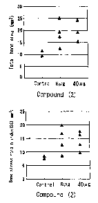
EXPERIMENTAL EXAMPLE 6: Regeneration of Radius Defect in
Rabbit
(Acclimation)
Japanese White rabbits (males 11-week-olds 4
CA 02447619 2003-11-18
49
rabbits/group) were housed for seven days at room
temperature (23 ~ 2°C) and 55 ~ 15 o humidity. During the
housing period, the rabbits were free to access
commercially available food (from Oriental Bio Service;
LRC4 ) .
(Fracture Healing)
Under pentobarbital sodium anesthesia, radium was
separated from muscle tissue of right forearm of a rabbit;
periosteum was pealed off, and removed 10 mm of diaphysis
by cutting with a bone cutter. To a gelatin capsule
(CAPSUGEL, size 5) were filled microspheres prepared in
Example 7-(1), which contains 8~cg or 40~cg of Compound (2),
and the total amount of microspheres contained in the
capsule was adjusted to 15 mg by filling microspheres
prepared in Control Example 2. One each capsule was placed
at the bone defect portion, the periosteum was repositioned
to include the capsules and sutured. In control group,
gelatin capsules filled with microspheres prepared in
Control Example 2 in the same manner were used. Muscle and
skin were each sutured and sterilized. At six weeks after
sutura, under pentobarbital anesthesia, rabbits were
sacrificed by bleeding and the radiuses were excised.
(Experimental Results)
As to the excised radium of right forearm, the
shoulder-side on the fracture line was defined as the
proximal end and the point 5 mm apart from the proximal end
toward wrist defined as the distal end. The total (cross
sectional) bone area (mm2) and stress-strain index (SSI:
mm3) at the distal end were determined using pQCT (Norland
Stratec; XCT-960A) (Clinical Calcium Vo1.10, 35-41, 2000).
CA 02447619 2003-11-18
The results are shown in Fig 2. As shown in Fig 2, in
the group treated with microsphere preparation containing a
PDE4 inhibitor of the present invention, both the total
cross sectional bone area and stress-strain index improved
5 compared with control group. These results show the
effectiveness of PDE4 inhibitor.
EXPERIMENTAL EXAMPLE 7: Acceleration of Fracture Healing in
Diabetic Rat
10 (Acclimation)
CD (SD) IGS rats (Charles River Japan, Inc.~ male; 7
week-old) were housed for seven days at room temperature
(23 ~ 2°C) and 55 ~ 15 ~ humidity. During the housing
period, the rats were free to access commercially available
15 food (from Oriental Bio Service; CRF-1).
(Induction of Diabetes)
Streptozotocin (Sigma), which induce diabetes, was
dissolved in citrate-buffered saline (pH 4.5) to obtain
0.05 M streptozocin solution and injected intravenously to
20 each rat at 60 mg/kg. One week later, blood was collected
from tail end and blood-glucose level was determined with a
blood-glucose monitor (Molecular Devices; M-SPmax250).
Based on the measurements, rats were divided into groups so
that a significant difference in glucose level may not
25 occur among groups. The average blood-glucose level was
from 426.12 to 428.23 mg/dl.
(Fracture Healing)
Under ether anesthesia, the left-lower legs of rats
were shaved and sterilized with 70 % aqueous ethanol,
30 fibulas were exposed with scissors and cut with nail
CA 02447619 2003-11-18
51
scissors (Natsume Seisakusyo; B17). The cut sections of
the fibula were re-matched with tweezers. For the test
groups (12 rats/group), the drug-containing microspheres
prepared in Example 7-(1), which contains 0.03 or 0.1 mg of
Compound(2), were placed around the cutting site using
spatula followed by suturing with silk thread. For the
control group (12 rats/group), the same amount of drug-free
microspheres prepared in Control Example 2 were placed
around the cutting site using spatula followed by suturing
with silk thread. Following the suturing, every animal was
sterilized with 70 ~ aqueous ethanol. After six weeks from
the sutura, under ether anesthesia, rats were sacrificed by
laparotomy with bleeding, and the fibulas were excised.
(Experimental Results)
Measurements of Fibula Bone Mineral Content
Eight fibulas were randomly selected from those
excised at 6 weeks after sutura and subj ected to DXA bone
densitometer (from Aloka; DCS-600) to determine bone
mineral density and bone mineral content at fracture region
(scanning width: 1 mm). The results are shown in Table 12.
TABLE 12
Drug Drug Content Bone mineral content (mg)
Control 0 10.25 1.27
Compound(2) 0.03 mg 13.69 0.96
Compound(2) 0.1 mg 14.60 0.69
As shown in Table 12, it is found that PDE4 inhibitor
has bone mineral content increasing effects in a dose-
dependent manner even in the case of bone fracture of
diabetic subjects, of which healing is known to be delayed,
CA 02447619 2003-11-18
52
as demonstrated in the diabetic model animals .
(Measurements of Fibula Strength)
The fibula used in the determination of mineral
content was subjected to a three-point bending test with
bone strength tester (from Muromachi Kikai Co., Ltd.; TK-
252C) to determine bone strength. Briefly, the fibula was
supported by two supports apart from each other by 8 mm and
the cut section was positioned at the middle of these
supports, i.e., 4 mm apart from the respective two supports.
Loading (3 mm/minute) from upper direction was kept on the
middle point (fracture section) of fibula until fibula
begins to fracture. The maximum pressure necessary to
break the bone was defined as the breaking force. The
total energy spent to break the bone was defined as the
breaking energy. The results are shown in Table 13.
TABLE 13
Drug _ Drug Content Breaking Force (N)
Control 0 6.25 0.70
Compound(2) 0.03 mg 8.13 1.20
Compound(2) 0.1 mg 11.75 2.14
Drug Drug Content Breaking Energy (mJ)
Control 0 0.69 0.08
Compound(2) 0.03 mg 1.53 0.39
Compound(2) 0.1 mg 2.91 0.82
As shown in Table 13, it is found that PDE4 inhibitor
increased the breaking force and the breaking energy even
in the case of bone fracture of diabetic subjects, of which
healing is known to be delayed, as demonstrated in the
diabetic model animals .
CA 02447619 2003-11-18
53
(X-Ray Photography)
Four fibulas excised at 6 weeks after sutura was
photographed with micro focus magnification radiography
system (Fuji Photo Film Co. Ltd.; uFX-1000; tube voltage:
25kV; tube current: 80uA; 2b seconds).
In the control group, the void of bone defect was not
filled. In contrast, the void of bone defect was filled
and bulging bone was observed in every sample from the test
groups.
EXPERIMENTAL EXAMPLE 8: Increase of cAMP Content at
Fracture Region in Normal Rats
(Acclimation)
CD (SD) IGS rats (Charles River Japan, Inc.; male; 7-
week-old) were housed for seven days at room temperature
(23 ~ 2°C) and 55 ~ 15 % humidity. During the housing
period, the rats were free to access commercially available
food (from Oriental Bio Service; CRF-1).
(Fracture Healing)
Under ether anesthesia, the left-lower legs of rats
were shaved and sterilized with 70 o aqueous ethanol,
fibulas were exposed with scissors and cut with nail
scissors (Natsume Seisakusyo; B17). The cut sections of
the fibula were re-matched with tweezers. For the treated
group (test compound-administered group) (6 rats/group),
the drug-containing microspheres prepared in Example 7-(1),
which contains 0.1 mg of Compound(2), were placed around
the cutting site using spatula followed by suturing with
silk thread. For the non-treated group (6 rats/group), the
same amount of drug-free microspheres prepared in Control
CA 02447619 2003-11-18
54
Example 2 were placed around the cutting site using spatula
followed by suturing with silk thread. Regarding control
group (6 rats/group), the cut sections were re-matched with
tweezers and sutured with silk thread without any treatment.
Following the suturing, all animals were sterilized with
70 ~ aqueous ethanol. At 0, 3, 7, 14, 28 or 42 days after
suturing, each one rat from each group was sacrificed by
laparotomy with bleeding under ether anesthesia and the
fibula was excised.
(Measurement of CAMP Content)
The fracture segment of the excised fibula was cut
into 1 cm sections and frozen with liquid nitrogen. The
resultant sections were milled at -80°C, suspended in 300
u1 of 6 % trichloroacetic acid, and sonicated. The
suspension was centrifuged at 12000 rpm for 15 minutes.
The supernatant was extracted with ether to remove
trichloroacetic acid and incubated at 75°C for 5 minutes to
remove ether from the supernatant. The CAMP in the
resultant supernatant was measured using cAMP EIA system
(Amersham Pharmacia Biotech).
(Experimental Results)
The results of CAMP measurement are shown in Fig 3.
As shown in Fig 3, in the PDE4-non-treated group (~) and
control group ( O ), the quantity of cAMP (CAMP content)
gently increased and, after the peak on day 7, decreased
gradually. On the other hand, in the treated group( ~ ),
cAMP content increased remarkably to reach its peak on day
7 and promptly returned to the same level as the control
group. These results suggest that the decomposition of
intracellular cAMP is prevented by a PDE4 inhibitor, which
CA 02447619 2003-11-18
led to the increase of CAMP content at the fracture region.
EXPERIMENTAL EXAMPLE 9: Increase of cAMP Content at
Fracture Region in Diabetic Rat
5 (Acclimation)
CD (SD) IGS rats (Charles River Japan, Inc. ~ male: 8
week-old) were housed for seven days at room temperature
(23 ~ 2°C) and 55 ~ 15 o humidity. During the housing
period, the rats were free to access commercially available
10 food (from Oriental Bio Service; CRF-1).
(Induction of Diabetes)
Streptozotocin ("STZ", Sigma), which induce diabetes,
was dissolved in citrate-buffered saline (pH 4.5) to obtain
0.05 M streptozocin solution and injected intravenously to
15 each rat at 60 mg/kg. One week later, blood was collected
from tail end and blood-glucose level was determined with a
blood-glucose monitor (Molecular Devices; M-SPmax250).
Based on the measurements, rats were divided into groups so
that a significant difference in glucose level may not
20 occur among groups. The average blood-glucose level was
from 404.5 to 410.00 mg/dl.
(Fracture Healing)
Under ether anesthesia, the left-lower legs of
diabetic rats (5 groups, 5 rats/group) and normal (non
25 treated) rats (5 groups, 5 rats/group) were shaved and
sterilized with 70 ~ aqueous ethanol, fibulas were exposed
with scissors and cut with nail scissors (Natsume
Seisakusyo; B17). The cut sections of the fibula were
matched with tweezers followed by suturing with silk thread.
30 Following the suturing, all animals were sterilized with
CA 02447619 2003-11-18
56
70 ~ aqueous ethanol. At 0, 3, 7, 14 or 28 days after
suturing, five rats from each group were sacrificed by
laparotomy with bleeding under ether anesthesia and the
fibula was excised.
(Measurement of cAMP Content)
The fracture segment of the excised fibula was cut
into 1 cm sections and frozen with liquid nitrogen. The
resultant sections were milled at -80°C, suspended in 300
u1 of 6 ~ trichloroacetic acid, and sonicated. The
suspension was centrifuged at 12000 rpm for 15 minutes.
The supernatant was extracted with ether to remove
trichloroacetic acid and incubated at 75°C for 5 minutes to
remove ether from the supernatant. The CAMP in the
resultant supernatant was measured using CAMP EIA system
(Amersham Pharmacia Biotech).
(Experimental Results)
The results of cAMP measurement (showing the cAMP
content at the fracture region) in STZ-treated group
(diabetic rats) ( O ) and non-treated group (normal
rats) ( ~ ) after treatment with PDE4 inhibitor are shown in
Fig 4.
EXAMPLE 1
( 1 ) To 0 . 1 g of Compound ( 1 ) and 1. 9 g of lactic acid
glycolic acid copolymer (lactic acid: glycolic acid
50:50; average molecular weight 20,000; PLGA5020: Wako Pure
Chemical Industries, Ltd.) was added 4.0 g of methylene
chloride, and the mixture was shaken for 30 minutes
thoroughly to form an oil phase (0).
(2) The oil phase was added to 8 ml of 0.5 o aqueous
CA 02447619 2003-11-18
57
solution of polyvinyl alcohol (POVAL PVA-220C: Kuraray Co.,
Ltd.) and emulsified at 25°C for 5 minutes with homogenizer
(Polytron, Kinematica A.G.) to form (0/W) emulsion, wherein
the oil phase is dispersed in the water phase.
(3) The emulsion was added to 1000 ml of distilled
water, stirred at 400 rpm with Three-one motor (Shinto
Scientific Co., Ltd.) and subjected to in-water drying
method at 25°C for 3 hours to remove methylene chloride.
(4) The resultant microsphere suspension was filtered
through 150 um filter to remove aggregates and filtered
under reduced pressure through 20 um filter to remove the
water phase. The resultant microsphere was combined with a
little amount of distilled water and lyophilized to give
1.6 g of microsphere.
Ten mg of the resultant microsphere was dissolved in 3
ml of acetonitrile. The solution was combined with 7 ml of
0.5 M aqueous sodium chloride solution, stirred with a
mixer (Touch mixer MT-51: YAMATO Scientific Co., Ltd.) and
then centrifuged at 2000 rpm for 5 minutes to separate
supernatant. A portion of supernatant was loaded on FL-
HPLC (column; Hypersil 5-ODS, diameter: 4 mm, length: 300
mm, GL Sciences, Inc., excitation wavelength: 315 nm,
fluorescence wavelength: 465 nm) and the drug concentration
in the supernatant was determined by comparing with a
standard curve prepared separately with a drug solution.
On the basis of the resultant concentration and the volume
of supernatant, the drug content in microsphere was
estimated as 4.21 %.
An adequate amount of resulting microsphere was
dispersed in a dilute solution of polyoxyethylene sorbitan
CA 02447619 2003-11-18
58
fatty-acid ester (Tween 80: Nikko Chemicals Co., Ltd.) The
particle distribution was measured with a particle size
analyzer SALD-1100 (Shimadzu Corporation), and the average
particle size was calculated. The average particle size
was 57 um.
(5) The microsphere obtained in (4) above was added to
physiological saline (dispersion medium) containing 0.5
carboxymethyl cellulose sodium (Nichirin Chemical
Industries) and 0.1 °~ polyoxyethylene sorbitan fatty acid
ester (Tween 80: Nikko Chemicals Co., Ltd.) at final drug
concentration of 2.5 mg/ml, and the mixture was stirred
with a mixer (Touch mixer MT-51: YAMATO Scientific Co.,
Ltd.) thoroughly to yield microsphere dispersion.
EXAMPLE 2
(1) Microsphere (1.6 g) was prepared in a manner
similar to that described in Example 1-(1) to (4) except
that a mixture of 0.57 g of lactic acid-glycolic acid
copolymer (lactic acid:glycolic acid - 50:50; average
molecular weight 20,000; PLGA5020: Wako Pure Chemical
Industries, Ltd.) and 1.33 g of lactic acid polymer
(average molecular weight 20,000; PLA0020: Wako Pure
Chemical Industries, Ltd.) was used.
The drug content and the average particle size of
microsphere were measured in a manner similar to that
described in Example 1-(4) and proved to be 3.70 o and 47.7
a m, respectively.
(2) The microsphere obtained in (1) above was treated
in a manner similar to that described in Example 1-(5) to
give microsphere dispersion (drug rate: 2.5 mg/ml).
CA 02447619 2003-11-18
59
EXAMPLE 3
(1) Microsphere (1.5 g) was prepared in a manner
similar to that described in Example 1-(1) to (4) except
that lactic acid polymer (average molecular weight 20,000;
PLA0020: Wako Pure Chemical Industries, Ltd.) was used.
The drug content and the average particle size of
microsphere were measured in a manner similar to that
described in Example 1-(4) and proved to be 3.73 ~ and 52.2
um, respectively.
(2) The microsphere obtained in (1) above was treated
in a manner similar to that described in Example 1-(5) to
give microsphere dispersion (drug rate: 2.5 mg/ml).
EXAMPLE 4
(1) To 0.2 g of Compound(1) and 0.3 g of lactic acid
polymer (average molecular weight 20,000; PLA0020: Wako
Pure Chemical Industries, Ltd.) was added 1.0 g of
methylene chloride, and the mixture was shaken with a mixer
(Touch mixer MT-51: YAMATO Scientific Co., Ltd.) thoroughly
to form an oil phase (O).
(2) The oil phase was added to 4 ml of 0.25 ~ aqueous
solution of methyl cellulose (METOLOSE: Shin-Etsu Chemical
Co., Ltd.) and emulsified at 25°C for 5 minutes with
homogenizer (Polytron, Kinematica A.G.) to form (O/W)
emulsion, wherein the oil phase is dispersed in the water
phase.
(3) The emulsion was added to 400 ml of distilled
water, stirred at 400 rpm with Three-one motor (Shinto
Scientific Co., Ltd.) and subjected to in-water drying
CA 02447619 2003-11-18
method at 25°C for 3 hours to remove methylene chloride.
(4) The resultant microsphere suspension was filtered
through 150 um filter to remove aggregates and filtered
under reduced pressure through 20 um filter to remove water
5 phase. The resultant microsphere was combined with a
little amount of distilled water and lyophilized to give
microsphere. The drug content and the average particle
size of microsphere were measured in a manner similar to
that described in Example 1-(4) and proved to be 39.6 ~ and
10 33.4 um, respectively.
EXAMPLE 5
(1) To 0.05 g of Compound(1) and 0.45 g of lactic
acid-glycolic acid copolymer (lactic acid:glycolic acid
15 50:50; average molecular weight 20,000; R202H: Boehringer
Ingelheim Co., Ltd.) was added 1.0 g of methylene chloride,
and the mixture was shaken with a mixer (Touch mixer MT-51:
YAMATO Scientific Co., Ltd.) thoroughly to form an oil
phase (0).
20 (2) The oil phase was added to 40 ml of 0.5 $ aqueous
solution of polyvinyl alcohol (GOHSENOL EG-40: The Nippon
Synthetic Chemical Industry Co., Ltd.) and emulsified at
25°C for 4 minutes with homogenizer (Polytron, Kinematica
A.G.) to form (0/W) emulsion, wherein the oil phase is
25 dispersed in the water phase.
(3) Emulsion was poured into a cylindrical airtight
container (inside diameter: 110 mm; volume 1,000 ml)
containing 400 ml of purified water, and methylene chloride
was removed from the container by stirring at 25°C and 400
30 rpm using 4-bladed propeller (diameter: 50 mm, propeller R
CA 02447619 2003-11-18
61
type: HEIDON) equipped with Three-one motor (BL-600;
HEIDON) while supplying nitrogen gas into hollow fibers of
cylinder-type hollow fiber membrane module made of silicone
rubber (NAGAYANAGI Co., Ltd.) inserted in the container
(gas flow rate is 2 L/minute). This procedure was
conducted for 1 hour.
The cylindrical hollow fiber membrane module made of
silicone rubber used in this procedure is cylinder type
NAGASEP M60-1800 of the following specification.
Cylinder diameter :100 mm
Cylinder length :120 mm x 120 mm
Membrane thickness of hollow fiber :60 um
membrane
Inside diameter of hollow fiber :200 um
membrane
Outside diameter of hollow fiber :320 um
membrane
Number of hollow fiber :1800
Effective membrane area of hollow :0.15 mz
fiber membrane
(4) The resulting microsphere suspension was filtered
through 150 um filter to remove aggregates and filtered
under reduced pressure through 20 um filter to remove water
phase. The resultant microsphere was combined with a
little amount of distilled water and lyophilized to give
0.26 g of microsphere. The drug content and the average
particle size of microsphere were measured in a manner
similar to that described in Example 1-(4) and proved to be
3.07 % and 71.7 um, respectively.
EXAMPLE 6
(1) To 0.05 g of Compound(2) and 0.45 g of lactic
CA 02447619 2003-11-18
62
acid-glycolic acid copolymer (lactic acid:glycolic acid -
50:50; average molecular weight 20,000; RG502H: Boehringer
Ingelheim Co., Ltd.), 2.5 g of methylene chloride was added
and shaken with a mixer (Touch mixer MT-51: YAMATO
Scientific Co., Ltd.) thoroughly to form an oil phase (0).
(2) The oil phase was added to 3 ml of 0.5 ~ aqueous
solution of polyvinyl alcohol (POVAL PVA-220C: Kuraray Co.,
Ltd.) and emulsified at 22°C for 5 minutes with homogenizer
(Polytron: Kinematica A.G.) to form (0/W)emulsion, wherein
the oil phase was dispersed in the water phase.
(3) The above procedures (1) and (2) were repeated
five times. The resultant emulsions (from 5 trials) were
combined, added to 1000 ml of distilled water, and stirred
at 400 rpm with Three-one motor (Shinto Scientific Co.,
Ltd.) to remove methylene chloride by conducting in-water-
drying at 25°C for 1.5 hours, at 40°C for 1 hour and at
25°C for 0.5 hours.
(4) The resultant microsphere suspension was filtered
through 150 dun filter to remove aggregates and filtered
under reduced pressure through 20 um filter to remove water
phase. The resultant microsphere was combined with a
little amount of distilled water and lyophilized to give
2.3 g of microsphere.
Ten mg of the resultant microsphere was dissolved in 3
ml of acetonitrile. The solution was combined with 6 ml of
0.5 M aqueous sodium chloride solution, stirred with a
mixer (Touch mixer MT-51: YAMATO Scientific Co., Ltd.) and
then centrifuged at 2000 rpm for 5 minutes to separate
supernatant. A portion of supernatant was loaded on W
HPLC (column; Hypersil 5-ODS, diameter: 4 mm, length: 300
CA 02447619 2003-11-18
63
mm, GL Sciences, Inc., detection wavelength: 240 nm) and
the drug concentration in the supernatant was determined by
comparing with a standard curve prepared separately with a
drug solution. On the basis of the resultant concentration
and the volume of supernatant, the drug content in
microsphere was estimated. Further, the average particle
size was measured in a manner similar to that described in
Example 1-(4). As a result, the drug content was 9.9 ~ and
the average particle size was 26.4 um.
(5) The microsphere obtained in (4) above was treated
in a manner similar to that described in Example 1- (5) to
give microsphere dispersion (drug rate: 0.1 mg/ml).
EXAMPLE 7
(1) Microsphere (2.2 g) was prepared in a manner
similar to that described in Example 6-(1) to (4) except
that lactic acid-glycolic acid copolymer (lactic
acid:glycolic acid - 75:25; average molecular weight
20,000; PLGA7520: Wako Pure Chemical Industries, Ltd.) was
used and that 2.0 g of methylene chloride was added.
The drug content and the average particle size of
microsphere were measured in a manner similar to that
described in Example 6-(4) and proved to be 10.1 ~ and 27.0
um, respectively.
(2) The microsphere obtained in (1) above was treated
in a manner similar to that described in Example 6-(5) to
give microsphere dispersion (drug rate: 0.1 mg/ml).
CONTROL EXAMPLE 1 (Control of Example 2)
(1) To 0.6 g of lactic acid-glycolic acid copolymer
CA 02447619 2003-11-18
64
(lactic acid:glycolic acid 50:50; average molecular
weight 20,000; PLGA5020: Wako Pure Chemical Industries,
Ltd.) and 1.4 g of lactic acid polymer (average molecular
weight 20,000) was added 4.0 g of methylene chloride, and
the mixture was shaken for 30 minutes thoroughly to form an
oil phase (0). In accordance with the procedures described
in Example 1- ( 1 ) to ( 4 ) , 1 . 7 g of microsphere free of drug
was obtained.
(2) Preparation of Placebo Dispersion Solution
The microsphere obtained in (1) above was treated in a
manner similar to that described in Example 1-(5) to
prepare microsphere dispersion.
CONTROL EXAMPLE 2 (Control of Example 7)
To 0.45 g of lactic acid-glycolic acid copolymer
(lactic acid:glycolic acid - 75:25; average molecular
weight 20,000; PLGA7520: Wako Pure Chemical Industries,
Ltd.) was added 2.0 g of methylene chloride, and the
mixture was shaken with a mixer (Touch mixer MT-51: YAMATO
Scientific Co., Ltd.) thoroughly to form an oil phase. In
accordance with the procedures described in Example 6-(2)
to (4), 2.2 g of microsphere free of drug was obtained.
TEST EXAMPLE 1
To 10 mg of microsphere in a test tube was added 10 ml
of phosphate buffered saline (pH 7.4) containing 0.05 0
Tween 80, and stirred with a rotating cultivator at 25 rpm
in an air-temperature-controlled cabinet at 37°C. When a
defined period of time passed from the initiation of
stirring, test tube was centrifuged (2000 rpm, 5 min) and 9
CA 02447619 2003-11-18
ml of supernatant was sampled and loaded on FL-HPLC
(column: Hypersil 5-ODS, diameter: 4 mm, length: 300 mm, GL
Sciences, Inc., excitation wavelength: 315 nm, fluorescence
wavelength: 465 nm) and the drug content was determined by
5 comparing with a standard curve prepared separately with a
drug solution. On the basis of the result and the sampling
volume, the elution amount of drug was estimated.
Further, the estimation of elution amount of drug was
repeated regularly by adding 9 ml of phosphate buffered
10 saline (pH 7.4) to the test tube after sampling, and
conducting the same procedures under the same conditions,
which comprises stirring, sampling, and estimating.
After the final sampling, the remaining eluate was
removed from the test tube and the drug content in the
15 residual microsphere was determined according to the method
described in Example 1-(4).
The above procedures were carried out on the
microspheres obtained in Examples 1 to 3. The results are
shown in Fig.5.
20 The elution rate was calculated based on the
assumption that the sum of drug eluted from and remained in
the microsphere being 100 %.
TEST EXAMPLE 2
25 Male SD rats (7-weeks-old, 3 rats/group, Japan SLC)
were conditioned for a week by housing at room temperature
(23 ~ 2°C) under 12 hours light-dark cycle while feeding
with food and water ad libitum. Each rat then received
rapid-injection of Compound(1) (1 mg/ml) dissolved in
30 physiological saline containing 10 % polyethylene glycol
CA 02447619 2003-11-18
66
400 ((Wako Pure Chemical Industries, Ltd.) from femoral
vein at 0.5 ml/animal (total drug dosage: 0.5 mg/rat).
After drug administration, under ether anesthesia
blood samples were collected at regular time intervals from
jugular vein with a syringe containing heparin and
centrifuged to obtain plasma samples. To 0.1 ml of plasma
were added 0.2 ml of internal standard solution and 1 M
dibasic potassium phosphate and then 7.0 ml of chloroform.
The mixture was shaken for 10 minutes and centrifuged for 5
minutes to separate 5 ml of organic phase. The resultant
organic phase was evaporated to dryness at 40°C under
nitrogen atmosphere, re-dissolved in 0.5 ml of mobile phase
and then loaded on FL-HPLC (column; Hypersil 5-ODS,
diameter: 4 mm, length: 300 mm, GL Sciences, Inc.,
excitation wavelength: 315 nm, fluorescence wavelength: 465
nm) to determine the plasma concentration. The results are
shown in Fig.6.
TEST EXAMPLE 3
Male SD rats (7-weeks-old, 5 rats/group, Japan SLC)
were conditioned for a week by housing at room temperature
(23 ~ 2°C) under 12 hours light-dark cycle while feeding
with food and water ad libitum. Each rat then received
subcutaneously microsphere dispersion obtained in Examples
1-(5), 2-(2) or 3-(2) from back at 2 ml per rat (total drug
dosage: 5 mg/rat). After drug administration, under ether
anesthesia, blood samples were collected at regular time
intervals from jugular vein with a syringe containing
heparin and centrifuged to obtain plasma samples. The
concentration of the compound in plasma was determined in a
CA 02447619 2003-11-18
67
manner similar to that described in Test Example 2. As a
result of formulating PDE4 inhibitor into microsphere, the
maximum plasma concentration of PDE4 inhibitor could be
reduced to 1/25 to 1/100, even when compared with that
achieved by intravenous injection of saline containing only
a tenth amount of PDE4 inhibitor (Test Example 2). The
results are shown in Fig.7.
TEST EXAMPLE 4
Male SD rats (7-weeks-old, 5 rat per group, Japan SLC)
were conditioned for a week by housing at room temperature
(23 ~ 2°C) under 12 hours light-dark cycle while feeding.
with food and water ad libitum. Each rat then received
subcutaneously microsphere dispersion obtained in Example
2-(2) from back at 2 ml per rat (total drug dosage: 5
mg/rat).
At days 3, 7, 10, 14, 21 and 35 after drug
administration, microspheres were collected from the sites
of administration. To the collected microspheres, 5 ml of
acetonitrile containing internal control substance was
- added and dissolved with homogenizer (Polytron: Kinematica
A.G.). After centrifugation at 3,000 rpm, 5 minutes, 3 ml
of supernatant was collected, combined with 7 ml of 0.5 M
aqueous sodium chloride solution, stirred with a mixer
(Touch mixer MT-51: YAMATO Scientific Co., Ltd.) and then
centrifuged at 2,000 rpm for 5 minutes to separate
supernatant. A portion of supernatant was filtrated
through KC prep-omni 13 (Katayama Chemistry Inc.) and
loaded on FL-HPLC (column; Hypersil 5-ODS, diameter: 4 mm,
length: 300 mm, GL Sciences, Inc., excitation wavelength:
CA 02447619 2003-11-18
68
315 nm, fluorescence wavelength: 465 nm). The drug
concentration was determined by comparing with a standard
curve prepared separately with a drug solution. On the
basis of the resultant concentration and the volume of
supernatant, the residual rate of a drug remaining in
microsphere was calculated. The results are shown in Fig.8.
TEST EXAMPLE 5
Male SD rats (7-weeks-old, Japan SLC) were conditioned
for a week by housing at room temperature (23 ~ 2°C) under
12 hours light-dark cycle while feeding with food and water
ad libitum. Each rat then received subcutaneously
Compound(2)-containing microsphere dispersions obtained in
Examples 6-(5) and 7-(2) at 1 ml per rat (total drug
dosage: 0.1 mg/rat) from back.
Microspheres were collected at regular time intervals
from the administration site. To the collected
microspheres, 10 ml of acetonitrile was added and dissolved
with homogenizer (Polytron: Kinematica A.G.). After
centrifugation at 3,000 rpm for 5 minutes, 3 ml of
supernatant was collected, combined with 6 ml of 0.5 M
aqueous sodium chloride, stirred with a mixer (Touch mixer
MT-51: YAMATO Scientific Co., Ltd.) and then centrifuged at
2000 rpm for 5 minutes to separate supernatant. A portion
of supernatant was filtrated through KC prep-omni 13
(Katayama Chemistry Inc.) and loaded on W-HPLC (column;
Hypersil 5-ODS, diameter: 4 mm, length: 300 mm, GL Sciences,
Inc., detection wavelength: 240 nm). The drug
concentration was determined by comparing with a standard
curve prepared separately with a drug solution. On the
CA 02447619 2003-11-18
:'
69
basis of the resultant concentration and the volume of
supernatant, the residual rate of a drug remaining in
microsphere was calculated. The results are shown in Fig.9.
INDUSTRIAL APPLICABILITY
The bone fracture healing accelerating composition of
the present invention comprises a PDE4 inhibitor as an
active ingredient, which, when administered locally to the
fracture region, can promote the fracture healing by
accelerating the endochondral ossification in the
reparative phase without producing side effects due to
systemic action of PDE4 inhibitor, and can accelerate the
healing of bone fracture of elderly people, and diabetic or
osteoporosis patients in the early stage, which bone
fracture is becoming a major social issue of recent years,
whereby exerts an effect of preventing the patients from
becoming bedridden and ensures their normal daily life.
Still higher effect can be achieved by formulating a
composition containing a PDE4 inhibitor and a biocompatible
and biodegradable polymer into a depot preparation,
especially into an injectable microsphere preparation and
administering the same locally to a fracture region thereby
allowing efficacy to last.