Note : Les descriptions sont présentées dans la langue officielle dans laquelle elles ont été soumises.
CA 02448390 2003-11-24
WO 02/095375 PCT/1L01/00475
SEMEN ANALYSIS
FIELD OF THE INVENTION
This invention relates to semen analysis.
BACKGROUND OF THE INVENTION
According to WHO statistics, 8-10% of all married couples consult medical
to professionals after failing to conceive. Over 40 million couples are
currently being
treated for infertility. Among these infertile couples, it is estimated that
the
infertility in 40% of the couples is due to male originating causes, and
another 20%
is due to combined male and female originating causes. Semen analysis is a
major
technique in evaluating male originating causes.
Standard semen analysis protocol involves the determination of at least three
major semen parameters:
1. total sperm concentration (TSC);
2. percentage of motile sperm; and
3. percentage of normal sperm morphologies.
For all practical purposes, semen analysis, a key factor in human male
fertility medicine, has not changed since the 1930's and is still done today
by
microscopic inspection. In fact, it is one of the very few remaining in vitro,
body
fluid analysis still performed almost solely via manual methods.
This manual methodology involves carefully observing the sperm cells,
counting them to determine their concentration, classifying their motility,
identifying their morphology, etc. This work requires high expertise, is very
labor
intensive and if done according to standard protocols, takes at least an hour
per test.
CA 02448390 2003-11-24
WO 02/095375 PCT/1L01/00475
-2-
Manual assessments are known to be quite inaccurate due to numerous
sources of error. The main sources of error are:
Subjectivity of the observer.
The varying criteria used in the different labs and by different observers.
. The large statistical errors due to the limited number of sperm analyzed.
The WHO manual (WHO laboratory manual for the examination of human
semen and sperm-cervical mucus interaction. 4th edition, Cambridge University
Press, 1999) recommends observing not less than 200 sperm and classifying the
morphology and motility of each. This itself is an error introducing procedure
due
to the tediousness and time consuming nature of the task. In practice, 50 to
100
sperm cells at most are analyzed. Even if the observer introduces no errors,
the
statistical error alone reaches tens of percentages.
As a result of the above methodology, semen analysis test results are
globally recognized to be highly subjective, inaccurate and poorly
reproducible.
Inter lab and inter technician variations are of such proportions that this
issue is of
major concern in male fertility medicine and the unresolved subject of
discussion in
the vast majority of symposiums, congresses and conventions on the subject.
In order to overcome these difficulties, medical instrumentation companies
have introduced dedicated computerized systems based on image analysis (CASA -
Computer Assisted Semen Analyzers). These systems require an extremely high
quality image because all their results are based on image processing.
Although
these systems have attempted to replace manual analysis and establish industry
accepted standards, they have not succeeded in either of these objectives.
The first objective could not be achieved because analysis results continue
to be dependent on manual settings and on the different makes of equipment.
Replacing routine manual analysis is totally impracticable because the systems
are
extremely expensive, complex and difficult to use. The fact is that such
systems are
generally not found in routine semen analysis but have rather established
their
niche almost solely in research centers, university hospitals and occasionally
in
3o highly specialized fertility centers.
CA 02448390 2010-01-25
-3-
An additional approach for semen measurements is described in U.S.
Patent Nos. 4,176,953 and 4,197,450. These patents describe a method for
measuring sperm motility using electro-optical means and an analog signal
analyzer. A suspension of sperm cells is continuously examined in a
predetermined field in order to detect variations in optical density by the
motion of the sperm. An amplitude-modulated analog electrical signal is
generated in response to the variations, and the peaks and valleys of this
signal
are counted over a predetermined time period to provide an abstract parameter
termed Sperm Motility Index (SMI). This parameter is related to motility and
gives readings which are proportional to the number of motile cells multiplied
by their respective velocity.
An automatic sperm analyzer called the Sperm Quality Analyzer (SQA),
which provides the SMI parameter, has been on the market for a number of
years.
The analyzer is used in the following manner: a sperm specimen is taken up by
a
is disposable chamber which has a rubber bulb at one end to aspirate the
sample, and
a thin measuring compartment at the other end. After aspirating the sample,
the
measuring compartment is inserted into the SQA and the SMI of the sample is
automatically determined. The SMI parameter, although useful in some
applications, was not significantly accepted by the medical community as a
viable
alternative to the conventional microscopic semen measurements.
It is common knowledge that in some fields of veterinary fertility analysis,
total sperm concentration (TSC), is evaluated by measuring optical turbidity
of the
specimen. The physical principle behind this approach is that sperm cells are
more
opaque than the surrounding seminal plasma, and absorption of a light beam by
the
specimen is therefore proportional to the TSC.
For example, U.S. Patent No. 4,632,562 discloses a method of measuring
sperm density by measuring the optical absorbance of a sperm containing sample
and relating the absorbance output signal to the density by using at least
three
summing channels. The disclosed method is intended for use in artificial
CA 02448390 2003-11-24
WO 02/095375 PCT/1L01/00475
-4-
insemination in the cattle breeding industry, and measures the optical
absorbance in
the range of 400-700 nm.
This technology however, has not and could not be adopted for human use
for the following reasons:
(1) Human sperm concentrations in the normal range (and even in higher
than normal cases), are more than an order of magnitude lower than in most of
their
veterinary counterparts - where this technology has been adopted.
(2) Human cases are treated even when sperm concentrations are far below
their normal levels. This of course is not the case for animals. Infertile
animals are
to normally culled - in any case, they are not treated for infertility.
(3) TSC in humans is a parameter, which in itself, is totally insufficient for
fertility investigations, and microscopic analysis is in any case required for
all the
other data in the standard semen analysis protocol. To a large degree, this
also holds
for veterinary applications. This fact made optical absorption measurements
superfluous, and no real effort has been invested in this field.
There is thus a need for a simple, objective technique for measuring TSC in
human semen.
According to the WHO manual, sperm motility assessment (considered by
most to be the most important single semen parameter) can be carried out
manually
using a grid system under the microscope or, alternatively, by use of CASA.
CASA provides some advantages over manual methods. However, accuracy
and provision of quantitative data are totally dependent on precise semen
preparation techniques and instrument settings. These factors (high expertise
and
sophisticated environment) along with the prohibitive cost of such
instrumentation,
rule out for all practical purposes their application for routine semen
analysis.
U.S. Patent No. 4,896,966 discloses a motility scanner for characterizing the
motion of sperm, bacteria and particles in fluid. The scanner comprises an
optical
system including a collimating lens, condensing lens, imaging lens and a pair
of
reflecting elements, a source of illumination, radiation sensing means, signal
CA 02448390 2003-11-24
WO 02/095375 PCT/1L01/00475
-5-
processing means, and display means. The imaging lens has a useful depth of
field
at its object plane of at least about 0.2 nu-n.
SUMMARY OF THE INVENTION
It is an object of the present invention to provide a method for measuring
TSC.
It is a further object of the invention to provide a method for determining
the
motile sperm concentration (MSC) and % motility.
It is a still further object of the invention to provide a sampling device for
use in the determination of semen parameters.
It is another object of the invention to provide a system for the
determination of semen parameters.
In a first aspect of the invention, there is provided a method for measuring
the total sperm concentration (TSC) in a sample. The method comprises (i)
placing
the sample in a transparent container between a synchronically pulsed light
source
and a photodetector; and (ii) measuring the optical absorbance of the sample
in the
range of 800-1000 nrm, the TSC of the sample being proportional to the
absorbance.
The method of the invention provides an objective measurement of TSC
which is not dependent on image analysis, and which can measure human TSC.
However, the method may also be used to measure animal TSC.
In a second aspect of the invention, there is provided a sampling device for
use in optically analyzing a biological fluid comprising:
(i) an aspirator for aspirating the fluid into the device;
(ii) a thin measuring chamber having an upper and lower wall, the distance
between the walls being in the range of 100-500 microns;
(iii) a thick measuring chamber having an upper and lower wall, the distance
between the walls being in the range of 0.5-3 cm; and
(iv) means for excluding air from the measuring chambers.
In a preferred embodiment, the biological fluid is semen, most preferably
human semen. The device serves both as a sampler and dual test chamber,
enabling
CA 02448390 2003-11-24
WO 02/095375 PCT/1L01/00475
-6-
simultaneous testing of TSC and MSC. No dilution is required for any of the
measurements. This not only saves labor but also eliminates a significant
source of
errors - namely, dilution inaccuracy.
The device also enables (when required) built-in visualizations of the
specimen without transferring it to a separate viewing chamber. The thick
chamber
is also referred to as an optical densitometer.
In a third aspect of the invention, there is provided a method for measuring
motile sperm concentration (MSC) in a semen sample comprising:
(i) placing the sample in a transparent container between a light source and
a photodetector, wherein the sperm motion in the sample modulates the
light transmitted therethrough, thereby generating a signal;
(ii) sampling the signal so as to produce a plurality of signal samples;
(iii) selecting acceptable signals;
(iv) calculating an absolute value for each of the acceptable signal samples;
(v) calculating an average a of the absolute values; and
(vi) calculating the MSC based on the average a.
It has now been discovered that analysis of waveforms of the analog signals
derived from a light beam which traverses a semen sample can provide an
indication of the MSC. Using appropriately selected criteria, excellent
correlation
was found to exist between the averaged area covered by the waveform and the
MSC. The MSC of a sperm sample is obtained in accordance with the invention by
analyzing optical properties of the sample, which vary over time due to the
motility
of the sperm. This is in fact, the average signal amplitude in the relevant
portions of
the waveform, as will be described in more detail below.
In a fourth aspect of the invention, there is provided a method of
determining the average velocity (AV) of sperm cells comprising:
(i) obtaining a Sperm Motility Index (SMI) of the sperm cells as defined in
US Patent No. 4,176,953;
(ii) obtaining the MSC; and
CA 02448390 2003-11-24
WO 02/095375 PCT/1L01/00475
-7-
(iii) calculating AV using an algebraic expression involving the ratio
SMI/MSC.
Reference is made here to US Patent No. 4,176,953 issued Dec. 4, 1979, and
which has been implemented in various versions of Sperm Quality Analyzers
produced by Medical Electronic Systems, Israel. This patent, when applied to
semen analysis, provides a parameter called SMI (Sperm Motility Index). As
disclosed in the above patent and proven in numerous supporting studies, SMI
is a
function of both the concentration of motile cells (what is referred to as
MSC) and
their average velocity (AV). For the sake of simplification, we can say that
SMI is a
to function of MSCxAV, or AV is a function of SMI/MSC. The average velocity of
a
sperm sample can provide an indication of the quality of the motility of the
sperm.
Not withstanding that which is stated above, SMI as a function of MSC and
AV is more complex than a direct multiplication. After observing, analyzing
and
measuring over a hundred semen samples, the correct inter-relationship
(formula)
between them has been developed. In general terms, the formula for extracting
the
average velocity can be defined as: AV=f(SMI/MSC), "f ' being a polynomial of
the third degree. Working with f(x) =1/1000x3 +1/10x2 +0.89x, provided a
correlation factor of r = 0.82.
It should be noted that most semen analysis protocols require data on the %
of sperm having progressive motility rather than their average velocity.
Progressive
motility is defined as those sperm having an average velocity of 5
microns/second
or more. This parameter too, can readily be extracted from the average
velocity if a
normal spread of velocities is assumed around the average. Even in cases where
the
velocity spread is not normal, the error in calculating the % of progressively
motile
sperm is not significantly affected. Moreover, when different minimal
velocities are
defined as progressively motile, this varying threshold is readily entered
into the
calculation, thereby giving extra flexibility in providing this parameter.
This is
important when working in different diluting media, ambient temperatures or in
fact different species in vet measurements.
CA 02448390 2003-11-24
WO 02/095375 PCT/1L01/00475
In a fifth aspect of the invention, there is provided a system for analyzing
sperm viability comprising:
(i) means for measuring TSC;
(ii) means for measuring MSC; and
(iii) a video visualization system.
The system of the invention combines the measurement of the two major
sperm parameters TSC and MSC, with the traditional visualization of the sperm,
thus enabling acquiring the third parameter - sperm morphology. In a preferred
embodiment, TSC and/or MSC are determined according to the methods of the
lo invention. In another preferred embodiment, the system further comprises
the
sampling device of the invention.
It should be emphasized that there is a basic difference between the video
visualization system used in the system of the invention and other sperm
visualization systems (such as CASA). The other systems require extremely high
quality images because all their results are built on image processing. In the
present
invention, on the other hand, visualization is used only as a complementary
tool to
view atypical or suspect cases, to add confidence to processed results, to
identify
specific pathologies and to enable manual sperm morphology assessment, when
required.
In order to fulfill these tasks, the video visualization system used in the
invention is designed as a compact, inexpensive subsystem, which although of
limited use as a stand-alone, precisely fills a complementary role in the
system of
the invention. An additional important advantage of the visualization system
as
compared to microscopic procedures, is that pipetting, preparation of slides,
dilutions and filling of hemocytometers is unnecessary. Use, together with the
video
visualization system, of the device of the invention, which doubles as a
complete
test chamber, obviates all of the above. These features, in effect permit and
enable
the use of the system of the invention in any small clinic or even office
environment.
CA 02448390 2003-11-24
WO 02/095375 PCT/1L01/00475
-9-
The video visualization system allows one to obtain the following
supplementary information regarding the tested sample:
1. Measurement of very low sperm concentrations
Measuring TSC at very low concentrations (below 5 million sperm/ml) is
inherently limited in accuracy. This is due to the fact that light absorption
by factors
other than sperm cells, may become relatively significant at these low levels.
Light
absorption may be due to seminal plasma variability or to the presence of
cells
other than sperm. The latter include WBCs (white blood cells indicating
infections)
and other immature or non-spermic cells from various sources, etc. Since
according
1o to the invention TSC is measured by optical absorption, without
visualization there
would be a possibility for ambiguity in the very low ranges due to the above
mentioned considerations. When TSC is considered important in the low ranges,
visualization enables differentiation between the different cells contributing
to the
light absorption. Since MSC is measured independently of light absorption, the
%
motility (MSC/TSC) can be calculated using the visually determined TSC
parameter.
2. Identifying foreign cells in the semen
The system is useful in identifying the presence of other cells which may
have an effect on semen quality and/or assist in diagnosing patient ailment.
For
instance, leukocytes indicate infection, immature cells indicate a problem in
spermatogenesis, agglutination may be due to a number of causes, etc.
3. Manual sperm morphology assessment
Although the system of the invention automatically assesses the % of sperm
with normal morphologies, it does so according to a given criteria (e.g. the
WHO
criteria). Regretfully this criteria is not universally accepted. Such
universally
accepted criteria do not yet exist, and are often a factor of application. For
example,
morphology criteria for IVF and ICSI applications are normally far stricter
than in
normal cases. Other international standards (such as strict or Krueger
criteria) are
also widely applied. Visualization allows the fertility practitioner to select
his own
CA 02448390 2003-11-24
WO 02/095375 PCT/1L01/00475
-10-
criteria as well as to identify the specific defects present (head deformity,
tail
problem, etc.)
4. Vasectomy validation and Azoospermia diagnosis
In order to fully validate the outcome of vasectomy or to obtain a conclusive
diagnosis of azoosperinia, it is necessary to determine that there are
absolutely no
sperm in the semen under evaluation. This is generally not possible with the
light
absorption technology, because the concentrations that are to be measured can
be
very low. In this case, manual visualization is necessary in order to
carefully scan
large fields of view in search of individual sperm cells. The sperm
visualization
to system used in the system of the invention is specifically tailored to
optimally
address these applications.
5. Hard copy
The video visualization system enables "freezing" a given selected view (or
a few views) which may then be printed and attached to the Semen Analysis
Report. This is of great value for consultations and validation of treatment
efficacy.
A by-product of the freezing option is viewing the semen sample under static
conditions. This strongly facilitates analysis and counting. In microscopic
assessments, this can only be done by demobilizing (killing) the sperm prior
to
viewing. Even then, all dead sperm will end up in one layer, a condition which
normally complicates analysis due to high concentration and sperm overlap in
the
said layer.
BRIEF DESCRIPTION OF THE DRAWINGS
In order to understand the invention and to see how it may be carried out in
practice, preferred embodiments will now be described, by way of non-limiting
examples only, with reference to the accompanying drawings, in which:
Fig. 1 is block diagram illustrating one embodiment of the method of
measuring TSC according to the invention;
Fig. 2 is a perspective top view of one embodiment of a sampling device
according to the invention;
CA 02448390 2003-11-24
WO 02/095375 PCT/1L01/00475
-11-
Fig. 3 is a partial side sectional view of the device of Fig. 2;
Fig. 4 is a sectional view of the separating valve rotated 900 from the view
of Fig. 3;
Fig. 5 is a schematic illustration of a system for semen analysis according to
one embodiment of the invention;
Fig. 6 is a flow chart illustrating an algorithm for calculating the MSC;
Fig. 7 is a flow chart illustrating an algorithm for calculating the average
velocity;
Fig. 8 illustrates a typical analog signal of motile sperm as a function of
time;
Fig. 9 is a correlation curve of the MSC with average analog signal; and
Fig. 10 is a block diagram illustrating one embodiment of a video
visualization system according to the invention.
DETAILED DESCRIPTION OF THE INVENTION
Example 1
As stated above, the automatic optical measurement of TSC in human
semen samples as opposed to animal samples has been hampered in the past due
to
the low concentration of sperm cells. This, together with the high background
electronic and optical noise due e.g. to seminal plasma variability has
prevented the
application of methods routinely used in veterinary fertility analysis. The
method of
the present invention comes to overcome these obstacles by combining the
following features:
(i) the sample is placed in a transparent container between a synchronically
pulsed light source and a synchronically enabled photodetector. The use
of a synchronically pulsed light source and photodetector enables the
distinction of sperm cells at low concentrations over electronic noise
levels.
CA 02448390 2003-11-24
WO 02/095375 PCT/1L01/00475
-12-
(ii) measuring the optical absorbance of the sample in the range of
800-1000 run. It has been found that measuring the absorbance in the
near infrared region provides the optimal conditions for obtaining strong
absorption by sperm cells and low absorption by seminal plasma.
Preferably the measured range is 850-950 nm. Most preferably, the
range is 880-900 nm.
By using the method of the invention, the TSC of a sample may be
determined as a function of the absorbance. Although the method of the
invention
is preferably used with samples of human semen or human sperm, it may also be
1o used with animal semen and animal sperm, preferably after appropriate
dilution.
An example of an optical system using one embodiment of the method of
the invention is illustrated in Fig. 1. The system, indicated generally by the
numeral 2, comprises a light source 4, a photodetector 6 and a sample holder 8
interposed therebetween. A preferred light source may be a fast-switching
synchronically pulsed light emitting diode (LED) which emits light in the near
infrared region. The light source may be controlled by a light intensity
controller 10
which in turn is regulated by a modulator 12. The photodetector is capable of
detecting synchronically pulsed light. The photodetector transmits the
measured
analog signals to a demodulator 14, which is also regulated by the modulator
12,
and from there to output 16 of the signal in digital form.
The beam path through the sample is preferrably vertical. The length of the
beam path through the sample is generally between 5 and 15 mm, preferably 10
nun. The sample holder must be fully transparent to light waves in the near
infra-red region of between 800 and 1000 nm. The plastic material from which
the
sample holder is made must be totally non toxic to sperm cells. A preferred
material
is polystyrene PG-79. The sample holder should preferably be designed to
totally
prevent penetration and forming of air bubbles in the sample, which interefere
with
the optical measurement.
CA 02448390 2003-11-24
WO 02/095375 PCT/1L01/00475
-13-
By using the method of the invention, TSC detection levels down to appr. 2
million cells/ml. have been achieved. This level already indicates extreme
semen
pathology.
Example 2
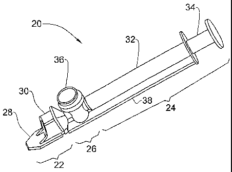
Fig. 2 illustrates one embodiment of a sampling device 20 according to the
invention, for use in measuring semen. The device comprises an anterior
optical
viewing section 22, a posterior aspirating section 24 and an intermediate air
exclusion section 26.
The optical viewing section 22 comprises a thin measuring chamber 28 and
a thick measuring chamber 30. The thin chamber is used to measure MSC and/or
for visualization, while the thick chamber is used to measure TSC. In this
way,
multiple parameters can be measured simultaneously using the same sampling
device and sampling step.
The aspirating section 24 comprises a cylinder 32 and a plunger 34 slidingly
inserted therein. These parts match each other and function as in a standard
syringe.
This section serves for the aspiration of the semen sample into the measuring
chambers
The air exclusion section 26 comprises a separating valve 36 for separation
of the measuring chambers from the cylinder volume after filling. The
aspirator,
thin measuring chamber, thick measuring chamber and air exclusion section are
all
in fluid communication.
An adapter 38 in the form of a rectangular rail extends along one side of the
device 20 and serves for the correct sliding in and aligning of the device
upon
insertion into an optical instrument by which the sample is evaluated. It also
provides the mechanical support and stability required for precision electro-
optical
measurements.
The parts of the device may be seen more clearly in Fig. 3. The thin
measuring chamber 28 is an internal cavity having an upper 40 and a lower 42
parallel transparent wall through which the optical beam may pass. The
distance
CA 02448390 2003-11-24
WO 02/095375 PCT/1L01/00475
-14-
between the walls is in the range of 100-500 microns, preferably 250-350
microns,
most preferably approximately 300 microns. In the later case, the volume of
liquid
in the chamber is approximately 25 1. The anterior end 44 of the chamber has
an
aperture through which the sample may be drawn into the device. In the
illustrated
embodiment, the chamber is approximately 4 mm wide.
The thin measuring chamber serves for evaluation of sperm motility and
may be positioned between a light source e.g. opposite the lower wall 42 and a
photodetector e.g. opposite the upper wall 40. It will be understood that the
light
source and photodetector may also be positioned on the opposite sides of the
io chamber. A light beam is transmitted through the chamber containing a semen
sample. The detector on the other side of the chamber registers optical
density
variations caused by moving sperm cells. The optical density variations are
translated into an electrical signal by the photo-detector which is then
routed to the
electronic circuits to be filtered, digitized and processed so as to indicate
the MSC.
The thin measuring chamber may also be used with a video visualization system,
as
will be further explained below.
The thick measuring chamber 30 has an upper 46 and a lower 48 transparent
wall through which an optical beam may pass. The distance between the walls is
in
the range of 0.5-3 cm, preferably 0.8-1.2 cm, most preferably approximately 1
cm.
The approximate volume held by the thick compartment in the latter case would
be
approximately 0.5 ml.
This chamber serves for electro-optical absorption measurements of sperm
concentration. A light beam, which may be the same or different from that of
the
thin chamber 28, is transmitted through the upper and lower walls of the
chamber
and detected by a photo-detector. The chamber volume should be completely
filled
with a sperm sample in order to avoid inaccuracies due to air bubbles. The
attenuation of the light beam as it passes through the chamber is proportional
to the
sperm concentration. The light beam intensity is measured after passing
through the
chamber and translated to units of TSC by electronic means. The order of the
chambers in the sampling device may be exchanged.
CA 02448390 2003-11-24
WO 02/095375 PCT/1L01/00475
-15-
The cylinder 32 is in fluid communication with the two measuring chambers
28 & 30, so that by drawing the plunger 34, fluid is drawn into the chambers.
This
method of aspiration allows large sample volumes to be aspirated into the
device.
In order to prevent air bubbles from remaining in the measuring chambers, a
s separating valve 36 is interposed between the cylinder and the measuring
chambers,
and is in fluid communication with them. The valve is shown in detail in Fig.
4 and
comprises a piston 50 slidingly held in a valve housing 52. A connecting bore
54
connecting between the measuring chamber 30 and the cylinder 32 passes through
the piston 50.
When the valve is in the upper position, there is a connection between the
measuring chambers and the aspirating cylinder. Pressing the valve down breaks
that connection and ensures that no air remains in the measuring chambers
where
the samples are measured and no leakage will occur even when there is a
temperature variation. This technique is equivalent to positive displacement
since
air is excluded from the measured fluid volumes (except at the anterior end
44).
This design enables working with samples of virtually all viscosities, while
at the
same time preventing leakage and the penetration of air bubbles into the
specimen
volumes to be analyzed.
Although the means for excluding air from the measuring chambers has
been exemplified by a separating valve, other means may also be used, such as
a
positive displacement pipette
All parts of the device may be manufactured from any material which is not
toxic to the measured cells. Preferably, the material is relatively cheap,
such as
plastic materials, so that the device can be disposable. An example of a
polymer
which may be used to produce the device is polystyrene PG79. The separating
valve, cylinder and piston may be made from polypropylene. The thin measuring
compartment is by far the most toxi-sensitive part of the device due to the
very high
area to volume ratio of the seminal liquid in that section.
In order to aspirate a sample into the device 20, the tip 44 of the thin
measuring compartment 28 is dipped approximately 5 mm deep into the semen
CA 02448390 2003-11-24
WO 02/095375 PCT/1L01/00475
-16-
sample, which is then aspirated into the device past the separating valve 36.
Only
app. 0.6cc are required for a complete filling of the device. The separating
valve is
then pushed down, and the device may be inserted into an optical measuring
apparatus.
Example 3
As mentioned above, determination of the MSC according to the invention
requires the generation of a voltage signal which is proportional to the MSC.
Fig. 5
shows one embodiment of a system for semen analysis capable of generating such
a
to signal.
An optical capillary 100 having a rectangular cross-section is used to hold a
semen sample 102. The capillary 100 is illuminated with an incident light beam
105
produced by a light source 110. The capillary 100 has an optical path of 300
PM
through which the light beam 105 passes. After passing through the capillary,
the
scattered beam 106 is collimated by a round aperture 108 having a diameter of
70
dun. The collimated beam 107 impinges upon a photodetector 115. The
photodetector 115 produces an analog voltage signal 120 proportional to the
intensity of the beam 107. The analog signal varies in time due to the
motility of the
sperm in the semen sample 102, as shown for example in Fig. 8. The analog
signal
120 is inputted to an analog-to-digital converter 125 that samples the analog
signal
120 at a rate of e.g. 8000 Hz and generates a digital output signal 128. The
digital
output signal may be stored in a memory 130. Sperm motion in the sample 102
leads to a modulation in the intensity of the beam 107, which in turn affects
the
analog signal 120 and digital signal 128.
A processor 135 is configured to carry out an analysis of data stored in the
memory 130 in order to produce an analysis of the semen sample 102. The
results
of the analysis may be displayed on any display device such as a CRT screen
140 of
a personal computer 145, or on an internal LCD screen 148 of the measuring
device.
CA 02448390 2003-11-24
WO 02/095375 PCT/1L01/00475
-17-
Fig. 6 shows a flow chart diagram for one embodiment of an algorithm for
calculating the MSC as carried out by e.g. the processor 135 of Fig. 5, in
accordance with the invention.
In step 200, the digital signal 128 of Fig. 5 is digitally filtered in order
to
remove high and low frequencies that are not relevant to the dominant
frequency of
the signal, which is determined by the motility characteristics of the semen
sample
102. This is done in order to optimize the signal to noise ratio. The DC
component
of the signal 128 is also removed. For human sperm samples, for example, the
optimal relevant frequency range was found to be between 5 and 30 Hz. In step
205, digital samples having an absolute value below a first predetermined
threshold, which may be determined empirically, are excluded. In step 210 the
same
threshold value is subtracted from all remaining samples.
In step 215, a waveform selection procedure is carried out to discard all
waveforms due to artifacts such as from non-relevant cells, etc. A preferred
embodiment of waveform selection with human sperm is to eliminate all
waveforms not satisfying the following criteria:
Minimum height - 10 millivolts.
Minimum width - 37.5 milliseconds.
Maximum width - 500 milliseconds.
Minimum rise/fall time - 2.5 milliseconds.
The correct definition (and detection) of the beginning and end of sperm
associated waveforms are defined as those where significant changes of
waveform
direction occur. The time difference between two such points defines the time
width of a given wave. The manner of selection may be understood by way of
example with reference to Fig. 8 (not drawn to scale), which shows the
amplitude
of the analog signal (120 in Fig. 5) as a function of time. The threshold 302
is
determined empirically to provide optimal linearity between the output signal
and
the microscopically measured MSC. The waveforms that are used for the
calculation of MSC are labeled 304, 305, 306 and 307. The other waveforms have
CA 02448390 2003-11-24
WO 02/095375 PCT/1L01/00475
been rejected for various reasons: 308 because its peak is less than the
threshold;
310 because it is too wide; and 312 because it is too narrow.
In step 220 of Fig. 6 the absolute value of all selected samples is
calculated,
and in step 225, the average a of the absolute values is calculated. In step
230, the
MSC of the sample 102 is calculated based upon the average a. For example, it
was
found that the dependency of MSC on a can be described by a linear equation of
the form:
MSC=aa
where a is an empirically derived constant. In a preferred embodiment, the
io dependency of MSC on a may be described by a quadratic equation of the
form:
MSC=Aa2+Ba
With reference to Fig. 9, a specific human sperm sample was analyzed in
accordance with the invention. It was found that the dependency of MSC on a
could be described by the following algebraic expression:
is MSC = 0.0047a2 + 0.869a (I)
A good linear correlation was found to exist for small values of a. Using
formula
(I), the correlation factor (r) for fresh sperm over the entire range was >
0.98.
Analysis of treated semen samples with varying viscosity was also
perfonned using thawed samples, washed sperm, diluted samples (both in 3%
20 Sodium Citrate and Test Yolk buffer) as well as with samples containing up
to 20%
glycerol having artificially raised viscosity. It was found that varying
sample
viscosity (and therefore sperm velocity), did not significantly affect the
correlation
between MSC and average signal ("r" in all case remained above 0.96).
Using centrifugal enrichment techniques, a very wide range of motile human
25 sperm concentrations were measured (up to 250 MIml). No significant
saturation
was found. The slight non-linearity at the highest ranges is easily corrected
by a
simple second-degree polynomial correction - given above.
Analysis of bovine semen was also carried out and correlation factors
between bovine MSC and identically averaged signals (same methodology as for
3o humans) provided similarly excellent results. It is to be noted however,
that bovine
CA 02448390 2003-11-24
WO 02/095375 PCT/1L01/00475
-19-
semen has to be diluted prior to measurements. This is due to their MSC being
typically an order of magnitude above that of human.
Example 4
As explained above, the average velocity is a function of SMI and MSC.
With reference to Fig. 7, the SMI is calculated in step 235. This may be done,
for
example, as disclosed in U.S. Patent No. 4,176,953, or using an SQA analyzer.
In
step 240 the MSC is calculated by any known method. In a preferred embodiment,
MSC is calculated by the algorithm of the invention (see Example 3 above). In
step
245 the average velocity AV is calculated using an algebraic expression
involving
the ratio SMT/MSC. In one embodiment AV is calculated using the algebraic
expression:
2
SMI
AV = 0.001 SMI + 0.1 SMI +0.89
MSC MSC MSC
In step 250 the results are displayed on the display device 145 or 148 (Fig.
5).
Example 5
One embodiment of a video visualization subsystem (WS) which may be
used with the analyzing system of the invention is illustrated in Fig. 10. A
semen
sample 300 is placed before a diffused, phase contrasted illuminator 305. The
sample may be held in a standard laboratory slide or smear, or may be held in
a
sampling device according to the invention. Light from the illuminator 305
passes
through the sample 300 and through a switchable dual lens system 310,
preferably
with amplifications of 20 and 40. The amplified light is then conveyed to a
miniature CCD video camera 315. The resulting image may be displayed on a
built-in internal viewing screen 320 or on external displaying means 325 such
as
PCs, screens, printing devices, etc.
CA 02448390 2003-11-24
WO 02/095375 PCT/1L01/00475
-20-
In a preferred embodiment, the VVS is built around the sampling device of
the invention, and particularly the thin measuring compartment. The object of
this
feature is that no extra preparations will be necessary to incorporate this
function to
the normal testing procedure. One simply takes the semen filled device on
which
the automated test is performed and inserts it -as such, into the viewing
port.
However, the VVS is not limited to use with the sampling device of the
invention,
and may be used with standard laboratory slides or smears.
The front end of the VVS is similar to that of the microscope. Two objective
lenses are selectable for optimizing magnification and field of view,
according to
to the application (x20 or x40). However, instead of the eyepieces of the
microscope,
the image from the objective is conveyed to a miniature CCD video camera. The
size of the CCD (diagonal) is 6mm. The viewing screen is a 100mm LCD. This
provides a video amplification of app. 17. This in effect gives a potential
overall
amplification of 340 or 680. Although amplification factors of only 200 and
400
are required, this set up is selected so that the above amplification could be
reached
in a much smaller construction. This is desirable e.g. for a compact and
robust
desk-top unit (decreasing the specified image distance decreases the
amplification
to what is required).
The lenses and their magnification set-up may be selected so that the
"Working distance" (from object to lenses) can be varied to enable scanning
throughout the whole depth of the thin measuring compartment (e.g. 300
microns).
This is opposed to normal microscopic viewing which does not require such
scanning, because the object is normally enclosed in a slide which is just 20
microns deep and the whole depth can be viewed without scanning or refocusing.
As mentioned above, an overall amplification factor of 200 or 400 may be
selected. An amplification of 400 will be the choice when it is necessary to
identify
non-spermic cells (white blood cells, round cells, etc.), as well as to
investigate and
evaluate various morphological pathologies of sperm cells (agglutinations,
immature cells, sperm head or tail defects, etc.). An amplification of 200
will be
preferable for cell counting - irrespective of whether they are sperm or
others. The
CA 02448390 2003-11-24
WO 02/095375 PCT/1L01/00475
-21-
lower amplification provides a larger field of view (4 times larger) and
thereby
improved counting statistics. The possibility of freezing images greatly
enhances
both applications.
In order to facilitate cell counts and acquire a truly quantitative result
using
the VVS, in a preferred embodiment a calibrated grid may be charted directly
on
the LCD viewing screen. The grid comprises 2 cm squares which are equivalent
to
a pre-amplification size of 0.1nnn in the semen filled measuring compartment
(amplification factor of 200). This approach precludes the very difficult task
of
precisely charting a minute grid on the measuring compartment itself. The
latter
to expensive solution is incorporated in the Mackler Counting Chamber as well
as
some other hemacytometers -- precluding their use as disposables. In the
present
invention this is unnecessary and the VVS allows the grid to be a part of the
viewing screen.
The WS may be useful in the following applications:
(a) Measuring very low sperm concentrations.
(b) Identifying foreign cells in the semen (other than sperm cells).
(c) Manual morphology analysis according to any selected criteria.
(d) Vasectomy efficacy validation.
(e) Diagnosing Azoospermia.
(f) On the spot comparison of computerized results with visual analysis.
(g) Providing hard copy "Snap shots" of immobilized images of various
semen layers. The immobilization is achieved by electronic freezing of
the images.