Note : Les descriptions sont présentées dans la langue officielle dans laquelle elles ont été soumises.
CA 02449509 2003-11-19
Description
Method and Device for the Three-Dimensional Mappi~and Digitization of a
Plaster or Positive
Model
The invention relates to a method and a device for the three-dimensional
mapping and digitization
of a plaster or positive model for producing dental prostheses.
In particular, the invention relates to the field of producing basic
structures for dental prostheses, in
particular for dental crowns and/or bridges for fastening to prepared natural
and/or artificial tooth
I 0 stumps or the like.
A number of devices and methods for producing artificial dental bridges and
crowns are known.
Generally, after the dental preparation in which the teeth used for anchoring
are prepared by grinding
for receiving a crown or bridge or for which, e.g. a pin is implanted, an
impression of the tooth
15 stump, the surrounding area and jaw is made. This is usually done with
silicone sealing compounds,
but other materials are also known.
A so-called master model can be made from the impression (shows the situation
in the patient's
mouth negatively) by means of a plaster cast. This model shows the situation
in the patient's mouth
20 positively. In this model, the dental technician with his handicraft skills
fashions a model ofthe basic
structure of the dental prosthesis from wax or from plastic which melts at a
low temperature or
hardens in a polymerizing manner (positive model). In this case, the dental
technician can also take
the counter occlusion of the other jaw into account by means of the plaster
model in hand.
25 Traditionally, the model produced by the dental technician is embedded and
melted in heat-resistant
substances. The basic structure can be made of in conventional metal dental
alloys by precision
casting in the mold thus produced .
For cosmetic reasons, a facing in ceramic or plastic is usually also made, at
least in the area of the
30 front teeth.
It is known from WO 99/47065 to completely digitalize the outer and inner
surface after a wax model
(positive model) has been formed. A model which inadequately reflects the
situation in the patient's
mouth is then mathematically completed with respect to the three-dimensional
outer and inner
CA 02449509 2003-11-19
-2-
surface. The result of the digitization and a calculated supplementation
should represent a digital
description of the complete surface of the basic structure of the prosthesis
to be produced. The
positive model can thereby be turned in steps of up to 180° to
digitalize the occlusively and cavitally
accessible surfaces. The digitization described in the embodiment in WO
99/47065 of a wax model
(positive model) of a tooth bridge construction should take place by sinuous
line scanning of the wax
body from two sides by the positive model being clamped between two waves.
The digitization is thereby accomplished mechanically or optically. For this
purpose, reference is
made to methods for digitization in the mouth of a patient on a prepared tooth
stump or to models
which, for example, are known from US 4,182,312 with respect to a mechanical
digitization and from
EP 0 054 785 Al with respect to an optical digitization.
The fundamental disadvantage of the mechanical digitization known from US
4,182,312 is in the
fixing of the mechanical scanning device to the patient, since the scanning is
to take place directly
in the oral cavity of the patient. The secure handling of the device in the
narrow oral cavity is equally
problematic. A processing machine for producing dental prostheses should be
controlled directly
with the scanning of teeth and surrounding tissue as in a duplicating mill.
To this end, a probe having a transmission rod securely fixed to it must be
moved by the dentist over
the surfaces in the patient's mouth that are of interest. A complete detection
of the surface requires
very many scanning movements, which is very stressful for the patient due to
the time needed.
Furthermore, the probe tips must be changed, depending on the shape of the
processing tool.
With the method described in EP 0 054 785 Al, an image recording head is to be
inserted into a
patient's mouth. This image recording head is to detect a three-dimensional
image of a tooth cavity
or the like. For this purpose, the image data is to be displayed on a computer
screen, so that a dentist
can check to see whether the positioning of the image recording head enables a
sufficiently accurate
image. If necessary, the more favorable positioning of the image recording
head can be changed
accordingly.
When a proper position has been obtained, a three-dimensional image of the
tooth cavity or the like
should - without further explanation - be formed spatially true to size. The
appropriate data is then
to be completed by interpolation and manual processing of the data set along
the lines of a CAD
construction, until a corresponding dental prosthesis body has been completely
formed. The
corresponding data should then be used to work on a suitable blank in order to
produce a suitable
dental prosthesis directly from the image while avoiding the aforementioned
skilled production steps.
CA 02449509 2003-11-19
-3-
The awkward manipulation with the camera in the patient's mouth was also found
to be
disadvantageous in practice with this method, and in particular, it requires
great discipline on the part
of the patient.
Furthermore, as described in the aforementioned document, it is necessary to
coat the tooth which
is to be mapped with a powder to obtain defined reflection conditions, since
the natural dental
material has translucent properties. Due to the translucent properties, light
could otherwise penetrate
partially uncontrolled into the tooth stump to be measured and perhaps be
reflected in deeper layers
which would result in an inaccurate result. However, the coating with a
reflection powder
simultaneously increases the inaccuracy by the application of the powder which
will inherently and,
based on the restricted conditions in the patient's mouth, always be irregular
in practice. The limited
resolving power of the image recorder and the difficult lighting conditions in
the mouth to be mapped
are also disadvantageous.
Furthermore, other methods for the optical digitization of workpieces in the
field of dental
technology are also known in which a clamped workpiece is shown in typically 8
to 16 different
angular positions and the data thus obtained is mathematically compiled to
form a volume model.
In addition to high demands for accuracy of the devices used, this method
causes substantial
computing requirements with considerable sources of error due to the many and,
thus, long
measurements. On the whole, therefore, these methods are very expensive and
time consuming.
A method and an arrangement for the non-contact three-dimensional measurement
of denture
models is known from DE 43 O1 538 Al. For this purpose, the object to be
measured is placed on
a rotary table in order to measure it according to the triangulation
principle.
A drill template for implanting artificial teeth by means of CAD/CAM
technology is produced by
laser scanning of a working model according to DE 100 29 256 A1.
A machine tool as well as a method for producing basic structures for dental
prostheses is known
from WO 01/39691 Al. For this purpose, a dental preparatory model of the basic
structure is
preferably scanned in a tactile manner to produce, from the digitization data
thereby obtained, a
blank for producing the basic structure. For the scanning, the preparatory
model can be set in two
positions turned by 180°.
The object of the invention is to provide an improved method, especially with
respect to handling
and cost efficiency, for mapping a plaster or positive model and a device for
carrying out the method.
CA 02449509 2003-11-19
-4-
According to the invention, the object is essentially solved by:
clamping the plaster or positive model in a mounting which is rotatable about
an axis of
rotation in a defined orientation;
irradiation of the plaster or positive model by means of a radiation source
and receiving the
radiation reflected by the plaster or positive model;
evaluating the reflected radiation by a scanning unit and generating a
distance information;
defined movement of the plaster or positive model relative to the radiation
source along a
plane and/or first axis (y) which extends perpendicular or almost
perpendicular to the
direction of radiation;
linking a signal for detection of the rotation with the path and distance
information for
forming a three-dimensional volume model of the plaster or positive model,
whereby the
distance between the mounting and scanning unit in direction of the optical
axis (z axis) of
the scanning unit remains unchanged or essentially unchanged during the
digitization of the
plaster or positive model along a scanning path s, where s >_ 1 mm.
In particular, the scanning path s corresponds to the entire or almost the
entire scanning distance
along a side of the model to be scanned. A turning is not required for the
measurement and
digitization of a plaster model. With a positive model, it is necessary to
turn it by approximately 180°
about the axis of rotation of the mounting, which extends perpendicular to the
direction of radiation.
With the method according to the invention, very accurate results can be
obtained with relatively
simple constructions, in addition to which the method is not very prone to
error sources.
Furthermore, the computational effort for forming a data model of the body to
be mapped is much
less compared to the known methods since a plurality of different views no
longer have to be
mathematically interlinked, given that the positive model is measured in only
two positions displaced
by 180° and the plaster model only in one position. Furthermore, the
distance between the mounting
or the plane mounted by it or a plane passing through the axis of rotation and
the scanning unit, in
particular when scanning a side, remains constant or almost constant, at least
however in a direction
of scanning along the plaster or positive model or along a scanning path.
A software for realizing the data processing can be created substantially more
easily with this
measure, as a result of which the speed of operation increases, the hardware
requirements are
reduced in favour of a more advantageous price and, due to the simpler
structure of the software, the
danger of programming and calculating errors is considerably reduced.
CA 02449509 2003-11-19
-5-
In an especially advantageous embodiment, the radiation is performed by a line
scanner. By
designing the method of the invention in this manner, only a mechanical
movement in one axis has
to be carried out for the three-dimensional digitization of a body or its
surface, as a result of which
the equipment requirement can be even further reduced and lowered in cost and,
at the same time,
possible errors are reduced by mechanical tolerances.
In particular due to the currently still moderate resolving power of line
scanners, however, it can also
be advantageous for obtaining a high accuracy if the radiation takes place by
a laser with an almost
point-like beam.
In this case, it is especially advantageous if the method also comprises the
step of a defined
movement of the body relative to the radiation source along a second axis
almost perpendicular to
the direction of radiation and linking the second travel path with the
scanning information and the
first path.
To determine absolute values of height information of the body, it is
advantageous if the distance
information is standardized to a reference point of the body, in particular,
if the reference point is
almost that point of the body which delivers the lowest distance value, which
is advantageously
ascertained by a preliminary run-through of the method. Absolute information
about the height of
the body can be read directly from the data thus obtained which can e.g. be
used to select the blank.
Furthermore, according to the invention, the object is solved by a device
comprising a mounting for
accommodating a plaster or positive model to be mapped and digitalized, a
scanning unit for
optically scanning the plaster or positive model, the mounting with the
plaster or positive model
being displaceable in at least one direction relative to, and at a right angle
or almost at a right angle
to, the optical axis of the scanning unit, and a device for detecting the path
of the mounting with the
plaster or positive model which is rotatable at a right angle about the
optical axis by at least 180° or
almost 180° in the at least one direction, whereby the scanning unit
comprises a CCD image recorder,
a birefractive crystal and an objective in the ray path. The possibility of
turning the mounting is not
required when measuring and digitalizing a plaster model.
In an especially advantageous embodiment of the device, the scanning unit also
comprises a laser
diode as well as a device for the imaging of the light of the laser diode into
the path of rays of the
scanning umt.
CA 02449509 2003-11-19
-6-
Further details, advantages and features of the invention can be found not
only in the claims, in the
features to be found therein - separately and/or in combination - but also in
the following description
of the preferred embodiments found in the drawings, in which:-
Fig. 1 shows a schematic view of a device for carrying out the method
according to the invention;
Fig. 2 shows the device from Fig. 1, in which each of the mountings have been
omitted, so that the
optical scanning unit and the cutting tool can be seen;
and
Fig. 3 shows a simplified side view of the device of Figs. 1 and 2.
The orientations of a coordinate system noted in the following relate to the
illustration in the attached
drawings and serve only to describe the invention.
If the invention is described essentially with reference to a positive model,
this does not, however,
restrict the invention. The same applies analogously to a plaster model.
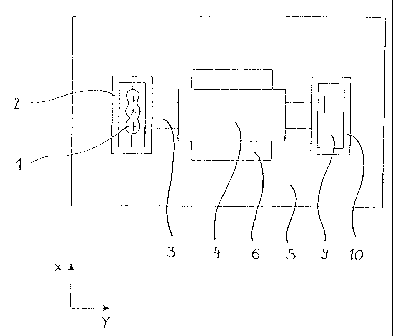
In the first embodiment, a positive model in the form of a wax model 1 of a
dental bridge is clamped
in a mounting 2 of a device according to the invention, as shown in the
figures. The mounting 2 is
mounted on a shaft 3 which enables a rotation of the mounting 2 by
180°. Furthermore, the shaft 3
is mounted on a table 4 which can travel precisely in three axes x, y, z. The
axis of rotation of the
shaft 3 extends, for example, in the y direction. The drive of the table 4 is
mounted in an equipment
housing 5. The opening in the equipment housing 5 required for the travel
movement of the table
4 can be covered in any known manner, e.g. by a bellows or by a sleeve 6.
Furthermore, an optical scanning unit 7 is accommodated in the equipment
housing 5 for measuring
distance. The scanning unit 7 comprises a laser beam source (not shown in
greater detail), e.g. a laser
diode, as well as advantageously a device for reflecting the light of the
laser diode into the ray path
of the scanning unit 7 and further optical elements as well as a CCD camera
adjusted in its sensitivity
to the laser. A birefractive crystal which splits the laser light reflected by
the wax model 1 (positive
model) into a regular portion and an irregular portion is arranged in front of
the CCD camera, as a
result of which holograms with border areas are generated on the CCD image
recorder which can be
accurately measured and with reference to which the exact distance to the
measured point can be
3 5 determined.
CA 02449509 2003-11-19
_7_
The scanning unit 7 is fastened in the equipment housing 5 in such a way that
an emitted laser beam
runs along the z axis. After a single calibration during assembly, the
scanning unit 7 delivers absolute
information about the distance to an object reflecting the laser beam, e.g. a
wax model 1 (positive
model) clamped in the mounting 2, according to the so-called conoscopic
holography. Details of this
measuring method are described, for example, in WO 99/64916, US 5,953,137, WO
99/42908, US
5,892,602, US 5,291,314, EP 0 394 137, EP 0 394 138 and US 4,976,504.
1'he high intensity of the laser light enables the use of an image forming
objective with a relatively
small opening, so that a field depth is produced which is larger than, for
example, the typical height
of a dental bridge or the wax model 1 thereof or the dental stump of a plaster
model, e.g. 15 mm.
Since the previously described scanning unit 7 gives measured values about the
absolute distance
of the point lit by the laser beam based on the reflection as measured value,
when mounting a device
according to the invention, not only is the scanning unit 7 adjusted such that
the laser beam is parallel
to the z axis of the table 4, but the scanning unit 7 is also calibrated via a
reference plate which is
clamped in the mounting 2. Thus, the area of the tolerable blur (field depth)
can thereby be
simultaneously determined by moving the table 4 accordingly in the z
direction.
During a later mapping of the positive model 1 such as the wax model or
plaster model, the mounting
2 is moved over the table 4 along the z axis of the table 4, accordingly in
the focus range of the
scanning unit 7. The plaster or positive model 1 is now digitalized by moving
the mounting 2 and
the table 4 in a defined manner along the x and y axis, e.g. by line or in
columns, and this information
is linked with the distance information determined by the scanning unit 7. The
position of the table
4 and with it of the model 1 to be mapped in the z direction, is subtracted
from the distance value
which the scanning unit 7 gives to form the measured data set. During scanning
of the model 1, the
table 4 is not moved along the z axis but only in x and y direction.
By linking the x and y position values with the distance information of the
scanning unit 7, a data
pattern is produced which reproduces the three-dimensional design of the side
of the plaster or
positive model 1 facing the scanning unit 7.
For the complete three-dimensional appraisal of the entire model 1, the
positive model 1 together
with the mounting 2 is turned about the y axis through 180° after one
side has been scanned and the
rear side of the positive model 1 is mapped in the same manner.
CA 02449509 2003-11-19
_g_
However, a prescan (preliminary run-through of the method) can also be
undertaken prior to starting
the measurement of the first side of the positive model 1 to determine an
extreme value of the
positive model 1 in the z direction, e.g. the model point with the least
distance to the scanning unit
7 and the associated z value of the coordinate as reference value and thus the
distance information
standardized to the model point as reference point. This reference value can
be adopted for forming
a reference plane perpendicular to the z axis. In this way, the maximum
extents of the mapped
model can be derived directly from the data set generated.
If redundant measured data is generated by the mapping of two sides, these can
be removed later by
appropriate reprocessing by software when forming the volume model to avoid
malfunctions during
later control of a processing machine or a processing tool such as a milling
tool 8.
A milling tool 8 of this type is advantageously integrated in a housing 5, for
example, relative to the
table 4, opposite the optical scanning unit 7. Advantageously, the milling
tool 8 has a stationary
spindle. A ceramic blank 9, for example, consisting of a presintered yttrium
oxide stabilized
zirconium oxide, is clamped in a further mounting 10 which is connected with
the rear end of the
shaft 3. The forward movements in the x, y and z directions required for
processing the side of the
blank 9 facing the milling tool 8 are carried out by corresponding movement of
the table 4 with the
shaft 3 and the mounting 10. When the processing of the side of the blank 9
facing the milling tool
8 is finished, the blank 9 can be moved away from the milling tool 8 in z
direction by a forward
movement and the mounting 10 turned by 180°, as during scanning of the
positive model 1, to
process the other side of the blank 9.
Instead of a ceramic blank 8, a blank consisting of any other suitable
material, e.g. a metal, plastics
or composite materials, can also be used.
In a further embodiment of the invention (not shown in the figures), the use
of a so-called line
scanner is provided instead of the laser beam with almost point-like cross
section, whereby the line
width should correspond to at least the width of the model to be scanned, e.g.
in the order of 100
mm. With a line scanner of this type, which can, moreover, work similarly to
the scanning unit 7
already described above, it would then be possible to completely digitalize
three-dimensionally a
positive model 1 or also a plaster stump or a plaster model of the jaw by
moving the table 4 with the
mounting 2 and the plaster or positive model 1 along an axis. If a positive
model is scanned, both
sides are measured by turning the mounting through 180°. With a plaster
model, only the side with
the dental stumps is scanned.
CA 02449509 2003-11-19
-9-
For example, a wax model of a bridge construction, which has three-
dimensionally formed
functional or connecting surfaces both on the upper side and on the lower
side, scanned from both
sides by a line scanner of this type after a rotation of the mounting through
180°, as previously noted.
The scanning of a three-dimensional plaster or positive model 1 according to
the invention by
displacement along only one or at most two axes, with an additional turning of
the model 1 through
180°, also represents, with respect of the computational effort
required to form a three-dimensional
data model of the measured object, significant progress compared to the known
optical scanning
devices, in which the object to be scanned is usually tilted several times and
the data pattern of the
various "views" thus obtained must be linked with one another by appropriate
computational
operations to produce a volume model of the measured object.
However, care must be taken that, for a sufficiently reliable reflection and
thus a reliable distance
adjustment by the scanning unit 7 with typical materials for the modelling in
the dental field, the
surfaces to be mapped form an angle of at least about 0.1 °, preferably
of at least 1 °, to the z axis with
the optical axis of the laser beam. Nevertheless, the angle should not exceed
20°. However, this does
not represent a limitation in practice since, at the latest for mounting the
dental prosthesis onto the
prepared tooth stump or the implant, at least such an inclination is required
for the proper cementing
of the prosthesis as would be required for a shape inclination of a
conventional cast prosthesis.
Undercuts may not occur in any event in prostheses of these types, since
cavities between the
prosthesis and tooth stump could form in this case, which would inevitably
lead to further damage
of the tooth stump, for example, by caries bacteria remaining in the cavity
thus produced.
To ensure a sufficiently accurate clamping of the plaster or positive model 1
to be mapped in the
mounting 2, this can, for example, be accomplished with aid of a
parallelometer, in which an
apparent undercut, caused by an inclined position of the model or a tangential
run of the laser beam
of the scanning unit 7, can be prevented with very good reliability and
reproducibility with aid of the
adjustment of the so-called light-gap method.
It is understood that it can also be provided that the scanning unit 7 moves
in the z direction instead
of the table 4 being moved in the z direction or even that a movement in the z
direction can be
entirely omitted for the scanning if the scanning unit 7 is equipped with
interchangeable objectives
of various focus lengths for adapting the working distance or with a zoom
optic having an adjustable
focal length.