Note : Les descriptions sont présentées dans la langue officielle dans laquelle elles ont été soumises.
CA 02449705 2003-11-10
WO 02/091930 PCT/USO1/15390
TRACTION TROCAR APPARATUS AND METHOD
Background of the Invention
Field of Invention
This invention relates generally to trocar systems and more specifically to
obturator apparatus and methods for placing a trocar cannula across a body
wall.
Discussion of the Prior Art and Related Technology
It is generally well known that holes can be created through body tissue
either by cutting the tissue or by mechanically parting the tissue along lines
of weakness.
Where tissue is cut, it is severed along a line, which is determined by the
direction of the
cutting implement. Where tissue is parted, it separates along natural tissue
planes such as
those defined by muscle fibers or differing layers of tissue such as skin and
muscle.
Tissue that is mechanically separated tends to heal better than tissue that is
cut. with
tissue that is mechanically separated the healing process requires only that
the affected
tissues re-approximate each other. with cut tissue, and in particular muscle
fibers, the
healing process must reconstruct the damaged tissue, often with resultant
scaring and
incomplete reconstruction. It has been shown for laparoscopic surgery in
particular, that
trocar wound sites of 10 millimeters in diameter and higher, made by cutting
obturators,
require suturing to prevent incisional hernias from occurnng. It has also been
shown that
where the same size would site is created by expanding or parting the wound
from a cut
of 3 millimeters, for example, that the wound site does not require stitching
and tends to
heal faster.
-1-
CA 02449705 2003-11-10
WO 02/091930 PCT/USO1/15390
For laparoscopic surgery there is a requirement that instrument ports in the
form of cannulas be placed in the patient's abdominal wall. These cannulas are
then used
as access ports for the surgeon to place instruments such as scissors and
graspers. In the
past these cannulas have been introduced by using a sharp cutting obturator,
placed
within the cannula, to cut a line or hole for advancing the cannula through
the abdominal
wall. The obturator is then removed from the cannula and the cannula is left
in place for
the duration of the surgery.
For most surgeries the cutting obturator is only used after the abdomen has
been insufflated with carbon dioxide gas. There is then separation between the
abdominal wall and the underlying anatomical structures and organs. Even with
this
separation, however, there is a risk that the patient will be injured by the
sharp cutting tip
of the obturator as it breaks through the abdominal wall. To help resolve this
issue a
variety of mechanical shielding mechanisms have been employed to cover the
cutting
element once it breaks through the abdominal wall. Tt has been noted and
observed that
even with these mechanical shield mechanisms that the risk is not completely
eliminated
and that the rigid shields themselves can cause damage to internal organs and
structures.
Other methods have been used as well. For example, optical trocars have
been provided with a clear plastic cutting tip. This allows the suxgeon to
view the tissue
layers as they are cut, and in principle to better control the timing of
insertion forces.
These plastic tips, however, are not as sharp as the metal bladed variety and
therefore
require a higher insertion force which in turn increase abdominal wall
distortion. This
distortion or tenting brings the obturator tip into closer proximity with the
internal organs
-2-
CA 02449705 2003-11-10
WO 02/091930 PCT/USO1/15390
and increases the chances for potential damage. The wound created by such a
device is
still a cut and not a mechanical separation, as it still suffers from the
above-mentioned
disadvantages.
Another manufacturer employs a multistage system whereby a sheath is
S inserted over a veress needle. The needle is then removed and a conical
obturator, placed
inside a cannula, is inserted through the sheath thereby expanding it to the
desired
cannula size. The obturator is then removed leaving the cannula in place. This
offers the
advantage of a smaller initial incision with the veress needle. However, the
needle still
presents a risk to internal organs, and the system is more expensive and
complex than
those associated with the cutting obturator devices.
W all of these systems of the past, a cutting element is employed to either
create the final size of the wound site or to make a smaller initial wound
site that is then
expanded to the final size. The use of sharp cutting elements common to all
systems
presents an unavoidable risk to the patient.
1S
Summary of the Invention
These deficiencies of the prior art are overcome with the present invention
which
provides for the parting rather than cutting of tissue, and, the use of
opposing radial
forces which precede the tip of the obturator shaft. After the tissue is
parted, it is drawn
proximally along the outer surface of the shaft as the shaft is moved distally
through the
body wall. The resulting counter forces can produce a net proximal force on
the body
wall with a minimal distal or penetration force.
-3-
CA 02449705 2003-11-10
WO 02/091930 PCT/USO1/15390
In a preferred embodiment a tubular mesh sleeve is initially disposed in
the hollow shaft of the obturator. This sleeve is pulled out of a hole at the
tip of the
obturator shaft and drawn radially and proximally along the outer surface of
the shaft.
The mesh sleeve inverts at the distal tip facilitating its movement interiorly
of the shaft
and its traction with the parted tissue exteriorly of the shaft.
In one aspect of the invention, an apparatus is provided for creating an
opening through body tissue. The apparatus includes a shaft having an axis and
a channel
extending axially between a proximal end and a distal end. The shaft has a
distal tip and
a hole in the tip communicating with the channel of the shaft. Portions of the
tip define a
leading surface of the tip. Means is disposed along this leading surface and
is moveable
relative to the tip for creating generally opposing forces on the body tissue
which tend to
part the body tissue and thereby create the opening through the body tissue.
In another aspect of the invention, a surgical instrument is used for
creating an opening through an abdominal wall retaining internal organs. The
instrument
includes a shaft having an outer surface and a tip. A sheath initially
contacting the body
tissue generally at a point extends proximally from the point along the outer
surface of
the shaft. The shaft is operable to create a distal force on the body tissue
while the sheath
is operable to create a proximal force on the body tissue. The proximal force
is greater
than the distal force in order to create a net proximal force on the abdominal
wall tending
to separate the abdominal wall from the internal organs as the opening is
created.
In another aspect of the invention, a flexible sheath having a tubular
configuration extends from an axial channel of the shaft through the distal
tip of the shaft.
A handle is attached to the sheath exteriorly of the shaft and is moveable
proximally
-4-
CA 02449705 2003-11-10
WO 02/091930 PCT/USO1/15390
relative to the shaft to withdraw the sheath from the channel and to
progressively invert
the sheath at the tip of the shaft.
hl another aspect of the invention, the shaft of the surgical instrument has
a tubular configuration with an outer surface, an axial channel, and a distal
tip. At least
one flexible traction tread is carried within the axial channel and extends
outwardly of the
shaft at the distal tip. A handle attached to the traction tread exteriorly of
the shaft is
moveable proximally to withdraw the traction tread distally through the distal
tip.
An associated method of operation includes even further aspects of the
invention. For example, a method fox creating an opening in body tissue
includes the
steps of providing opposing traction treads extending from the axial channel
of the shaft
outwardly through the hole in the tip of the shaft. The body tissue is
contacted with the
traction treads at the tip of the shaft and the traction treads are moved
radially outwardly
from the hole in the tip. During this moving step, the body tissue is engaged
at the tip to
produce parting forces on the body tissue tending to separate the body tissue
and thereby
create the opening through the body tissue.
In another method of operation, first and second cannulas are inserted
through body tissue by providing an obturator having a shaft with an outer
surface and a
traction tread moveable relative to the outer surface. Placing the obturator
in the first
cannula, the body tissue is engaged with the tread and the tread is moved
relative to the
outer surface of the shaft to facilitate penetration of the body tissue by the
shaft and the
first cannula. The obturator is then removed from the first cannula and placed
in the
second cannula where again the traction thread engages the tissue and
facilitates
penetration of the body tissue by the shaft in the second cannula. Removing
the obturator
-5-
CA 02449705 2003-11-10
WO 02/091930 PCT/USO1/15390
from the second cannula leaves both the first cannula and the second cannula
operatively
disposed across the body wall.
In another method associated with the invention, removal of a trocar
cannula from a body wall is facilitated by placing a mesh sleeve between the
cannula and
the body wall. The sleeve is provided with properties which exert a radial
force on the
cannula tending to resist removal of the cannula from the body wall. However,
an axial
force can be applied to the sleeve to reduce the radial force of the sleeve on
the cannula.
During this step of applying the axial force, the cannula can be removed from
the body
wall.
hl a method for inserting an obturator, the obturator is provided with a
shaft having an outer surface and a traction tread moveable along the outer
surface of the
shaft. The tread is carried within the shaft. As the obturator is moved
through the body
wall, a first force is applied to the obturator in a first direction and a
second force is
applied to the obturator in a second generally opposing direction. As the
obturator is
moved distally relative to the body wall, it engages wall portions which face
the outer
surface of the shaft and pulls those wall portions proximally along the shaft.
These and other features and advantages of the invention will be better
understood with reference to preferred embodiments of the concept and
reference to the
associated drawings.
Description of the Drawings
Fig. 1 is a side elevation view of a patient with insufflated abdomen and
trocars in
the process of being placed using the trocar system of the present invention;
-6-
CA 02449705 2003-11-10
WO 02/091930 PCT/USO1/15390
Fig. 2 is a side elevation view of a prior art trocar system involving an
incision
and blunt tip obturator;
Fig. 3 is a side elevation view of a prior art trocar system involving an
obturator
with cutting wings;
Fig. 4 is a side elevation view of a prior art trocar system including a
cutting tip;
Fig. 5 is a side elevation view of the system of Fig. 4 invading an interior
organ;
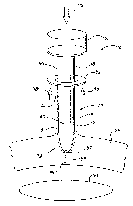
Fig. 6 is a side elevation view of the system of the present invention
including an
inverting sheath operable with counter forces which can produce a net proximal
force on
the body wall;
Fig. 7 is a side elevation view of an embodiment including a single traction
tread;
Fig. 8 is a radial cross section view taken along lines 8-8 of Fig. 7;
Fig. 9 is a side elevation view of an embodiment including a pair of opposing
traction treads;
Fig. 10 is a radial cross section view taken along lines 10-10 of Fig. 9;
Fig. 11 is a side elevation view of an embodiment having more than two
traction
treads equally circurriferentially spaced;
Fig. 12 is a radially cross section view taken along lines 12-12 of Fig. 11;
Fig. 13 is a side elevation view wherein the traction tread is radially
continuous
and forms a tube or traction sheath;
Fig. 14 is an axial cross section view taken along lines 14-14 of Fig. 13;
Fig. 15 is an end view taken along lines 15-15 of Fig. 14;
Fig. 16 is a side elevation view of a further embodiment of the obturator
wherein
opposing traction treads are axially continuous;
CA 02449705 2003-11-10
WO 02/091930 PCT/USO1/15390
Fig. 17 is an axial cross section view taken along line 17-17 of Fig. 16;
Fig. 18 is an end view taken along line 18-18 of Fig. 17;
Fig. 19 is a front elevation view similar to Fig. 16 and illustrating an
embodiment
including axially continuous traction treads;
Fig. 20 is an axial cross section view taken along lines 19-19;
Fig. 21 is an end view taken along lines 21-21 of Fig. 20;
Fig. 22 is a side elevation view of the embodiment of Fig. 13 placed in
initial
contact with an abdominal wall to produce opposing parting forces;
Fig. 23 is a side elevation view similar to Fig. 22 and showing the obturator
with
the abdominal wall being drawn upwardly onto the cannula of the trocar system;
Fig. 24 is a side elevation view similar to Fig. 23 and illustrating the
abdominal
wall fully parted by the trocar cannula;
Fig. 25 is a side elevation view of the embodiment illustrated in Figure 24
showing the abdominal wall drawn proximally onto the trocar cannula by the
traction
sheath;
Fig. 26 is the side elevation view of the system illustrated.in Figure 25 with
the
traction sheath fully deployed to maintain traction between the abdominal wall
and the
cannula, and with the obturator removed to vacate the working channel of the
cannula;
Fig. 27 is a side elevation view of the system illustrated in Fig. 26 with the
trocar
cannula removed from the traction sheath;
Fig. 28 is a side elevation view of the system illustrated in Fig. 27 showing
the
traction sheath removed from the opening leaving parted surfaces to promote
healing;
_g_
CA 02449705 2003-11-10
WO 02/091930 PCT/USO1/15390
Figs. 29-32 illustrate a series of side elevation views showing progressive
steps
for operating an embodiment wherein the inverting sheath is disposed outwardly
of the
obturator but inwardly of the trocar cannula;
Fig. 33 is a side elevation view of a blunt-nose obturator having windows to
facilitate the return of the inverting sheath to an interior channel of the
obturator;
Fig. 34 is a cross section view taken along lines 34-34 of Fig. 33;
Fig. 35 is a side elevation view similar to Fig. 33 and showing an obturator
tip
with converging planes;
Fig. 36 is a front elevation view taken along lines 35-35 of Fig. 34;
Fig. 37 is an axial cross section view of an obturator similar to that of Fig.
34 and
including a biasing means for returning the inverting sheath to its initial
position;
Fig. 38 is an axial cross section view similar to Fig. 37 arid illustrating
the biasing
means stretched to a final position of the inverting sheath;
Fig. 39 illustrates a fabric adapted for use as a traction tread or sheath,
the fabric
being illustrated in a normal state;
Fig. 40 is a side elevation view of the mesh of Fig. 39 axially stretched;
Fig. 41 is a side elevation view of the mesh of Fig. 39 radially stretched;
Fig. 42 is an end view of the traction sheath formed of the mesh of Fig. 39;
Fig. 43 is an end view similar to Fig. 41 of the inverting sheath forming
pleats to
provide texture for traction;
Fig. 44 is a side elevation view of a further embodiment of the obturator
including
a blunt tip with a conical point for microscopic puncture;
Fig. 45 is an end view taken along lines 45-45 of Fig. 44;
_g_
CA 02449705 2003-11-10
WO 02/091930 PCT/USO1/15390
Fig. 46 is a perspective view of an application adapted for use in placing
the trocar system of the present invention;
Fig. 47 is a schematic axial cross section view of a further embodiment
including gears with circumferential teeth;
Fig. 48 is an end view of the embodiment illustrated in Fig. 47;
Fig. 49 is a schematic axial cross section view of a further embodiment
including a single oscillating gear; and
Fig. 50 is an end view of the embodiment of Fig. 49.
Description of Preferred Embodiments
and Best Mode of the Invention
A trocar system of the present invention is illustrated generally in Figure I
and designated by the reference numeral 10. The system 10 includes a trocar
cannula 12
having a seal housing 14, and an obturator 16 with a shaft 18 and handle 21,
and
including a traction mechanism 23 of particular interest to the present
invention. The
obturator 16 is used in placing the cannula 12 across a body wall such as an
abdominal
wall 25, associated with a patient 27. In the case of the abdomen, the wall 25
defines an
abdominal cavity 29 which includes many organs such as that designated by the
reference
numeral30.
In less evasive laparoscopic procedures, multiple cannulas 32 and 34 are
used to provide access across the abdominal wall 25 to facilitate surgical
procedures
within the abdominal cavity 29. By way of example, the removal of a
gallbladder is
-10-
CA 02449705 2003-11-10
WO 02/091930 PCT/USO1/15390
typically accomplished with such a laparoscopic procedure. Initially, cannulas
12, 32 and
34 are placed across the abdominal wall 25, each providing a working channel
through
which various instruments can be inserted and surgically manipulated. For
example, the
cannula 32 is shown with a grasper 36 which can be inserted through the
cannula to grasp
the organs 30 or other tissue within the abdominal cavity 29. A fiber-optic
scope 38 is
illustrated in Figure 1 operatively disposed through the cannula 34 and across
the
abdominal wall 25 to provide visualization witlun the abdominal cavity 29.
As further background to the trocar system 10 of the present invention,
Figures 2-5 are provided and to illustrate the deficiencies of trocars and
obturators of the
prior art.
One trocar of the prior art is illustrated in Figure 2 and is designated by
the
reference numeral 41. This trocar includes a cannula 43 and blunt obturator
45. In the
placement of this device, an incision 47 is cut entirely through the abdominal
wall 25
using a scalpel 50. All of the deficiencies previously discussed with respect
to cutting
rather than parting the abdominal wall 25 impact this procedure. After the
incision 47 is
cut, the blunt obturator 45 is moved through the incision to place the cannula
43 across
the wall 45. Due to the delicate cutting required by this procedure, placement
of this
trocar 41 may take as long as 10 minutes. In a procedure requiring the
placement of four
trocars, this time intensive procedure would require as much as 40 minutes,
for example.
In comparison, placement of a self cutting obturator may require only one
minute of time. In a procedure requiring the placement of four trocars, this
part of the
procedure may require only four minutes of time as opposed to the 40 minutes
of time
required for the precut procedure of Figure 2.
-11-
CA 02449705 2003-11-10
WO 02/091930 PCT/USO1/15390
A self cutting trocar system 52 of the prior art is illustrated in Figure 3.
This system 52 includes a cannula 54 and an obtuxator 56 having a pair of
opposing
wings 58. These wings 58 are provided with sharpened outer edges so that they
tend to
cut a path through the abdominal wall 25. Again, the disadvantage of cutting
an incision
through the abdominal wall 25 also impacts this embodiment.
Perhaps the most widely used embodiment of a trocar is that illustrated in
Figures 4 and 5. In this case, a trocar 61 includes a cannula 63 and obturator
65 having a
sharpened point 67. A safety shield 70 associated with the obturator 65 is
biased to move
over the point 67 and to protect the interior organs 30 upon penetration of
the abdominal
wall 25. In the process associated with this instrument, the trocar 61 is
forced through the
abdominal wall 25 creating a significant distal force on the wall 25. This
distal force
provides the wall 25 with a concave shape, commonly referred to as tenting,
and tends to
bring the point 67 into close proximity to the interior organs 30.
As with the prior art embodiment of Figure 3, the point 67 precedes the
remainder of the trocar 61 as it cuts the tissue of the wall 25. Therefore, of
the
deficiencies previously discussed with reference to cutting are applicable to
this prior art
embodiment. Furthermore, the significant force required for penetration, a
force typically
as high as ten pounds, coupled with close proximity of the concave wall 25 to
the organs
30, tends to provide little time for the safety shield 70 to cover the tip 67.
As a
consequence, damage to the interior organs 30 has been severe notwithstanding
the
presence of the safety shield 70.
The high forces required for penetration are particularly applicable to
those trocar systems which require both penetration forces as well as cutting
forces.
-12-
CA 02449705 2003-11-10
WO 02/091930 PCT/USO1/15390
In all of these embodiments of the prior art, it will be noted that cutting of
the abdominal wall is required. Furthermore, all forces associated with
movement of the
trocars 41, 52 and 61 through the abdominal wall 25 produce a distal force as
great as ten
pounds which tends to move the abdominal wall 25 into a concave shape and into
close
proximity with the interior organs 30.
The advantages of the trocar system 10 of the present invention will be
readily apparent with reference to the obturator 16 of Figure 6 and a
comparison with the
prior art devices illustrated in Figures 2-5. As previously discussed, this
obturator 16
includes the shaft 18, handle 21 and traction mechanism 23. In this case, the
traction
mechanism 23 may include a fabric 72 having the configuration of a tube with a
first end
74 and a second end 76. In this context, the word "fabric" refers to any
flexible sheet
material. The shaft 18 can be solid, but in a preferred embodiment it is at
least partially
hollow to receive the first end 74 of the fabric 72 within the shaft 18. The
shaft I8
extends to a distal tip 78 having a wall 81 that defines an internal channel
83 and an axial
hole 85. This wall 81 is defined by a leading surface 87.
The tubular fabric 72 is initially disposed with its first end 74 positioned
in
the interior channel 83. The fabric 72 extends distally outwardly through the
hole 85
where it inverts and extends proximally along the leading surface 87 and the
outer surface
90 of the shaft 18. At the second end 76, the tubular mesh is preferably
attached to a
finger engagement means, such as a projection, tab, flange or ring 92.
In operation, the handle 21 of the obturator 16 is placed in the palm of the
user's hand and his/her fingers are extended to engage the ring 92. In a
common and
familiax motion, the hand of the user is closed drawing the fingers towards
the palm of
-13-
CA 02449705 2003-11-10
WO 02/091930 PCT/USO1/15390
the hand. This moves the ring 92 toward the handle 21 and draws the tubular
fabric 72
distally through the hole 85 and proximally along the outer surface 90 of the
shaft 18. As
the ring 92 moves proximally upwardly in Figure 6, the first end 74 of the
fabric 72 is
pulled toward the distal tip 78 where the fabric 72 exits the hole 85 and
inverts to move
along the outer surface 90. With the fabric 72 disposed between the shaft 18
and the
abdominal wall 25, it tends to grip the abdominal wall 25, and move the wall
25
proximally along the shaft 18. As the wall 25 moves upwardly in Figure 6 along
the shaft
18, it tends to part at the leading edge 87 along a line of weakness
designated generally
by the reference numeral 94. It is of particular importance to note that the
wall 25 is
parted rather than cut in order to achieve the advantages previously
discussed. In this
particular embodiment, there is no structure which works to cut the abdominal
wall 25 or
otherwise force the obturator 16 along a predetermined path. Rather, the
obturator 16
fords its own path along the line of weakness 94.
Notwithstanding this significant aspect of the present invention, perhaps
the greatest advantage is achieved with a net zero or even proximal force on
the wall 25.
As previously noted, the prior art produced only a distally directed force in
creating an
incision while moving an obturator through the abdominal wall. This tended to
move the
abdominal wall toward a concave shape and into proximity with the internal
organs.
With the present embodiment, the handle 21 can be held stationary with a
distally
directed force, shown by an arrow 96, while a counter proximal force of equal
or greater
magnitude is applied to the ring 92, as shown by the arrows 98.
Since these forces, shown by the arrows 92 and 98, are applied in different
directions, they tend to offset each other so that the net distal force
applied to the
-14-
CA 02449705 2003-11-10
WO 02/091930 PCT/USO1/15390
abdominal wall 25 can actually be negative. Note for example, that if the
handle 21 is
maintained stationary, and the ring 92 is moved upwardly, the net force on the
abdominal
wall 25 is a proximal force directed upwardly in Figure 6. As a result, the
abdominal
wall 25 can be moved toward a convex shape and a spaced relationship with the
interior
organs 30.
It will be appreciated from the foregoing discussion, that the counter
forces which are of particular advantage to the present invention can be
produced from a
variety of structures. More specifically, the tubular fabric 72 discussed with
reference to
Figure 6 can be any material capable of being pulled along the outer surface
90 of the
shaft 18. This material could be organic or inorganic and will generally be
elongate so
that it can be pulled with some magnitude of force in the axial, proximal
direction. For
example, the tubular fabric 72 of Figure 6, although preferred for that
embodiment, could
be replaced with just a single traction tread 101 as illustrated in Figure 7.
With this
traction tread 101 disposed between the shaft 18 and the abdominal wall 25,
the traction
tread 101 will engage the tissue of the abdominal wall 25 and pull it
proximally relative
to the shaft 18 .
Forces will be more balanced if at least two traction treads, such as the
tread 101 and a second tread 103, were diametrically opposed as illustrated in
Figure 9
and 10. With this configuration, the abdominal wall 25 (Figure 1) is engaged
on both
sides of the shaft 18 and pulled proximally relative to the shaft 18.
Other embodiments of the invention might include three traction treads,
such as the treads 101 and 103 and a third tread I05, equally spaced around
the
circumference of the shaft 18. Such an embodiment is illustrated in Figures 11
and 12.
-15-
CA 02449705 2003-11-10
WO 02/091930 PCT/USO1/15390
While independent and discrete traction treads, such as the treads 101, 103
and 105, will function to produce the discrete counter forces, a single
traction tread that is
radially continuous, as illustrated in Figures 13-I5, may be preferred as it
provides
complete isolation of the shaft I8 from the abdominal wall 25 (Figure 1).
Where the
shaft 18 of the obturator 16 is cylindrical and the distal tip 78 is conical
or convex as it as
illustrated in Figure 13, this tubular configuration for the traction tread
107 is particularly
desirable. With this configuration, the traction tread 107 passes through the
axial hole 85
where it inverts and travels radially as shown by arrows 110 in Figure 15.
From this
point, the traction tread 107 travels proximally along the surface 90 of the
shaft 18,
upwardly in Figure 14.
In a further embodiment of the invention, the distal tip 78 of the obturator
16 is formed as a pair of planar surfaces 112 and 114 which converts distally
in the nature
of a flathead screwdriver. This configuration lends itself to the opposing
pair of tractor
treads 101 and 103 previously illustrated in Figures 9 and 10. With this
construction, the
traction treads 101 and 103 separate generally at an exit slot 116 best shown
in Figure 18.
In this view the opposing forces are shown by the arrows 118 and 121 which
produce the
tissue parting results of particular advantage to the present invention.
With reference to Figures 19, 20 and 21, it can be seen that a similar
embodiment including the converging surfaces 112 and 114 can be accommodated
with
traction treads 201 and 103 which are axially continuous. Thus, each of these
treads 101
and 103 forms a continuous band 123 and 125 respectively. The two bands 123,
I25
-16-
CA 02449705 2003-11-10
WO 02/091930 PCT/USO1/15390
counter rotate through the slot 116, and extend proximally along the surface
90, returning
to the interior channel 83 through opposing windows 127 and 130 in the wall 81
of the
obturator.
With respect to the embodiment of Figure 6, the method of operation will
now be discussed with reference to Figures 22-28. In these views, the
abdominal wall 25
is further defined by a fascia 141, muscle tissue 143, and a peritoneum 145.
In this case,
the shaft 18 and fabric 72 of the obturator 16 can be inserted through the
seal housing 14
and into the cannula 12. A distal end 147 of the cannula 12 is disposed
through the ring
92 and into the associated tubular fabric 72. In operation, the trocar system
10 functions
by pulling the ring 92 proximally along an outer surface 149 of the cannula
12, upwardly
in Figure 22.
In an initial step of the process, a cut 152 can be made in the skin or fascia
141. This cut 152 is preferably made to gain access to the muscle tissue 143
which is
more easily parted. The cut 152 also marks the desired location for insertion
of the trocar
system 10. As the ring 92 is drawn upwardly along the cannula 12, the tubular
fabric 72
exits the distal hole 85, inverts and follows the ring 92 upwardly along the
outer surface
149. At the leading surface 187, the inverting fabric 72 produces opposing
radial forces
shown by arrows 152 and 154. With these opposing forces, the tissue 143 is
parted along
the line of weakness 94 (Figure 6) as the trocar system 10 is moved relatively
into the
abdominal wall 25. It will be noted that the arrows 152 and 154 are merely
representative of all of the radial forces which emanate from the hole 85 as
shown by the
arrows 110 in Figure 15.
-17-
CA 02449705 2003-11-10
WO 02/091930 PCT/USO1/15390
With reference to Figure 23, it can be seen that the ring 92 and inverted
tubular fabric 72 are preferably drawn proximally by the arrows 98 while the
cannula 12
and obturator 16 are held stationary as shown by a pair of arrows 156. This
produces the
counter forces previously described and elevates the abdominal wall 25 as it
is pulled
proximally upwardly along the cannula 12 by the fabric 72.
Full penetration of the abdominal wall 25 including the peritoneum 145 is
illustrated in Figure 124. It is interesting to suspend further description at
this point and
note that on the distal side of the abdominal wall 25, the trocar system 10
presents no
shazp objects that might be detrimental to the interior organs 30. There is no
scalpel
(Figure 2), no sharp wings 56 (Figure 3), and no sharp cutting point 67
(Figure 5)
characteristic of this prior art. Furthermore, the space between the abdominal
wall 25 and
the interior organs 30 is actually increased by the net proximal force
associated with
operation of the trocax system 10. This space can be even further increased as
illustrated
in Figure 25 by merely pulling on the trocar system 10 to further elevate the
convex
abdominal wall 25 into a more conical configuration.
Once the trocar system 10 has fully penetrated the abdominal wall 25, the
ring 92 can be drawn further upwardly along the cannula 12 into contact with
the seal
housing 14. In a preferred embodiment, this disposition of the ring 92 will
cause the first
end 74 of the tubular fabric 72 to exit the axial hole $5 of the shaft 18
(Figure 25). At
this point, the obturator 16 can be removed, leaving the seal housing 14,
associated
cannula 12 and tubular fabric 72. With the obturator 16 removed, the interior
working
channel of the cannula 12 is vacated to facilitate access with surgical
instruments, such as
the endoscope 38 and grasper 36 illustrated in Figure 1.
-18-
CA 02449705 2003-11-10
WO 02/091930 PCT/USO1/15390
Even during this stage of the process, the trocar system 10 of the present
invention offers significant advantages. Noteworthy in this embodiment is the
fact that
the tubular fabric 72 remains between the cannula 12 and the abdominal wall 25
even
after the obturator 16 is removed. In this position, the high traction
characteristics which
facilitated penetration of the abdominal wall 25 by the trocar system 10,
remains to
ensure that the cannula 12 stays in place during the insertion and removal of
surgical
instruments. The structure that aided in penetration of the abdominal wall 25
now aids in
maintaining the cannula 12 in its preferred operative disposition.
When the surgical operation is complete, the cannula 12 and associated
valve housing 14 (Figure 26) can be removed, from the ring 92 and attached
tubular
fabric 72. This removal of the cannula 12 may be inhibited in an embodiment
wherein
the tubular fabric 72 is automatically biased to a reduced profile. This bias
tends to exert
radial forces on the cannula increasing the amount of friction which must be
overcome to
separate the cannula 12 from the tubular fabric 72. In such an embodiment, it
has been
found that application of an axial force on the ring 92 and attached tubular
fabric 72, will
tend to radially expand the fabric 72. In Figure 26, this axial force is
represented by an
arrow 160. With this radial expansion of the fabric 72, the cannula 12 and
associated
valve housing 14 can be removed from the tubular fabric 72.
Without the Iarge cannula 12 radially stretching the fabric 72, the tubular
configuration will automatically be drawn down to a reduced diameter as
illustrated in
Figure 27. This lower profile greatly facilitates removal of the tubular
fabric 72 as
illustrated in Figure 28 by an arrow 161. It will be noted that once the
tubular fabric 72 is
-19-
CA 02449705 2003-11-10
WO 02/091930 PCT/USO1/15390
withdrawn, the abdominal wall 25 is left with the parted line of weakness 94
initially
discussed with reference to Figure 6.
An additional embodiment of the invention is illustrated in the progressive
views of Figures 29-32, wherein elements of structure similar to those
previously
discussed are designated with the same reference numeral followed by the lower
case
letter "a." Thus the trocar system 10a is shown with the cannula 12a and
associated seal
housing 14a. The obturator 16a includes the shaft 18a and handle 21a, as well
as the
axial hole 85a. The tubular fabric is designated with a reference numeral 72a.
Note that
in this embodiment the tubular fabric 72a also extends through the valve
housing 14a to
the ring 92a which is disposed proximally of the valve housing 14a.
This embodiment differs from that previously disclosed in that the
obturator 16a and tubular fabric 72a are disposed entirely within the working
channel of
the cannula 12a. Thus, the obturator I6a with fabric 72a is inserted into the
cannula 12a
in the initial step of operation. It will be noted that with this
construction, the fabric 72a
is exposed to the abdominal wall 25 (Figure 1) only in a distal region 163
where the
obturator shaft 19a is exposed distally of the end 132a of the cannula 12a.
Since this
region produces the parting forces represented by the arrows 152 and 154 in
Figure 22, as
well as the proximal counter forces, represented by the arrows 98 in Figure 6,
this
embodiment provides many of the advantages previously discussed.
In operation, the obturator 16a with tubular fabric 72a is disposed in the
cannula 12a. The leading edge 87a is brought into contact with the body wall
25a and the
ring 92a is drawn proximally toward the handle 21a as illustrated in Figure
30. As the
tubular mesh emanates from the axial hole 85a it inverts in the manner
previously
-20-
CA 02449705 2003-11-10
WO 02/091930 PCT/USO1/15390
discussed pulling the abdominal wall 25a upwardly onto the cannula 12a. The
ring 92a is
drawn proximally into an abutting relationship with the handle 21 a as
illustrated in Figure
31. At this point, the cannula 12a should be fully inserted through the
abdominal wall
25a. Following this step in the surgical procedure, the obturator 16a as well
as the
tubular fabric 72a can be entirely withdrawn leaving the cannula 12a
operatively disposed
across the abdominal wall 25a as illustrated in Figure 32.
One of the significant advantages associated with this embodiment is that
the obturator 16a and tubular fabric 72a can be repeatedly used in the
placement of
multiple cannulas, such as the cannula 12a. Thus, a first cannula can be
placed through
the abdominal wall using the obturator 16a. Upon removal of the obturator 16a,
the first
cannula can be left in place as illustrated in Figure 32. Then the obturator
16a can be
inserted into a second cannula to facilitate its placement across the
abdominal wall. The
same obturator 16a can then be removed to facilitate placement of additional
cannulas.
A further embodiment of the invention is illustrated in Figures 33-35
where elements of structure similar to those previously discussed are
designated with the
same reference numeral followed by the lower case letter "b." This embodiment
is
similar to that discussed with reference to Figure 29 in that the obturator
16b and tubular
fabric 72b are formed as a subassembly which is inserted into the cannula 12b.
Thus, the
tubular fabric 72b is only exposed in the distal region 163b distally of the
distal end 132b
of the cannula 12b.
The embodiment of Figure 33 differs from that of Figure 29 in that the
tubular fabric 72b moving proximally is not disposed between the obturator
shaft 18b and
the cannula 12b. Rather, the proximally moving tubular fabric 72a is disposed
exteriorly
-21 -
CA 02449705 2003-11-10
WO 02/091930 PCT/USO1/15390
of the shaft 18b only in the distal region 163b. At the proximal end of this
region 163b,
in proximity to the distal end 132b of the cannula 12b, the proximal moving
tubular
fabric 72b is directed through the windows 127b and 130b back into the
interior channel
83b of the shaft 18b.
Within the channel 83b, the second end 76b of the tubular fabric 72b is
attached to the ring 92b. This calls for a special construction of the shaft
18b and ring
92b which is best described with reference to the radial cross section. view
of Figure 34.
In order to attach the second end 76b of the tubular fabric 72b (which is
disposed
interiorly of the shaft 18b) to the ring 92b (which is disposed exteriorly of
the shaft 82b),
some structure is required to extend through the wall 81b of the shaft 18b.
Initially, the
shaft 18b can be formed with axial slots 165 which extend along the shaft 18b
beneath
the ring 92b. These axial slots 165 are preferably equally spaced around the
circumference of the shaft 18b. Spokes 167 integrally molded with the ring
92b, can be
positioned in the slots 165 of the shaft 18b to extend from regions exterior
of the shaft
18b to regions interior of the shaft 18b. Within the channel 83b, the second
end 76b of
the tubular mesh can be attached to the spokes 167.
A similar embodiment of the invention is illustrated in Figure 5 where
elements of structure similar to those previously discussed are designated by
the same
reference numerals followed by the lower case letter "c." Tn this case, the
obturator shaft
18c is formed at its distal end with a structure similar to that illustrated
in Figure 16.
Thus, the shaft 18c is formed with the converging planar surfaces 112c and
114c and the
separate traction treads lOlc and 103c, best illustrated in Figure 36. As
previously
discussed, this embodiment requires both of the windows 127c and 133c, as well
as the
- 22 -
CA 02449705 2003-11-10
WO 02/091930 PCT/USO1/15390
slot 116c. This embodiment of the trocar system 10c offers a further advantage
that the
obturator 16c can be used repeatedly with multiple cannulas 12c.
For those embodiments which offer this choice of repeated use, such as
the embodiments of Figures 29, 33 and 36, it may be desirable to provide some
mews for
recycling the obturator 16 as illustrated in Figure 37. In this case, a
tension spring 170 is
fixed at one end to the handle 21c and at the other end to the first end 74c
of the tubular
fabric 72c. In operation, the ring 92c is drawn proximally toward the handle
21c along
with the second end 76c of the tubular fabric 72c. This causes the first end
74c of the
tubular fabric 72c to move distally stretching the spring 170. The spring 170
is stretched
even further (Figure 38) as the ring 92c is drawn proximally and the traction
treads lOlc
and 103c pass outwardly through the axial slot 116c and inwardly through the
windows
127c and 130c, respectively. When this operation is completed and the
associated
cannula 12c is placed across the abdominal wall 25c, the obturator 16c can be
withdrawn
and the ring 92c released from its proximal-most position (Figure 38). At this
point, the
bias of the spring 170 will pull the first ends 74c of the traction txeads l
Olc, 103c
proximally. As the obturator 16c is reset, the treads lOlc, 103c will pass
outwardly
through the windows 127c, 130c, respectively, and inwardly through the axial
slot 116c.
This will enable the spring 170 to return to its normal, non-stretched state
with the ring
92c disposed in its distal most position.
It can be appreciated that the spring 170 could be replaced with any
biasing means which mechanically, electrically or elastomericallyo, for
example, would
bias the first end 74c in the proximal direction.
- 23 -
CA 02449705 2003-11-10
WO 02/091930 PCT/USO1/15390
With the foregoing description of these preferred embodiments, it can be
appreciated that the structure forming the tubular fabric 72 as well as the
various traction
treads 101, 103, 105 and 107, is of particular importance to the present
invention. This
structure is preferably formed as a sheet material and is flexible and
elongate with at least
one tractive surface. These characteristics will be appreciated particularly
in those
embodiments involving the traction treads 101 and 103 where the width of the
treads
remains generally constant. In these cases, the tread is able to maintain its
width as it
exits the distal slot 116 and enters the windows 127 and 130.
For those embodiments involving the distal exit hole 85, it may be further
desirable if the structure of the fabric is capable of radially expanding and
contracting.
Particularly if the mesh is biased to the contracted low-profile state, it
will occupy less
space within the interior channel 83 and more easily feed through the exit
hole 85. A bias
to the contracted state will also facilitate removal of the fabric 72 as
illustrated in Figure
28.
I 5 As noted, the fabric 72 preferably has a sheet configuration and can be
either woven or non-woven. It can be formed with filaments, which in the
preferred
embodiment of Figure 39, are divided into filament groups 170 and 172 that
extend in
transverse directions. Thus the filaments in the group I70 may extend, in a
normal state,
perpendicular to the filaments in the group 172, as illustrated in Figure 39.
In order to facilitate the traction characteristics of the material 168, the
filament groups 170 and I72 can be woven to form points of intersection I74
where the
filaments cross and spaces or interstices 176 between the filaments. At the
points of
intersection, the fabric 72 will have a thickness equal to the sum of the
diameters of a
-24-
CA 02449705 2003-11-10
WO 02/091930 PCT/USO1/15390
single filament in the group 170 and a single filament in the group 172.
Between the
points of intersection, the filaments in the groups 170 and 172 will provide
the fabric 72
with only a single thickness. In the interstices 176 between the filaments of
the groups
170 and 172, the material 168 will have zero thickness. Thus, the woven
configuration of
even this simple embodiment will provide the fabric 72 with three different
thicknesses
greatly facilitating the traction between the material 168 and the tissue
associated with
the abdominal wall 25 (Figure 1).
Even those significant traction characteristics can be dramatically
increased with simple variations in the weave parameters. Consider for example
the
effect of making the various filament groups 170, 172 with different
diameters. With an
appropriate weave, this could add two additional levels of thickness to the
fabric 72.
Thus it is contemplated that any of the weaves known in the textile industry
could
provide multiple levels of thickness having dramatic effects on the traction
of the fabric
72 relative to the tissue of the abdominal wall 25 (Figure 1).
It should also be considered that any one of the filaments in the groups
170 and 172 can be formed from a different material. Solid, non-resilient
materials, such
as monofilament, will tend to maintain their shape providing more of a
mechanical
traction to the tissue. The filaments could also be formed from fibrous
materials, such as
cotton, in which case traction would be further enhanced by capillary action.
The
filaments of the groups I70, 172 could also be individually varied in their
diameters or
thicknesses, or provided with a more tractive surface, shape or coating.
In a preferred embodiment, the filaments forming the group 170 include
monofilaments which are alternated with cotton filaments. The same alternation
of
-25-
CA 02449705 2003-11-10
WO 02/091930 PCT/USO1/15390
filament materials is applied to the filaments of the group 172. With even a
simple
weave of these filament groups 170 and 172, significant variations in
thickness occur due
to the fixed diameter of the monofilaments and the variable diameter of the
cotton
filaments. The resulting material 168 provides many different thicknesses for
high
mechanical traction and additionally provides the capillary action associated
with fibrous
cotton material.
Thermoplastic materials can also be used for the filaments in the groups
170 and 172. These materials will permit the fabric 72 to be biased to a
compacted state
as illustrated in Figure 40 and stretched to an expanded state as illustrated
in Figure 41.
This thermoplastic bias facilitates movement of the tubular fabric 72 between
a low
profile state interiorly of the shaft 18b, and an expanded high profile state
exteriorly of
the shaft 18b. With a bias to the low profile state, the tubular fabric 72
will automatically
contract to achieve the advantages previously discussed.
The fabric 72 can also be woven in a manner that the filaments of the
group 170 are fixed to the filaments of the group 172 at each point of
intersection 174.
This feature will tend to make the fabric 168 more rigid so that it does not
tend to close
down on the surface of the cannula 12 or shaft Z 8 as it is drawn proximally.
The
resulting fabric 72 will also have less of a tendency to expand or contract.
This may tend
to produce pleats in the fabric 72 particularly where it emanates from the
axial hole 85b.
With reference to Figure 42, it can be seen that these pleats 178 can provide
the fiuther
advantage of texture variations at the critical leading surface 87 of the
obturator 16. This
additional texture can even fiuther enhance the traction with the tissue where
the
important parting of the tissue is taking place.
-26-
CA 02449705 2003-11-10
WO 02/091930 PCT/USO1/15390
Alternatively, the filaments forming the group 170 and 172 can remain
disconnected at their points of intersection 74. This will enable the
filaments to move
over each other enhancing their ability to expand and contract. The
characteristic of this
weave is best illustrated in Figure 43 where the fabric 168 tends to maintain
its
cylindrical configuration as it passes through the axial hole 85 and moves
from the low
profile state to the high profile state.
In a particular embodiment of the invention it may be desirable to control
the stretchability of the fabric 168 in different directions. For example, it
may be
desirable to facilitate radial expansion while inhibiting axial expansion. The
radial
expansion might be desirable as it facilitates the transition of the tubular
fabric 72 from
the low profile state at the exit hole 85b, to the high profile state
exteriorly of the casmula
12 or shaft 18. At the same time, it might be desirable to inhibit expansion
or contraction
in the axial direction. Alternatives for providing different stretch
characteristics in
different directions are well known in the textile industry and include
formation of the
fabric 72 with filaments of different material and shape as well as
orientation of the
filaments relative to the cut of the fabric 168.
A further embodiment of the invention is illustrated in Figure 44 where
elements of structure similar to those previously discussed are designated
with the same
reference numeral followed by the lower case letter "d." In the side elevation
view of
Figure 44, the obturator 16d is illustrated with its wall 81b extending along
an axis 181 to
form a blunt tip 183 and the exit hole 85d. A needle 185 having a sharp
conical tip 187 is
supported within the interior channel 83d to extend slightly through the axial
hole 85d. Tn
this embodiment; the tubular mesh 82d is disposed around the needle 185 and
exits
_27_
CA 02449705 2003-11-10
WO 02/091930 PCT/USO1/15390
through the axial hole 85d and proximally along the wall 81d in the manner
previously
discussed. The needle 185 can be fixed to the obturator 16b, or can be
moveable distally,
either manually or automatically, to facilitate penetration of the wall 25.
With this construction, the needle 185 can provide a microscopic puncture
which precedes the fabric 72d as it exits from the hole 85d. This microscopic
puncture
can provide the initial cut 152 in the fascia 141 and/or facilitate puncture
of the
peritoneum 145 (Figure 22). Even in this embodiment it is desirable that the
parting
forces represented by the arrows 152 and 154 of Figure 22 predominate over any
cutting
associated with the conical tip 187. This will ensure that the obturator 16
progresses
along the line of weakness 94 to achieve the advantages previously discussed
with
reference to Figure 6.
An insertion apparatus 201 adapted for use with the trocar system 10 of
the present invention is illustrated in Figure 46. This particular embodiment
of the trocar
system 10 includes the obturator handle 21, valve housing 14, cannula 12 and
ring 92
coupled to the tubular fabric 72. The insertion apparatus 201 includes a frame
203 fixed
to a longitudinal tray 205 that extends along an axis 206 to a distal radial
wall 207. The
frame 203 includes a palm handle 210 and finger handle 212 which operate a
ratchet
mechanism 214 to move a plunger 216 and a distal engagement pad along the tray
205.
In operation, the trocar system 10 is placed within the tray 205 and aligned
axially with its cannula 12 extending through a hole 221 in the distal wall
207.
Importantly, the ring 92 is disposed on the proximal side of the wall 207.
Mechanical, electrical, or hydraulic operation of the handles 210, 212
moves the plunger 218 axially distally bringing the engagement pad 217 into
contact with
_ 28 _
CA 02449705 2003-11-10
WO 02/091930 PCT/USO1/15390
the handle 21 of the trocar system 10. Further operation of the handles 210
and 212
operates the ratchet assembly 214 to move the cannula 12 distally within the
tray 205 of
the insertion apparatus 201. With distal movement of the ring 92 inhibited by
the wall
207, the ring 92 moves proximally relative to the advancing cannula 12. This
deploys the
tubular fabric 72 and causes it to move proximally relative to the outer
surface of the
cannula 12.
Use of this insertion apparatus 201 can significantly aid in placement of
the trocar system 10. It not only provides some mechanical advantage to the
process but
is also operable by a single hand of the user.
In a further embodiment of the invention illustrated in Figure 47, elements
of structure similar to those previously discussed are designated by the wane
reference
numeral followed by the lower case letter "D." Thus, this embodiment includes
the
obturator 16b with shaft 18b having the distal tip 78b. In this embodiment, a
pair of
gears 230 and 234 are rotatable on the shaft 18b and disposed radially with
respect to
each other. Teeth 234 and 236 on the circumference of the gears 230 and 232
extend
beyond the distal tip 78b and form the leading surface 87b of the obturator
16b. The
teeth 234 and 236 mesh between the gears 230 and 232 so that these gears turn
in
opposing directions generating the parting forces illustrated by the arrows
152b and 154b.
A pilot gear 138 can be used to rotate one of the gears 130, 132 which in turn
rotates the
opposing gear 232 or 230 respectively. The pilot gear 238 can be rotated by
any suitable
mechanism, such as a belt 241 receiving an applied force from the proximal end
of the
obturator 16b. In this case it can be seen that the traction treads mentioned
with respect
to previous embodiments take the form of the gear teeth 234 and 236 which are
axially
- 29 -
CA 02449705 2003-11-10
WO 02/091930 PCT/USO1/15390
continuous and produce the parting forces at the leading surface 187b. An end
view of
this embodiment is illustrated in Figure 48. A further embodiment of the
invention is
illustrated in the axial cross section view of Figure 49 and the associated
end view of
Figure 50, where elements of structure similar to those previously disclosed
are
designated by the same reference numeral followed by the lower case letter
"E." Thus,
the obturator 16e includes a single gear 243 exposed at the distal tip 78e. In
this case, the
idle gear 238e is rotatable by the belt 241e alternately clockwise and counter-
clockwise.
This oscillating movement is transferred to the gear 230e causing its teeth
234e to move
back and forth at the leading surface 87e. This oscillating movement is
illustrated by an
arrow 245 in Figures 49 and 50.
Many alterations and modifications can be made to the foregoing preferred
embodiments without departing from the spirit and scope of the invention.
Therefore it
must be understood that the illustrated embodiments have been set forth only
by way of
example, and should not be taken as limiting the invention. For example,
notwithstanding the fact that the claims set forth below recite certain
elements and
combinations, it must be expressly understood that the invention includes
other
combinations of fewer, more or different elements, which are not disclosed
above even
when not initially claimed in such combinations.
In addition, the words used in this specification to describe the invention
and its various embodiments are to be understood not only in the sense of
their
commonly defined meanings, but also in the sense of any special definitions
used in this
specification, which may extend beyond the scope of the commonly defined
meanings.
Thus if an element can be understood in the context of this specification as
including
-30-
CA 02449705 2003-11-10
WO 02/091930 PCT/USO1/15390
more than one meaning, than its use in the claims must be understood as being
generic to
all possible meanings supported by the specification and by the word itself.
The definitions of the words or elements of the following claims are,
therefore, defined in the specification to include not only the combination of
the elements
which are literally set forth, but all equivalent structure, material or
method steps for
performing substantially the same function, in substantially the same way, to
obtain
substantially the same way to obtain substantially the same result. In this
sense it is
therefore contemplated that an equivalent substitution of two or more elements
may be
made for any one of the elements in the claims below or that a single element
may be
substituted for two or more elements in a claim. Insubstantial changes from
the claimed
subject matter, now known or later devised, are expressly contemplated as
being
equivalently within the scope of the claims. Therefore, obvious substitutions
now or later
known to one with ordinary skill in the art are deemed to be within the scope
of the
defined elements.
1 S The claims are thus to be understood to include what is specifically
illustrated and described above, What is conceptually equivalent, what can be
obviously
substituted, and also what essentially incorporates the idea of the invention.
Many alterations and modifications can be made to the foregoing preferred
embodiments without departing from the spirit and scope of the invention.
Therefore it
-31-
CA 02449705 2003-11-10
WO 02/091930 PCT/USO1/15390
must be understood that the illustrated embodiments have been set forth only
by way of
example, and should not be taken as limiting the invention. For example,
notwithstanding the fact that the claims set forth below recite certain
elements and
combinations, it must be expressly understood that the invention includes
other
32