Note : Les descriptions sont présentées dans la langue officielle dans laquelle elles ont été soumises.
CA 02449783 2003-12-O1
WO 02/098357 PCT/US02/17363
MICROFABRICATED TISSUE AS A SUBSTRATE FOR PIGMENT
EPITHELIUM TRANSPLANTATION
FIELD OF THE INVENTION
[0001] The present invention relates generally to the field of treatment of
eye
disorders, in particular retinal disorders such as age-related macular
degeneration, retinitis
pigmentosa, and other retinal diseases. In addition, the invention relates to
methods and
apparatus for modifying tissues, and for the transplantation of cells and
tissues.
BACKGROUND OF THE INVENTION
[0002] Diseases of the retina, such as age-related macular degeneration (AMD),
retinitis pigmentosa (RP), and other diseases, are the leading cause of severe
visual impairment
or blindness in the industrialized world. One hallmark of AMD, as in RP, is
the degeneration
and loss of cells of the retinal pigment epithelium (RPE). Bruch's membrane is
also thought
to be damaged; such damage may be the initiating stimulus for RPE demise. RPE
cells are
vital to the survival and proper functioning of retinal photoreceptors, which
are the only cells
in the eye which directly sense light. RPE degeneration in retinal diseases
such as AMD and
RP is related to the loss of photoreceptor function and the visual impairment
that is associated
with these diseases.
[0003] The RPE is located adjacent to the neural retina, directly opposed to
the retinal
photoreceptors. RPE cells in vivo form a one cell thick cobblestone-like
tissue linked together
by tight junctions, with differentiated apical and basal membranes. The RPE
cells in vivo
grow tightly packed together at high density to form a tight epithelium that
acts as a barrier
regulating transport between the photoreceptors and the underlying Bruch's
membrane,
choroid and the choroidal vasculature. The apical portion of the RPE is
adapted to surround
and engulf photoreceptor outer segments, in order for it to perform its vital
functions of
phagocytosis and digestion of shed photoreceptor tips, and of recycling
retinal for re-use in
photopigments. The basal portion of the RPE is apposed to Bruch's membrane, a
highly
vascularized supporting membrane which supplies the RPE and photoreceptors
with needed
oxygen and nutrients, and prevents the accumulation of carbon dioxide and
other waste
products which would otherwise impair retinal function. Damage to Bruch's
membrane,
which may occur due to accumulation of waste products from outer segment
metabolism, for
-1-
CA 02449783 2003-12-O1
WO 02/098357 PCT/US02/17363
example, prevents the exchange of oxygen, growth factors and waste products.
Such
impaired exchange leads to hypoxia in the photoreceptors. In response, it is
thought that
survival signals are sent out to initiate the in-growth of neovascular
vessels, and so to the wet
form of AMD.
[0004] The iris pigment epithelium (1PE), which, like the RPE is derived from
the
neuroectoderm of the embryo, is located adjacent to the iris at the part of
the eye opposite to
the retina. Thus, in place in the intact eye, IPE cells are remote from
retinal photoreceptors.
Although much about IPE cell physiology and function remains unknown, like RPE
cells, IPE
cells in culture have been shown to be capable of phagocytosis of
photoreceptor outer
segments.
[0005] RPE cells may be grown on artificial substrates (Pfeffer, B. A.,
Chapter 10,
"Improved Methodology for Cell Culture of .Human and Monkey Retinal Pigment
Epithelium," Progress in Retinal Research, Vol. 10 (1991) Ed. Osborn, N., and
Chader, J.;
Lu et al., J. Biomater. Sci. Polymer Edn. 9:1187-1205 (1998), and Lu et al.,
Biomaterials
20:2351-2361 (1999). In addition, there have been attempts to use lens capsule
tissue as a
substrate for growing RPE and IPE cells (Hartman et al., Graefe's Archiv Clin
Exp
Ophthalmol 237:940-945 (1999); Nicolini et al., Acta Ophthalmol Scand 2000
Oct;78(5):527-31)).
[0006] Many approaches have been tried in the treatment of degenerative and
progressive retinal diseases. For example, attempted treatments for AMD
include
photodynamic therapy, radiation therapy, and macular relocation in order to
repair, retard the
progression, or compensate for the eiI'ects of the disease. However, such
approaches have not
met with great success.
[0007] Since RPE cell loss occurs in many retinal diseases, the
transplantation of cells
has great attraction as a therapy and possible cure for AMD and other
diseases. . Direct
transplantation of RPE cells into the retina has been attempted in order to
replace lost RPE
cells. However, this approach, has not succeeded in the past, due in part to
the failure of the
transplanted cells to function properly and in part due to rejection of the
cells by the host
animals.
[0008] Transplantation of RPE cells has been suggested as a therapy for
retinal
dystrophy (U.S. Patent No. 5,962,027 to Hughes and U.S. Patent No. 6,045,791
to Liu). All
patents and publications named herein, both supra and infra, are hereby
incorporated by
-2-
CA 02449783 2003-12-O1
WO 02/098357 PCT/US02/17363
reference in their entirety. In addition, experimental evidence that IPE cells
could substitute
for RPE cells has led to preliminary attempts to transplant IPE cells in
animals and in order to
ameliorate symptoms of AlVID (Abe et al., Tohoku J. Exp. Med. 189:295-305
(1999), Abe et
al., Cell Transplantation 8(5):501-10 (1999); Schraermeyer et al., Invest.
Opth. his. Sci.
40(7):1545-56 (1999); Thumann et al., TrafZSplantatiorz 68(2)195-201 (1999);
Abe et al.,
Tohoku J. Exp. Med. 191:7-20 (2000); Abe et al., Current Eye Research
20(4):268-275
(2000); Lappas et al., Graefes's Arch Clin Exp Ophthalmol 238:631-641 (2000),
Thumann,
et al., Arch. Ophthalmol. 118:1350-1355 (2000)).
[0009] However, challenges to both IPE and RPE transplantation methods include
i)
difficulty in repairing the diseased Bruch's membrane, ii) inability to secure
and position newly
transplanted cells, and iii) lack of control over extracellular matrix
signaling molecules that are
critical to the structure, function, and survival of the pigment epithelial
cell. For these and
other reasons, techniques for IPE and RPE transplantation using antibiotics or
immunosuppressants have not been successful. There has been no demonstration
of
significant visual improvement with these approaches, and problems of tissue
reintegration
remain. Thus, despite the apparent promise of the transplantation approach,
AMD and other
retinal diseases remain without successful therapeutic interventions.
[0010] Accordingly, there is need in the art for novel methods and apparatus
for
modification of tissues for transplantation and for transplantation of such
tissues for the relief
1 of retinal diseases.
BRIEF SUMMARY OF THE INVENTION
[0011] The present invention is directed to methods, apparatus, and related
products
for modifying tissues and growing cells for transplantation. In particular,
the invention is
directed towards methods, apparatus, and related products for transplantation
of cells and
tissues into the retina for treatment of retinal diseases such as AMD and RP.
Tissues modified
by the novel methods disclosed herein are termed microfabricated tissues. The
invention
includes microfabricated membranous tissues, including microfabricated ocular
membranous
tissues, for example microfabricated lens capsule tissues, microfabricated
inner limiting
membrane tissues, microfabricated Bruch's membrane tissues, and other tissues.
The
invention further includes microfabricated membranous tissues for use in
transplantation,
methods for microfabricating membranous tissues, methods for using
microfabricated
-3-
CA 02449783 2003-12-O1
WO 02/098357 PCT/US02/17363
membranous tissues, methods for growing cells on microfabricated membranous
tissues, and
methods for transplanting microfabricated tissues and cells into the eye of an
animal. For
example, the animal may be a human.
[0012] A microfabricated membranous tissue embodying features of the invention
may
be prepared by contacting membranous tissue with a substrate including a
bioabsorbable
material, which may be submersed in phosphate bufFered saline, or by coating a
surface of a
membranous tissue with a bioabsorbable material, and modifying the membranous
tissue either
before or after coating or contacting the tissue with the substrate. Suitable
bioabsorbable
materials include collagen; glycosaminoglycans; chitosan;
poly(hydroxyalkanoates); poly(a-
hydroxy acids); polyglycolic acid (PGA); polylactic acid (PLA); polylactide-
polyglycolide
(PGA-PLA) mixtures, alloys and copolymers (PLGA); poly(dioxanones); poly(g-
caprolactone); poly(ortho esters); poly(anhydrides); poly(phosphazenes);
poly(amino acids);
and other compounds, polymers, copolymers, alloys, mixtures and combinations
of these
compounds. Suitable membranous tissue includes lens capsule, inner limiting
membrane,
Bruch's membrane, corneal tissue, amniotic membrane, serosal membrane tissue,
mucosal
membrane tissue, and other tissue including neurological tissue.
[0013] A microfabricated membranous tissue, coated with, in contact with, or
placed
on a substrate, may further have cells grown upon it, by a method including
coating
membranous tissue or contacting membranous tissue with a substrate, the tissue
optinoally
being submersed in phosphate buffered saline or other physiological solution,
modifying the
membranous tissue, and applying cells (such as IPE and RPE cells) to the
modified
membranous tissue. A microfabricated membranous tissue may also be modified by
partly
covering the membranous tissue with a stencil and growing cells on the exposed
surface of the
membranous tissue.
[0014] Methods for modifying membranous tissues may include mechanical methods
including mechanical ablation, mechanical contact, and photoablation methods.
The methods
of the invention for modifying membranous tissues may be applied to a variety
of tissues,
including ocular membranous tissues. For example, the methods of the invention
include
methods for modifying lens capsule tissue, such as human lens capsule tissue,
and for
modifying inner limiting membrane tissue, such as human inner limiting
membrane tissue.
[0015] Methods for modifying membranous tissues include bulk modification
methods
and surface modification methods. Surface modification methods and bulk
modification
-4-
CA 02449783 2003-12-O1
WO 02/098357 PCT/US02/17363
methods may be applied alone, or may each be applied together to the same
membranous
tissue. Modification of the surface and bulk properties of the membranous
tissue improves the
tissue's suitability for transplantation into an animal. Such tissue
modification may improve
the ability of cells to attach and grow on the tissue, and may improve the
permeability
properties of the tissue so that nutrients, electrolytes, and other desired
substances are better
able to pass through the modified tissue.
[0016] The methods of the invention, whether bulk or surface modification
methods,
include removal of membranous tissue, such as a lens capsule or an inner
limiting membrane,
from an eye, flattening the membranous tissue onto a glass or plastic
substrate, such as a
coverslip, submersed in phosphate buffered saline, or flattening the
membranous tissue onto a
temporary dissolvable polymer for ease of surgical transplantation. The
modified tissue
provides a suitable substrate for cells, and may be exposed to cells which may
attach and
grow. The modified tissue, with adherent cells if any were applied to and
grown on the tissue
and/or with polymer, if any, may next be transplanted into a desired location
within the body
of an animal. Following transplantation, where the modified tissue has been
prepared with a
dissolvable polymer, the polymer will dissolve and be absorbed by the body of
the animal into
which the tissue has been transplanted, leaving the transplanted tissue and
cells in place.
[0017] Suitable dissolvable polymers include poly-lactic acid, polyglycolic
acid,
polyorthoesters, poly anhydrides, polyphosphazines, poly-lactic acid glycolic
acid copolymers
(PLGA), including PLGA (e.g., a 50:50 mixture of lactic to glycolic acid
copolymer, a 90:10
mixture, or other proportions), poly-lactic acid polymers (PLLA), polyethylene
glycol/polylactic acid copolymer (PEG/PLA), and blends and co-polymers
thereof.
[0018] Bulk modification methods are those where substantial portions of the
membranous tissue, not limited to surface portions of the tissue, are modified
by the method.
Surface modification methods are those where the membranous tissue is modified
at and near
to the surface, but is not greatly modified in other portions of the tissue.
[0019] Bulk modification methods for modifying membranous tissue, including
ocular
membranous tissue such as lens capsule tissue and inner limiting membrane
tissue, include
methods for modifying the thickness, permeability, and other properties of the
tissue. Bulk
modification methods include mechanical ablation, including rubbing, scraping,
cutting, and
applying tension, contacting the membranous tissue with a contacting surface
such as a stamp,
and producing a micropattern in the membranous tissue. In one embodiment of
the bulk
-5-
CA 02449783 2003-12-O1
WO 02/098357 PCT/US02/17363
modification method, treatment after removal and flattening of the membranous
tissue includes
use of a laser or ion beam to modify the surface of the membranous tissue to
reduce the
overall thickness of the tissue. For example, the lens capsule, which can
normally be up to
about ~ to 14 micrometers (gym) thick, may be ablated by photoablation with an
excimer laser
to be about 2 to 5 p.m thick, so that the overall thickness of the altered
lens capsule mimics the
thickness of Bruch's membrane (about 2 to 4 Vim).
[0020] In another embodiment of the bulk modification method, such further
treatment
includes photoablation using a laser, such as an excimer, titanium sapphire,
or YAG laser, or
ion beam treatment, to produce micropores or pits in the membranous tissue.
The micropores
may be sized on the order of a few micrometers or less in diameter. A
micropattern of
micropores or pits produced in the membranous tissue by such treatment.
[0021] Membranous tissue may be treated by impregnation with a deactivated
collagenase enzyme that is activated by laser light illumination. For example,
very small
regions sized less than a micrometer in diameter of tissue may be activated by
illumination
with a 2-photon confocal laser system.
[0022] Enzymes may be deposited onto the membranous tissue effective to
biologically etch the surface and interior of the membranous tissue effective
to provide desired
topology and surface adhesion properties to the tissue. In some embodiments of
this method,
the deposition step includes contacting the membranous tissue with a
contacting surface, such
as a microcontact printing stamp, carrying enzymes effective to biologically
etch the surface
and interior of the membranous tissue.
[0023] Treatments may include surface modification of the membranous tissue as
well.
For example, treatment may include deposition of patterns of biomolecules onto
membranous
tissue, and production of patterns of pores or pits or other surface features
by laser or ion
beam treatment. In some embodiments of this method, the patterns are sized on
the order of a
few micrometers or less. In other embodiments of this method, the biomolecules
include
peptides and small chain polymers effective to deactivate selective cell
attachment sites on
membranous tissue.
[0024] In one embodiment of the surface modification method, microcontact
printing
techniques are used to fabricate chemical micropatterns of biomolecules on the
membranous
tissue. Membrane surfaces may also be modified by mechanical ablation methods
including
rubbing, scraping and flowing solutions over the surface.
-6-
CA 02449783 2003-12-O1
WO 02/098357 PCT/US02/17363
[0025] In another embodiment of the surface modification method, the surface
of the
membranous tissue is masked to cover part, but not all, of the surface of the
tissue, and then
irradiated with ultraviolet (UV) radiation effective to denature the
extracellular matrix (ECM)
of the exposed portions of tissue. In order to activate only certain portions
of the surface of
r
the membranous tissue, a deactivating substance such as polyvinyl alcohol
(PVA) or mucilage
can be applied to the surface of the tissue, and an excimer laser can be used
to ablate a
micropattern on the membranous tissue surface through an irradiation mask. .
[0026] The masking step may be performed by placing a grid onto the surface of
the
membranous tissue, or by using microcontact printing techniques to apply a
pattern of
protecting molecules onto the surface of the membranous tissue effective to
prevent ECM
denaturation in regions covered by the protecting molecules or grid.
[0027] Cells may be grown on microfabricated membranous tissues. For example,
cells may be applied to microfabricated membranous tissues which may have
patterns on their
surfaces. In further embodiments, the microfabricated membranous tissue may be
lens capsule
tissue or inner limiting membrane tissue, and the cells may be IPE cells. In
yet other
embodiments of the invention, the microfabricated membranous tissues and the
cells may be
obtained from the same animal. In this last case, transplantation of the
modified tissue and
cells into that animal would be autologous transplantation, which would not
suffer from
rejection by the animal's immune system.
[002] The invention also provides methods for using microfabricated membranous
tissues, including surgical methods for transplanting microfabricated
membranous tissues into
an animal. The methods for transplanting microfabricated membranous tissues
into an animal
include surgical methods for transplanting microfabricated membranous tissues
into the eye of
an animal, such as transplantation of microfabricated lens capsule tissue or
microfabricated
inner limiting membrane tissue near to or into the retina of an animal. The
transplanted tissue
may further include cells grown on microfabricated lens capsule tissues or
microfabricated
inner limiting membrane tissues. In preferred embodiments, the transplanted
microfabricated
membranous tissue includes IPE cells grown on microfabricated lens capsule
tissues or
microfabricated inner limiting membrane tissues, and may be autologous tissue
and cell
transplants.
[0029] The invention also provides products useful in fabricating and using
microfabricated tissues. Such products include products and tools for making
modified ocular
_7_
CA 02449783 2003-12-O1
WO 02/098357 PCT/US02/17363
membranous tissues, including microfabricated lens capsule and inner limiting
membrane
tissues, and products and tools for transplanting the transplanted tissues and
cells into the eye
of an animal.
[0030] The present invention is directed to methods and related products for
treating
retinal diseases such as AMD, RP, and other retinal diseases. For example, one
therapy for
AlVID is to transplant suspensions of either retinal pigment epithelial (RPE)
cells, iris pigment
epithelial (IPE) cells, stem cells, or other cells, to rescue the diseased
retina. The present
invention provides novel tissue engineering techniques to precision engineer
autologous
human tissues (e.g., human lens capsule) as a substrate for transplanting
cells, such as IPE
cells, RPE cells, stem cells, and other cells. Transplanted pigment epithelium
cells grown on
the modified tissue and substrates of the invention are able to grow to high
density and to
exhibit features indicative of differentiation, important characteristics of
these cells in normal
retinas. In addition, unlike prior methods, the modified membranous tissues
(including
modified ocular membranous tissues, such as lens capsule, inner limiting
membrane, and other
substrates provided by the present invention, and such substrates with growing
epithelial cells)
are effective to replace many of the functions of Bruch's membrane, which may
be damaged in
degenerative retinal diseases. Thus the present methods and apparatus for
transplantation of
pigment epithelial cells provide transplanted cells which grow to high density
and are able to
perform needed physiological functions lacking in patients with retinal
degenerative diseases.
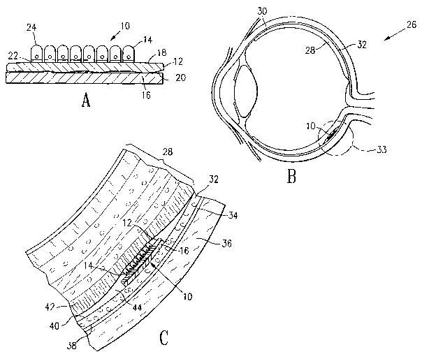
BRIEF DESCRIPTION OF THE DRAWINGS
[0031] Figure 1A is a cross-sectional view of microfabricated membranous
tissue on a
dissolvable substrate embodying features of the invention.
[0032] Figure 1B is a cross-sectional view of an eye of an animal having
microfabricated tissue on a dissolvable substrate implanted in the subretinal
space of its eye.
[0033] Figure 1C is a detailed cross-sectional view of the microfabricated
tissue, retina
and subretinal space of the eye shown in Figure 1.
[0034] Figure 2 illustrates a microcontact printing stamp useful for producing
microfabricated membranous tissue embodying features of the invention.
[0035] Figure 3 illustrates microfabricated lens capsule tissue after contact
with a
microcontact printing stamp embodying features of the invention.
_8_
CA 02449783 2003-12-O1
WO 02/098357 PCT/US02/17363
[0036] Figure 4 illustrates a poly(dimethylsiloxane) (PDMS) stamp for
micropatterning membranous tissue according to methods embodying features of
the
invention.
[0037] Figure 5 illustrates cells growing on a lens capsule micropatterned
with the
PDMS stamp illustrated in Figure 4.
[0038] Figure 6 is a photomicrograph of a microfabricated lens capsule on a
poly-
lactide/polyglycolide earner matrix.
[0039] Figure 7 is a photomicrograph of a section of rabbit retina containing
a
microfabricated lens capsule on a poly-lactide/polyglycolide earner matrix one
week after
implantation.
DETAILED DESCRIPTION OF THE INVENTION
[0040] The present invention provides methods and apparatus for modifying
tissues
and cells for transplantation. The methods of the invention for modifying
tissues may be
applied to a variety of tissues from a variety of organs. The following
definitions are helpful in
describing the invention.
[0041] The term "autologous" is used herein to refer to cells or tissues
derived from
the same animal as other cells or tissues; thus, with respect to a tissue,
cells are autologous
cells when they are derived from the same animal as the tissue is derived
from; analogously,
the tissue is autologous tissue with respect to the cells when the cells and
tissue are derived
from the same animal.
[0042] The term "biomolecule" is used herein to mean a molecule that has a
biological
activity. Thus, a biomolecule is one that, when in contact with a cell or
tissue, acts on or
aiTects that cell or tissue.
[0043] The term "bulk modification" is used herein to mean the modification of
the
properties of substantial portions of a tissue, where such modification is not
limited to the
surface portions of the tissue.
[0044] The term "surface modification" is used herein to mean the modification
of the
properties of a tissue at and near to the surface of the tissue.
[0045] The term "membranous tissue" is used herein to mean any tissue of an
animal
that forms a sheet or sheath; membranous tissue commonly encloses or delimits
a tissue, or
divides an organ or tissue into separate compartments. "Ocular membranous
tissue" is used
-9-
CA 02449783 2003-12-O1
WO 02/098357 PCT/US02/17363
herein to mean membranous tissue derived from the eye of an animal; lens
capsule tissue and
inner limiting membrane are examples of ocular membranous tissue, as are
corneal
membranes, Bruch's membrane, and other membranous tissues of the eye.
[0046] The term "ablation" is used herein to mean the alteration of a tissue,
not
necessarily including the reduction in the size or the removal of tissue. As
used herein,
"mechanical ablation" means alteration, reduction, or removal of tissue by
mechanical action,
such as scraping, scoring, contacting with a contacting surface (such as a
stamp), applying
tension, or other mechanical method. As used herein, "photoablation" means
irradiation by
ultraviolet light, laser light, or other radiation, such as by light from an
excimer, titanium
sapphire, YAG or other laser, effective to alter the surface or bulk
properties of a tissue. "Ion
ablation" is used herein to refer to surface or bulk modification effected by
ion beam treatment
of a membranous tissue.
[0047] A "proteolytic enzyme," or a "protease," is a type of molecule that is
effective
to at least partially digest (cut into pieces) a protein or peptide molecule.
Examples of
proteases and proteolytic enzymes include, but are not limited to,
collagenase, trypsin,
chymotryptsin, dispase, liberase, thermolysin, pepsin, and papain.
[0048] The term "transplantation" is used herein to mean the insertion,
deposition or
positioning of cells or tissues into an animal. The deposition of cells
growing on modified lens
capsule tissue into the subretinal space is an example of transplantation.
[0049] The term "microcontact printing" is used herein to mean deposition of
desired
molecules onto a surface in a pattern with features sized on the order of
several tens of
micrometers or smaller.
[0050] The term "microfabrication" is used herein to mean the production of
modified
tissues by surface modification, bulk modification, or both.
[0051] The term "microfabricated tissue" is used herein to mean a tissue that
has been
altered or modified by microfabrication methods.
[0052] A "contacting surface" is a surface configured for contacting a second
surface
and for depositing molecules initially present on the contacting surface onto
the second
surface. A "stamp," "microfabrication stamp," "microcontact printing stamp,"
"microcontact
stamp," or "microfabrication printing stamp" is a contacting, surface, and the
terms "stamp,"
"microfabrication stamp," "microcontact printing stamp," "microcontact stamp,"
and
-10-
CA 02449783 2003-12-O1
WO 02/098357 PCT/US02/17363
"microfabrication printing stamp" are used herein to mean a device adapted to
deposit desired
molecules in a pattern with features sized on the order of several tens of
microns or smaller.
[0053] The term "micropattern" is used herein to mean a pattern, such as an
ordered
array, design or contour with features sized on the order of several tens of
microns or smaller.
[0054] By "dissolvable polymer" is meant a polymer that is biodegradable, and
that
upon introduction into an animal may at least partially dissociate and
disperse into fluids and
tissues of the animal.
[0055] A "laser" may be an excimer laser, a titanium sapphire laser, an
yttrium-
aluminum-garnet (YAG), or other laser. A laser is capable of emitting a
powerful beam of
coherent light produced by light amplification within the laser cavity or
crystal of the laser.
[0056] As used herein "excimer laser" means a laser light source that provides
laser
light of a wavelength below about 400 nanometers (nm). Excimer lasers may be
xenon,
krypton, or fluorine lasers, or, more preferably may be an argon fluoride
laser. An argon
fluoride laser provides laser light in the ultraviolet, typically with a
wavelength of about 193
nm, suitable for ablation of epithelial, connective, and other tissues. For
use in tissue
modification, such as tissue ablation, laser light may be pulsed at between
about 1 to SO Hz
with each pulse having a duration of between about 1 to 200 nanoseconds (ns),
preferably
between about 10 to 20 ns. Laser beams, such as produced by argon fluoride
lasers, are
typically sized on the order of a few millimeters to several tens of
millimeters.
[0057] A titanium-sapphire (TiS) laser is a tunable laser capable of emiting
infra-red
laser light with wavelengths ranging from about 700 to about 1100 nm.
[0058] An yttrium-aluminum-garnet (YAG) laser, such as a neodimium YAG, a
horonium YAG, or an erbium YAG laser, is a solid-state laser emitting laser
light at a
wavelength on the order of a micron. Water molecules absorb energy at micron
wavelengths;
water preferentially absorbs energy at wavelengths near 3 p.m, and erbium-
doped YAG lasers
emit light with a wavelength of 2.94 p,m, making them particularly suitable
for use in
photoablation by rapid, local vaporization of water present in cells and
tissues, causing rapid
expansion and ablation of tissue.
[0059] An ion beam is a beam of ionized gas molecules, typically excited by
radio-
frequency energy and directed at a target. Ion beam sources used in the
practice of the
present invention may be of any kind; an ion beam source is described, for
example, in U.S.
Patent 5,216,330 to Ahonen. Ion beams may be used to create holes in
materials. U.S. Patent
-11-
CA 02449783 2003-12-O1
WO 02/098357 PCT/US02/17363
6,093,445 to Nawate describes an ion beam method for producing rectangular and
circular
holes sized from about 10 nm to about 2 p.m.
[0060] A tissue implant 10 having microfabricated lens capsule tissue 12 with
attached
cells 14 and a dissolvable substrate 16 is shown in cross-section in Figure
1A. Cells 14 are
attached to and growing upon upper surface 18 of the microfabricated lens
capsule tissue 12.
Lower surface 20 of the microfabricated lens capsule tissue 12 is in contact
with the
dissolvable substrate 16. Cells 14 are iris pigment epithelial cells, which
have apical 22 and
basal 24 membranes, with basal membranes 22 in contact with upper surface 18
of the
microfabricated lens capsule 12. Expression of the proper cellular
differentiation into basal 22
and apical 24 membranes, as is found in pigment epithelial cells in vivo, is
indicative of the
proper functioning of the epithelial cells growing on the microfabricated lens
capsule.
[0061] Figure 1B illustrates, in cross-section, an eye 26 of a mammalian
animal into
which the tissue implant 10 has been surgically placed. Figure 1C is a detail
of the region
within circle 33 of eye 26 including neural retina 28 and tissue implant 10.
Shown in Figures
1B and 1C are the neural retina 28, the iris pigment epithelium (IPE) 30, the
retinal pigment
epithelium (RPE) 32 growing on Bruch's membrane 34 which separates the choroid
36 from
the basal membrane 38 of the RPE. The apical membrane 40 of the RPE has
numerous
processes which enfold and surround the light-sensitive portions of the
photoreceptors in the
photoreceptor layer 42 of the neural retina 28. The space between the apical
membranes of
the RPE 40 and the photoreceptors 42 is the subretinal space 44. The choroid
36 serves to
maintain an environment capable of supporting the high metabolic demands of
the
photoreceptor layer 42 in particular and the neural retina 28 in general by
allowing the passage
of nutrients and electrolytes to, and removal of waste products from, the
subretinal space 44.
[0062] In a healthy eye, the subretinal space 44 is only a virtual space,
there being only
minimal separation between photoreceptors 42 and apical portions of the RPE
40. However,
in many eye disorders, such as retinal detachment, the photoreceptors 42 may
become
separated from the apical RPE membranes 40. In addition, the neural retina 28
and the
pigment epithelium 30 and 32 may be artificially separated during eye surgery
if desired. As
shown in Figures 1B and 1C, a tissue implant 10 may be placed into the
subretinal space 44.
Microfabricated lens capsule 12 with adherent IPE cells 14 is shown implanted
into eye 26
where the IPE cells 30 are able to contact photoreceptors 42 and provide
metabolic support.
Dissolvable substrate i6 is shown between the lower surface 20 of the
microfabricated lens
-12-
CA 02449783 2003-12-O1
WO 02/098357 PCT/US02/17363
capsule 12 and the choroid 36, as it is initially after placement of the
tissue implant 10.
However, the dissolvable substrate 16 will dissolve and be removed from the
subretinal space
44 leaving only the transplanted microfabricated lens capsule 12 and attached
IPE cells 14 in
place in the subretinal space 44.
[0063] Figure 2 is a scanning electron micrograph (SEM) of a
poly(dimethylsiloxane)
(PDMS) microfabrication stamp 46 embodying features of the invention. The grid-
lines 48 are
about 5 ~.m wide and are separated by about 50 pm. The structural height of
the PDMS
stamp is 7 Nxu from the base to the face. Grid lines 48 may be coated with
compounds, for
example, poly-L-lysine, for placement onto a surface by contacting the surface
with the stamp
46.
[0064] Figure 3 is a SEM of a microfabricated lens capsule tissue after
contact with
the microfabrication stamp 46 shown in Fig. 2 that had been coated with a
mixture of 2%
polyvinyl aclohol (PVA) and 0.1 mg/mL fluorescein. The bright areas show
microprinting of
the fluorescein solution. Micropattern lines 50 follow the same pattern and
spacing as the
stamp 46 that produced them by contact with the lens capsule surface.
[0065] Figure 4 shows a SEM of a PDMS stamp 52 that has a stamp surface 54
with a
topology given by an array of circular wells 56. Thus, the stamp surface 54 of
stamp 52 has
circular depressions 56 that will not receive a coating, while the rest of the
stamp surface 54
does receive a coating of molecules which may then be transferred to any
surface with which it
becomes in contact. Coating the stamp 52 with molecules, such as PVA,
mucilage, or other
inhibitory molecules, and then placing the stamp 52 in contact with a surface,
such as a lens
capsule surface, leaves a pattern of those molecules on the surface everywhere
but on the
circles themselves. Such a micropattern of inhibitory molecules allows cells
growing on the
surface to attach only on these circular areas. Because the unmodified lens
capsule surface
actively allows growth of cells adherent to it, inhibitory patterns are
required for patterned
growth. Each circle 56 is about 50 p,m in diameter.
[0066] Figure 5 is a SEM of a human lens capsule S8 that has received a
micropattern
60 of PVA inhibitory molecules from the PDMS stamp 52 as illustrated in Figure
3. The scale
bar is 25 p.m. It is evident that RPE cells 62 are growing in a pattern
determined by the
micropattern 60 deposited on lens capsule 58 by stamp 52. Cells remained
viable and in this
pattern for as long as 24 days. By controlling the width of the grid lines,
cells can be
-13-
CA 02449783 2003-12-O1
WO 02/098357 PCT/US02/17363
separated to a greater or lesser degree. Thinner grid lines allow growing
cells to touch each
other, allowing formation of a transporting epithelial layer from the
contacting, growing cells.
[0067] Attachment of cells onto the microfabricated tissue substrate may be
speeded
or enhanced by placement of the microfabricated tissue within a flat-bottomed
centrifuge tube
along with cells to be grown on the microfabricated tissue. Centrifugation at
low speed, such
as, for example, between about 5,000 to about 15,000 revolution per minute
rapidly deposit
the cells onto the microfabricated tissue and aid the directed growth of
deposited cells onto
the microfabricated tissue.
[0068] Placement of microfabricated tissue onto, or coating a microfabricated
tissue
with, a carrier matrix aids in its processing and in its implantation into the
body of an animal.
Microfabricated tissue may be coated on one side only, or, in some embodiments
of the
invention, microfabricated tissue may be coated on both sides. The processing
and
implantation of microfabricated lens capsule, microfabricated inner limiting
membrane,
microfabricated Bruch's membrane, or other microfabricated membranous tissue
may be aided
in this way; for example, a carrier matrix makes microfabricated tissues more
rigid and easier
to handle. In addition, a carrier matrix is effective to prevent folding and
curling of the tissue,
allowing implantation of a flat, spread-out tissue sheet. Such a spread-out
configuration
provides maximal surface area for growth of implanted cells, and provides the
implanted cells
with maximal access to fluids and surrounding tissues. Biodegradable carrier
matrices
embodying features of the invention are flexible, fitting easily to the
contours of the retina.
Preferably, the carrier matrix is biodegradeable, and so may be resorbed by
the host body
within a desired time period after placement in the eye. A desired time may be
about a week
to a few months, preferably a few weeks to about two months, more preferably a
carrier
matrix embodying features of the invention biodegrades after implantation in a
retina within
about two weeks to about six weeks.
[0069] Biodegradable matrix materials suitable for assisting in the processing
of tissues
and in the implantation of tissues into the eye include, for example:
collagen;
glycosaminoglycans; chitosan; poly(hydroxyalkanoates); poly(a-hydroxy acids),
including but
not limited to polyglycolic acid (PGA), polylactic acid (PLA), and polylactide-
polyglycolide
(PGA-PLA) mixtures, alloys and copolymers (PLGA); poly(dioxanones); poly(s-
caprolactone); poly(ortho esters); poly(anhydrides); poly(phosphazenes);
poly(amino acids);
and other compounds, polymers, copolymers, alloys, mixtures and combinations
of these
-14-
CA 02449783 2003-12-O1
WO 02/098357 PCT/US02/17363
materials. Figure 6 shows a lens capsule (stained blue) on a carrier matrix of
poly-
lactide/polyglycolide. The scale bar is 1 mm in length. Carrier matrix
substrates and coatings
may be dyed (e.g., with trypan blue or rhadamine), improving visualization of
the tissue to be
implanted during implantation surgery. Such coatings and substrates may be
used for lens
capsule, inner limiting membrane, Bruch's membrane, and other membranous
tissue, including
corneal tissue, amniotic membrane,serosal membranes, mucosal membranes, and
neurological
tissue.
[0070] Figure 7 shows a section of rabbit retina containing a human lens
capsule on a
poly-lactide/polyglycolide carrier matrix. The retinal section shown was taken
one week after
implantation of the lens capsule tissue in the subretinal space between the
neural and
pigmented retinal cells in a rabbit eye. The lens capsule has a flat
configuration, showing no
folding or curling that would interfere with the flow of nutrients and waste
products to and
from the transplanted cells.
[0071] Tissues to be modified may be obtained by means known in the art, such
as
excision, biopsy, at surgery or at autopsy. As will be understood by those of
ordinary skill in
the art, care should be taken to avoid damage or contamination of the
membranous tissue
during procedures for obtaining it, as by following standard sterile operating
procedures. It
will be understood that the methods and apparatus are suitable for modifying
any membranous
tissue, including but not limited to ocular membranous tissue.
[0072] In the following discussion, methods and apparatus for modifying tissue
will be
discussed using primarily lens capsule tissue as exemplary membranous tissue.
The methods
and apparatus are thus also suitable for modifying inner limiting membrane
tissues and other
tissues, and may be used to modify inner limiting membrane and other tissues
as well. The
tissue modification provided by the methods of the invention is effective to
alter the properties
of the subject tissue to provide a more favorable substrate for cell
attachment and growth, and
to alter the physical and biochemical properties of the lens capsule tissue to
allow more ready
exchange of fluid and solutes across the tissue.
[0073] Membranous tissue such as lens capsule tissue and inner limiting
membrane
may be obtained from donor eyes, or from the patient (autologous tissue) by
techniques
known in the art, such as following lens extraction for cataract surgery. For
example, lens
capsule tissue may be obtained from an eye after a cataract incision has been
made (either a
scleral incision or a corneal incision). In this method, viscoelastic is next
placed in the anterior
-15-
CA 02449783 2003-12-O1
WO 02/098357 PCT/US02/17363
chamber following making an incision. The viscoelastic is usually either
Healon~ (Pharmacia,
Kalamazoo, MI) or Viscoat~ (Alcon, Fort Worth, TX). The capsulotomy is then
performed
by using a cystotome needle. This needle is used to puncture the anterior
capsule centrally,
creating a capsule flap. This flap is then raised using the cystotome needle.
Utrata forceps are
used to grasp the flap of the capsule and it is pulled in a circular fashion.
Pulling of the
capsule for 360° in a controlled fashion will result in a round
continuous capsulorhexis,
exposing the cataract. The lens and lens capsule may then be removed.
[0074] Once removed, the membranous tissue (e.g., lens capsule, inner limiting
membrane, or other eye tissue) may be maintained irz vitro or prepared for in
vivo
transplantation. Membranous tissue is then placed on a glass, plastic, or
polymer substrate.
The glass substrate may be, for example, a glass cover slip. The plastic
substrate may be, for
example, a tissue culture dish. The polymer substrate, for example, may be a
biodegradable
polymer. Biodegradable polymer films may include poly-lactic acid, poly-
glycolic acid, poly-
lactic acid glycolic acid copolymers (PLGA), including PLGA (50:50 lactic to
glycolic acid
copolymer), poly-lactic acid polymers (PLLA), or polyethylene
glycol/polylactic acid
copolymer (PEG/PLA), polyorthoesters, polyanhydrides, polyphosphazines and
blends and
copolymers thereof. Methods for using biodegradable polymer films may be found
in , e.g.,
U.S. Patent 5,512,600 to Mikos et al.
[0075] For example, the methods discussed in U.S. Patent 5,512,600 and in .l.
Bio»aedical Materials Research, Vol 34:87-93 (1997) by Giordano et e1. may be
used to
maintain healthy lens capsule, inner limiting membrane, or other membranous
tissue in vitro
and in vivo. Biodegradable (e.g., dissolvable after placement in an animal)
polymer films
comprising poly-lactic acid polymers (PLL.A), poly-glycolic acid polymers,
polyorthoesters,
polyanhydrides, polyphosphazines, poly-lactic acid glycolic acid copolymers
(PLGA),
including PLGA (50:50 lactic to glycolic acid copolymer), and polyethylene
glycol/polylactic
acid copolymer (PEG/PLA) films may be placed on the bottom of plastic petri
dishes. The
lens capsule or other membranous tissue is then placed onto the surface and
smoothed down
with the use of a pipette. The membranous tissue and polymer film are
transplanted together.
The film dissolves in vivo leaving the membranous tissue behind. The film
provides a greater
ease of manipulation for the membranous tissue; for example, polymer films
prevent lens
capsule from curling, which is a problem observed with prior art methods. In
addition, further
treatment of the membranous tissue may be applied following these steps.
-16-
CA 02449783 2003-12-O1
WO 02/098357 PCT/US02/17363
[0076] Lens capsule tissue (or other membranous tissue) may be placed in an
environment suitable for cell growth, such as a tissue culture incubator or
environmental
chamber. In one embodiment, lens capsule tissue is immersed in a phosphate
buffered saline
solution (PBS) arid maintained at 37 °C in a 95% 02-5% COZ atmosphere.
Following
incubation, the PBS is removed with a' sterile pipette and the lens capsule is
allowed to lie flat
on the bottom of a sterile petri dish. The lens capsule is then soaked in
trypsin-EDTA for 1
hour to remove any lens epithelial cells and subsequently,
penicillin/streptomycin for 30
minutes for sterility. The lens capsules are then rinsed three times in PBS
followed by three
rinses in distilled water. Each rinse is performed carefixlly with sterile
pipettes. Finally, the
lens capsule and the petri dish it rests on are sterilized under UV light for
at least three hours.
[0077] In another embodiment, an interface chamber is used, wherein lens
capsule
tissue (or other membranous tissue) is placed on wetted filter paper covering
a dish filled with
phosphate buffered saline, and maintained at 37 °C in a 95% OZ-5% CO~
atmosphere. It will
be understood that various saline solutions known in the art, such as
bicarbonate-buffered
saline, or other saline solutions, may be substituted for PBS. Alternatively,
culture medium
(such as, for example, those as RPMI, DMEM or Hamm's F1~ (Life Technologies,
MD)) may
be added to or may replace the saline in the methods, and growth factors,
antibiotics, serum,
and other materials may be added to the saline or culture medium used in
maintaining lens
capsule tissue.
[0078] Methods for modifying tissues include bulk modification methods and
surface
modification methods. Bulk modification methods include methods where
substantial portions
of the tissue, not limited to surface portions of the tissue, are modified by
the method. Surface
modification methods include methods wherein the tissue is modified at and
near to the
surface of the tissue, but is not greatly modified in other portions of the
tissue.
[0079] The methods of the invention as applied to lens capsule tissue, whether
bulk or
surface modification methods, include removal of a lens capsule from an eye,
flattening the
lens capsule onto on a~ sterile glass or plastic substrate, such as a culture
dish, microscope slide
or a glass coverslip, that is submersed in phosphate buffered saline or other
suitable solution,
followed by fi~rther treatment of the lens capsule. It will be understood that
similar treatments
may be applied to inner limiting membrane tissue, Bruch's membrane, amniotic
membrane, or
other tissue.
-17-
CA 02449783 2003-12-O1
WO 02/098357 PCT/US02/17363
[0080] Plastic substrates such as culture dishes and glass substrates such as
microsope
slides may be sterilized by standard procedures, such as by irradiation with
ultraviolet light,
immersion in acid followed by repeated washing in sterile distilled water, or
other procedures
known in the art. In addition, plastic or glass substrates may be used with or
without surface
coatings. Surface coatings may include collagen, collagen gel, fibronectin,
laminin, a silane
coating such as polymethyl silane, a polymer coating such as ~ poly-L-lysine,
or other coating
known in the art.
[0081] In embodiments of the invention, the substrate is prepared for the
membranous
tissue. For example, tissue-culture plastic may be rinsed in a 70% ethanol
solution to remove
dust and oils and allowed to air dry. Following the drying step, the tissue
culture plastic may
be covered with a solution comprising a desired extracellular matrix molecule
(e.g., 4 mg/ml
collagen, type I rat tail in PBS, 1 p,g/ml laminin from human placenta in PBS,
or 25 ~,g/ml
fibronectin from human plasma in PBS) (collagen and fibronectin may be
purchased from
Sigma, St. Louis, MO). After one hour, the plastic may be rinsed in sterile
distilled water
twice and allowed to dry under UV overnight. If the lens capsule substrates
are not
immediately stamped, they are stored at 4 °C.
[0082] Bulk modification methods for modifying membranous tissue such as lens
capsule tissue include methods for modifying the thickness, permeability, and
other properties
of the lens capsule tissue. In one embodiment of the bulk modification method,
such further
treatment includes use of an excimer laser to ablate the surface of the lens
capsule so that the
overall thickness of the lens capsule is reduced. For example, the lens
capsule may be ablated
by a laser or ion beam, or by mechanical methods, so that the overall
thickness mimics the
thickness of Bruch's membrane.
[0083] A laser, such as an excimer laser (e.g., an argon fluoride laser
(Lambda Physik,
Model 201E)) may be used to provide pulses of laser light ei~ective to ablate
the surface of a
lens capsule. For example, pulse of between about 10 to 20 ns duration,
delivered at a
frequency of about 1 to 50 Hz, with pulse energy densities of between about
300 to 500
millijoules per square centimeter (mj/cm2) are ei~ective to ablate the surface
of a lens capsule
in a desired manner. Each pulse is effective to ablate the tissue to a depth
of between about 5
to 50 microns. Accordingly, repeated pulses are effective to reduce the
thickness of the lens
capsule tissue to a desired overall thickness. Methods as have been applied to
the cornea may
be followed or adapted and are suitable for use in photoablation of lens
capsule tissue. Such
-18-
CA 02449783 2003-12-O1
WO 02/098357 PCT/US02/17363
methods of corneal photoablation are disclosed in, e.g., U.S. Patent 4,665,913
to
L'Esperance, U.S. Patent 5,634,920 to Hohla, and U.S. Patent No. 5,735,843 to
Trokel.
[0084] ~ In another embodiment of the bulk modification method, such further
treatment
following placement of tissue on a glass substrate includes use of a laser,
such as, e.g., a YAG
laser to produce micropores in the lens capsule. Such bulk modification by
providing
micropores alters the properties of the lens capsule tissue so as to provide a
more favorable
substrate for cell attachment and alters the biochemical properties of the
lens capsule tissue to
allow more ready exchange of fluid and solutes across the tissue. In
embodiments of the
invention, the micropores are sized on the order of l Os of nanometers (nm) or
less in diameter.
Thus, micropores produced by the bulk modification methods may range in size
between
about 0.01 micron to about 10 microns, preferably between about 0.1 micron to
about 1
microns. An erbium YAG laser can be used to provide pulses of between about 10
to SO ns
duration, at energy levels of between about 1 to 50 mj, preferably between
about 1 to about
20 mj, effective to ablate holes in lens capsule tissue according to the
methods of the
invention.
[0085] In another embodiment of the bulk modification method, such further
treatment
following placement of membranous tissue on a glass substrate includes use of
an ion beam to
produce micropores in the lens capsule to provide a more favorable substrate
for cell
attachment and to allow more ready exchange of fluid and solutes across the
tissue. See, for
example, Goplani et al. J ~Llembr. Sci 178:93-98 (2000), Xu et aL, in Material
Research
Societ~Symposium Proceeding Vol. 540 "Microstructural Processes in Irradiated
Materials ",
pages 255-260 (1999), and Ohmichi et al., ,~ Nuclear Materials 248:354-359
(1997). In
embodiments of the invention, the micropores are sized on the order of lOs of
nms to a few
Tm in diameter.
[0086] The membranous tissue may be freeze dried for purposes of exposing to
the ion
beams. Alternatively, the membranous tissue may be dried out entirely, then
rehydrated after
the micropores are made. An ion beam, such as a 120 MeV beam of Si28 ions, may
be used to
irradiate the tissues. Following exposure to the ion beam, the membranous
tissues may be
rehydrated. Biological etching using collagenase and other proteases or
proteolytic enzymes,
as discussed below, may be used to enlarge the microholes if larger holes are
desired.
[0087] In another embodiment of the bulk modification method, treatment of the
membranous tissue includes deposition of proteolytic enzymes onto the
membranous tissue
-19-
CA 02449783 2003-12-O1
WO 02/098357 PCT/US02/17363
effective to biologically etch the surface and interior of the membranous
tissue to provide
desired topology and surface adhesion properties to the tissue. In some
embodiments of this
method, the deposition step includes contacting the lens capsule or other
membranous tissue
with a microcontact printing stamp carrying enzymes effective to biologically
etch the surface
and interior of the tissue. After stamping of the enzymes onto the tissue,
albumin or an
enzyme inhibitor may be used to stop the reaction after a given time. For
example, incubation
with collagenase is preferentially carried out for various periods up to 26 h
at 20 °C in a
constant temperature water bath, and the collagenase reaction stopped by the
addition of
EDTA to a final concentration of 50 mM. Incubation with trypsin (e.g., 0.25%
trypsin in a
balanced salt solution without calcium or magnesium) may be performed at about
0 to 5 °C for
about 6 to about 18 hours. Following this incubation with trypsin, the trypsin
solution may be
removed and the membranous tissue incubated at 37°C for 20 to 30
minutes before washing
with a wash solution containing divalent rations (such as calcium and
magnesium) in the
amount of about 1 to about 5 mM (and optionally containing a trypsin inhibitor
such as
soybean trypsin inhibitor). Alternatively, membranous tissues may be incubated
with dispase
(about 0.5 to about 3 U/ml) or other proteolytic enzymes in a balanced salt
solution that is
substantially divalent ration-free at 37 °C for up to several hours
before removal of the
solution and washing of the membranous tissue with a balanced salt solution
containing about
1 to about 5 mM divalent rations.
[0088] In embodiments of the bulk modification methods, for example, agents
such as
collagenase, trypsin, chymotryptsin, dispase, liberase, thermolysin, pepsin,
papain, and other
proteases may be applied as solutions in distilled water, phosphate-buffered
saline, or other
buffered solution, at concentrations ranging between about 0.01 mg/mL to about
100 mg/mL,
preferably between about 1 mg/mL to about 20 mg/mL, to the surface of a
microcontact
printing stamp. The surface of the tissue, such as lens capsule tissue, may be
contacted in air
or while immersed in a saline solution. Where the protease is active in the
absence of calcium,
such as for trypsin, chelating agents such as EDTA and EGTA, preferably at
concentrations in
the range of between about 1 to about 10 mM, may be included in the solutions.
In such
cases, enzymatic action may be halted when desired by the addition of calcium
and or
magnesium to the solution. In any case, enzymatic action may be stopped by
dilution with
excess of enzyme-free solution or by addition of an appropriate enzyme
inhibitor. (For
-20-
CA 02449783 2003-12-O1
WO 02/098357 PCT/US02/17363
example, trypsin may be inhibited by a trypsin inhibitor such as soybean
trypsin inhibitor (T-
9003, Sigma Chemical Co. St. Louis, MO).)
[0089] In another embodiment of the bulk modification method, treatment of
inner '
limiting membrane or lens capsule tissue includes impregnation of the tissue
with a deactivated
enzyme, such as a deactivated collagenase enzyme, that is activated by laser
light illumination.
For example, in one embodiment very small regions sized less than a micron in
diameter of
tissue are activated by illumination with a 2-photon confocal laser system.
Enzymes activated
in this way are effective to degrade or otherwise alter tissue in the small
region where
activation occurs, while nearby regions not activated by the confocal laser
system remain
unaltered. The activated enzyme may be flushed out or deactivated by water.
Enzymes
suitable for the practice of the invention include but are not limited to
collagenase, trypsin,
chymotrypsin, dispase, liberase, papain, pepsin, thermolysin, and other
proteases.
[0090] In one embodiment of the surface modification method, microcontact
printing
techniques are used to fabricate chemical micropatterns of biomolecules onto
tissue. For
example, surface modification of lens capsule tissue may include deposition of
patterns of
biomolecules onto lens capsule tissue. Such patterns may include repeated
iterations of
geometric or linear patterns, or may include only a few, or a single, pattern
not made up of
smaller pattern units. Thus, patterns of surface modification may include
linear arrays of
biomolecules deposited onto a tissue surface, or curved arrangements of
biomolecules, series
of circularly-shaped patterns, such as rings or dots, of biomolecules, or a
series of other
shapes, including multiple shapes in a single pattern, of biomolecules.
Alternatively, such
patterns may include extended areas substantially covered by deposited
biomolecules, or
extended areas substantially devoid of deposited biomolecules. It will be
understood that the
methods include any suitable pattern comprising lines, shapes, or regions of
deposited
molecules, including regions devoid of deposited molecules situated between
regions with
deposited biomolecules. Such micropatterns may, .in general improve cell
attachment and
growth on the modified membranous surface. However, in embodiments of the
invention,
micropatterns are produces where regions of the modified membranous tissue are
rendered
less suitable, or unsuitable, for cell attachment and growth. In this way,
cell attachment and
growth may be directed to and limited to those regions of the membranous
tissue that have not
been so treated.
-21-
CA 02449783 2003-12-O1
WO 02/098357 PCT/US02/17363
[0091] Microcontact printing stamps may include the entire pattern to be
deposited
onto target tissue, or may include a portion of the desired pattern. Where the
stamp includes a
portion of the desired pattern, multiple applications of the microcontact
printing stamp to the
tissue surface are effective to provide a desired pattern of biomolecules on
the tissue surface.
Where. the stamp includes the entire pattern, biomolecules may be deposited
onto the
microcontact printing stamp itself in the desired pattern.
[0092] The patterns of biomolecules on a microcontact printing stamp may be
determined by directed placement of the biomolecules on the stamp, or may be
determined by
the surface geometry of the stamp. Where the pattern of biomolecules is
determined by the
surface geometry of the stamp, the geometric pattern may include locally-
raised ridges, where
contact of the stamp with a source of biomolecules is effective to deposit
such biomolecules
onto the raised surfaces, with substantially no biomolecules being deposited
on other, non-
raised portions of the surface. In such a microcontact stamp, the pattern of
biomolecules
deposited onto a tissue would follow the pattern of the raised surfaces
Alternatively, the
pattern may include depressions, valleys or fissures, such as scratches made
into a surface,
where contact of the stamp with a source of biomolecules is effective to
deposit such
biomolecules onto a major portion of the surface, with substantially no
biomolecules being
deposited on the depressed portions of the surface. In such a microcontact
stamp with
depressions, biomolecules would be deposited over a substantial portion of the
tissue, with
regions substantially lacking deposited biomolecules following the pattern of
the depressed
surfaces.
[0093] In some embodiments of this method, the patterns are sized on the order
of a
few microns or less. Accordingly, in embodiments of the surface modification
methods of the
invention, the individual patterns of which the overall patterns are comprised
may range in size
between about 0.1 micron to about 20 microns, preferably between about 0.5
microns to
about 5 microns.
[0094] Biomolecules suitable for deposition onto tissue surface include
proteins,
peptides, organic molecules, oligosaccharides, and small chain polymers,
including but not
limited to collagen, hyaluronic acid, keratin sulfate, glycosaminoglycan,
methylacrylate, poly
(methyl methacrylate), polystyrene, poly(methyl styrene), polylysine,
polylactic glycolic acid
(PLGA)-derivatized polylysine, polylysine peptides, and silane polymers such
as
octadecyltrichlorosilane (OTS). Surface modification comprising deposition of
liiomolecules
-22-
CA 02449783 2003-12-O1
WO 02/098357 PCT/US02/17363
is effective to alter biological properties of the tissue, such as the ability
or ease of attachment
by cells placed onto microfabricated tissues. For example, deposition of
hydrophobic
molecules is effective to deactivate selective cell attachment sites on lens
capsule tissue.
[0095] Microcontact printing stamps may be made of any material capable of
retaining
a suitable pattern, such as glass, ceramic, metal, plastic, polymer, or other
material. In
presently preferred embodiments of the method, microcontact printing stamps
include
poly(dimethylsiloxane) (PDMS), which is commercially available (e.g., Sylgard
184 from Dow
Corning, Midland MI 48640) . Microcontact .printing stamps may be cast in PDMS
from
masters containing desired patterns, such as, for example, a grid pattern of
lines.
Alternatively, where the pattern to be formed is determined by the pattern of
deposition of
biomolecules onto a tissue, the stamp may include a simple surface, such as a
flat surface,
suitable for carrying biomolecules. Such stamps may include pins, slotted
pins, bars or rods,
for example, and may have circular, triangular, square, rectangular, other
polygonal or
irregularly shaped perimeters.
[0096] In embodiments of the surface modification method, the surface of the
lens
capsule tissue is masked to cover part, but not all, of the surface of the
lens capsule tissue, and
then irradiated with ultraviolet (I1V) radiation effective to denature the
extracellular matrix
(ECM) of the exposed portions of tissue. This deactivates molecules specific
for cell
adhesion, and to inhibits or prevents cell adhesion and growth in the exposed,
but not the
covered, regions. Thus, in this embodiment of the methods of the invention,
portions of the
substrate are rendered unsuitable for cell attachment and growth. In this way,
growing cells
can be directed to desired regions, and away from undesired regions.
[0097] In embodiments of the invention, the entire substrate surface may be
deactivated to prevent attachment or growth of cells, and then specific
regions reactivated.
By deactivating proteins that are specific for cellular adhesion, the growth
of cells may be
limited to confined regions. A deactivating substance is one that prevents the
attachment, the
spread, or both, of growing cells. For example, 0.2% polyvinyl alcohol (PVA)
solution and
mucilage are effective deactivating substances.
[0098] A surface may be deactivated, and a portion of that surface
reactivated, by
application of a deactivating substance to the surface. For example, 0.2 % PVA
applied to the
surface of the lens capsule is effective to deactivate the surface of the lens
capsule. Exposure
of the deactivated lens capsule surface to a micropattern of light from an
excimer laser is
- 23 -
CA 02449783 2003-12-O1
WO 02/098357 PCT/US02/17363
effective to ablate a micropattern on the lens capsule surface. For example, a
micropattern
may be produced on the lens capsule surface by illumination of the lens
capsule surface
through an irradiation mask. The ablated micropattern, by removing or altering
the
deactivating substance, reactivates portions of the substrate to allow cell
growth and
spreading into the ablated regions, thereby directing cell growth to follow a
desired pattern.
[0099] The masking step may~include placement of a grid onto the tissue, where
the
grid includes a material effective to prevent irradiation of the surface by a
source of radiation,
such as UV radiation. The grid may be made of materials including metal,
glass, plastic,
ceramic, polymer, protein, or other material effective to absorb or reflect UV
radiation.
[0100] In an alternative embodiment of the masking method, the masking step
includes using
microcontact printing techniques to apply a pattern of protecting molecules
onto the surface
of the lens capsule tissue effective to prevent ECM denaturation in regions
covered by the
protecting molecules. Thus, the grid of a masking step may include a coating
on the surface
effective to screen the surface from irradiation. Such a coating may include a
protein,
preferably one rich in tyrosine and other amino acid residues that absorb
ultraviolet light, a
polymer effective to absorb UV light, or a small molecule effective to screen
UV light, such
as, for example, para-amino benzoic acid (PABA).
[0101] It will be understood by one of skill in the art that surface
modification
methods and bulk modification methods may each be applied to a single tissue.
Thus, for
example, the same lens capsule tissue may be treated with both surface
modification and bulk
modification methods effective to provide microfabricated lens capsule tissue.
[0102] Microfabricated tissues are suitable substrates for growing cells. A
method for
growing cells on microfabricated tissues includes providing a microfabricated
tissue produced
by one of the methods described above, and applying cells to the
microfabricated tissue. For
example, the microfabricated tissue may include a microfabricated lens capsule
with a pattern
on its surface, such as a pattern of collagen, and the cells may include IPE
cells, RPE cells,
stem cells, or other cells. In preferred embodiments of the invention
comprising autologous
tissue and cells, the microfabricated tissues and the cells are obtained from
the same animal.
[0103] The invention also provides methods for using microfabricated tissues,
comprising surgical methods for transplanting microfabricated tissues into an
animal. In
preferred embodiments, the methods for transplanting microfabricated tissues
into an animal
include surgical methods for transplanting microfabricated tissues into the
eye of an animal. In
-24-
CA 02449783 2003-12-O1
WO 02/098357 PCT/US02/17363
most preferred methods, the transplantation of microfabricated tissues into
the eye of an
animal includes transplantation of microfabricated lens capsule tissue near to
or into the retina
of an animal. In some embodiments, the transplanted tissue further includes
cells grown on
microfabricated lens capsule tissues. In other embodiments, the transplanted
tissue includes
RPE cells, IPE cells, stem cells, or other cells grown on microfabricated lens
capsule tissues.
Alternatively, dissolvable polymer substrates may be used for growing cells
for
transplantation. In further embodiments, the transplanted tissue includes RPE
cells, IPE cells,
stem cells, or other cells grown on microfabricated membranous tissues or on
dissolvable
polymer substrates, where the cells and tissues are taken from the same animal
as the animal
into which they are transplanted (autologous tissue).
[0104] Methods for isolating or removing RPE cells from an eye may be found in
Pfeffer, B. A., Chapter 10, "Improved Methodology for Cell Culture of Human
and Monkey
Retinal Pigment Epithelium," Progress in Retinal Research, Vol. 10 (1991) Ed.
Osborn, N.,
and Chader, J.; these methods may also be applied to IPE cells. The cells may
be removed
from a donor eye, or from the intact eye of a patient, including the eye that
will ultimately
receive a transplant of microfabricated tissue with cells. Methods for
harvesting cells obtained
in a biopsy, as for an autologous transplantation procedure, may be found in
Lane, C., et al.
Eye 3:27-32 (1989). Further methods for procurement of RPE and IPE may be
found, e.g., in
Abe et al., 1999, Thumann, et al., 1999; Lappas et al., 2000; and in Thurmann
et al., 2000.
[0105] The IPE cells, RPE cells, stem cells, or other cells may be dispersed
in saline,
such as phosphate-buffered saline, at a density of between about 104 cells/mL
to about 107
cellslmL. Isolated RPE cells, IPE cells, stem cells or other cells may be
applied to
microfabricated tissue, for example, to microfabricated lens capsule tissue by
gently pipetting a
solution containing IPE cells, RPE cells, stem cells or other cells onto the
microfabricated
tissue immersed in PBS, followed by maintenance of the cells and tissue at 37
°C in a sterile
95% Oa-5% C02 atmosphere for 12 hours. The PBS may be removed with a sterile
pipette
and the lens capsule allowed to lie flat on the bottom of a sterile petri dish
or other container.
The lens capsule may then be soaked in trypsin-EDTA for 1 hour to remove any
lens epithelial
cells and subsequently, penicillinlstreptomycin for 30 minutes for sterility.
Following this, the
lens capsules may then be rinsed three times in PBS followed by three rinses
in distilled water.
Each rinse should be performed carefully with sterile pipettes. Finally, the
lens capsule and its
support are sterilized under LTV light for at least three hours.
- 25 -
CA 02449783 2003-12-O1
WO 02/098357 PCT/US02/17363
[0106] Before the application of cells, microfabricated tissues, such as lens
capsule,
inner limiting membrane, Bruch's membrane, and other tissues, may be modified
and
microfabricated as described above. Alternatively, or in addition to such
modification and
microfabrication, a microfluidic channel or pattern of microfluidic channels
may be placed
onto a membrane surface to be modified, and a suspension of cells or molecules
may be
delivered to the membrane surface. For example, a microfluidic network as
described by
Delamarche et al. (Science 276:779-781 (1997)), herein incorporated by
reference in its
entirety, may be applied to a membrane surface in order to modify the
membrane. In such a
procedure, a trough or series of troughs may be formed in PDMS or other
biocompatible
material, the troughs configured to form conduits upon placement of the PDMS
onto a '
membrane surface, with the membrane surface serving as a conduit wall. Cells
or
biomolecules may be brought into contact with the membrane surface by flowing
a solution
containing the cells or biomolecules, or containing both cells and
biomolecules, through the
conduits. The cells and biomolecules may thus be deposited onto, or may
otherwise modify,
the exposed surface of the membrane that forms a wall of the conduit.
[0107] Isolated RPE cells, IPE cells, stem cells, or other cells may also be
applied to a
membranous tissue which has been partially covered by a stencil. A stencil
suitable for the
practice of the invention is configured with a pattern of holes or passages
passing through its
surface. Such a stencil covers underlying membranous tissue when the stencil
is applied to a
membranous tissue, while the pattern of holes or passages is effective to
leave portions of
underlying membranous tissue exposed. A stencil for microfabricating tissue
may have a rim
thicker than the bulk of the stencil in order to help provide mechanical
strength. A stencil
having such a pattern of holes or passageways may be applied to a surface of
membranous
tissue to be microfabricated, effective to direct the growth of cells on the
membranous tissue
or to modulate the exposure of the membranous tissue to external agents and
treatments. In
some embodiments of this method, the patterns may be sized on the order of a
few microns or
less, or on the order of several microns, so that patterns may range in size
between about 0.1
micron to about 100 microns, preferably between about 1 micron to about 75
microns, more
preferably between about 5 microns to about 50 microns. A stencil may be
formed of any
suitable biocompatible material, such as, for example, PDMS or PLGA/PEG
copolymer. A
suitable stencil material may be solid or gelatinous, and is preferably
flexible. A stencil
material may also be biodegradable (e.g., PLGA/PEG copolymer). Stencils
suitable for
-26-
CA 02449783 2003-12-O1
WO 02/098357 PCT/US02/17363
application to membranous tissue for transplantation into the eye of an animal
may be made by
methods similar to those described in, for example, Folch et al. J. Biomed.
Mater. Res.
52:346-353 (2000), hereby incorporated by reference herein in its entirety.
[0108] Transplantation of microfabricated lens capsule tissue into the
subretinal space
may be effected by any means providing access to the subretinal space. Access
to the
subretinal space may be provided, for example, by a scleral incision placed
laterally on the eye,
or via the, vitreous humor by a more frontal incision. Procedures providing
access to, and
transplantation into, the retina, including the subretinal space, have been
described; see, for
example, Abe et al., Tohoku J. Exp. Med. 189:295-305 (1999), Abe et al.,
Tohoku J. Exp.
Med. 191:7-20 (2000), Lappas et al., Gfaefes's Arch Clin Exp Ophthalnzol.
238:631-641
(2000), Thumann, et al., Arch. Ophthalmol. 118:1350-1355 (2000), U.S. Patent
No.
5,962,027 to Hughes and U.S. Patent No. 6,045,791 to Liu.
[0109] Alternatively, a microcontact printing stamp, or a stencil, may be
applied to an
ocular membrane itz vioo. For example, access to Bruch's membrane within an
intact, living
eye may be had by standard surgical procedures, including formation of a bleb
by infusion of a
gas, saline, mineral oil, or other biocompatible liquid into the subretinal
space of an eye, and
placement of a microcontact printing stamp or of a stencil onto Bruch's
membrane. Such
application of a microcontact printing stamp, or of a stencil, may be
performed wet, that is in
the presence of normal bodily fluids, PBS or other artificial physiological
solution, mineral oil,
or other biocompatible liquid. Alternatively, such application of a
microcontact printing
stamp, or of a stencil, may be performed dry, that is in the absence of normal
bodiy fluids or
artificial physiological solution, by, for example, filling the subretinal
space with an inert gas
such as nitrogen or argon.
[0110] An intact Bruch's membrane may be prepared for in situ microfabrication
by
scraping or otherwise debriding Bruch's membrane to remove RPE cells before
application of
a stamp or a stencil. A stamp or stencil may then be used to provide a desired
pattern onto a
surface of the membrane. A PDMS stencil, for example, having a pattern of
passages may be
applied to the surface of Bruch's membrane. Cells are able to attach and grow
on the
membrane surfaces exposed by the passages. Similarly, biomolecules in a
solution on contact
with the membrane surface are able to contact and modify or adhere to the
membrane surfaces
exposed by the passages. The pattern of passages is effective to provide a
pattern suitable for
directing the depostion of biomolecules, and of directing the growth of added
cells, such as
_27_
CA 02449783 2003-12-O1
WO 02/098357 PCT/US02/17363
RPE, IPE, stem cells, or other added cells, or the regrowth of endogenous
cells from other
regions of the eye. Alternatively, or in addition, a microcontact printing
stamp carrying
laminin, fibronectin, or other desired coating agent, may be applied to the
ocular membrane
surface. In further alternative methods embodying features of the invention,
internal ocular
membranes may be accessed via the sclera, as, for example, by scleral
puncture, scleral
incision, formation of a scleral window, or other method. In methods taking
advantage of
scleral access, there may be no need to traverse the vitreous humor in order
to access ocular
membranes for treatment.
EXAMPLE 1
[0111] Microcontact printing was used to deposit micron-sized patterns of
biomolecules onto lens capsule tissue. Poly(dimethyl siloxane) (PDMS) stamps
were cast
from masters containing a topological pattern of grid lines spaced 50 microns
apart. The
PDMS stamp was made from a master that was microfabricated from a silicon
wafer. PDMS
stamps were used to microfabricate patterns onto lens capsule tissue. Shown in
Figure 1 is a
scanning electron micrograph (SEM) of a PDMS stamp used to deposit a
micropattern onto a
piece of human lens capsule tissue.
The PDMS stamp shown in Figure 2 has a surface topology given by a hexagonal
array of 5 p;m-wide lines. Each line is separated by approximately 50 pm.
Figure 3 shows a
human lens capsule stamped with the PDMS stamp shown in Figure 1. The PDMS
stamp was
used to deposit hexagonal patterns of a PVA and fluorescein solution (2% PVA
and 0.1
mg/mL fluorescein) onto the lens capsule. This example shows that the stamp is
effective to
produce a pattern on a surface corresponding to the pattern of the stamp.
EXAMPLE 2
[0112] A SEM of a PDMS stamp with circular patterns used for micropatterning
tissue is shown in Figure 4. As shown, the stamp has a surface topology given
by an array of
circular wells of approximately 50 p,m in diameter. When the relief pattern is
coated with an
inhibitory molecule, such as PVA or mucilage, and the stamp applied to a lens
capsule, the
inhibitory molecules are transferred to the lens capsule in the pattern shown.
Figure 5 shows
the surface of a lens capsule that has been patterned with a PDMS stamp having
a pattern as
shown in Figure 2 and RPE cells grown on it. This example shows that the stamp
is effective
_28_
CA 02449783 2003-12-O1
WO 02/098357 PCT/US02/17363
to place a pattern on the lens capsule surface that corresponds to the pattern
of the stamp, and
for cell growth to be patterned according to the pattern of the stamp.
[0113] Thus, application of the stamps of the invention are able to deposit
inhibitory
molecules in patterns that can direct the growth of cells growing on a
patterned substrate.
Because the lens capsule actively allows growth, patterns of inhibitory
molecules, such as
PVA, are preferred for patterned growth. Use of the stamp on substrates
treated to inhibit
growth would require the use of activating molecules to pattern growth on the
substrate.
EXAMPLE 3
[0114] Masking of the surface of lens capsule tissue and then irradiating the
exposed
surface, but not the masked surface, with UV radiation is accomplished by
placement of a
SEM grid onto the surface of lens capsule tissue. A SEM grid with spacing of
50 microns is
placed onto the exposed surface of an excised lens capsule tissue resting on a
glass coverslip
immersed in phosphate-buffered saline. The surface of the lens capsule tissue
and the SEM
grid are not immersed in the phosphate-buffered saline, but rise above the
level of the
phosphate-buffered saline. UV light is directed onto the exposed surface of
the lens capsule
tissue effective to irradiate the lens capsule tissue not resting immediately
below the SEM grid
material. After irradiation, the SEM grid is removed. The lens capsule surface
includes a
micropattern of lines comprising tissue not irradiated (regions under SEM grid
material)
enclosing regions comprising irradiated tissue.
EXAMPLE 4
[0115] Growth of monolayer cultures of retinal pigment epithelium and iris
pigment
epithelium cells is facilitated by flat substrate and by insuring that the
substrate does not curl
or fold upon implantation in the subretinal space or other region of the eye.
A biodegradable
matrix coating was found to prevent folding and curling of lens capsule
tissue. Such a
biodegradable matrix coating which prevents substrate curling or folding is
suitable for use as
a substrate for the growth of monolayer cultures of retinal pigment epithelium
and iris pigment
epithelium cells for implantation into an eye.
[0T16] A biodegradable polymer matrix of poly(dl-lactidelglycolide) was made
by
dissolving 50 mg of a 90:10 mixture of poly(dl-lactide/glycolide)
(Polysciences, Inc.,
Warrington PA 18976) in different amounts of dichloromethane to make solutions
of 100
-29-
CA 02449783 2003-12-O1
WO 02/098357 PCT/US02/17363
mg/mL (0.5 mL dichloromethane), 150 mg/mL (0.33 mL dichloromethane), and 200
mg/mL
(0.25 mL dichloromethane).
[0117] Human lens capsules obtained during cataract surgery were stored in
phosphate buffered saline at 4° C prior to sterilization under
ultraviolet light (254 nm for three
hours) and treatment with 0/0S% trypsin-(ethylene diamine tetraacetic acid)
for ten minutes at
37 °C to remove native epithelial cells. Treated lens capsules were
spread in a single layer on
Parafllm~ (Pechiney Plastic Packaging, Inc., Neenah, WI 54956) in a Petri dish
and coated on
one side with the poly-d-lactyl glycolic acid (PLGA) biodegradable matrix of a
single
concentration by dispensing S p.L,, 10 p.I, or 20 p.L of the PLGA solution
from a pipette onto
the lens capsule surface. The PLGA solution was allowed to spread over a 5 mm
diameter
circular area containing the flattened lens capsule. The solvent was
evaporated in a chemical
hood.
[0118] Five New Zealand White rabbits weighing 2.5 to 3.5 kg underwent
implantation of the lens capsule/PLGA complex following ketamine (40 mglkg)
and Xylazine
(5 mg/kg) anesthesia. Tropicamide 0.5% and Phenylephrine 2.5 % eyedrops were
instilled
into the conjunctival sac of the left eye every five minutes for three doses.
Standard three-port
pars plans vitrectomy was performed, and a retinal bleb was inflated in the
macular area by
injection of approximately 0.5 mL of balanced salt solution througha 42-gauge
needle. A
retinotomy 1 mm in diameter was created, and the lens capsule/PLGA was
inserted into the
subretinal space through the retinotomy with subretinal forceps. The retina
was then
reattached by air-fluid exchange.
[0119] The operated eyes were removed one week after implantation of the lens
capsule/PLGA and fixed in 1.25 % glutaraldehyde/ 1% paraformaldehyde in
cacodylate bufer
(pH 7.4). The eyes were then cut open, fixed, post-fixed in osmium tetroxide,
dehydrated in a
graded series of ethanol, embedded in epoxy resin, cut into 1 pm sections and
stained with
toluidine blue.
[0120] Histological studies performed one week post-implantation demonstrated
that
the lens capsule remained flat on Bruch's membrane. This is illustrated by a
section of a rabbit
retina having a lens capsule/PLGA implant taken 1 week after implantation is
shown in Fig. 7.
There was local disruption of the photoreceptor layer, and an overlaying
rtinal detachement,
presumably in the area previously occupied by the bleb. The PLGA was almost
completely
dissolved in all cases. The five implantations demonstrated that lens capsule
coated with
-30-
CA 02449783 2003-12-O1
WO 02/098357 PCT/US02/17363
PLGA was easy to handle during surgery, and had sufficient rigidity so that
the implants
remained flat within the subretinal space. No evidence of significant
inflammatory reaction
was noted.
[0121] This example shows that PLGA greatly improves the surgical handling of
lens
capsule during subretinal implantation and allows the lens capsule to be
implanted without
curling. While untreated lens capsule may roll into multiple layers or fold
during implantation,
lens capsule treated with a bioabsorbable matrix coating such as the PLGA
coating used in this
example remains relatively flat in the subretinal space after implantation.
Because the PLGA
degrades within a few weeks, concerns for late ~ immune reactions to an
implant are allayed.
This example demonstrating improved mechanical characteristics of coated
microfabricated
lens capsule illustrates that coating microfabricated tissues such as lens
capsule, inner limiting
membrane, Bruch's membrane, and other tissues, overcomes the limitations of
the mechanical
weakness of the untreated tissue and provides an improved substrate for
implantation of
tissues and cells.
EXAMPLE 5
[0122] One therapy for AMD is to transplant suspensions of either RPE cells or
IPE
cells to rescue the diseased retina. The present invention provides novel
tissue engineering
techniques to precision engineer autologous human tissues as a substrate for
transplanting
cells, such as IPE cells, RPE cells, stem cells, and other cells. Suitable
tissues include
membranous tissues, such as lens capsule (e.g., human lens capsule), inner
limiting membrane
tissue, Bruch's membrane tissue, corneal tissue, amniotic membrane tissue,
serosal membrane
tissue, mucosal membrane tissue, and neurological tissue.
[0123] A microgeometry of inhibitory molecules is arranged onto the surface of
a
suitable substrate. Suitable substrates include human lens capsule, collagen
gel, collagen-,
fibronectin-; and laminin-coated plastic, and a dissolvable polymer such as
PLGA or PLLA.
Human lens capsules may be obtained during cataract surgery. Cultures of
experimental RPE
cells are grown on these microengineered surfaces and analyzed using scanning
electron
microscopy, atomic force microscopy, and fluorescence microscopy. Comparisons
between
microfabricated surfaces of autologous tissue and synthetic surfaces and
membranes are then
made.
-31 -
CA 02449783 2003-12-O1
WO 02/098357 PCT/US02/17363
[0124] These comparisons demonstrate that individual RPE cells may be directed
to
grow in microenvironments on the respective biological surfaces. Although all
surfaces
studied are amenable to micromachining, including human lens capsule, it will
be understood
that different tissue engineering methods may be used to vary cell-to-cell
distance and the
microenvironment of growth factors and cell adhesion molecules.
EXAMPLE 6
[0125] In this example, isolated RPE cells are applied to a lens capsule
membrane
tissue which has been partially covered by a stencil and their growth on the
lens capsule is
directed by the stencil pattern. Fibronectin (from human plasma, 25 p,g/ml in
PBS) is coated
onto an excised lens capsule tissue resting on a glass coverslip immersed in
phosphate-
buffered saline. A PDMS stencil having a regular pattern of hexagonal holes of
about 50 pxn
across, the holes being spaced about 10 p,m apart, is sterilely placed onto
the excised lens
capsule tissue. The fibronectin promotes the adherence of the stencil to the
lens capsule as
well as promoting the adherence of added cells. RPE cells dispersed in cell
growth medium
(10' cells/mL) are sterilely added to the saline solution onto the surface of
the lens capsule that
is partially covered by the stencil. The lens capsule, stencil, glass
coverslip, added cells, and
cell growth medium are maintained in a tissue culture dish in a tissue culture
incubator and are
maintained under suitable culture conditions at approximately 37° C in
a 95% 02-5% COZ
atmosphere. The stencil's pattern of hexagonal holes leaves portions of
underlying
membranous tissue exposed . to the RPE cells. The RPE cells adhere to the
exposed lens
capsule tissue, and grow on it in hexagonal patterns directed by the stencil.
The resulting
microfabricated lens capsule tissue with adherent RPE cells is suitable for
transplantation into
the eye of an animal.
EXAMPLE 7
[0126] In this example, a microcontact printing stamp is applied to an ocular
membrane in vivo, providing a microfabricated membranous surface suitable for
growth of
cells. Such a microfabricated membranous surface suitable for the growth of
cells aids in the
treatment of eye diseases or conditions stemming from defects of ocular
membranes or ocular
cells. For example, in an eye having a region of diseased RPE cells, or RPE
cells which are
not functioning properly, removal of the defective RPE cells and
microfabrication of the
-32-
CA 02449783 2003-12-O1
WO 02/098357 PCT/US02/17363
underlying Bruch's membrane, optionally with the addition of cells, is
effective to treat the
eye.
[0127] A subretinal bleb is used to access a retinal region in an eye of a
patient having
diseased RPE cells. The bleb is formed by infusion of sterile saline into the
subretinal space
following a 3-port pars plans vitrectomy and puncture of a small pathway
through the neural
retina. RPE cells exposed by the bleb are removed from a portion of Bruch's
membrane by
scraping or otherwise debriding Bruch's membrane, such as by techniques used
in choroidal
neovascular surgery. A microcontact printing stamp having a pattern of 50 pm-
diameter
circles spaced 10 pm apart is coated with 25 ~,glml fibronectin from human
plasma in PBS,
rolled into a tubular configuration, and inserted into a needle. The needle is
connected to a
syringe containing PBS. The microcontact printing stamp and PBS are gently
injected into the
bleb within the subretinal space, and the stamp unrolled by action of the
needle and of PBS
delivered by the needle. The microcontact printing stamp is placed in contact
with the
exposed Bruch's membrane. Such placement of a coated microcontact printing
stamp is
effective to deposit a pattern of fibronectin onto the exposed Bruch's
membrane and to
prepare the intact Bruch's membrane in situ for microfabrication effective to
provide a pattern
suitable for directing the growth of added cells or the regrowth of endogenous
cells from
other regions of the eye. After 15 minutes, the microcontact printing stamp is
removed from
the bleb via the pathway through the neural retina. In alternative treatments,
the microcontact
printing stamp is left in contact with Bruch's membrane for periods varying
between about 1
minute up to about 1 hour. Following removal of the microcontact printing
stamp, a
dispersion of RPE cells in PBS (10~ cells/mL) is gently infused into the bleb.
In further
alternative treatments, in which the microcontact printing stamp is made of
biodegradable
materials, the microcontact printing stamp is not removed, but may remain in
place as cells are
added and afterwards. The instruments are then removed from the eye, the
incisions are
closed and standard post-operative care is given to the patient. The RPE cells
proliferate to
cover the exposed portion of Bruch's membrane and aid in maintaining the
health of Bruch's
membrane and in the support of overlying neural retina.
- 33 -
CA 02449783 2003-12-O1
WO 02/098357 PCT/US02/17363
E~~AMPLE 8
[0128] In this example, a stencil is placed onto an ocular membrane irz vivo,
providing
a microfabricated membranous surface suitable for growth of cells. A
subretinal bleb is used
to access a retinal region in an eye of a patient having diseased RPE cells.
The bleb is formed
by infusion of sterile saline into the subretinal space following a 3-port
pars plana vitrectomy
and puncture of a small pathway through the neural retina. RPE cells exposed
by the bleb are
removed from a portion of Bruch's membrane by scraping or otherwise debriding
Bruch's
membrane, such as by techniques used in choroidal neovascular surgery.
Fibronectin (from
human plasma, 25 pg/ml in PBS) is diffused into the bleb to coat the exposed
Bruch's
membrane and to promote adherence of an added stencil. A stencil made from
PDMS and
having a pattern of hexagons measuring 60 ~m across at their widest dimension,
and spaced
pm apart is rolled into a tubular configuration in PBS and inserted into a
needle. The
stencil and PBS are gently injected into the bleb within the subretinal space,
and the stencil
unrolled by action of the needle and PBS delivered by the needle. The stencil
is placed onto
the exposed Bruch's membrane. Such placement of a stencil onto Bruch's
membrane in situ is
effective to provide a pattern on the exposed Bruch's membrane effective to
direct the growth
of added cells or the regrowth of endogenous cells from other regions of the
eye. Following
placement of the stencil, a dispersion of IPE cells in PBS (105 cellslmL) is
gently infused into
the bleb. The instruments are then removed from the eye, the incisions are
closed and
standard post-operative care is given to the patient. The IPE cells
proliferate to cover the
portions of Bruch's membrane exposed by the gaps of the stencil and aid in
maintaining the
health of Bruch's membrane and in the support of overlying neural retina.
-34-