Note : Les descriptions sont présentées dans la langue officielle dans laquelle elles ont été soumises.
CA 02482849 2004-10-15
WO 03/087825 PCT/IL03/00257
METHOD, SYSTEM AND KIT FOR DETECTING
AN ANALYTE IN A SAMPLE
FIELD OF THE INVENTION
The present invention concerns detection of analytes in fluid samples by
formation of a thin layer of aggregates comprising the analyte, and analysis
of the
aggregates thus formed, preferably by the use of image analysis.
s BACKGROUND OF THE INVENTION
Early detection of an active disease in a patient is an essential factor for a
successful treatment. Provided that a rapid and correct diagnosis is
established, it is
possible to slow down the progress and at times cure patients from a disease.
Traditional methods for the detection of infectious diseases include serology
to assays, e.g. complement fixation (CF), indirect and direct fluorescent
antibody (IFA
and DFA, respectively), enzyme-linked immunosorbent assay (ELISA) and latex
agglutination; culturing assays in which the infectious microorganism
recovered
from a patient during acute infection is cultured and then identified; and
assays
involving the use of monoclonal antibodies specific against the infectious
agent.
1 s Flow cytometry analysis is also used for disease detection and involves
the
measuring of certain physical and chemical characteristics of cells or
particles,
including cell size, shape and internal complexity or any other cell component
that
can be detected by a fluorescent compound, as the cells or particles travel in
suspension one by one past a sensing point. The use of flow cytometry for
detection
2o methods has been described, for example in W099/47933. This publication
describes a method for the detection of surface antigens by contacting an
antibody-
CA 02482849 2004-10-15
WO 03/087825 PCT/IL03/00257
-2-
coupled bead with a test sample and, if the target antigen is present in the
sample, a
bead-antibody-antigen complex is thus formed and detected by flow cytometry.
US 6,159,748 describes a kit for the detection of antibodies in serum
samples using a flow cytometer. In particular, the kit is provided with beads
coated
s with a series of antigens, each having a different bead size and carrying a
different
antigens. The beads are used for the detection of different antibodies,
including
auto-antibodies.
As appreciated by those versed in the art, when utilizing a flow cytometry
instrument, the cell sample preparation, data collection and data analysis
must be
io performed by a specially trained technician. The flow cytometry instrument
includes a laser and complex optical system, a high-power computer and
electrical
and fluidic systems. The component systems of the flow cytometry instrument
must
be properly maintained and calibrated on a regular and frequent basis. The
high
cost of the instrument and the expertise required to correctly operate such
I s instrument render detection by flow cytometry convoluted and expensive.
Evidently, this rational also applies to many other tests and instruments,
including,
inter alia, Enzyme-Linked Immunosorbent Assay (ELISA).
WO 01/33215 and WO 02/79749 describe systems for generating a profile
of particulate components of a body fluid sample. The systems include in
general a
2o device for causing controlled flow of the body fluid sample on a substrate,
the
controlled flow of the body fluid sample leading to a differential
distribution of the
particulate component on the substrate, and a magnifying device being for
providing a magnified image of differentially distributed particulate
components on
the substrate. The magnified image represents a profile of the particulate
2s components of the body fluid sample. The systems described may further
comprise
an image analyzer for analyzing the profile of the particulate component in
the
body fluid sample.
CA 02482849 2004-10-15
WO 03/087825 PCT/IL03/00257
-3-
SUMMARY OF THE INVENTION
The present invention aims for providing a rapid, sensitive and easy-to-
perform method of detecting in vitro analytes in a fluid sample making use of
an
optical image analyzer. The method of the invention is preferably aimed for
s therapeutic diagnosis, however, may be suitable for other applications, e.g.
ecological, environmental, etc.
Thus, according to a first of its aspects, the present invention provides a
method for detecting an analyte in a fluid sample comprising:
(a) mixing said fluid sample with a reagent comprising a capturing
to agent which is a first member of a binding couple that can bind to an
analyte, the analyte being a second member of the binding couple, such
that if the analyte is present in the fluid sample, particulates of the
binding
couple are formed;
(b) treating said mixture so as to form on a solid substrate a thin layer
Is of said particulates, if formed as a result of said mixing;
(b) obtaining an optical image of the thin layer; and
(c) analyzing said image so as to determine therefrom the absence or
presence of particulates formed as a result of the association between the
binding couple, the presence of particulates in the sample indicating the
2o presence of said analyte in the sample; or to determine therefrom at least
one parameter of said particulates.
A parameter according to the invention may be, without being limited
thereto: size of particulates or size distribution of the particulates formed
as a
result of the association between the binding couple; particulates' count;
2s particulates shape; and/or spatial distribution of the formed particulates.
The invention further provides a system for performing the method of the
invention, the system comprising:
(a) holding means for holding a solid substrate carrying a thin layer of
particulates;
CA 02482849 2004-10-15
WO 03/087825 PCT/IL03/00257
-4-
(b) an optical image acquisition device for capturing an image of the thin
layer on the solid substrate;
(c) an image analysis device coupled to said image acquisition device
and for analyzing an image obtained by the image acquisition device.
s The system optionally comprises a magnifying device.
Yet further, the invention provides a kit for use in the method of the
invention, the kit comprising:
(a) at least one reagent comprising a capturing agent being a first
member of a binding couple and comprising at least two capturing moieties
to which binds an analyte if present in a fluid sample, the analyte being a
second member of the binding couple; and
(b) a solid substrate for carrying a thin layer of particulates.
BRIFE DESCRIPTION OF THE FIGURES
In order to understand the invention and to see how it may be carried out in
1 s practice, some embodiments will now be described, by way of non-limiting
examples only, with reference to the accompanying Figures, in which:
Figs. lA-1C show light microscope images of plasma samples incubated
with microbeads coated with multiple copies of an antibody directed against D-
dimer. The plasma samples containing a very low level of D-dimer (Fig. lA), an
2o intermediate level of D-dimer (Fig. 1B) or a high level of D-dimer (Fig.
1C).
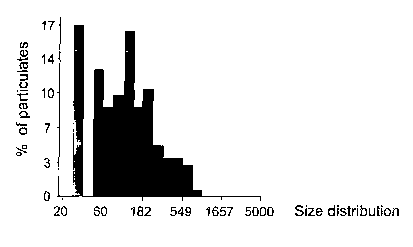
Fig. 2A-2C are bar representations of the size distribution of particulates
formed as a result of binding of D-dimer to microbeads coated with antibodies
directed against D-dimer. Microbeads coated with multiple copies of antibodies
directed against D-dimer were incubated with samples containing low levels of
D-
2s dimer (Fig. 2A), intermediate levels of D-dimer (Fig. 2B) or high levels of
D-dimer
(Fig 2C).
Fig. 3 shows the average size of particulates obtained as a result of
titration
CA 02482849 2004-10-15
WO 03/087825 PCT/IL03/00257
-5-
of plasma samples containing different concentrations of D-Dimer.
Fig. 4 shows the average size of particulates obtained with heparin induced
thrombocytopenia (HIT) samples by use of the Diamed kit with negative samples
(n=4) as well as positive samples (n=10).
s Fig. 5A-SB show light microscope images of EDTA-anticoagulated type A
blood samples mixed with a dilution buffer (0.9 % NaCI and 2% bovine albumine)
to give a final dilution of 100-fold and anti blood group A (Fig. SA) or anti
blood
group B (Fig. SB) antibodies are added to a final concentration of 0.1 to 0.25
mg/mL, the mixture is than incubated for 1 min with gentle mixing followed by
Io examination by light microscope. Fig. SB shows an image of a negative
response
while Fig. SA shows an image of a positive response.
Fig. 6 is a graph showing the average size of blood aggregates of blood
groups O, A and AB incubated with antibodies against A or B groups obtained
and
determined by performing the method of the invention.
is DETILED DESCRIPTION OF THE INVENTION
The present invention provides a rapid and easy in vitro method of
diagnosing an analyte in, preferably, a biological fluid sample obtained from
a
subject, without the need of sophisticated equipment or professional skills to
analyze the sample. Moreover, the method of the invention is sensitive and
allows
2o detection at an early stage of disease and provides a tool to follow a
patient from
onset to the recovery from a specific disease and to monitor the effectiveness
of a
chosen treatment against a disease. The sensitivity of the method of the
invention
arises from the creation of a thin layer of the particulates formed in the
analyzed
sample (after being mixed with a suitable reagent), if the analyte is present
in the
2s sample. The formation of a thin layer enables the accurate image analysis
of the
particulates so as to obtain a qualitative as well as a quantitative
determination with
respect to the analyte.
CA 02482849 2004-10-15
WO 03/087825 PCT/IL03/00257
-6-
It is to be understood that both the forgoing general description and the
following description of some specific embodiments of the invention have been
provided merely for the purpose of explanation and are in no way to be
construed
as limiting the present invention as claimed.
s Thus, according to one aspect of the invention there is provided a method
for detecting an analyte in a fluid sample, the method comprising:
(a) mixing said fluid sample with a reagent comprising a capturing
agent which is a first member of a binding couple that can bind to an
analyte, the analyte being a second member of the binding couple, such
Io that if the analyte is present in the fluid sample, particulates of binding
couples are formed;
(b) treating said mixture so as to form on a solid substrate a thin layer
of said particulates, if formed as a result of said mixing;
(c) obtaining an optical image of the thin layer; and
is (d) analyzing said optical image so as to determine therefrom the
absence or presence of particulates formed as a result of the association
between the binding couple, the presence of particulates in the sample
indicating the presence of said analyte in the sample; or to determine from
said image at least one parameter of said particulates.
2o The term "detect" or "detection " as used herein refers collectively to
both
a qualitative and quantitative determination of the presence of an analyte in
a
sample.
Thus, the method of the present invention also provides analytical
(quantitative) detection of a target analyte in a fluid sample. According to
this
2s embodiment, i.e. the quantitative detection, one or more parameters
characterizing the particulates formed as a result of association of the
binding
couple is determined. Such parameters can be easily defined by a man skilled
in
the art, and include, for example determination of the size of the
particulates
CA 02482849 2004-10-15
WO 03/087825 PCT/IL03/00257
-7-
formed as a result of association between the first and second members of the
binding couple, size distribution of the particulates, the number of
particulates
formed (particulate count), the pattern of distribution, etc. In order to
analytically
determine the above parameters, it is essential that a thin layer of the fluid
sample
s and reagent is formed, as explained hereinafter.
According to the invention, the association (binding or complexing)
between an analyte to a respective capturing agent results in the formation of
a
complex. The capturing agent and the analyte constitute together a binding
couple. The binding couple may, for example, be one of the couples selected
from the group of receptor-ligand, sugar-lectin, antibody-antigen (the term
"antibody" should be understood as referring to a polyclonal or a monoclonal
antibody, to a fraction of an antibody comprising the variable, antigen-biotin
binding portion, etc.).
The term "analyte", according to a first aspect of the invention, refers to a
is cellular or microorganism component such as proteins (e.g. antibodies,
cytokines,
receptors), glycoproteins, peptides, low molecular weight compounds, the
detection of which in a sample obtained from a subject is indicative of
whether
the subject has a specific disease or disorder. The term "analyte" may refer
also
to a synthetic or natural chemical, or a drug or a toxin. The analyte
according to
2o this aspect of the invention contains at least two binding sites
(recognition sites)
to which two individual and separate capturing agents may bind. The results of
binding to each binding site of the analyte to an individual capturing agent
thus
results in the formation of aggregates of binding couples (referred to herein
also
by the term "particulates").
2s According to another embodiment, the analyte refers to particles
presenting on their surface at least two binding (recognition) sites. For
example,
the analyte may include antigen-presenting particles, e.g. antigen presenting
cells,
viruses or other infectious microorganisms, which present on their surface
more
than one copy of a specific antigen to which the capturing agent binds.
CA 02482849 2004-10-15
WO 03/087825 PCT/IL03/00257
-g-
The term "capturing agent" according to the invention refers to any bi or
multifunctional agent, which can bind, preferably with specificity, to two or
more
analytes in a sample, thereby forming aggregates of binding couples.
Accordingly, the capturing agent includes dimmeric, trimeric or multimeric
s molecules presenting, respectively, two, three or more capturing sites which
can
bind independently to an analyte in the fluid sample. For example, the agent
may
be a dipeptide or diprotein bridged by a linker. According to a preferred
embodiment, the capturing agents are microbeads coated with specific capturing
moieties. The capturing agent comprises a "capturing moiety" which is, in
to principle, a binding site which the analyte has an affinity and the
association
between the two is as a result of association between the said capturing
moiety
(site) of the capturing agent and the recognition site of the analyte. The
meaning
of the above terms may be further understood in view of the following non-
limiting examples.
is According to one embodiment of the invention, the capturing agent is a
particle comprising at least two epitopes and the analyte is an antibody
(comprising two binding sites) to which antigenic epitopes of different
capturing
agents binds, or vice versa, the agent is an antibody (comprising two
recognition
sites) and the analyte is an antigen comprising at least two antigenic
epitopes or a
2o particle presenting on its surface at least two antigenic epitopes to which
two or
more antibodies can bind.
According to a second aspect of the methods of the invention, the
capturing agents are microbeads coated with capturing moieties. The microbeads
may comprise on their sensing interface a single type of capturing agent or
2s several types of capturing agents so as to enable the use of the coated
microbeads
in different detection assays. The "sensing interface" refers preferably to
the
outer surface of the beads, which is coated with the capturing agents) so as
to
allow the formation of the resulting particulates.
CA 02482849 2004-10-15
WO 03/087825 PCT/IL03/00257
-9-
Microbeads which are used according to the invention may be made of
polymer such as polystyrene, latex etc., which are coated with the capturing
agent
either by simple adsorption, by the aid of cross-linking agents or any other
method of conjugating the capturing agent to the microbeads, as known by those
s versed in the art. The microbeads according to the invention may also be
referred
to as affinity beads and according to one embodiment the microbeads are
immunobeads.
The sample according to the invention refers preferably to a fluid
biological sample and more preferably to any body fluid, including blood
(plasma
to and serum), saliva, urine or cellular moieties derived from body fluids
(e.g. blood
cells), or cellular components which may be obtained from a tissue or from
body
cavities and then suspended in a suitable medium for detection by the method
of
the present invention. Nevertheless, the sample according to the invention may
also be of other sources, e.g. for the detection of analytes in sewages, water
is reservoirs, chemical solutions, etc. Therefore, while the following
examples refer
to biological samples, the invention should be construed as applying also to
detection of analytes in non-biological samples, such as chemicals.
The optical image obtained may be a magnified image of the thin layer of
the sample and the magnification can be achieved by the use of a light
2o microscope lens.
The light microscope lens may be constructed within a light microscope
device, or within any other technical means known in the art for optically
viewing
a micro-image within a sample. The light microscope lens may be coupled to an
optical image acquisition device.
2s The term "associate" or "association" as used herein in connection with
the capturing agent and recognition site on the analyte (so as to form the
binding
couple) refers to any form of combination between the first and second member
of the binding couple, which results in the formation of the optically
detectable
CA 02482849 2004-10-15
WO 03/087825 PCT/IL03/00257
-1~-
particulate matter comprised of the binding couple. The term "associate" thus
includes all types of chemical bonding, e.g. ionic bond, covalent bond,
metallic
bond, hydrogen bond, Yan der Waals bond and electric dipoles. Thus, the
association between the binding couple may be a strong association (e.g. in
case
s of covalent bonding) or a week association (e.g. hydrogen bond) and in any
case
the association is sufficiently stable to allow the imaging of the particulate
formed.
According to the method of the invention it is essential that a thin layer of
the particulate matter obtained from the mixture of the analyte in the fluid
sample
Io and the reagent is formed on a solid substrate, so as to enable the optical
imaging
and analysis of particulate matter formed within the fluid sample, in case the
analyte is present in said sample.
A "thin layer" according to the invention refers to a substantially uniform
layer of aggregates/particulates of binding couples formed as a result of
1 s association between the capturing agent (the first member of the binding
couple)
and the analyte (the second member of the binding couple). A substantially
uniform layer means that there is essentially no overlaying of one binding
couple
(or particulate comprising binding couples) on top of another binding couple
(or
particulate comprising binding couples) and that there is substantially only
one
20 (single) particulate/objectlaggregate at the vertical dimension of the
layer.
According to one embodiment of the invention, a thin layer is a monolayer.
There are several methods of obtaining a substantially uniform layer or
monolayer of particulates, such as those formed according to the instant
invention. For example, a thin layer of particulates may be obtained by
fixation of
2s the particulates to the solid substrate, e.g. by saturating the solid
substrate
carrying the sample-reagent mixture with a spray fixative or by immersion of
the
mixture with a suitable fixative solution; by the use of capturing agents
immobilized to the solid substrate; by the use of high specific gravity
capturing
CA 02482849 2004-10-15
WO 03/087825 PCT/IL03/00257
-11-
agents (e.g. high specific gravity beads coated with capturing moieties that
precipitate by gravity force to the bottom of the testing chamber); by the use
of
magnetic capturing agents (e.g. magnetic beads coated with capturing
moieties);
by applying mechanical pressure onto the sample-capturing agent mixture (e.g.
by
s applying a solid cover); by the use of a Cytospin technology which uses
centrifugal force to separate and deposit a monolayer of a substance,
typically
cells, on slides while maintaining the substance's integrity; etc.
In cases a binding couple is formed and a thin layer of binding-couple
particulate matter is created, the method of the invention may include the
to additional step of separating the thin layer from the fluid carrier.
Methods of
separating thin layers from fluid carriers have been developed, e.g. by
LaMina,
Inc. (Arlington, VA, e.g. in US patent No. 6,423,237; 6,106,483; 6,091,483 and
others, incorporated herein by reference).
The invention also provides a system for performing the method of the
t s invention, the system comprising:
(a) holding means for holding a solid substrate carrying a thin layer of
particulates;
(b) an optical image acquisition device for capturing an image of the thin
layer on the solid substrate;
20 (c) an image analysis device coupled to said image acquisition device
and for analyzing an image obtained by the image acquisition device.
The system may further comprise a magnifying device. According to one
preferred embodiment, the magnifying device comprises light microscope lenses
and according to a more preferred embodiment, the magnifying device is a light
25 microscope.
The optical image acquisition device may be any such device known in the
art of optical imaging, however, is preferably a camera. The image acquisition
device is coupled to said magnifying device if the latter is present.
CA 02482849 2004-10-15
WO 03/087825 PCT/IL03/00257
-12-
According to one embodiment, analysis of the image includes
determination of one or more parameters indicative of the presences and
concentration of the analyte in the sample, the parameter may be selected
from:
size distribution of particulates formed as a result of interaction between
the
s analyte in the sample and the capturing agent; number of particulates formed
as a
result of said interaction; shape of said particulates; and/or spatial
distribution of
the formed particulates.
The system of the invention is optionally equipped with a solid substrate.
The solid substrate according to the invention may include any carrier for
carrying the sample subject of detection and on which the association between
the
reagent comprising capturing agent and the analyte, if present in the sample,
may
be performed. The solid substrate is designed such that a thin layer of
particulates
of the binding couple may be formed thereon. The solid substrate thus may be,
without being limited thereto, a microscope slide, or a testing chamber. In
this
is connection, it should be understood that the mixing of the fluid sample and
the
reagent may be performed in a different carrier and a thin layer of
particulates
formed may then be transferred to the solid substrate for analysis.
Alternatively, the solid substrate may be a container at the bottom of
which the particulates are accumulated in the form of a thin layer.
2o Optical image acquisition devices (imaging sensors) are well known in the
art and thus should not be further detailed. One example of a device includes
video cameras (e.g. CCD or CMOS Camera), which may be mounted on the
microscope. The images obtained can be sent to a data processing unit and be
analyzed by any known image analysis software (e.g. an image analysis software
2s developed by Galai, Beit- Haemek, Israel or a specifically designed
software) to
determine the number of aggregates and the distribution of the particulate
sizes
formed as a result of aggregation. The distribution of the particulate size
correlated with the concentration of the analyte in the tested specimen and
with
CA 02482849 2004-10-15
WO 03/087825 PCT/IL03/00257
-13-
the number of complexes formed between capturing agents and analytes as a
result of incubation.
The invention also provides a kit for use in the method of the present
invention comprising:
s (a) at least one reagent comprising a capturing agent being a first
member of a binding couple and comprising at least two capturing moieties
to which binds an analyte, if present in a tested fluid sample, the analyte
being a second member of the binding couple,
(b) a solid substrate for carrying a thin layer of particulates.
to The kit may further comprise means for creating a thin layer of the mixture
comprising the fluid sample and the capturing agent. These means depend on the
type of solid substrate and/or capturing reagent employed. For example, with a
microscope slide or a testing chamber serving as a solid substrate, the thin
layer
may be created by applying a cover slide onto the sample-reagent mixture. The
~s pressure applied onto the fluid sample thus causes the formation of a thin
layer of
the latter. Alternatively, the kit may comprise fixation reagents for
fixating/immobilizing the capturing agent onto the solid substrate in a thin
layer
structure. Further, in case the capturing agent is comprised of magnetic
substance, they may be arranged in a thin layer by the use of magnetic forces.
2o The invention will now be illustrated by the following non-limiting
Examples with reference to the attached figures.
SPECIFIC EXAMPLES
Example 1
The following examples were performed using Latex-microbeads coated
2s with an antibody directed against D-dimer (Biopool International, Umea
Sweeden,
Cat# 150709, Example 1.1) or polymer beads, coated with heparin/PF4 complexes
(Dialled-ID PaGIA [Particle Gel Immuno Assay], Cat # 050051, Dialled AG,
CA 02482849 2004-10-15
WO 03/087825 PCT/IL03/00257
-14-
1785 Cressier s/Morat, Switzerland, Example 1.2).
In general, plasma samples were incubated with microbeads coated with the
specific capturing agents for a predetermined time period. After incubation,
each
sample was covered to form a thin layer of the mixture and placed on a light
s microscope slide and examined by a light microscope. Images of the resulting
thin
layer of the samples were captured by a video camera (CCD Camera) mounted on
the microscope. The images thus obtained were analyzed by an image analysis
software (Galai, Beit- Haemek, Israel), to determine the number of aggregates
and
the distribution of the particulate sizes formed as a result of aggregation.
The
to distribution of the particulate size correlated with the concentration of
the analyte in
the tested specimen and with the number of complexes formed between capturing
agents and analytes as a result of incubation.
Example 1.1
A D-dimer kit (Dade Behring Inc.) was used in order to determine the
1 s presence of D-dimer in plasma samples and operated according to
manufacturer's
instructions. In this specific assay three plasma samples: (i) containing a
very low
level of D-dimer, (ii) containing an intermediate level of D-dimer or (iii)
containing
a high level of D-dimer were tested for the presence of D-dimer by the use of
microbeads coated with antibodies directed against D-dimer. The microbeads
were
2o incubated with each sample for 1 minute, after which the samples covered
with a
cover-slide (to form a thin layer of the mixture) and transferred to
microscope
plates and analyzed as described above.
Figs. lA-1C and 2A-2C show the results obtained. In particular, a
microscope specimen taken from sample (i) after incubation with the
microbeads,
2s did not form substantial particulates as observed by the microscope (Fig.
lA). In
addition, analysis of the image obtained from this specimen revealed that the
average size of the particulates formed by complexing between D-dimer and the
microbeads is 21.6~1.8~n2 (Fig. 2A).
CA 02482849 2004-10-15
WO 03/087825 PCT/IL03/00257
-15-
A microscope specimen taken from sample (ii) containing intermediate
levels of D-dimer produced aggregates visible by the microscope (Fig. 1B). In
addition, analysis of the image obtained from this specimen revealed a shift
in the
distribution of the particulates size, with an average particulate size of
s 48.3~27.2~n2 (Fig. 2B).
Finally, a specimen taken from sample (iii) containing high levels of D-
dimer produced aggregates also visible by the microscope (Fig. 1C) and
analysis of
the image obtained from this specimen revealed an additional shift in the
distribution of the particulate size (as compared to Fig. 2B), with an average
to particulate size of 156~155~n2 (Fig. 2C).
These results obtained by detection of aggregates size distribution correlate
with the levels of D-dimer in the tested samples.
A titration curve of D-dimer concentrations in plasma samples was also
determined. In particular, plasma samples containing different concentrations
of D-
~ s dimer were incubated with anti-D-dimer antibody-coated beads for 1 minute
after
which specimens from the different samples were analyzed by the use of light
microscope. Fig. 3 presents the titration curve obtained immediately after
incubation period terminated and shows that there is a direct correlation
between
the D-dimer concentration in the samples and the average size of the
aggregates
2o formed as a result of complexing between D-dimer molecules present in the
sample
and the microbeads with which the sample was incubated. These results suggest
the
use of the method of the invention not only for qualitative determination but
also as
an analytical tool for quantitative determinations.
Example 1.2
2s HIT syndrome results from an immune response to complex of heparin and
platelet factor 4 (PF-4), which is located on the surface of platelet
membrane, in
some patients while treated by heparin. The result of this response is an
immune
CA 02482849 2004-10-15
WO 03/087825 PCT/IL03/00257
-16-
mediated thrombocytopenia, or, in fewer cases, also thrombosis of the skin or
other
organs. In the following assay beads coated with heparin and PF-4 are used,
which
interact with a patient's plasma. In the case of a positive response,
aggregates of
beads are captured.
s To this end, a HIT kit of Diamed (Dialled, Cressier, Switzerland) was used
in this assay and operated according to manufacturer's instructions in order
to
determine positive and negative samples. In general, plasma samples were mixed
with ID-PaGIA polymer particles, at a ratio of 5:1, and incubated at room
temperature for 5 minutes. Specimens from each sample were obtained for
further
to analysis as described above.
Fourteen plasma samples were tested, 4 of which were negative and 10
positive according to Diamed kit. 'The average size of the particulates
obtained by
the image analyzer is presented in Fig. 4, which shows that the average size
of the
particulates in the negative control group was 11.6~1.2~nz while in the
positive
is group 39.7~4.4~n2. Unpaired student-t test analysis of the data
demonstrated a
significant difference between the negative control group and the positive
group of
p<0.002.
Example 2
The method of the invention was also applied for typing of blood groups.
2o Accordingly, blood samples were taken from blood donors (with unknown blood
groups). The blood samples were treated with an anti-coagulating agent (EDTA)
and diluted ( 100 times) in a blood dilution buffer (0.9 % NaCI and 2% bovine
albumine). Drops (101) of the diluted blood were placed on slides pre-coated
with
an antibody. Coating was achieved either by placing anti-group A or anti-group
B
2s antibodies on the slide (10,1 of antibody at a concentration of 0.2 to 0.5
mg/mL and
allowing the slide to air dry.
In an alternative procedure, the blood samples were treated with an anti-
coagulating agent (EDTA) and diluted (50 times) in a blood dilution buffer,
the
CA 02482849 2004-10-15
WO 03/087825 PCT/IL03/00257
-17-
diluted blood samples (5~1) were then mixed with anti-group A or anti-group B
antibodies (5~,1; 0.2 to 0.5 mg/mL) and each drop of the mixed sample was
placed
on a slide.
In each case a thin layer of the tested mixture was created before analysis.
As control, anti-coagulated blood samples were placed on an uncoated slide
and without the presence of anti-group A or anti-group B antibodies. As a
further
control, blood samples of a known blood group were mixed with antibodies to
other blood groups (e.g. blood group A was mixed with antibodies to blood
group
B).
The blood samples (either the control or samples mixed with the antibodies)
were incubated for 15 seconds and then a cover slip was placed on the sample
drop
to form a thin layer of the sample (without direct contact with the slide,
e.g. at a
distanced of 0.5-1.0 mm from the slide). The covered samples were then exposed
to
ten light presses directed to the center of the blood drop and an optical
image was
1 s obtained by the use of a CCD camera connected to an Image analyzer (Galai)
Fig. SA shows an optical image of a negative response and in this particular
case, a response between blood group A and anti-B antibodies is shown. Fig. SB
shows a response between blood group A and anti-A antibodies, an image of a
positive response, which is exhibited by the formation of visible particulates
20 (aggregates) as a result of association between the antibody carrying two
capturing
agent and the blood cell carrying multiple copies of the corresponding
antigen.
The average size and surface coverage of the aggregates on the slide was
also determined. Table 1 and Fig. 6 presents the results, from which the blood
group was derived.
2s
CA 02482849 2004-10-15
WO 03/087825 PCT/IL03/00257
- Ig -
Table 1: Image analysis of blood
groups
Antiserum Average size Surface coverage Blood group
(llm2) (%)
A 64.9 4.1
A
B 22.7 1.9
A 19.2 1.7
B
B 83.9 4.6
A 68.5 5.4
AB
B 87.9 8.1
A 17.1 1.2
O
B 16.7 2.3