Note : Les descriptions sont présentées dans la langue officielle dans laquelle elles ont été soumises.
CA 02485478 2004-11-12
WO 03/096878 PCT/US03/14909
SYSTEM AND METHOD FOR HEALING SKIN INJURIES
BACKGROUND OF THE INVENTION
1. Field of the Invention
The present invention relates generally to systems and methods for healing
skin
injuries, and more particularly, to healing skin injuries caused by burns,
frostbite, or from a
prolonged exposure to abnormal pressure.
2. Prior Art
The. application of constant pressure over a period of several hours to an
area of
the skin can cause necrosis. This complication may be experienced by patients
who are
anaesthetized, and lying in one position without moving or who are elderly and
bedridden and
who lie on their back or side in such a way that pressure is applied to the
skin overlying a bony
prominence such as the sacrum, the femoral trochanter, or heel of the foot.
When this occurs, the
skin becomes necrotic, and a decubitus ulcer develops. The care of such
patients is extremely
prolonged and costly, and may eventually result in their death from chronic
infection.
In burned patients who sustain a deep dermal injury, the following sequence of
events can ensue. When the patient is first admitted to the hospital, the
affected areas appear to
be of a partial thickness nature, and would be expected to heal with
conservative therapy.
However, with the passage of up to 12-24 hours, it often becomes apparent that
the injury has
progressed to involve the full-thickness of the skin and that it will require
excision and grafting.
In the past, it has often been proposed that the injury to the skin has
"converted"
from a partial to full-thickness injury due to bacterial overgrowth of the
injured area.
CA 02485478 2004-11-12
WO 03/096878 PCT/US03/14909
SUMMARY OF THE INVENTION
The following mechanism, however, is considered to be the major cause of the
aforementioned "conversion" to full-thickness injury. The nutrition of the
slcin is via blood
flowing in the vessels that arise in the muscle and pass outward to the
subcutaneous tissues,
where they are of a fairly large caliber. As the vessels enter the dennis and
then proceed
peripherally to the outer layer of the dermis, they divide into smaller and
smaller branches. An
analogy would be that of a tree. The larger vessels are like the trunk of the
tree, and then they
progress to branches of decreasing caliber, finally reaching the periphery
where they become
very fine channels that can be easily occluded by increased pressure applied
external to their
walls by edema fluid.
In patients with large burns who receive approximately 4 cc of fluid per Kg by
weight per % burn, the edema of the burned areas contributes to the
progressive injury of the skin
by compressing the vessels supplying blood to the skin. For example, a patient
who weighs 70
Kg and sustains a 50% (%) body surface burn receives at least 4.0 cc x 70 Kg x
50 (%) i.e.,
14000 cc of LV. fluid in the first 24 hours after injury. The burned area
invariably swells,
because the capillaries that are injured by the heat of the bum, allow plasma
to escape into the
tissue. With time, in patients who recover, usually after about 5-7 days, the
tissue fluid is
reabsorbed from the burned tissue and is excreted via the urinary tract. But
in the case of the
injured skin, the damage to the burned area progresses and cannot be reversed.
In a very large
burn, the need for extensive surgery may in itself be life threatening,
especially if the patient is
elderly and suffers from other chronic illnesses, or is an infant.
Thermal injury occurs when the tissues are heated above a temperature of 40-
44°C for a sustained period. The relationship between the temperature
and the time of exposure
is well known in the art. As the temperature is sustained above 40°
to.44°C the enzyme systems
of the cells begin to malfunction and denaturation of protein occurs. Those in
the art have stated
-2-
CA 02485478 2004-11-12
WO 03/096878 PCT/US03/14909
that tissues such as skin in which water is the major component have a high
specific heat and a
low thermal conductivity. This explains the observation that skin overheats
slowly, and
conversely cools slowly. The duration of the overheating of skin endures
considerable longer
than the presence of the agent producing the burn. As a result, the applied
heat continues to
penetrate the depth of the tissues, and provides an explanation for the
profound physiologic
alterations caused by a burn in which tissues remote from the site of the burn
develop edema.
The bum wound can be thought of as an area of injury that is three-
dimensional.
The cells that are in direct contact with the intense heat go on to die. This
axes is called the "zone
of coagulation", and contains the destroyed skin or "eschar". Directly
surrounding the area of
coagulation is a zone of lesser injury called the "zone of stasis", thus
extending the severity of the
loss of tissue secondary to the bum. It has been demonstrated that the Po2
levels are consistently
at hypoxic levels at the edge of the edematous tissues, as well as at the
center of the burned
tissue. The impairment of blood flow is also aggravated by the formation of
microthorombi
secondary to platelet aggregation, neutrophil adherence to vessel walls,
fibrin deposition,
endothelial swelling and venous vasocontstriction. An additional factor which
impair the
delivery of oxygen to the tissues is that the erythrocytes that have been
exposed to the heat, lose
their ability to deform as they progress through the microcirculation.
Surrounding the "zone of stasis" is an area in which the circulation is
actually
increased. This area is tej-med the "zone of hyperemia"
The amount of edema which develops in the burned area and in the adjacent soft
tissues, is a major determinant of the fate of the much large volume of
tissues surrounding the
"zone of coagulation"; and influences whether the capillary stasis reverses
itself, or goes on to
necrosis. The new treatment attempts to control the formation of edema by the
application of
synchronous external pulsatile pressure thus restoring normal perfusion of the
skin.
-3-
CA 02485478 2004-11-12
WO 03/096878 PCT/US03/14909
The various factors which control the production of burn wound edema will now
be considered.
Burn wound edema develops when the rate at which fluid is filtered from the
vessels into the tissues exceed the rate by which fluid leaves the tissues and
enters into the lymph
channels (JL) which drain that area. Following a burn, the rate of formation
of edema increases
immediately. It has been observed experimentally, that there is a 70-~0%
increase in the water
content (i.e. edema) of a full thickness burn by 30 minutes post burn. The
rate of edema
formation then continues, but more gradually, both into the burned and the
surrounding unburned
tissue for the following 24 hours. The amount of edema that is formed is
proportional to the
extent of the burn and its depth. The depth is dependent upon the bunting
agent, and for how
long it is in contact with the skin, i.e., water, oil, gasoline, or the vapors
of an explosive agent.
The edema formation is also influenced by the administration of resuscitation
fluid. The amount
of fluid usually administered immediately post bum to correct hypovolemia and
maintain normal
parfusion of vital organs is Lactated Ringers Solution in the amount of
4°°/kg/% burn. However,
the large amount of fluid that is given, also serves to augment the edema.
The physical forces that govern the movement of tissue fluids between the
vascular and extra-vascular compartments are described by the Landis-Starling
equation: Jv = I~f.
~(p~ P;f) - O (np n;f)~. Edema occurs when the lymphatic drainage (JL) does
not keep pace with
the increase in J~, the volume of fluid that crosses the microvasculature
barrier; K,. is the capillary
filtration coefficient, which is the product of the capillary surface area and
the hydraulic
conductivity; P~ is the capillary hydrostatic pressure; P;f is the
interstitial hydrostatic pressure; O
is the osmotic reflection coefficient; r~, is the interstitial fluid
hydrostatic pressure of plasma, and
n;f is the correct osmotic pressure of interstitial fluid.
Edema will occur if I~., P~, or n;f are increased; or if O, P;f, or nP are
decreased. In
-4-
CA 02485478 2004-11-12
WO 03/096878 PCT/US03/14909
a severe burn, all of the above variables change significantly in the
direction that results in
increased fluid filtration, J~, and edema formation.
Capillary Filtration Coefficient (I~,.)
Immediately after the burn, there is a two-to-three-fold increase in the
capillary
filtration coefficient (Kf), indicating that there is an increase in the water
permeability or/in the
hydraulic conductivity of the capillary wall. But since Kf is also a function
of the capillary
surface area, local vasodilatation may also contribute to the increased I~.,
since the over-all size
of the capillary bed is increased. Another contributing factor may be that the
heat created during
the burn damages the capillary and venular/endothelial cells, and causes them
to swell. This
swelling disrupts the intercellular connections and creates avenues for fluid
loss. The release
from the injured tissue of brady kinins, and oxygen free radicals probably
also contributes to the
increased capillary permeability.
Those in the art have measured measured Kf. values and the rate of edema
formation and calculated the changes in transcapillary pressure that would be
required to account
for capillary filtration. These calculations indicate that transcapillary
pressure gradients of 100-
250 mm Hg occurred in the first 10 minutes after a burn. It was then concluded
that only a small
fraction of the early burn edema could be attributed to changes in
permeability, (I~.) which
suggested that osmotically active molecules were released from cells damaged
by burning which
were responsible for generating large osmotic resorption pressures.
Studies of capillary pressure, P~, in the scalded hind limb of dogs showed
that P
doubled to 45-40 mm Hg in the first 30 minutes after a burn and then slowly
returns to the
baseline value over a 3-hour period.
-5-
CA 02485478 2004-11-12
WO 03/096878 PCT/US03/14909
Interstitial hydrostatic pressure: P;f, Others have demonstrated that the
interstitial
hydrostatic pressure which is normally - lmm Hg becomes very negative and
reaches - 100 mm
Hg in isolated skin preparations. Again it is postulated that the very
negative values are a result
of the denaturation of collagen. The data point to the highly negative values
of P;f which in
conjunction with the increased capillary pressure P~, are the predominant
mechanisms
responsible for the rapid development of wound edema secondary to a burn.
The plasma proteins normally exert an osmotic effect across the capillary wall
trending to maintain the intravascular volume. An osmotic reflection
coefficient, O, of 0.1
represents a membrane which is impermeable to protein, while a value of 0
represents a
membrane completely permeable to protein. Pitt° measured a 0 of 0.85
for the normal hind paw
skin of a dog. This value fell by half or to 0.45 after a scald injury.
Plasma colloid osmotic pressure rrp
The normal plasma protein concentration of 6-8g/dl and its associated np of 20-
30
mm Hg produces a significant transcapillary resorptive force limiting fluid
filtration out of the
microvasculature. Plasma colloid osmotic pressure decreases in non-
resuscitated animals as a
protein-rich fluid extravasates into the burn wound further reducing the
plasma colloid osmotic
pressure np in the burn wound. At the same time, a protein-poor fluid is
resorbed in nonburned
tissues further reducing the plasma colloid osmotic pressure np. The plasma is
fixrther diluted and
the np is further reduced by resuscitation with large amounts of crystalloid
solutions. In
resuscitated buried patients, the plasma oncotic pressure is reduced from 20-
30 mm Hg to 10-15
mm Hg. The osmotic pressure gradient, np - n;f, can actually be reserved in
such patients and
favors filtration and edema formation.
Interstitial colloid osmotic pressure n;f is normally about 10-15 mm Hg, or
about
-6-
CA 02485478 2004-11-12
WO 03/096878 PCT/US03/14909
one half that of plasma. Direct measurements of n;f using wiclc sampling
~'8°3''~ show only modest
increases of n;F of 1-4 mm Hg in the early non-resuscitated phase after the
burn injury.
The cause of the very early increase in extravascular osmotic activity in the
damaged tissues is still not fully elucidated. Those have stated that the
magnitude of the
transcapillary driving force for fluid transfer in the burn in the post-burn
period is in the order of
250 to 300 mm Hg, and postulated that this may be due to leakage of
intracellular split products
into the interstitial space. Still others showed experimentally that thermal
degradation of
collagen is the main mechanism which is responsible for the generation of
increased inbibition
pressure: It has been postulated that the burn injury causes partial
denaturation of collagen as a
result of loss of cross-linking between each element in the triple-helix
structure. The subsequent
movement of water into this expanded space, and the concentration of the
macromolecules in this
space result in an increase in the colloid osmotic pressure of the
interstitial fluid.
The altered physical factors that have been described above account for the
formation of edema in the burn wound. However, after a maj or burn edema also
forms in
unburned tissue. Those in the art have reported an increased water content in
non-burned skin
even after only a 10% burn; reaching its maximum at 12 hours. Still others
measured changes in
lymph flow and protein transport in non-injured tissues for 12 hours post-burn
and found that
skin and muscle permeability were elevated for up to 12 hours post-burn for
molecules the size
of albumin and immunoglobulin G. It is postulated that the sustained increase
in water content
and the increased lymph flow of these tissues is probably caused by the
persistent
hypoproteinemia.
The above discussion explains how each of the physical components of the
vasculature and the surrounding interstitial tissues contribute to the
formation of burn edema. In
summary, the sequence leading to edema is as follows.
CA 02485478 2004-11-12
WO 03/096878 PCT/US03/14909
1. Increased loss from the capillary system because of increase of the
capillary
filtration coefficient (I~F) the loss of albumin into the interstitial
tissues.
2. Increase in capillary hydrostatic pressure secondary to vasodilatation and
resuscitation fluids.
3. Decreased interstitial fluid hydrostatic pressure allowing fluid to enter
the
interstitium from the capillaries.
4. And, a decrease in the osmotic reflection coefficient, O, of the capillary
wall to
half the normal value because of loss of albmnin molecules.
5. At the same time the interstitial osmotic pressure n;f rises immediately
and
dramatically because of the osmotic activity exerted by the collagen particles
denatured y the
burn. The net effect is to create a force of the magnitude of 250 to 300 mm
Hg. driving fluid out
into the tissues. The edema interferes with the circulation and nutrition of
the tissues of the
tissues in the "zone of stasis", where cells are initially viable and often
results in necrosis.
'Therefor e, there is a need in the art for a system and method for
facilitating the
healing of damaged skin due to frostbite, burns, and/or prolonged periods of
abnormal pressure.
Considering the aforementioned theory that the obligatory edema of the skin
and
deeper adj scent tissues has a deleterious effect on the nutrition and
viability of the burned skin,
and that it causes the "conversion" from partial to full thickness injury,
then by improving
circulation to increase arterial inflow and promote venous outflow, the
viability of the skin will
be preserved.
Therefore, the methods and apparatus of the present invention preserve the
_g_
CA 02485478 2004-11-12
WO 03/096878 PCT/US03/14909
viability of the integument of the body when certain portions of the skin are
either subjected to
injury from extremes of temperature experienced either in burns or frost bite,
or from injury that
may occur because the blood flow is decreased by an abnormal amount of
pressure is exerted
over a period of time upon a portion of the slcin.
The theory behind the operation of the methods and apparatus of the present
invention is that the application of positive and/or negative relative (gage)
external pressure to
the skin at risk enhances the inflow of blood from the subcutaneous tissues
and the dermis to the
epidermis or outer layer of the skin, thus enhancing the circulation to the
outer layers of the skin
which have been injured.
The positive pressure should be applied in a sequential manner, i.e., the
positive
pressure should begin at the most distal portion of the injured area and then
either return to
atmospheric or zero pressure, or be subjected to a negative pressure.
Following this, the positive
pressure should be applied more proximally and so on, up to the most proximal
portion of the
injured area. The rationale for the sequential nature of the application of
the pressure is that it
prevents the valuing or trapping of venous blood distally which probably would
occur if the
entire injured area were to be subj ected simultaneously to a positive
pressure.
Therefore it is an obj ect of the present invention to provide a method and
apparatus for facilitating the healing of damaged skin by enhancing blood flow
to outer layers of
the damaged skin. In addition, the methods and devices of the present
invention both prevents
and inhibits the formation of edema in the injured tissues.
It is another obj ect of the present invention to provide a system that
applies
positive and/or negative relative pressure to the desired surface area of the
body by creating
positive and negative relative air pressure within an enclosed volume over the
desired surface of
the body.
-9-
CA 02485478 2004-11-12
WO 03/096878 PCT/US03/14909
It is yet another object of the present invention to provide a control means
to
regulate the generation of the positive and negative relative pressure cycles
and preferably
synchronize them with the pulses of blood flow to the affected region of the
body.
It is yet another obj ect of the present invention to provide means to
regulate the
temperature of the enclosed volume by means of one or more temperature sensors
positioned to
sense the said chamber air temperature and to control the heat produced by one
or more heating
elements that preferably heat either the air entering the chamber or the air
already within the
chamber.
It is still yet another object of the present invention to provide a means to
deliver
sterile air to the enclosed volume with controlled humidity and or appropriate
medication may
also be mixed with the supplied air in the form of a mist or gas or introduced
directly into the
enclosed volume via appropriately positioned ports in the enclosing shell.
Accordingly, a method for facilitating the healing of damaged skin of a
patient is
provided. The method comprises: isolating the damaged skin in an enclosure
having an air-tight
seal between a portion of the enclosure and adjacent skin, the enclosure and
skin forming a
chamber; and applying cycles of positive and negative pressure in the chamber
to enhance blood
flow to outer layers of the damaged skin.
The method preferably further comprises: detecting a cardiac cycle of the
patient
wherein the application of the positive and negative pressure in the chamber
are synchronized
with the detected cardiac cycle. The synchronizing preferably comprises
applying the positive
pressure when the cardiac cycle is allowing blood to exit from the damaged
skin and the negative
pressure is applied when the cardiac cycle is pumping blood into the damaged
skin.
Preferably, the applying step comprises pumping a gas into the chamber to
apply
the positive pressure and withdrawing the gas to apply the negative pressure.
The gas is
-10-
CA 02485478 2004-11-12
WO 03/096878 PCT/US03/14909
preferably sterile air. The method preferably further comprises heating the
gas prior to pumping
it into the chamber. More preferably, the temperature inside the chamber is
detected; and the
heating of the gas is controlled based on the detected temperature.
The method can also preferably further comprise applying a medicine into the
chamber. The applying of the medicine preferably comprises introducing the
medicine directly
into the chamber. Alternatively, the applying of the medicine comprises
introducing the
medicine into the chamber with the gas.
Preferably, the method further comprises at least partially filling the
chamber with
an air permeable material and/or covering the damaged skin with a flexible
material. The
flexible material can alternatively be medicated.
'The method also preferably further comprises providing a viewing port on the
enclosure and in communication with the chamber to view the damaged skin. The
entire
enclosure can also be transparent in which case the viewing port comprises the
entire enclosure.
Also provided is an apparatus for facilitating the healing of damaged skin of
a
patient. The apparatus comprising: an enclosure for isolating the damaged skin
and for forming a
chamber between a wall of the enclosure and the damaged skin, the enclosure
having means for
sealing a portion thereof to a portion of skin adj acent to the damaged skin;
and means for
applying cycles of positive and negative pressure in the chamber to enhance
blood flow to outer
layers of the damage skin.
The apparatus preferably further comprises: a sensor for detecting a cardiac
cycle
of the patient; and means for synchronizing the application of the positive
and negative pressure
in the chamber to the detected cardiac cycle.
Preferably, the means for applying cycles of positive and negative pressure in
the
chamber comprises means for directing pressurized gas into the chamber to
apply the positive
-11-
CA 02485478 2004-11-12
WO 03/096878 PCT/US03/14909
pressure and means for withdrawing the gas to apply the negative pressure.
Preferably, the gas is
sterile air. Preferably, the apparatus further comprises a heater for heating
the gas prior to
pumping it into the chamber. More preferably, the apparatus further comprises:
a heat sensor for
detecting the temperature inside the chamber; and a controller for controlling
the heater based on
the detected temperature.
The apparatus preferably further comprises means for applying a medicine into
the chamber. Preferably, the means for applying the medicine into the chamber
comprises at
least one medicine port formed in the wall of the enclosure for introducing
the medicine directly
into the chamber. Where the means for applying cycles of positive and negative
pressure in the
chamber comprises means for pumping a gas into the chamber to apply the
positivepressure and
means for withdrawing the gas to apply the negative pressure, the means for
applying the
medicine into the chamber preferably comprises a means for introducing the
medicine into
tubing used to carry the gas into the chamber.
The apparatus also preferably further comprises an air permeable material for
at
least partially filling the chamber and/or a flexible material for covering
the damaged skin.
Preferably, the flexible material further comprises a medicine disposed
thereon.
Preferably, the apparatus further comprises one or more viewing ports formed
on
the wall of on the enclosure and in communication with the chamber to view the
damaged skin.
The enclosure of the apparatus preferably has at least two segments formed in
the
wall and joined by a hinge for forming the enclosure to the shape of the body
adjacent to the
damaged skin. The hinge is preferably a living hinge. The at least two
segments preferably
comprise a plurality of segments formed in a first direction, each segment
being joined to an
adj acent segment by the hinge. More preferably, the at least two segments
comprise a plurality
of segments formed in both first and second directions, each segment being
joined to an adjacent
segment by the hinge.
-12-
CA 02485478 2004-11-12
WO 03/096878 PCT/US03/14909
Still yet provided is an enclosure for covering a body portion. The enclosure
comprises: a wall having a portion thereof for providing a seal between the
enclosure and the
body portion for isolating the body portion in a chamber formed between the
body portion and
the wall; and at least two segments formed in the wall and j oined by a hinge
for forming the
enclosure to the shape of the body portion. The hinge is preferably a living
hinge. The at least
two segments preferably comprise a plurality of segments formed in a first
direction, each
segment being joined to an adjacent segment by the hinge. More preferably, the
at least two
segments comprise a plurality of segments formed in both first and second
directions, each
segment being joined to an adjacent segment by the hinge.
BRIEF DESCRIPTION OF THE DRAWINGS
These and other features, aspects, and advantages of the apparatus and methods
of
the present invention will become better understood with regard to the
following description,
appended claims, and accompanying drawings where:
Figure 1 illustrates the apparatus of the present invention, shown having an
enclosure isolating the chest of a patient.
Figure 2 illustrates a schematic of a preferred implementation of the
apparatus of
Figure 1.
Figure 3 illustrates sectional view of the enclosure of the apparatus of
Figure 1,
shown on a body portion.
Figure 4 illustrates an alternative configuration of the enclosure of the
apparatus
of Figure 1.
Figure 5 illustrates a sectional view of the enclosure of Figure 4 as taken
along
line 5-5 of Figure 4.
-13-
CA 02485478 2004-11-12
WO 03/096878 PCT/US03/14909
Figure 6 illustrates a yet another alternative configuration of the enclosure
of the
apparatus of Figure 1.
Figure 7 illustrates still yet another alternative configuration of the
enclosure of
the apparatus of Figure 1.
Figure 8 illustrates a preferred configuration for securing the enclosure of
Figure 7
to the body of the patient.
Figure 9 illustrates a plan view of an enclosure wall having segments and
hinges
formed therein in a first direction.
Figure 10 illustrates a sectional view of the enclosure of Figure 9 as taken
along
line 9-9 in Figure 9.
Figure 11 illustrates am alternative configuration of the enclosure of Figure
9,
wherein the segments and hinges are formed in first and second directions.
Figure 12 illustrates a schematic diagram of a prefers ed valve unit of Figure
2.
DETAILED DESCRIPTION OF THE PREFERRED EMEODIMENT
Although this invention is applicable to numerous and various types of skin
injuries, it has been found particularly useful in the environment of burns,
frostbite, and injuries
due to prolonged periods of abnormal pressure. Therefore, without limiting the
applicability of
the invention to bums, frostbite, and injuries due to prolonged periods of
abnormal pressure, the
invention will be described in such environment.
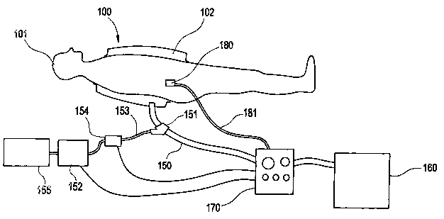
Referring now to Figure 1, a general schematic of a preferred implementation
of
an apparatus of the present invention is shown therein and generally referred
to by reference
numeral 50. Apparatus 50 consists of an enclosure 100 that seals a segment of
the body 101 to
-14-
CA 02485478 2004-11-12
WO 03/096878 PCT/US03/14909
form an enclosed chamber 102. A tubing system 150 preferably consists of one
or more tubes to
provide an inflow of gas into the enclosed chamber 102, preferably at a high
relative (gage)
pressure and to also provide for an outflow of gas from the enclosed chamber
102 to generate a
relative (gage) negative pressure within the enclosed chamber 102. A means 160
for generating
the required relative vacuum and pressurized gas is also provided as is a
control unit 170. The
control unit 170 has a valve system and preferably electronic control system,
which is preferably
equipped with a microcomputer to regulate the supply of pressurized air and
vacuum to the
enclosed chamber 102. One or more sensors 180 are provided to sense the blood
flow pulses and
send appropriate signals through the one or more signal lines 181 to the
control unit 170 to
preferably synchronize the pressurization and vacuum generation cycles within
the enclosed
chamber 102 with the pulses of the blood flow. Preferably, the synchronization
is achieved by
detecting the pulse near the injured area since there is a delay between the
cardiac and local
pressure pulses. In the schematic of Figure 1 and for the sake of simplicity,
only one enclosing
means 100 which is supplied by only one tubing system 150 are shown. It is
however,
understood that more than one enclosing means 100 may be applied to more than
one segment of
the patient body and that each enclosed chamber 102 may be supplied with more
than one tubing
system 150, means for generating the and vacuum and pressurized gas 160, and
control unit 170.
In the present descriptions, air is considered to be the medium that is
injected into
the enclosed volume to generate the desired internal pressure. It should
however be appreciated
that any appropriate gas or fluid may also be similarly used. However, sterile
air with a
controlled amount of humidity and temperature is preferred in most situations.
It may also be
desirable to add an appropriate amount of medicating substances such as
antimicrobial oils or
similar liquids, preferably in the form of a gaseous substance or fluid mist,
to the inflow stream.
Preferably, the medicine is added to the inflow stream of gas at a port 151,
for example by a
pump 152. A tubing line 153 connecting the outlet of the pump 152 to the port
151 preferably
has a valve 154 which closes when the apparatus is in the vacuum cycle and
opens when
-15-
CA 02485478 2004-11-12
WO 03/096878 PCT/US03/14909
medicine is to be added to the inflow stream of gas. The pump 152 is
preferably connected at its
inlet to a medicine supply 155. Both the pump 152 and valve 154 are connected
to the control
unit 170, which syncluonizes theirs to deliver medicine to the inflow stream
of gas when needed
and to prevent the flow of medicine when the vacuum cycle is applied.
Alternatively, medicine
can be manually injected into the port 151 or directly into the enclosure by
any means known in
the art, such as by a syringe (not shown).
Alternatively, a balloon (not shown) can be utilized in the enclosure 100
which is
selectively inflated with a fluid to minimize the volume of the enclosed
chamber 102. In this
way, an enclosure can be used on various size limbs or other body parts
without the need for
customization according the particular shape or size of the patients injured
area. For instance, an
enclosure 100 for a patients ann can be made relatively large to fit the
largest of a person's arm
and the same enclosure can be used on patients having smaller arms by
inflating the balloon
inside the enclosed chamber 102 to minimize the volume of the enclosed chamber
102.
The pressurization and vacuum cycles are preferably synchronized with the
cardiac systole and diastole so that as the blood is being pumped into the
burned region, a
vacuum is generated within the enclosed chamber 102 to assist in the inflow of
the blood and the
enclosed chamber 102 is pressurized to assist the flow of the blood out of the
burned region. The
syncluonization may be with each cardiac cycle, or with a cardiac cycle after
skipping one or
more number of cycles. However, the apparatus may be operated without this
synchronization,
in which case the sensor 180 component of the apparatus is not required. The
sensor to detect
the patient's pulse 180 is preferably one of the commonly used sensors in
medial practice, such as
an EKG or pressure sensor that senses the pulse at the location of the sensor.
A sensor signal is
sent from the sensor 180 to the control unit 170 that processes the signal to
synchronize the
relative vacuum generation and pressurization cycles by properly operating the
control unit
valves and the means of introducing various treatment substances into the
enclosed chamber 102.
Preferably, the negative pressure is applied as the blood is being pumped in
and the positive
-16-
CA 02485478 2004-11-12
WO 03/096878 PCT/US03/14909
pressure is applied as the blood is pumped out of the injured region.
Refen-ing now to Figure 2, an example of a configuration of the means 160 for
generating the required relative vacuum and pressurized gas, the control unit
170, and the tubing
system are shown in more detail. The pressurized air is preferably supplied by
an air compressor
161. In certain cases, the amount of pressure that is required may be within
the range of fan or
turbo or other similar types of air flow generation devices that may then be
utilized. The vacuum
is also preferably provided using a vacuum pump 162. Each of the air
compressor 161 and
vacuum pump 162 are connected to a respective tank 163, 164 by appropriate
plumbing 165. The
air compressor tank 163 must be fabricated to withstand high pressure, while
the vacuum tank
164 must be fabricated to withstand a high vacmun. The plumbing 165 connects
each tank 163,
164 to the valuing of the control unit 170. However, when the amount of
pressurized air to be
delivered to the enclosed chamber 102 is relatively small, the required air
may be delivered fiom
essentially closed one or more chambers which are preferably sealed and are
constructed with
one or more flexible walls and are used to pump their enclosed air in and out
of the enclosed
chambers 102. Such "pumps" are preferably constructed with bellows and are
operated with
electrically driven actuation means. However, other constructions of such
enclosures with one or
more flexible walls may be utilized and be driven by electric, pneumatic or
other actuation
means. In general, by pumping an appropriate amount of air from the enclosed
chamber 102
using the above essentially closed circuit pumping systems, the required level
of vacuum may
also be generated within the enclosed chamber 102. In general, wherever the
volume of the
enclosed chamber 102 is small enough to allow the use of the above air
pressure and vacuum
generation system, the use of such systems are preferred over conventional
compressors and
vacuum pumps.
The control unit 170 preferably comprises a programmable controller 171, such
as
a PC, and a valve unit 172. The programmable controller is programmable to
operate the desired
operation sequence and timing of the air compressor, 161, vacuum pump,
medicine pump 152,
-17-
CA 02485478 2004-11-12
WO 03/096878 PCT/US03/14909
and assorted valves. In Figure 2, valve 154 is not shown because it is
preferably incorporated
into the valve unit 172. Referring now to Figure 12, there is shown a
preferred implementation of
the valve unit 172. The valve unit 172 is preferably constructed and operates
as follows. One or
more solenoid valves 402 controls the flow of pressurized air into the
enclosure 100 from the
tank 163 through a pressure regulator 401 via piping 406. The operator of the
solenoid of the
valve 402 is achieved by the signal from the programmable controller 171. 'The
outflow of the air
from the enclosure 100 into the vacuum tank is controlled by one or more open-
closed solenoid
valves 404. The air is exhausted into the vacuum tank 164 via piping 410. More
than one
pressurized air inlets 406 and valves 402 may be used along the length of the
enclosure 100 to
achieve the sequential pressurization of the enclosure as previously
described. In a similar
manner, more than one vacuum outlet may be used to provide for the sequential
negative
pressure application to the injured area as previously described. When the
free volume within
the enclosure 100 is relatively large, the outflow of air may be accelerated
and the capacity of the
vacuum pump 162 and the vacuum tank 164 may be significantly reduced by
providing an
exhaust outlet operated by an exhaust fan 415 and one or more relatively large
diameter solenoid
valves, with the piping 411.
When utilized, the valves 412 are turned on first and when a considerable
amount
of the required air is exhausted, the valve 412 is closed and the valve 404 is
then opened. One or
more pressure sensors 416 are used to measure the pressure within the
enclosure 100 and send
the measurement by line 417 to the programmable controller 171. The solenoid
valves 402, 404,
and 412 are operated by signals sent by the programmable controller via lines
419, 418, and 420,
respectively.
A first variation of the enclosure 100 is shown in the schematic of Figure 3.
In
Figure 3, a segment of the body, e.g., a segment of the leg or the arm or the
trunk 201, is shown
enclosed within a relatively rigid outer shell 202. The outer shell 202 must
be rigid so as not to
deform under the pressurization or vacuum within the chamber 102. The outer
shell 202 is
-18-
CA 02485478 2004-11-12
WO 03/096878 PCT/US03/14909
constructed with an outer wall 203 and sides 204. The sides 204 have walls 205
to keep the outer
wall 203 at a certain distance from the body segment surface (slcin) and
provide the enclosed
volume 207 of the chamber 102. Lips 206 projecting from the walls 205 are also
provided on the
sides 204 to provide a relatively large surface area for contact with the body
surface (skin) to
distribute the contact forces over a large enough surface area during the
operation of the
apparatus 50. The sides 204 and the outer wall 203 are preferably integrally
formed.
The lips 206 of the sides 204 are preferably sealed to the sunace of the body
segment to provide the sealed volume 207. A layer of a relatively soft sealing
material 212, such
as soft rubber, may be placed between the lips 206 and the body surface to
conform to the body
surface, to assist the sealing action, and to distribute the load more evenly
over the body surface.
The layer 212 and the sides 204 may also be integral. Medical adhesive tape
208 is preferably
used to secure the enclosure 100 to the patient, if necessary.
The outer shell 202 may be constructed as one piece or may be made out of one
or
more segments that are attached and sealed together during the assembly. The
outer wall 203
and/or the sidewalls 205 are provided with one or more openings with ports 209
to allow gas
inflow and outflow from tubing system 150. In the preferred embodiment, gas
flows in from one
or more ports while the air flows out from one or more other ports that are
situated away from
the inflow ports. One or more heating unit 210 may be provided in one or more
inflow air
streams and one or more temperature sensors 211 may be provided to measure the
temperature
within the enclosed volume 207 for the purpose of regulating the temperature
of the air within
the enclosed volume 207 and to keep the enclosed volume 207 close to a set
temperature. The
temperature sensor 211 preferably generates a signal indicative of the
temperature within the
chamber 102 and outputs the signal to the heating unit 210 either directly if
the heating unit 210
has a processing capability or through the programmable controller 171, which
assumes control
of the heating unit 210.
-19-
CA 02485478 2004-11-12
WO 03/096878 PCT/US03/14909
Appropriate medication may be mixed with the inflow air through one or more
ports 151 located on or near one or more air inlets 209 as described above, or
may be introduced
directly into the enclosed chamber through one or more sealed ports 213.
The surface (skin) of the segment of the body 201 located within the enclosure
100 may be covered by a soft and flexible material 103 such as fabric, sponge,
or silicon rubber
or the like by specially constructed and possibly medicated material. The
enclosed volume 207
may be partially or fully filled with an air permeable sponge type of material
104 (shown in
Figure 5) or the like to provide support for the outer wall 203, and/or reduce
the amount of
required air inflow and outflow to produce the desired positive and negative
relative pressure
within the enclosed volume 207 to support the surface of the body. The air
permeable material
can also be spherical or other shaped pellets, as are known in the art.
The shell 202 of the enclosure may be constructed in a tubular shape to go
around
a segment of the body such as arm, leg, thigh or the trunk as shown in Figure
3. The shell 202 of
the enclosure 100 may also be used to cover a certain area of the surface of
the body 250 as
shown schematically in Figure 4, the cross-section 5-5 of which is shown in
Figure 5. In Figures
3 and 5, like elements are indicated by like reference numbers and perform in
a like manner. The
enclosure 100 of Figure 4 functions as described for the enclosure of Figure
3. In Figures 4 and
5, the peripheral elements 209-211 and 213-214 are not shown for the sake of
simplicity but are
understood to be included and function as previously described. The enclosure
100 may also be
used on an extremity such as a foot, in which case it is preferably
constructed with one opening
With side structure 204 as shown in cross-sectional schematic of Figure 6. In
the schematic of
Figure 6, for the salve of simplicity, only a small number of components of
the enclosure are
shown. But it is understood that all the components shown in Figure 3 are also
present and
utilized in the salve manner in this variation of the enclosure 100 design.
When the surface area of the outer wall of the enclosure shell 203 is small or
has a
-20-
CA 02485478 2004-11-12
WO 03/096878 PCT/US03/14909
shape that renders it relatively stiff to deformation into the enclosed volmne
207 (Figure 4), when
the negative relative pressure is applied to the enclosed volume and when it
is also relatively stiff
and resists outward deformation when the positive relative pressure is applied
to the enclosed
volume 207, then a simple plate with an appropriate thickness that is cut and
formed to the
required shape would be sufficient to form the outer surface 203 of the
enclosure 202 and is also
preferred. The outer wall 203 is preferably constructed with easily deformed
and sterilized plate
material such as Plexiglas or other relatively hard plastics or metals such as
stainless steel. A
clear plastic port 1 OS for easy viewing of the covered surface is, however,
preferred for at least a
portion of the outer wall 203 surface to provide for a viewing window.
Referring now to Figure 7, there is shown another version of the enclosure
100.
The enclosure of Figure 7 is particularly well-adapted to appendages such as
the arm or leg and is
shown therein for use with the arm. The enclosure 100 of Figure 7 is
constructed of a body 300,
a closed end fitting 302, and preferably an open end fitting 304. The body 300
preferably
comprises at least one tubular rigid section. In the preferred implementation
shown for adapting
to an arm of a patient, two such rigid tubular sections 306, 308 are shown.
The sections 306, 308
are preferably joined by a coupling 310. The rigid sections 306, 308, closed
end fitting 302,
open end fitting 304, and coupling 310 are joined so as to provide an
appropriately sealed
chamber 102. In this configuration, the rigid sections 306, 308 can be
appropriately sized to
provide more or less volume as needed in a particular area of the appendage.
In Figure 7, for the
sake of simplicity, only a small number of components of the enclosure are
shown. Bit it is
understood that all of the components shown in Figure 3 are also present and
utilized in the same
mmner in this variation of the enclosure 100 design.
Referring now to Figure 8, there is shown the enclosure 100 of Figure 7 having
a
means for supporting the enclosure 100 on the patient. Since the enclosure is
pressurized at
some points during treatment, and since the enclosure 100 of Figure 7 is
closed as one end, it
may have a tendency to fly off of the patient during the pressurization cycle.
Furthermore, the
-21-
CA 02485478 2004-11-12
WO 03/096878 PCT/US03/14909
enclosure may tend to move upwards towards the amnpit of the patient during
the vacuum cycle.
Therefore, it is important that the enclosure 100 be properly supported and
secured to the patient.
Preferably, this support is provided by a support bracket 312 and support
strap 314. The support
bracket 312 is preferably fabricated from a rigid material and having an "L"
shape. A first leg
316 of the "L" shape is fastened to the enclosure 100 and a second leg 318 of
the "L" shape rests
against an adjacent side of the patient. The first leg 316 may be adjustably
connected to the
enclosure 100 to vary the distance between the enclosure 100 and the side of
the patient. The
support strap is preferably fabricated from a flexible material that wraps
around the torso of the
patient and is attached to the enclosure at both ends 320 (one of which is
shown). The support
strap 314 also preferably has an adjustment means, such as a belt buckle (not
shown) to vary its
length.
Referring now to Figures 9-11, another variation of the enclosure of the
present
invention is shown. In this variation, the outer wall of the enclosure shell
203 is constructed with
variously shaped bubbles 251 that are hinged together, preferably with living
hinges 253, to
allow them to conform to the shape of the body, leaving a relatively small
space between the
outer walls of the enclosure and the body surface. The cross-section of such
an enclosure 202 is
shown schematically in Figure 10. The bubbles 251 with sides 252 and living
hinges 253 may
extend in a first direction to cover the entire length of the enclosure or a
portion thereof. The top
view of a first variation of the bubble configuration is shown in Figure 9.
This construction is
preferred for covering limbs such as legs or arm. The bubbles 251 may extend
in a second
direction along the length of the enclosure as shown in Figure 11. The second
variation of the
bubble configuration shown in Figure 11 is preferred for covering surfaces
such as the back or
chest so that the enclosure can conform more closely to the body surface. The
bubbles also
function as stiffeners to limit the inward and outward deformation of the
outer surfaces of the
enclosure during the application of relative vacuum and pressures,
respectively. In addition, the
shape of the bubbles are shown to be nearly square and/or rectangular and
having orthogonal tops
-22-
CA 02485478 2004-11-12
WO 03/096878 PCT/US03/14909
and sides. In practice, however, the bubbles may be provided in any shape and
their side 252 or
top surfaces may be tapered to allow better conformation to the commonly
tapered limbs of the
body.
Method of Treatment: The following method of treatment is given by way of
example only and
not to limit the spirit or scope of the present invention in any way.
The device which will apply external synchronous pulsatile pressure to either
the
whole body or portions of the body has as its goal the preservation of injured
areas of tissues of
the body, particularly in the zone of stasis. This will be accomplished by
controlling the edema,
which begins to form in the tissues immediately after the burn.
The pulsatile external pressure will vary from -25mm Hg., +300 mm Hg. and will
be applied synchronous with the cardiac cycle. The positive phase will be
applied during cardiac
diastole and the negative phase during cardiac systole. The positive phase
will enhance venous
drainage from the wound, and the negative phase will enhance arterial inflow
into the subdermal
plexus.
The dermis is divided into a tlun, superficial layer called the papillary
dermis and
a deeper layer called the reticular dennis. There is a large plexus of vessels
beneath the dermis,
known as the subdermal plexus, which sends vessels towards the periphery to
form a plexus
between the reticular and papillazy dermis. More superficially there is a
plexus of vessels called
the papillary plexus. The blood supply to all of these small vessels becomes
occluded as a result
of the edema caused by the factors that were described earlier in this
document; and is further
aggravated by the infusion of large amounts of crystalloid solution which
quickly extravasates
into the interstitial tissues and augments the volume of edema.
-23-
CA 02485478 2004-11-12
WO 03/096878 PCT/US03/14909
The pulsatile pressure system will be applied as soon after the burn occurs as
is
possible, and will preferably be applied for up to 4 days, the period during
which edema
normally continues to form and finally is stabilized. The pulsatile pressures
will be applied
continuously, and interrupted as frequently as is necessary to inspect and
treat the wound surface,
i.e. 2-3 times daily.
Ancillary measures:
Those in the art have showed that capillary stasis can be reversed by careful
maintenance of hydration of the wound surface, and by avoiding over or under
hydration during
the resuscitation phase after the burn.
Since the internal setting for thermal control of the body is set at a higher
level in
burned patients there is a significant evaporative water loss after 24 hours
which allows the body
to lose heat, the heat setting external to the body will be kept at a
sufficiently high level to
prevent shivering and to maintain a normal body temperature.
The wound surface will be washed several times a day with soap and will be
treated with topical antiinicrobial agents, and with either a plastic film
such as "Biobrane" or
cultured alografts, in order to prevent desiccation of the skin surface.
Systemically, heparin will be administered in a doses sufficient to provide
prophylaxis against thrombus formation. The resuscitation regimen will be
primarily with
Lactated Ringer's solution- given in a dose of 4cc/Kg body wt% burn; or as
3cc/Kg% Lactated
Ringer's with plasma in a dose of 1 cc/Kg% burn.
Systemic antibiotics will be withheld during this period unless there is a
specific
-24-
CA 02485478 2004-11-12
WO 03/096878 PCT/US03/14909
indication.
While there has been shown and described what is considered to be preferred
embodiments of the invention, it will, of course, be understood that various
modifications and
changes in form or detail could readily be made without departing from the
spirit of the
invention. It is therefore intended that the invention be not limited to the
exact forms described
and illustrated, but should be constructed to cover all modifications that may
fall within the
scope of the appended claims.
-25-