Sélection de la langue

Search

Sommaire du brevet 2488201 

Énoncé de désistement de responsabilité concernant l'information provenant de tiers

Une partie des informations de ce site Web a été fournie par des sources externes. Le gouvernement du Canada n'assume aucune responsabilité concernant la précision, l'actualité ou la fiabilité des informations fournies par les sources externes. Les utilisateurs qui désirent employer cette information devraient consulter directement la source des informations. Le contenu fourni par les sources externes n'est pas assujetti aux exigences sur les langues officielles, la protection des renseignements personnels et l'accessibilité.

Disponibilité de l'Abrégé et des Revendications

L'apparition de différences dans le texte et l'image des Revendications et de l'Abrégé dépend du moment auquel le document est publié. Les textes des Revendications et de l'Abrégé sont affichés :

  • lorsque la demande peut être examinée par le public;
  • lorsque le brevet est émis (délivrance).
(12) Brevet: (11) CA 2488201
(54) Titre français: DISPOSITIF D'INSERTION D'UN GREFFON PAR ARTHROSCOPIE
(54) Titre anglais: ARTHROSCOPIC TISSUE SCAFFOLD DELIVERY DEVICE
Statut: Périmé et au-delà du délai pour l’annulation
Données bibliographiques
(51) Classification internationale des brevets (CIB):
  • A61B 17/56 (2006.01)
  • A61F 2/46 (2006.01)
  • A61M 5/00 (2006.01)
(72) Inventeurs :
  • ORR, KEITH M. (Etats-Unis d'Amérique)
  • HYMAN, ERIC (Etats-Unis d'Amérique)
  • BINETTE, FRANCOIS (Etats-Unis d'Amérique)
  • MCRURY, IAN D. (Etats-Unis d'Amérique)
  • LEPKE, STEVE (Etats-Unis d'Amérique)
  • PERKINS, ASH (Etats-Unis d'Amérique)
  • HWANG, JULIA (Etats-Unis d'Amérique)
(73) Titulaires :
  • DEPUY MITEK, INC.
(71) Demandeurs :
  • DEPUY MITEK, INC. (Etats-Unis d'Amérique)
(74) Agent: NORTON ROSE FULBRIGHT CANADA LLP/S.E.N.C.R.L., S.R.L.
(74) Co-agent:
(45) Délivré: 2008-08-26
(22) Date de dépôt: 2004-11-22
(41) Mise à la disponibilité du public: 2005-05-26
Requête d'examen: 2004-11-22
Licence disponible: S.O.
Cédé au domaine public: S.O.
(25) Langue des documents déposés: Anglais

Traité de coopération en matière de brevets (PCT): Non

(30) Données de priorité de la demande:
Numéro de la demande Pays / territoire Date
10/724,021 (Etats-Unis d'Amérique) 2003-11-26

Abrégés

Abrégé français

Un dispositif d'insertion de petit diamètre permettant d'insérer un greffon par arthroscopie à un défaut tissulaire ou un site de lésion, sans réduire la pression au site de la lésion, est fourni. Le dispositif d'insertion de greffon de la présente invention est constitué d'un système de piston qui comprend deux composants majeurs : un tube d'insertion et une tige d'insertion. Le tube d'insertion comporte une extrémité proximale évasée permettant de tenir le greffon avant l'insertion. Un corps creux de forme allongée se prolonge depuis l'extrémité évasée jusqu'à une extrémité distale du tube d'insertion, et définit un passage qui se prolonge dans le corps pour permettre l'insertion du greffon. La tige d'insertion comporte un corps de forme allongée qui se prolonge dans la poignée à une extrémité proximale et une pointe à une extrémité distale. La tige d'insertion est configurée de manière à être disposée de façon amovible à l'intérieur du tube d'insertion et à glisser dans le passage pour effectuer l'insertion du greffon à travers le tube d'insertion.


Abrégé anglais

A small diameter delivery device capable of delivering a tissue loaded scaffold arthroscopically to a tissue defect or injury site without reducing the pressure at the injury site is provided. The scaffold delivery device of the present invention comprises a plunger system that includes two main components: an insertion tube and an insertion rod. The insertion tube has a flared proximal end for holding a tissue scaffold prior to delivery. An elongate, hollow body extends from the flared proximal end to a distal end of the insertion tube, and defines a passageway that extends through the body for delivery of the tissue scaffold. The insertion rod has an elongate body that extends into a handle at a proximal end and a tip at a distal end. The insertion rod is configured to be removably disposed within the insertion tube for sliding along the passageway to effect delivery of the tissue scaffold through the insertion tube.

Revendications

Note : Les revendications sont présentées dans la langue officielle dans laquelle elles ont été soumises.


-7-
CLAIMS
1. A device for arthroscopically delivering a tissue scaffold to a damaged or
injured
tissue site, comprising:
a first component having a flared proximal end, a distal end, and an elongate,
hollow body extending therebetween, the elongate body defining a passageway
extending from the flared proximal end to the distal end; and
a second component having an elongate body with a tip at a distal end, the
elongate body having at least one sealing element formed at a location
proximal to the
distal end, the second component being configured to be removably disposed
within
the first component for sliding along the passageway.
2. The device of claim 1, wherein the passageway includes a first, flared
portion
extending into a second, tubular portion.
3. The device of claim 2, wherein the first, flared portion has a curved
tapered shape.
4. The device of claim 1, wherein the flared proximal end of the first
component has a
diameter in the range of about 15 mm to about 50 mm.
5. The device of claim 2, wherein the second, tubular portion has a diameter
in the range
of about 5 mm to about 17 mm.
6. The device of claim 1, wherein the tip of the second component comprises a
spherical
tip.
7. The device of claim 6, wherein the spherical tip has a diameter in the
range of about 6
mm to about 10 mm.
8. The device of claim 1, wherein the second component further includes at
least one
sealing ring around the elongate body proximal to the tip.
9. An instrument for arthroscopically delivering a tissue scaffold to a
damaged or injured
tissue site, comprising:
an insertion tube having a proximal end, a distal end and a passageway
extending therebetween; and

-8-
an insertion rod having an elongate shaft extending into a handle at a
proximal
end and a blunt tip at a distal end, the elongate shaft having at least one
sealing
element formed at a location proximal to the distal end, the insertion rod
being
configured to be removably disposed within the insertion tube for sliding
along the
passageway and contacting the tissue scaffold.
10. The instrument of claim 9, wherein the passageway includes a first, flared
portion
extending into a second, tubular portion.
11. The instrument of claim 10, wherein the first, flared portion has a
curved, tapered
shape.
12. The instrument of claim 10, wherein the second, tubular portion has a
diameter in the
range of about 6 mm to about 17 mm.
13. The instrument of claim 12, wherein the second, tubular portion has a
diameter in the
range of about 7 mm to about 9 mm.
14. The instrument of claim 9, wherein the proximal end is flared.
15. The instrument of claim 14, wherein the flared proximal end of the
insertion tube has
a diameter in the range of about 15 mm to about 50 mm.
16. The instrument of claim 9, wherein the blunt tip of the insertion rod
comprises a
spherical tip.
17. The instrument of claim 16, wherein the spherical tip has a diameter in
the range of
about 6 mm to about 10 mm.
18. The instrument of claim 17, wherein the spherical tip has a diameter in
the range of
about 6 mm to about 8 mm.
19. The instrument of claim 9, wherein the insertion rod further includes a
pair of sealing
rings around the elongate body.
20. The use of a delivery system for delivering a tissue scaffold to an
injured or defective
tissue, the delivery system comprising an insertion tube having a proximal
end, a
distal end and a passageway extending therebetween, and an insertion rod
having an
elongate shaft extending into a handle at a proximal end and a blunt tip at a
distal end,

-9-
the elongate shaft having at least one sealing element formed at a location
proximal to
the distal end, the insertion rod shaft being configured to be removably
disposed
within the insertion tube for sliding along the passageway;
wherein a tissue scaffold is loadable into the insertion tube from the flared
proximal end; and
the insertion rod is depressable against a loaded tissue scaffold and toward
the
distal end to cause the tissue scaffold to exit the distal end of the
insertion tube.
21. The use of claim 20, wherein the delivery system is for use
arthroscopically.
22. The use of the device according to any one of claims 1 to 8 for delivering
a tissue
scaffold to a damaged or injured tissue site.
23. The use of the instrument according to any one of claims 9 to 19 for
delivering a
tissue scaffold to a damaged or injured tissue site.

Description

Note : Les descriptions sont présentées dans la langue officielle dans laquelle elles ont été soumises.

CA 02488201 2004-11-22
-1-
ARTHROSCOPIC TISSUE SCAFFOLD DELIVERY DEVICE
FIELD OF THE INVENTION
This invention relates to surgical tools and devices for delivering a tissue
implant. More particularly, this invention relates to a delivery device for
arthroscopically delivering a tissue scaffold.
BACKGROUND
Injuries to soft tissue, such as cartilage, skin, muscle, bone, tendon, and
ligament
where the tissue has been injured or traumatized frequently require surgical
intervention
to repair the damage and facilitate healing. Such surgical repairs can include
suturing or
otherwise repairing the damaged tissue with known medical devices, augmenting
the
damaged tissue with other tissue, using an implant, a graft or any combination
of these
techniques.
One common technique for repairing diseased or injured tissue is to implant a
tissue scaffold at the defect site, either alone or along with cultured and
amplified cells.
In the past, such scaffolds have consisted mostly of two- or three-dimensional
porous
scaffolds that allow cell invasion and remodeling once the scaffold has been
combined
with living cells and has been delivered inside the patient. Another technique
is to load
the tissue scaffolds with tissue fragments and then implanting the tissue-
laden scaffold at
the defect site. Depending where the defect is located and the size of the
defect, these
tissue loaded scaffolds can vary from a few millimeters to several dozen
millimeters in
length and width.
The current method for implanting these tissue scaffolds is by an open or mini-
open surgical procedure, which increases recovery time for the patient.
Although a fully
arthroscopic procedure for delivering a tissue-loaded scaffold to a defect
site would be
advantageous because of its minimally invasive nature and reduced side
effects, there is
presently no convenient method for delivering the tissue-loaded scaffolds
through a
cannula to the defect site without the risk of damaging the scaffold in the
process.
Moreover, where it is necessary to deliver the scaffold to a defect in a
joint, there is
currently no acceptable way to arthroscopically deliver the scaffold without
reducing the
pressure inside the joint. It would therefore be desirable to provide a method
and device

CA 02488201 2004-11-22
-2-
which allows delivery of a tissue loaded scaffold in a fully arthroscopic
procedure. It
would also be advantageous to provide a delivery device which allows the
scaffold to be
delivered through a small diameter tube to a defect in a joint without
reducing the
pressure inside the joint.
SUMMARY OF THE INVENTION
The present invention provides a small diameter delivery device capable of
delivering a tissue loaded scaffold arthroscopically to a tissue defect or
injury site
without reducing the pressure at the injury site. The scaffold delivery device
of the
present invention comprises a plunger system that includes two main
components: an
insertion tube and an insertion rod. The first component, the insertion tube,
is
configured with a flared proximal end for holding a tissue scaffold prior to
delivery. An
elongate, hollow body extends from the flared proximal end to a distal end of
the
insertion tube. The elongate, hollow body defines a passageway that extends
through
the body for delivery of the tissue scaffold. The second component, the
insertion rod,
comprises an elongate body that extends into a handle at a proximal end and a
tip at a
distal end. The insertion rod is configured to be removably disposed within
the insertion
tube for sliding along the passageway to effect delivery of the tissue
scaffold through the
insertion tube.
The passageway of the hollow, elongate body includes a first, flared portion
that
gradually extends into a second, tubular portion. Prior to delivery, the
tissue scaffold
can be held inside the insertion tube at the first, flared portion until the
insertion rod is
placed on the scaffold and urged distally. The force of the tip of the
insertion rod
against the tissue scaffold moves the scaffold through the passageway and out
the distal
end of the insertion tube. The gradual taper of the passageway allows the
implant to be
delivered without incurring damage in the process. Due to the small diameter
of the
insertion tube, the tissue scaffold can be arthroscopically delivered without
reducing the
pressure at the implantation site.
BRIEF DESCRIPTION OF THE DRAWINGS
The invention can be more fully understood from the following detailed
description taken in conjunction with the accompanying exemplary drawing, in
which:

CA 02488201 2004-11-22
-3-
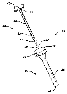
FIG. 1 illustrates an exploded view of the delivery device of the present
invention;
FIG. 2A illustrates a perspective view of the delivery device of FIG. 1; and
FIG. 2B is a cutaway view of the delivery device of FIG. 2A.
DETAILED DESCRIPTION OF THE INVENTION
Turning now to the drawings and particularly to FIG. 1, a delivery system 10
for
delivering a tissue scaffold 12 in accordance with the present invention is
shown. The
delivery system 10 comprises two main components. The first component is an
insertion tube 20 and the second component is an insertion rod 40. The
insertion tube 20
is defined by a flared proximal end 22 and a distal end 24. Extending between
these
ends 22, 24 is a hollow, elongate body 26. As shown in FIG. 2B, the hollow,
elongate
body 26 defines a passageway 28 comprising a first, flared portion 30 that
extends into a
second, tubular portion 32. The first, flared portion 30 can have a curved
taper in order
to provide a smooth transition between the two portions 30, 32 and to avoid
damage to
the tissue scaffold 12 during its delivery. The first, flared portion 30 can
be integral with
the second, tubular portion 32. Alternatively, the first, flared portion 30
can be provided
as a separate component which is then attached to the second, tubular portion
32 to
collectively form the insertion tube 20.
The second component of the delivery system 10, the insertion rod 40,
comprises
an elongate shaft 46 that extends into a handle 48 at a proximal end and a tip
50 at a
distal end. As shown, the handle 48 is configured as a contoured grip for ease
of
handling. It is understood, however, that the handle 48 can comprise other
configurations having any shape or size appropriate for the purposes of this
invention.
Meanwhile, the tip 50 can be blunt, having a spherical shape as illustrated in
FIG. 1 for
preventing damage to the tissue scaffold 12 during the delivery. The elongate
shaft 46 is
configured to be removably disposed within the insertion tube 20 for sliding
along the
passageway 28. Sealing rings 52 can be placed on the elongate shaft 46 to help
align the
insertion rod 40 and prevent backflow of fluid through the insertion tube 20
during the
delivery process. These sealing rings 52 can be made from a softer material
than the

CA 02488201 2004-11-22
-4-
insertion rod 40, and can be provided as separate components for adjustable
placement
on the insertion rod 40.
In one aspect of the present invention, the flared proximal end 22 of the
insertion
tube 20 has a diameter in the range of about 1 S mm to about 50 mm to
accommodate a
variety of sized and shaped tissue scaffolds 12 known in the art. The
passageway 28 of
the insertion tube 20 can have a second tubular portion 32 with a diameter in
the range
of about 6 mm to about 17 mm, preferably about 7 mm to about 9 mm. The
spherical tip
50 can have a diameter in the range of about 6 mm to about 10 mm, preferably
about 6
mm to about 8 mm. These small dimensions ensure not only that the scaffold
delivery
can be accomplished in a fully arthroscopic procedure, but that damage to the
tissue
scaffold 12 during the delivery process can be minimized. In addition, the
maximum
outer diameter of the elongate shaft 26, including the seating ring 52, should
approximate the diameter of the second tubular portion 32, so that backflow of
fluid can
be prevented and so that a closed volume within the insertion tube 20 can be
attained
when the insertion rod 40 is disposed within the passageway 28.
The delivery system 10 can be formed from any suitable biocompatible metal or
polymer. For example, the delivery system 10 can be formed from a medical
grade
surgical steel, or the delivery system 10 can be formed from a sterilizable,
medical grade
plastic such as polycarbonate. The sealing rings 52 of the insertion rod 40
can be
formed from a compliant material such as silicone. It should be understood
that the
present invention is not to be limited to these materials, and one skilled in
the art would
recognize that the delivery system 10 of the present invention can be made
from a
variety of other suitable materials.
The delivery system 10 of the present invention works in a plunger-like
manner.
In use, a patient having a damaged or diseased tissue site is prepared for
arthroscopic
surgery. With the insertion rod 40 separate from the insertion tube 20, a
tissue scaffold
12 can be loaded into the delivery system 10 by placing the scaffold 12 within
the flared
portion of the passageway 28. Depending on the dimensions and shape of the
scaffold
12, the scaffold 12 can be slightly folded or rolled into a configuration
similar to the one
illustrated in FIG. 1. The first, flared portion 30 of the passageway 28
should be
configured to sufficiently allow the scaffold 12 to seat within the insertion
tube 20 at the
flared, proximal end 22 without too much friction.

CA 02488201 2004-11-22
-$-
Next, the insertion rod 40 is placed onto the tissue scaffold 12, with the tip
50
contacting the tissue scaffold 12. Applying pressure against the handle 48,
the insertion
rod 40 is urged down the passageway 28 of the insertion tube 20, thus sliding
the tissue
scaffold 12 along with the depressed rod 40 into the insertion tube 20. The
tissue
scaffold 12 can be slid down the second tubular portion 32 of the passageway
28 until
the tissue scaffold 12 almost reaches the distal end 24 of the insertion tube.
The sealing
rings $2 on the elongate shaft 46 of the insertion rod 40 help align the rod
40 inside the
insertion tube 20 so that the movement of the insertion rod 40 and tissue
scaffold 12 run
parallel to the length of the insertion tube 20. In addition, the sealing
rings also serve to
prevent backflow of fluid up through the insertion tube and to maintain a
closed volume
between the delivery device 10 and the implant site during the delivery
process.
When the surgeon is ready to deliver the tissue scaffold 12 to the damaged or
injured tissue site, the insertion tube 20 along with the tissue scaffold 12
can then be
1$ inserted directly through the incision and positioned so that the distal
end 24 is located
where the scaffold 12 is to be delivered. The surgeon would then continue
depressing
the insertion rod 20 until the tissue scaffold 12 is fully exposed to the
implant site. After
the tissue scaffold 12 exits the distal end 24 of the insertion tube, the
insertion rod 40
can be removed and the delivery system 10 can be discarded, or it can be
reused if
~ desired. Because of the small size of the delivery system 10, it is
understood that the
entire delivery process can be accomplished in a fully arthroscopic procedure.
The delivery system 10 of the present invention is configured to accommodate
tissue scaffolds 12 of various sizes and shapes known in the art. Where the
tissue
scaffold 12 also requires an adhesive or glue component for securing the
scaffold 12 to
2$ the tissue defect or injury, the tissue scaffold 12 along with the glue
component can be
loaded into the insertion tube 20, preferably with the glue component on the
inside,
facing towards the passageway 28. Where it is desirable to use a fixation
device such as
a staple or tack to secure the scaffold 12, it is contemplated that the tissue
scaffold 12
can be loaded with the fixation device extending at least partially through
the tissue
scaffold 12. The scaffold 12 can be position within the first flared portion
30 such that
the fixation device is exposed. Then, the insertion rod 40 can be depressed
against the
fixation device to deliver both the tissue scaffold 12 along with the fixation
device to the
site of implantation. In another embodiment, the insertion rod 40 can have a
tip $0

CA 02488201 2004-11-22
-6-
which is configured (not shown) to attach to or mate with the fixation device,
thereby
allowing greater control over the delivery of the scaffold 12. Also, where the
tissue
scaffold 12 has a unique geometry, it is contemplated that the tip 50 can be
formed as a
piercing tip (not shown) in order to hold the scaffold 12 in place during the
delivery
process.
In yet another embodiment, the insertion rod 40 and the insertion tube 20 can
be
connected by a trigger mechanism (not shown) similar to the depression
mechanism of a
caulk gun. The trigger mechanism can include a handle or grip with an attached
pivoting trigger. The handle or grip can be fixedly attached to the insertion
tube 20,
while the trigger can be attached to the insertion rod 40. By depressing the
trigger either
forwards or backwards, the insertion rod 40 can be retract away from the
proximal end
22 of the insertion tube 20 to enable a tissue scaffold 12 to be loaded, or
advanced
towards the distal end 24 of the insertion tube 20 to effect movement of the
scaffold 12
through the distal end 24. It is contemplated that the configuration of the
insertion tube
and insertion rod 40 (including the sealing rings 52) would be similar to the
ones
previously described. This trigger mechanism would provide the surgeon with
greater
control over the advancement of the scaffold 12, and better ensure the
alignment of the
insertion rod 40 with respect to the insertion tube 20.
20 It will be understood that the foregoing is only illustrative of the
principles of the
invention, and that various modifications can be made by those skilled in the
art without
departing from the scope and spirit of the invention.

Dessin représentatif
Une figure unique qui représente un dessin illustrant l'invention.
États administratifs

2024-08-01 : Dans le cadre de la transition vers les Brevets de nouvelle génération (BNG), la base de données sur les brevets canadiens (BDBC) contient désormais un Historique d'événement plus détaillé, qui reproduit le Journal des événements de notre nouvelle solution interne.

Veuillez noter que les événements débutant par « Inactive : » se réfèrent à des événements qui ne sont plus utilisés dans notre nouvelle solution interne.

Pour une meilleure compréhension de l'état de la demande ou brevet qui figure sur cette page, la rubrique Mise en garde , et les descriptions de Brevet , Historique d'événement , Taxes périodiques et Historique des paiements devraient être consultées.

Historique d'événement

Description Date
Le délai pour l'annulation est expiré 2017-11-22
Lettre envoyée 2016-11-22
Accordé par délivrance 2008-08-26
Inactive : Page couverture publiée 2008-08-25
Inactive : Taxe finale reçue 2008-06-05
Préoctroi 2008-06-05
Modification reçue - modification volontaire 2008-06-05
Inactive : CIB attribuée 2007-12-07
Lettre envoyée 2007-12-07
Un avis d'acceptation est envoyé 2007-12-07
Inactive : CIB enlevée 2007-12-07
Inactive : Approuvée aux fins d'acceptation (AFA) 2007-09-10
Modification reçue - modification volontaire 2007-07-09
Inactive : Dem. de l'examinateur par.30(2) Règles 2007-01-09
Inactive : Supprimer l'abandon 2006-04-25
Inactive : CIB de MCD 2006-03-12
Inactive : Abandon. - Aucune rép. à lettre officielle 2006-02-23
Demande publiée (accessible au public) 2005-05-26
Inactive : Page couverture publiée 2005-05-25
Modification reçue - modification volontaire 2005-04-28
Inactive : CIB en 1re position 2005-02-28
Inactive : CIB en 1re position 2005-02-18
Lettre envoyée 2005-01-14
Exigences de dépôt - jugé conforme 2005-01-14
Lettre envoyée 2005-01-14
Inactive : Certificat de dépôt - RE (Anglais) 2005-01-14
Lettre envoyée 2005-01-13
Demande reçue - nationale ordinaire 2005-01-13
Exigences pour une requête d'examen - jugée conforme 2004-11-22
Toutes les exigences pour l'examen - jugée conforme 2004-11-22

Historique d'abandonnement

Il n'y a pas d'historique d'abandonnement

Taxes périodiques

Le dernier paiement a été reçu le 2007-10-18

Avis : Si le paiement en totalité n'a pas été reçu au plus tard à la date indiquée, une taxe supplémentaire peut être imposée, soit une des taxes suivantes :

  • taxe de rétablissement ;
  • taxe pour paiement en souffrance ; ou
  • taxe additionnelle pour le renversement d'une péremption réputée.

Veuillez vous référer à la page web des taxes sur les brevets de l'OPIC pour voir tous les montants actuels des taxes.

Titulaires au dossier

Les titulaires actuels et antérieures au dossier sont affichés en ordre alphabétique.

Titulaires actuels au dossier
DEPUY MITEK, INC.
Titulaires antérieures au dossier
ASH PERKINS
ERIC HYMAN
FRANCOIS BINETTE
IAN D. MCRURY
JULIA HWANG
KEITH M. ORR
STEVE LEPKE
Les propriétaires antérieurs qui ne figurent pas dans la liste des « Propriétaires au dossier » apparaîtront dans d'autres documents au dossier.
Documents

Pour visionner les fichiers sélectionnés, entrer le code reCAPTCHA :


Pour visualiser une image, cliquer sur un lien dans la colonne description du document. Pour télécharger l'image (les images), cliquer l'une ou plusieurs cases à cocher dans la première colonne et ensuite cliquer sur le bouton "Télécharger sélection en format PDF (archive Zip)" ou le bouton "Télécharger sélection (en un fichier PDF fusionné)".

Liste des documents de brevet publiés et non publiés sur la BDBC .

Si vous avez des difficultés à accéder au contenu, veuillez communiquer avec le Centre de services à la clientèle au 1-866-997-1936, ou envoyer un courriel au Centre de service à la clientèle de l'OPIC.


Description du
Document 
Date
(aaaa-mm-jj) 
Nombre de pages   Taille de l'image (Ko) 
Description 2004-11-22 6 311
Abrégé 2004-11-22 1 23
Revendications 2004-11-22 3 88
Dessin représentatif 2005-04-28 1 4
Page couverture 2005-05-10 1 38
Dessins 2005-04-28 2 22
Dessin représentatif 2007-07-23 1 5
Revendications 2007-07-09 3 100
Dessin représentatif 2008-08-13 1 6
Page couverture 2008-08-13 1 41
Accusé de réception de la requête d'examen 2005-01-13 1 176
Courtoisie - Certificat d'enregistrement (document(s) connexe(s)) 2005-01-14 1 105
Certificat de dépôt (anglais) 2005-01-14 1 158
Demande de preuve ou de transfert manquant 2005-11-23 1 102
Rappel de taxe de maintien due 2006-07-25 1 110
Avis du commissaire - Demande jugée acceptable 2007-12-07 1 163
Avis concernant la taxe de maintien 2017-01-03 1 178
Correspondance 2008-06-05 2 85