Note : Les descriptions sont présentées dans la langue officielle dans laquelle elles ont été soumises.
CA 02498934 2008-04-16
COMPOSITIONS FOR INDUCING INCREASED LEVELS OF (3-CHEMOKINES
AND METHODS OF USE THEREFOR
BACKGROUND OF THE INVENTION
Field of the Invention
The present invention generally relates to increased production of a-
chemokines, and
more particularly, to compositions comprising at least one GI phase arresting
agent
thereby resulting in increased levels and availability of 0-chemokines to
prevent or
treat viral infections and viral related cancers, such as HIV infections and
related HIV
cancers.
Background of the Related Art
The human immunodeficiency virus (HIVI has been implicated as the primary
cause
of the slowly degenerative immune system disease termed acquired immune
deficiency syndrome (AIDS). There are at least two distinct types of HIV: HIV-
l and
HIV-2. In humans, HIV replication occurs prominently in CD4 T lymphocyte
populations, and HIV infection leads to depletion of this cell type and
eventually to
immune incompetence, opportunistic infections, neurological dysfunctions,
neoplastic
growth, and ultimately death.
HIV is a member of the lentivirus family of retroviruses. Retroviruses are
small-
enveloped viruses that contain a single-stranded RNA genome, and replicate via
a
DNA intermediate produced by a virally encoded reverse transcriptase, an RNA-
dependent DNA polymerase.
The HIV viral particle comprises a viral core, composed in part of capsid
proteins,
together with the viral RNA genome and those enzymes required for early
replicative
events. Myristylated gag protein forms an outer shell around the viral core,
which is,
in turn, surrounded by a lipid membrane envelope. derived from the infected
cell
1
CA 02498934 2005-03-14
WO 2004/024683 PCT/US2003/028697
membrane. The HIV envelope surface glycoproteins are synthesized as a single
160
kilodalton precursor protein, which is cleaved by a cellular protease during
viral
budding into two glycoproteins, gp4l and gpl20. gp4l is a transmembrane
glycoprotein and gp 120 is an extracellular glycoprotein, which remains non-
covalently
associated with gp41, possibly in a trimeric or multimeric form.
HIV is targeted to CD4 cells because a CD4 cell surface protein (CD4) acts as
the
cellular receptor for the HIV-1 virus. Viral entry into cells is dependent
upon gp120
binding the cellular CD4 receptor molecules, explaining HIV's tropism for CD4
cells,
while gp41 anchors the envelope glycoprotein complex in the viral membrane.
While
these virus:cell interactions are necessary for infection, there is evidence
that
additional virus:cell interactions are also required.
HIV infection is pandemic and HIV-associated diseases represent a major world
health problem. Although considerable effort is being put into the design of
effective
therapeutics, currently no curative anti-retroviral drugs against AIDS exist.
The new
treatment regimens for HIV-1 show that a combination of anti-HIV compounds,
which target reverse transcriptase (RT), such as azidothymidine (AZT),
lamivudine
(3TC), dideoxyinosine (ddl), tenofovir, nevirapine, efavirenz, or anti-HIV
compounds
which target HIV protease such as saquinavir, nelfinavir, indinavir,
amprenavir, and
lopinavir. For example, impressive results have recently been obtained with a
combination of AZT, 3TC and a protease inhibitor as well as AZT, 3TC, and
Efavirenz have demonstrated potent antiviral activity. Unfortunately the
development
of viral resistance occurs in a significant number of treated patients. This
combined
with the development of anti-retroviral drug induced toxicity continues to
limit the
overall impact of current available treatments.
Moreover, long-term cytotoxic therapy may also lead to suppression of CD8+ T
cells,
which are essential to the control of HIV, via killer cell activity and by the
release of
suppressive factors, notably the chemokines.
Chemokines are a family of small cytokines that are released in response to
infection
together with other inflammatory cytokines. Chemokines are multiple mediators,
but
2
CA 02498934 2005-03-14
WO 2004/024683 PCT/US2003/028697
were first studied as inducers of chemotaxis of specific leukocytes. Further
studies
have revealed that chemokines also stimulate lymphocyte development,
angiogenesis,
degranulation of granulocytes, respiratory bursts and the release of lysosomal
enzymes
in monocytes.
Chemokines are divided into four different subfamilies, according to the
position of
the first two cysteines in their primary sequence: the a-chemokine subclass
bears a
CXC-motif, where the two cysteines are separated by one amino acid; the (3-
chemokines contain a CC motif; the y-subclass lacks one cysteine residue; and
in A.-
chemokines, or CX3C subclass, the two cysteines are separated by three amino-
acids.
These cysteine residues form disulfide bridges with two other cysteines
located further
downstream in the primary sequence, thus stabilizing the tertiary structure of
these
chemokines.
Recently, chemokines produced by CD8+ T cells have been implicated in
suppression
of HIV infection. The chemokines RANTES, MIP-la and MIP-1(3, which are
secreted by CD8+ T cells, were shown to suppress HIV-1 p24 antigen production
in
cells infected with HIV-1 or HIV-2 isolates in vitro (Cocchi, et al., 1995).
However,
levels of available chemokines are limited and the effectiveness of
introducing
exogenous chemokines is still in question because of the short serum half-life
of
exogenously administered chemokines.
Chemokine receptors are designated CXCR followed by a number when binding cc-
chemokines and CCR followed by a number when binding P-chemokines. The
importance of CCR5 for initial transmission of HIV-1 is highlighted by the
fact that
individuals lacking expression of CCR5 (the CCR5-A32 homozygous genotype) are
usually resistant to infection (Liu, et al., 1996). In addition, recent
studies show that
CCR5 cell-surface density correlates with disease progression in infected
individuals
(Lin, et al., 2002).
The natural ligands of CCR5 that include the P-chemokines macrophage
inflammatory
protein (MIP)-1 a, MIP-1 P, and RANTES, inhibit entry of CCR5 (R5) strains of
HIV-1
in both lymphocytes and macrophages (Cocchi, et al., 1995). The inhibitory
effect of
3
CA 02498934 2005-03-14
WO 2004/024683 PCT/US2003/028697
P-chemokines is proposed to act through blocking of CCR5 as well as through
down-
regulation of the coreceptor from the cell surface. Inhibition of viral entry
has also
been achieved by blocking the binding of the viral gp120 to the CCR5
coreceptor by
antagonist molecules such as TAK-779 (Baba, et al., 19990) or SCH-C (Strizki,
et al.,
2001). The P-chemokines RANTES (regulated on activation, normal T cell
expressed
and secreted), macrophage inflammatory protein-1 oc (MIP-1 oa), and MIP-1 R
are the
natural ligands of CCR5, the main coreceptor of non-syncytium-inducing HIV-1,
and
have been shown to inhibit the in vitro infection of lymphocytes (Cocchi, et
al., 1995).
The increase of RANTES, MIP-1a, and MIP-1R has been correlated with resistance
to
infection or a more favorable clinical prognosis, likely because of
competition of the
chemokines with HIV-1 for binding to CCR5 (Paxton, et al., 1996; Zagury, et
al.,
1998; Ferbas, et al., 2000; Cocchi, et al., 1995; and Adams, et al., 1997).
However,
natural levels and availability of chemokines are limited and as the effects
of HIV
infection increase the natural production of 0-chemokines is decreased.
Moreover,
attempts to increase levels of chemokines have been centered on administration
of
chemokines directly to a subject by means of administering exogenous proteins
directly to the subject. Depending on the mode of administration the amount of
chemokines delivered is variable.
Thus, it would be advantageous to identify compounds that induce the increased
levels
of endogenous chemokines through pathways that modulate the activity of
chemokines and increase the levels and availability thereof.
SUMMARY OF THE INVENTION
The present invention relates to enhancing levels and availability of anti-HIV
P-
chemokines by manipulating the cell cycle in activated lymphocytes by
administering
a composition that prolongs the G1 phase of the cell cycle, thereby increasing
production or available levels of P-chemokine by the activated lymphocytes,
which, in
turn reduces the effects of HIV and related complications, such as related
viral
cancers.
4
CA 02498934 2009-02-24
In one particular embodiment there is provided a pharmaceutical composition
for
increasing concentrations of chemokines to reduce entry of HIV virus into
mononuclear
cells through binding of chemokine binding receptors, the composition
comprising at
least one G1 phase arresting compound and at least one HIV viral entry
inhibitor agent
that inhibits entry of HIV to mononuclear cells, wherein the G1 phase
arresting
compound is in an amount sufficient to increase concentrations of
extracellular
(3-chemokines.
4a
CA 02498934 2005-03-14
WO 2004/024683 PCT/US2003/028697
In one aspect, the present invention provides for the modification of
synthesis of
known receptor ligands, such as chemokines, that alter the extracellular
recognition of
a receptor by an infectious agent, resulting in treatment of the disease or
condition.
Preferred receptors include: chemokine coreceptors, which mediate host cell
uptake of
viruses such as HIV. Examples of preferred receptor ligands include chemokines
and
receptor binding portions thereof. Particularly preferred chemokine ligands
include
MIP-la, MIP-1(3, and RANTES.
In another aspect, the present invention relates to enhancing levels of anti-
HIV (3-
chemokines by manipulating the cell cycle in activated T cells by
administering a
composition that prolongs the G 1 phase of the cell cycle, thereby increasing
the
overall P-chemokine production levels by activated T-cells, thereby inhibiting
binding
of HIV to (3-chemokine receptors which, in turn, prevents or reduces
replication of
HIV.
In another aspect, the present invention relates to compositions that inhibit
CCR5-
mediated viral entry of HIV by increasing the number and availability of 0-
chemokine
by exerting G1 cytostatic activity in mononuclear cells including, but not
limited to T
cells, activated T cells and macrophages.
In another aspect, the invention relates to a composition comprising a G1 cell
cycle
agent that delays entry of the S-phase in a mononuclear cell cycle thereby
increasing
the period of chemokine production in the Gl phase.
The G1 cell cycle agent may include any compound that arrests or prolongs the
G1
phase in the cell cycle of mononuclear cells, for example, including but not
limited to
sodium butyrate, aphidicolin, hydroxyurea (HU), olomoucine, roscovitine,
tocopherols, including alpha-tocopherol, beta-tocopherol, D-alpha-tocopherol,
delta-
tocopherol, gamma-tocopherol, tocotrienols, rapamycin (RAPA) and functional
analogs thereof.
The compositions of the present invention may further comprise at least one
antiviral
agent. The antiviral agent may include any agent that inhibits entry into a
cell or
5
CA 02498934 2005-03-14
WO 2004/024683 PCT/US2003/028697
replication therein of an infectious virus, and specifically retroviruses,
such as HIV
viruses. The antiviral agents include, but are not limited to nucleoside RT
inhibitors,
CCR5 inhibitors/antagonists, viral entry inhibitors and their functional
analogs.
Thus, in one aspect the compositions and methods of the present invention
further
comprise a therapeutically effective amount of at least one antiviral agent,
including,
but not limited to:
nucleoside RT inhibitors, such as Zidovudine (ZDV, AZT), Lamivudine (3TC),
Stavudine (d4T), Didanosine (ddl), Zalcitabine (ddC), Abacavir (ABC),
Emirivine
(FTC), Tenofovir (TDF), Delaviradine (DLV), Efavirenz (EFV), Nevirapine (NVP),
Fuzeon (T-20), Saquinavir (SQV), Ritonavir (RTV), Indinavir (IDV), Nelfinavir
(NFV), Amprenavir (APV), Lopinavir (LPV), Atazanavir, Combivir (ZDV/3TC),
Kaletra (RTV/LPV), Trizivir (ZDV/3TC/ABC);
CCR5 inhibitors/antagonists, such as SCH-C, SCH-D, PRO 140, TAK 779, TAK-220,
RANTES analogs, AK602, UK-427, 857, monoclonal antibodies;
viral entry inhibitors, such as Fuzeon (T-20), NB-2, NB-64, T-649, T-1249, SCH-
C,
SCH-D, PRO 140, TAK 779, TAK-220, RANTES analogs, AK602, UK-427, 857;
and functional analogs or equivalents thereof.
Another aspect of the present invention relates to a method to increase levels
of anti-
HIV 0-chemokines, the method comprising:
administering a composition comprising an effective amount of a compound
that arrests or prolongs the G1 phase of an activated T cell, thereby
increasing
production time for producing anti-HIV P-chemokines, wherein the increased
production time provides for increased levels of P-chemokines to reduce the
effects of
HIV and/or reduce replication of HIV.
In another aspect, the invention relates to a method that increases production
of
chemokines, the method comprising:
6
CA 02498934 2005-03-14
WO 2004/024683 PCT/US2003/028697
administering to a subject a composition comprising at least one G1 phase
arresting compound in an effective amount to delay entry of S-phase in the
cell cycle
of an activated T-cell, thereby increasing levels of chemokines in the G1
phase of cell
cycle.
In still another aspect, the present invention relates to a method of
combating a virus
infection, comprising:
administering to a patient a composition comprising an effective amount of a
G1 phase arresting compound to induce prolonged production of (3-chemokines
thereby increasing levels R-chemokines for antagonizing the function of a
chemokine
receptor.
In yet another aspect, the present invention relates to a method of
maintaining durable
viral control of HIV, the method comprising:
administering at least one antiviral agent and a G1 cell cycle arresting
compound.
The antiviral agent may be an HIV entry inhibitors, such as TAK 799 or SCH-C
both
of which block viral binding to CCR5 receptors. Viral resistance to these CCR5
antagonist molecules has been shown to result from more efficient use of CCR5
by
the virus (Trkola, et al., 2002). The fact that HIV-1 viruses that are
resistant to CCR5
blockers are still dependant on CCR5 receptors for infection suggests that the
increase
in extracellular (3-chemokines resulting from the use of G1 cell cycle agents
will
interfere with the growth and emergence of resistant viral variants, thereby
increasing
the antiviral durability of entry inhibitor therapy.
Another aspect of the present invention relates to a therapeutic method to
reduce
effects and replication of HIV in a HIV infected subject, the method
comprising
administering a G1 phase arresting agent alone and with at least one antiviral
agent in
a cyclic schedule or regime.
The cyclic schedule of the present invention may comprise:
7
CA 02498934 2005-03-14
WO 2004/024683 PCT/US2003/028697
a) administering a combination of at least one antiviral agent and at least
one
G1 phase arresting agent to the HIV infected subject for a first predetermined
time
period;
b) administering the at least one G1 phase arresting compound to the HIV
infected subject for a second predetermined time period;
c) administering the combination of the antiviral agent and Gl phase arresting
agent to the HIV infected subject for a predetermined third time period which
is less
than the first period;
d) administering the Gl phase arresting compound to the HIV infected subject
for a fourth predetermined time period, which is less than the second time
period; and
e) maintaining the cyclic schedule of steps c and d until an increase in
components indicates rapid replication of the HIV virus. The components may
include viral antigens, reduced T cells and any other indicator used by one
skilled in
the art to determine the progression of HIV.
The cyclic administering the antiviral agents and G1 phase arresting compounds
may
be maintained for an indefinite period of time with periodic evaluation of
viral load.
In still a further aspect, the present invention relates to a method of
preventing HIV in
a subject potentially exposed to the HIV, the method comprising:
administering to the subject at least one G1 phase arresting compound in an
effective amount to increase levels and/or availability of (3-chemokines
thereby
inhibiting binding of HIV to 0-chemokine receptors which, in turn, prevents
HIV viral
entry.
Another aspect of the present invention relates to a method to reduce
dependency
and/or effective amount of HIV antiviral agent by substituting the antiviral
agent with
a Gl phase arresting agent, augmenting the antiviral agent with a Gl phase
arresting
compound or substituting a portion of the antiviral agent with a G1 phase
arresting
compound. By substituting and/or augmenting antiviral agents with a G1 phase
arresting compound, antiretroviral ARV therapy may be discontinued, amounts of
antiviral agents can be reduced at least temporarily, and the ARV therapy is
deintensified and simplified.
8
CA 02498934 2005-03-14
WO 2004/024683 PCT/US2003/028697
Other features and advantages of the invention will be apparent from the
following
detailed description, drawings and claims.
BRIEF DESCRIPTION OF THE FIGURES
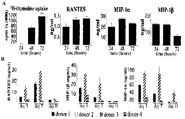
FIGs. 1A and B show kinetics of RANTES, MIP-la, and MIP-IR secretion in
activated PBMCs. (A) PBMCs (1 x 10⁶) were cultured in 1 ml of culture medium
in
the presence of PHA. Cultures were maintained for 72h. Every 24 h, the entire
culture medium was collected and replaced with fresh medium containing PHA.
Supernatants were assayed for chemokine content by ELISA. DNA synthesis was
measured by [3H]thymidine incorporation in PBMCs cultured in parallel under
identical conditions. Results are the mean SD of data obtained from three
different
donors. (B) PBMCs from four donors were cultured in the presence of PHA for 3
days and in the presence of IL-2 afterward. Culture supematants were assayed
for R-
chemokine production by ELISA on days 3, 7, and 11. Values from each donor are
the mean SD of triplicate wells.
FIGs. 2A, B and C show HU treatment of PHA-activated PBMCs results in
increased
levels of secreted P-chemokines. PBMCs were cultured in the presence of PHA
for 3
days and in the presence of IL-2 afterward. HU was added at the indicated
concentrations at the beginning of the experiment and added fresh every time
the
medium was changed. Culture supernatants collected on days 3, 8, and 14 were
assayed for chemokine production levels. Chemokine levels in the supematants
are
expressed as ng/ml (A) and as ng per 106 viable cells (B); cell number was
monitored
by trypan blue exclusion (C). Representative values of one of four
experiments, each
using PBMCs from a different donor, are shown. Values are means SD of
triplicate
wells. *, P < 0.01; #, P < 0.05, compared with untreated control by Student's
t test.
FIGs. 3A, B, C and D show treatment of activated PBMCs with G1 cytostatic
drugs
inducing G1 cell cycle arrest results in increased levels of extracellular P-
chemokines.
PHA-activated PBMCs were cultured in the presence of APH (A), SB (B), OL (C),
or
RC (D) at the indicated concentrations. Cultures were kept for 14 days, with
medium
9
CA 02498934 2005-03-14
WO 2004/024683 PCT/US2003/028697
changes every 3 or 4 days. Culture supernatants were tested for chemokine
content by
ELISA, and cell number was determined by trypan blue staining. Data show day 8
values for both APH and SB and day 3 values for RC and OL. Values are means
SD of triplicate wells. *, P < 0.01; #, P < 0.05, compared with untreated
control by
Student's t test.
FIGs. 4A, B, C, D and E show cell cycle arrest in G1, but not in G2, results
in
increased extracellular levels of P-chemokines. Purified CD8 lymphocytes were
activated by anti-CD3 and IL-2 treatment for 3 days. Activated cells were
cultured in
the presence of IL-2 medium containing HU at the indicated concentrations.
After 24
and 48 h of the addition of HU, cell number was evaluated by trypan blue
staining
(A), newly synthesized DNA was measured by [3H]thymidine incorporation (B),
percentage of cells in S phase was determined by propidium iodide staining
(C), and P
-chemokine levels were determined by ELISA (D). (E) Cell cycle arrest and
chemokine production levels in the presence of nocodazole 48 h after addition
of the
drug. Results are single data values, representative of three independent
experiments
for HU and representative of two independent experiments in the case of
nocodazole.
FIGs. 5 A and B show supernatants collected from PBMCs exposed to HU contain
suppressive factors that markedly inhibit HIV-1 BaL replication, whereas they
only
slightly affect the replication of HIV-1 IIIb. Activated lymphocytes from a
seronegative donor were infected with HIV-1 BaL (A) or HIV-1 IIIb (B).
Infected
cells were cultured in IL-2 culture medium supplemented by 50% with
supernatants
collected from HU-treated PBMCs (CM/HU) or supernatants collected from
untreated
PBMCs (CM/control). In addition, a culture containing 100 M HU in fresh
medium
was included. Virus replication was measured in the culture supernatant on day
7
after infection. Cell viability was assessed by the MTT assay. Data are means
SD
of triplicate wells.
FIGs. 6 shows that antiviral activity of supematants collected from HU-exposed
PBMCs is reversed by neutralizing antibodies against the P-chemokines RANTES,
MIP-1 a, and MIP-1 P. Activated lymphocytes from a seronegative donor were
infected
with HIV-1 BaL. Infected cells were cultured in the presence of supematants
CA 02498934 2005-03-14
WO 2004/024683 PCT/US2003/028697
collected from PBMCs that had been cultured for 7 days in the presence of 100
M
HU (CM/HU). CM/HU was preincubated with a mixture of neutralizing antibodies
(anti-RANTES, anti-MIPla, and anti-MIP1R; indicated as nAb) or an IgG control
before addition to the culture. Fresh medium containing CM/HU and the
correspondent antibodies was added again on day 3 after infection. On day 7,
viral
replication was measured by a p24 assay, and cell viability was assessed by
the MTT
assay. Data are mean values SD of duplicate wells.
FIG. 7 shows the effect of RAPA on proliferation of PBMCs. Purified PBMCs from
normal donors were cultured in the presence of IL-2 and RAPA. On day 7, the
extent
of cell proliferation was measured by the MTT assay. Representative results
obtained
on one of two independent experiments, each using cells from four donors, are
shown.
For each donor, data values are mean SD of three independent wells
FIGs. 8A and B show RAPA increases extracellular P-chemokine levels in PBMC
cultures. (A) Donor PBMCs were cultured in the presence of IL-2 and RAPA for
10
days, at which time supernatants were evaluated for 0-chemokine content by
ELISA
and then cells were also stained for CCR5 expression. Results shown in two
donors
are representative of four experiments using four different donors. *, P <
0.01; #, P <
0.05, compared with untreated control by Student's t test. (B) Effect of RAPA
on
extracellular levels of MIP-1P in cultures of CCR5-null PBMCs. Levels of MIP-
1p
protein in the presence and absence of RAPA were measured in supematants of IL-
2-
stimulated PBMCs from a normal donor and from a donor homozygous for the A32
mutation in the CCR5 gene. Values were obtained on day 10 of culture and are
means
SD of duplicate wells. Results are representative of two independent
experiments,
each using cells from two normal donors.
FIGs. 9 A, B and C show that RAPA inhibits HIV-1 replication in PBMCs, and the
antiviral activity in R5 HIV-1 is greater than in X4 HIV-1. (A) Seven-day RAPA-
treated PBMCs were infected with HIV-1 IIIb or HIV-1 ADA. Infected cells were
cultured in the presence of RAPA for 7 days, at which time virus replication
was
measured by p24 and cell viability was measured by the MTT assay. Results
(means
f SD of triplicate wells) are representative of seven independent experiments,
each on
11
CA 02498934 2005-03-14
WO 2004/024683 PCT/US2003/028697
cells from a different donor. (B) DNase-treated stocks of HIV-1 IlIb and HIV-1
ADA
were used to infect PBMCs that had been treated with or without 100 nM RAPA.
HIV-1 DNA sequences were amplified by PCR in cellular lysates prepared 24 h
after
infection. Amplified PCR products were detected with a radioactive probe. The
symbol "+" indicates presence of RAPA in the PBMC culture before and after
infection; "-" indicates no RAPA treatment. Amplification of P-actin sequences
indicated same amount of cellular DNA among the different cell lysates (data
not
shown). NC denotes PCR negative control. (C) The antiviral activity of low
concentrations of RAPA was investigated in a panel of R5 strains of HIV-1.
Cell
proliferation was assayed on uninfected cells from same donor cultured under
identical conditions. Results (means SD of triplicate wells) are
representative of
three independent experiments, each on different donor cells.
FIG. 10 shows that RAPA inhibits HIV-1 replication in MDMs. Purified monocytes
were cultured for 5 days in the presence of RAPA. On day 5, cells were
infected with
HIV-1 ADA and cultured in the presence of RAPA for 14 additional days. On days
7,
10, and 14 after infection, virus growth was measured by the RT assay. On day
14,
cell viability was determined by MTT. Results (means SD) are representative
of
data obtained in three independent experiments, each using cells from a
different
donor.
FIG 11 shows that RAPA enhances the antiviral activity of the CCR5 antagonist
TAK-779. PBMCs that had been cultured in the absence or presence of RAPA (1,
10,
and 100 nM) for 7 days were infected with HIV-1 ADA in the presence of 0.1 nM
TAK-779. Infected cells were cultured in the presence of RAPA and 0.1 nM TAK-
779. On day 7 after infection, virus production was measured by the p24 assay
in the
culture supernatant. Note the logarithmic scale in the y axis. Data represent
means
SD of triplicate wells. Representative results obtained in one of three
independent
experiments are shown.
FIG. 12 shows that treatment of activated PBMCs with Vitamin E resulted in
increased levels of (3-chemokines.
12
CA 02498934 2005-03-14
WO 2004/024683 PCT/US2003/028697
FIG. 13 shows the effects of Vitamin E(alpha-tocopherol) on HIV-1 production
upon
activation of patient's resting CD4 T cells. Virus production (p24 antigen)
was
measured in the culture supernatants by ELISA on day 14. Log transformation of
p24
values (pg/m/) were plotted. For statistical analysis, p24 negative samples
were
assigned a value of 6 pg/ml, which represents the detection limit of the p24
assay.
Data values for each patient culture (with and without VE) are represented by
symbols.
FIG. 14 shows the results of treatment with 2 mg/day of rapamycin and the
increase of
(3-chemokines (RANTES) in five subjects with a return to baseline at day 28.
(Results
normalized for (3-actin).
FIGs. 15 A and B show that Rapamycin increases extracellular (3-chemokine
levels in
cultured PBMCs. PBMCs from a healthy donor were cultured in the presence of IL-
2
and Rapamycin. On day 7, chemokines content in the supernatant was measured by
ELISA and cell viability was determined by the MTT assay.
DETAILED DESCRIPTION OF PREFERRED EMBODIMENTS
A method of treating a viral infection is meant herein to include
"prophylactic"
treatment or "therapeutic" treatment. A "prophylactic" treatment is a
treatment
administered to a subject who does not exhibit signs of a disease or who
exhibits early
signs of the disease for the purpose of decreasing the risk of developing
pathology
associated with the disease. The term "therapeutic," as used herein, means a
treatment
administered to a subject who exhibits signs of pathology for the purpose of
diminishing or eliminating those signs.
The term "therapeutically effective amount," as used herein means an amount of
compound that is sufficient to provide a beneficial effect to the subject to
which the
compound is administered. A beneficial effect means rendering virus
incompetent for
replication, inhibition of viral replication, inhibition of infection of a
further host cell,
or increasing CD4 T-cell count, for example.
13
CA 02498934 2005-03-14
WO 2004/024683 PCT/US2003/028697
The term "a virally-targeted cell," as used herein, means a cell in which
virus is
present and is infective or potentially infective and includes epithelial
cells, nervous
system cells, T-lymphocytes (activated or resting), macrophage, monocytes,
tissue
dendritic cells or the like.
The term "functional equivalent," as used herein, means that the agent retains
some or
all of the biological activity of the corresponding compound.
The term "functional analog," as used herein means compounds derived from a
particular parent compound by straightforward substitutions that do not result
in a
substantial (i.e. more than 100X) loss in the biological activity of the
parent
compound, where such substitutions are modifications well-known to those
skilled in
the art, e.g., esterification, replacement of hydrogen by halogen, replacement
of alkoxy
by alkyl, replacement of alkyl by alkoxy, etc.
The Invention:
G 1 Phase arresting compounds
The compositions of the present invention may include any G1 phase arresting
agent
that arrests, delays or prolongs cell-cycle activity in the G1 phase and/or Gl-
S
interface of mononuclear cells. G1 phase arresting agents may include, but are
not
limited to, sodium butyrate, aphidicolin, hydroxyurea (HU), olomoucine,
roscovitine,
tocopherols, tocotrienols, rapamycin (RAPA) and/or functional analogs thereof.
The present invention employs a G1 phase arresting compound for administration
to a
subject suffering from a viral infection, wherein the compound prolongs the G1
phase
of the cell cycle of an activated lymphocyte thereby providing an increase
number of
receptor-ligands to reduce replication of the viral infection.
Pharmaceutical Compositions Acceptable Derivatives and Salts
14
CA 02498934 2005-03-14
WO 2004/024683 PCT/US2003/028697
The present invention provides compositions comprising at least one G1 phase
arresting compound and optionally at least one antiviral agent, as well as
methods of
preventing, treating and/or reducing the effects of HIV. The methods comprise
administering said compositions comprising the G1 phase arresting compounds
and
optionally antiviral agents, wherein the two compounds can be administered,
separately, simultaneously, concurrently or consecutively.
Pharmaceutically Acceptable Derivatives and Salts
The term "pharmaceutically acceptable derivative" is used herein to denote any
pharmaceutically or pharmacologically acceptable salt, ester or salt of such
ester of a
compound according to the invention, or any compound which, upon
administration
to the recipient, is capable of providing (directly or indirectly) one ore
more of the
compounds according to the invention, or an antivirally active metabolite or
residue
thereof.
Preferred esters of the Gl phase arresting compounds of the invention include
carboxylic acid esters in which the non-carbonyl moiety of the ester grouping
is
selected from straight or branched chain alkyl. e.g. n-propyl, t-butyl, n-
butyl,
alkoxyalkyl (e.g. methoxymethyl), aralkyl (e.g. benzyl), aryloxyalkyl (e.g.
phenoxymethyl), aryl (e.g. phenyl optionally substituted by halogen, C1_4alkyl
or C1_4
alkoxy or amino); sulfonate esters such as alkyl- or aralkylsulfonyl (e.g.
methanesulfonyl); amino add esters (e.g. L-valyl or L-isoleucyl); and mono-,
di- or
triphosphate esters. The phosphate esters may be further esterified by, for
example, a
C1_20 alcohol or reactive derivative thereof, or by a 2,3-di C2_4 acyl
glycerol.
Pharmaceutically acceptable salts include, without limitation, salts of
organic
carboxylic acids such as acetic, lactic, tartaric, malic, isethionic,
lactobionic, p-
aminobenzoic and succinic adds; organic sulfonic acids such as
methanesulfonic,
ethanesulfonic, benzenesulfonic and p-toluenesulfonic acids and inorganic adds
such
as hydrochloric, sulfuric, phosphoric and sulfamic acids.
Anti-viral compounds
CA 02498934 2005-03-14
WO 2004/024683 PCT/US2003/028697
In one aspect the compositions and methods of the present invention further
comprise
a therapeutically effective amount of at least one antiviral agent, including,
but not
limited to nucleoside RT inhibitors, CCR5 inhibitors/antagonists, viral entry
inhibitors
and functional analogs thereof.
Preferably, the antiviral agent comprises nucleoside RT inhibitors, such as
Zidovudine
(ZDV, AZT), Lamivudine (3TC), Stavudine (d4T), Didanosine (ddl), Zalcitabine
(ddC), Abacavir (ABC), Emirivine (FTC), Tenofovir (TDF), Delaviradine (DLV),
Efavirenz (EFV), Nevirapine (NVP), Fuzeon (T-20), Saquinavir (SQV), Ritonavir
(RTV), Indinavir (IDV), Nelfinavir (NFV), Amprenavir (APV), Lopinavir (LPV),
Atazanavir, Combivir (ZDV/3TC), Kaletra (RTV/LPV), Trizivir (ZDV/3TC/ABC);
CCR5 inhibitors/antagonists, such as SCH-C, SCH-D, PRO 140, TAK 779, TAK-220,
RANTES analogs, AK602, UK-427, 857, monoclonal antibodies;
viral entry inhibitors, such as Fuzeon (T-20), NB-2, NB-64, T-649, T-1249, SCH-
C,
SCH-D, PRO 140, TAK 779, TAK-220, RANTES analogs, AK602, UK-427, 857;
and functional analogs thereof.
Antiviral Therapy
Although current treatment with antiretroviral (ARV) therapy causes
suppression of
HIV replication and results in improvements of immune function, it is limited
by high
costs, toxicities and is difficult to adhere to. Moreover, the chance of
achieving long-
term control of HIV infection with antiretroviral therapy alone seems very
unlikely.
To date, current antiretroviral therapy has been shown to be insufficient to
completely
eradicate HIV from infected individuals and there is no real data that the
amount of
residual virus is decreasing with time on typical antiretroviral therapy.
Further, after
stopping antiretroviral therapy, the viral load can rebound to higher levels
than
pretreatment viral loads (Davey, 1999; Dybul, et al., 2002 and 2001).
16
CA 02498934 2005-03-14
WO 2004/024683 PCT/US2003/028697
Antiretroviral therapy demands stringent adherence to complex dosing regimens.
The
rate of virological failure over a 6-month period of time has been
demonstrated to be
as high as 60% in patients that cannot achieve greater then 95% adherence. The
combination of multiple adverse side effects associated with antiretroviral
therapy and
the availability of this treatment to only 1 in 20 of the estimated 33 million
people
infected world wide has prompted us to reconsider the current strategies for
achieving
the goals of HIV therapy.
Moreover, HIV therapy is now thought to be a life-long process. Therefore, it
is
crucial to develop effective treatments that can be successfully administered
for long
periods of time for the suppression of retroviruses, and in particular, the
prevention
and/or inhibition of HIV. Further, it would be desirable to eliminate, or at
least
minimize, the cytotoxicity associated with the administration of antiviral
agents
otherwise determined to be effective. It is generally recognized that the
toxicity of an
antiviral agent may be avoided or at least minimized by administration of a
reduced
dose of the antiviral agent; however, it is also recognized that the
effectiveness of an
antiviral agent generally decreases as the dose is reduced.
Thus, one embodiment of the present invention provides for reducing the dose
of
antiviral agents while maintaining or reducing viral load by using cyclic
therapy and
introducing the G1 cell cycle agents of the present invention to a dosing
regime for an
HIV infected subject. Specifically, the use of the G1 phase arresting
compounds in
combination with antiviral agents has shown promise to maintain viral
suppression in
a cycle therapy dosing program. By using 50% less medication, side effects
associated with antiretroviral use have been shown to be reduced and adherence
has
shown to be increased. The other obvious impact is on overall cost of
medications,
which will facilitate expanding these drugs throughout the developed world.
By using our insight on the importance of G1 cell cycles in the treatment of
HIV,
manipulation of HIV cellular cycles can be used successfully to lengthen the
off
therapy periods of cyclic therapy. Further, HIV is decreased in the active and
resting
cell compartments. Resting lymphocytes are a major reservoir for HIV and thus
it is
important that antiretroviral therapy be capable of suppressing HIV in both
resting and
17
CA 02498934 2005-03-14
WO 2004/024683 PCT/US2003/028697
activated cells. Resting T cells can be infected by HIV at levels comparable
to that of
activated T cells. However, unlike the activated T cells, the viral DNA is
only
partially transcribed in resting T cells resulting in unintergrated proviral
DNA.
However, this proviral HIV DNA in the resting T cells may constitute a labile
but
inducible reservoir for activation. The importance of the activated cellular
state for
HIV replication coupled with the transient survival of replication competent
unintergrated proviral intermediates raises the possibility of successful
intervention
aimed at both depleting the HIV DNA from the resting cell pool and also
decreasing
the state of cellular activation.
Although attempts using primarily protease inhibitor containing regimens have
failed
to reduce the overall burden of HIV, therapeutic interventions that are
specifically
aimed at preventing the persistence and renewal of the resting cell reservoir
may be
successful but have not yet been pursued. The failure to decrease the overall
HIV
burden on typical antiretroviral therapy may be attributed to the inadequate
activity of
antiviral agents and protease inhibitors in resting cells.
Thus, in one embodiment of the present invention, cyclic therapy is employed
as an
alternative approach designed to increase activity of antiviral agents,
decrease drug
cost and toxicity. Furthermore, since one component of the compositions of the
present invention targets cellular machinery of the host, rather than the
virus, the
present inventors expect that viral resistance to this drug combination
essentially
would not occur.
A cycle antiviral therapy regime could run for about 12 weeks and then a Gl
phase
arresting compound is added or substituted for four weeks. If the viral load
remains
low or approximately constant, the cycles can be altered to reduce the time
period of
each cycle. The time period for consumption of the antiviral drugs can be
reduced, if
augmented with a G 1 phase arresting compound. Furthermore, a time period can
be
introduced that includes no antiviral drugs and only a G1 phase arresting
compound.
This time period wherein no antiviral agents are consumed by the subject,
provides
the biological system of the subject sufficient time to repair or compensate
for the
toxic effects of the antiviral compound.
18
CA 02498934 2008-04-16
A proposed dosing program may include one week of consumption of antiviral
agents
plus a G 1 phase arresting agent (3TC, Tenofovir (Tenofovir has improved
potency in
activated and resting T cells, and SustivaT1~ and GI pbrc aimsting compound
HU),
and then two weeks off of the antiviral agents but the Gi phase arresting
agent is still
consumed by the subject. Patients are monitored for immunological and
virological
parameters as well as the incidence of toxicity and side effects during both
the
treatment period and the interruption period. These cycles of I week on/2
weeks off
of antiretroviral medications will continue for an appropriate treatment
period with
constant reevaluation of viral loads. Obviously, each subject will respond
differently
to such cycles and a physician knowing the dynamics of the HIV infection can
determine the appropriate time period for each cycle.
Methods for Preventing and/or Treating a Viral Infection
The compositions and methods of the present invention can be used to prevent
viral
infection in a subject potentially exposed to the infection. The viral
infections
prevented by using the compositions and methods of the present invention are
preferably retroviral infections, and are more preferably, HIV infections. G1
cell
cycle agents for the prevention of HIV transmission either as single
therapeutic agents
or when used in combination with antiretroviral drugs and HIV vaccines may be
used
in the following settings:
1. Post blood borne exposure;
2. Post sexual exposure;
3. Mother to child transmission resulting from pregnancy, labor, delivery
and through breast milk transmission; and
4. Augmentation of preventive HN vaccine efficacy.
Further, the compositions and methods of the present invention can be used to
treat
HIV viral infections by reducing viral load and replication of the virus.
Still further the compositions and methods of the present invention can be
used in
combination with HIV vaccines to increase the efficacy of a vaccine in a
subject. The
19
CA 02498934 2005-03-14
WO 2004/024683 PCT/US2003/028697
methods comprise administering to a subject an immunizingly effective amount
of one
or more antigens against which an immune response is desired in the subject in
conjunction with an amount of a G1 phase arresting agent effective to enhance
the
immune response against the antigen by increasing the levels of chemokines in
the
subject. In one aspect, the GI phase arresting agent is administered to the
subject
concurrently with (e.g., in the same composition with) the antigen or antigens
against
which an immune response is desired.
In another, aspect, the G1 phase arresting agent is administered either before
or after
the administration of one or more antigens against which immunity is desired
in the
subject, but is administered within such time that the G1 phase arresting
agent
enhances the immune response to the one or more antigens. For example, the G1
phase arresting agent is suitably administered during the time that the
subject mounts
an immune response against the administered one or more antigens. The G1 phase
arresting agent is preferably administered within 30 minutes, 1 hour, 5 hours,
10
hours, 1 day, and/or 2 days of (preferably, after) administration of the one
or more
antigens against which immunity is desired.
In yet another aspect, the G1 phase arresting agent is suitably administered
for an
extended period of time after the vaccine is administered as a chemo-
prophylactic
agent that maximizes the effectiveness and long-term protection of the
vaccine.
The present invention further provides compositions comprising an immunizingly
effective amount of one or more antigens and an amount of at least one G1
phase
arresting agent effective to induce increased levels of chemokines.
Doses to be administered are variable according to the G1 phase arresting
agent, the
antiviral agent, the treatment period, frequency of administration, the host,
and the
nature and severity of the infection. The dose can be determined by one of
skilled in
the art without an undue amount of experimentation.
The compositions of the invention are administered in substantially non-toxic
dosage
concentrations sufficient to ensure the release of a sufficient dosage unit of
the present
CA 02498934 2005-03-14
WO 2004/024683 PCT/US2003/028697
combination into the patient to provide the desired inhibition of the HIV
virus. The
actual dosage administered will be determined by physical and physiological
factors
such as age, body weight, severity of condition, and/or clinical history of
the patient.
The active ingredients are ideally administered to achieve in vivo plasma
concentrations of an antiviral agent of about 0.01 uM to about 100 uM, more
preferably about 0.1 to 10 uM, and most preferably about 1-5 uM, and of a Gl
phase
arresting agent of about 1 uM-25uM, more preferably about 2-20 uM, and most
preferably about 5-10 uM.
For example, in the treatment of HIV-positive and AIDS patients, the methods
of the
present invention may use compositions to provide from about 0.005-500 mg/kg
body
weight/day of an antiviral agent, more preferably from about 0.1-200
mg/kg/day, and
most preferably 1-50 mg/kg/day; and from about 0.01-1000 mg/kg body weight/day
of
a G 1 phase arresting agent, more preferably from about 0.001-1000 mg/kg/day,
or
most preferably from about 0.5-50 mg/kg/day. Particular unit dosages of a G1
phase
arresting agent and an antiviral agent of the present invention include 50 mg,
100 mg,
200 mg, 500 mg, and 1000 mg amounts, for example, formulated separately, or
together as discussed infra.
It will be understood, however, that dosage levels that deviate from the
ranges
provided may also be suitable in the treatment of a given viral infection.
Therapeutic efficacy of the G1 phase arresting compounds can be determined by
standard pharmaceutical procedures in cell cultures or experimental animals,
e.g., for
determining The LD50 (The Dose Lethal To 50% Of The Population) and The ED50
(the dose therapeutically effective in 50% of the population). The dose ratio
between
toxic and therapeutic effects is the therapeutic index and it can be expressed
as the
ratio LD50/ED50. Compounds, which exhibit large therapeutic indexes, are
preferred. The data obtained from the cell culture assays and animal studies
can be
used in formulating a range of dosage for use in humans. The dosage of such
compounds lies preferably within a range of circulating concentrations that
include the
ED50 with little or no toxicity. The dosage may vary within this range
depending
upon the dosage form employed and the route of administration utilized. For
any
21
CA 02498934 2005-03-14
WO 2004/024683 PCT/US2003/028697
compound used in the method of the invention, the therapeutically effective
dose can
be estimated initially from cell culture assays. A dose may be formulated in
animal
models to achieve a circulating plasma concentration range that includes the
IC50
(i.e., the concentration of the test compound which achieves a half-maximal
inhibition
of symptoms) as determined in cell culture. Such information can be used to
more
accurately determine useful doses in humans. Levels in plasma may be measured,
for
example, by high performance liquid chromatography.
The desired dose is preferably presented as two, three, four, five, six or
more sub-
doses administered at appropriate intervals throughout the day. These sub-
doses may
be administered in unit dosage forms, for example, containing 0.01 to 1000 mg,
preferably 1 mg to 50 mg, depending on the number of sub-doses, of the GI
phase
arresting compound per unit dosage form.
While it is possible for the specific Gl phase arresting compound and
antiviral agent
to be administered individually, either sequentially or simultaneously, it is
preferable
to present them together, as combined in a pharmaceutical composition.
The compositions of the present invention may comprise both the above-
discussed
ingredients, together with one or more acceptable carriers thereof and
optionally other
therapeutic agents. Each carrier must be "pharmaceutically acceptable " in the
sense
of being compatible with the other ingredients of the formulation and not
injurious to
the subject.
The present invention provides a method for the treatment or prophylaxis of a
viral
infection such as retroviral infections which may be treated or prevented in
accordance with the invention include human retroviral infections such as
human
immunodeficiency virus (HIV), HIV-1, and HIV-2. The specific G1 phase
arresting
compounds, compositions and methods according to the invention are especially
useful for the treatment of AIDS and related HIV-positive conditions. The
compounds of the present invention are also useful for the treatment of
asymptomatic
infections or diseases in humans caused by or associated with human
retroviruses.
22
CA 02498934 2005-03-14
WO 2004/024683 PCT/US2003/028697
The therapeutic compositions according to the present invention may be
employed in
combination with other-therapeutic agents for the treatment of viral
infections or
conditions. Examples of such further therapeutic agents include agents that
are
effective for the treatment of viral infections or associated conditions such
as
immunomodulatory agents such as thymosin, ribonucleotide reductase inhibitors
such
as 2-acetylpyridine 5-[(2-chloroanilino) thiocarbonyl) thiocarbonohydrazone,
interferons such as alpha -interferon, 1- beta -D-arabinofuranosyl-5-(1-
propynyl)uracil, 3'-azido-3'-deoxythymidine, ribavirin and phosphonoformic
acid.
Routes of Administration
The compositions according to the present invention, may be administered for
therapy
by any suitable route including oral, rectal, nasal, topical (including
transdermal,
buccal and sublingual), vaginal and parenteral (including subcutaneous,
intramuscular,
intravenous and intradermal). It will be appreciated that the preferred route
will vary
with the condition and age of the recipient, the nature of the infection and
the chosen
active ingredient.
Pharmaceutical formulations of the present invention include those suitable
for oral,
rectal, nasal, topical (including transdermal, buccal and sublingual), vaginal
or
parenteral (including subcutaneous, intramuscular, intravenous and
intradermal)
administration.
The formulations may conveniently be presented in unit dosage form and may be
prepared by methods known in the art of pharmacy. Such methods include the
step of
bringing into association the Gl phase arresting compound and optionally an
antiviral
agent with the carrier. The carrier optionally comprises one or more accessory
ingredients. In general, the formulations are prepared by uniformly and
intimately
bringing into association the separate ingredients with liquid carriers or
finely divided
solid carriers or both, and then if necessary shaping the product.
Compositions suitable for transdermal administration may be presented as
discrete
patches adapted to remain in intimate contact with the epidermis of the
recipient for a
23
CA 02498934 2005-03-14
WO 2004/024683 PCT/US2003/028697
prolonged period of time. Such patches suitably contain the Gl phase arresting
compound and optionally an antiviral agent: 1) in an optionally buffered,
aqueous
solution; or 2) dissolved and/or dispersed in an adhesive; or 3) dispersed in
a polymer.
A suitable concentration of each synergistic ingredient is about 1% to 25%,
preferably
about 5 to 15%.
Formulations of the present invention suitable for oral administration may be
presented as discrete units such as capsules, caches or tablets, each
containing a
predetermined amount of the ingredients; as a powder or granules; as a
solution or a
suspension in an aqueous or non-aqueous liquid; or as an oil-in-water liquid
emulsion
or a water-in-oil liquid emulsion.
A tablet may be made by compression or molding, optionally with one or more
accessory ingredients. Compressed tablets may be prepared by compressing in a
suitable machine the Gl phase arresting compound and antiviral agent in a free-
flowing form such as a powder or granules, optionally mixed with a binder
(e.g.
povidone, gelatin, hydroxypropylmethyl cellulose), lubricant, inert diluent,
preservatives, disintegrant (e.g. sodium starch glycollate, cross-linked
povidone,
cross-linked sodium carboxymethyl cellulose) surface-active or dispersing
agent.
Molded tablets may be made by molding in a suitable machine a mixture of the
powdered compound moistened with an inert liquid diluent. The tablets may
optionally be coated or scored and may be formulated so as to provide slow or
controlled release of one or more of the synergistic ingredients therein
using, for
example, hydroxypropylmethyl cellulose in varying proportions to provide the
desired
release profile. Tablets may optionally be provided with an enteric coating,
to provide
release in parts of the gut other than the stomach.
Formulations suitable for topical administration in the mouth include lozenges
comprising one or more of the G1 phase arresting compounds and optionally an
antiviral agent in a flavored basis, usually sucrose or acacia; pastilles
comprising one
or more of the ingredients in an inert basis such as gelatin and glycerin, or
sucrose and
acacia; and mouthwashes comprising the one or more of the ingredients in a
suitable
liquid carrier.
24
CA 02498934 2005-03-14
WO 2004/024683 PCT/US2003/028697
Formulations for rectal administration may be presented as a suppository with
a
suitable base comprising, for example, cocoa butter or a salicylate.
Formulations suitable for vaginal administration may be presented as
pessaries,
tampons, creams, gels, pastes, foams or spray formulations containing, in
addition to
the one or more of the compounds of the present invention, such carriers as
are known
in the art to be appropriate.
Formulations suitable for parenteral administration include aqueous and non-
aqueous
isotonic sterile injection solutions which may contain anti-oxidants, buffers,
bacteriostats and solutes which render the formulation isotonic with the blood
of the
intended recipient; and aqueous and non-aqueous sterile suspensions which may
include suspending agents and thickening agents. The formulations may be
presented
in unit-dose or multidose sealed containers, for example, ampules and vials,
and may
be stored in a freeze-dried (lyophilized) condition requiring only the
addition of the
sterile liquid carrier, for example water for injections, immediately prior to
use.
Extemporaneous injection solutions and suspensions may be prepared from
sterile
powders, granules and tablets of the kind previously described.
For a perinatal subject, the drug combination of the present invention may be,
for
example, administered orally after 36 weeks of pregnancy and continued through
delivery. Interventions around the time of late gestation and delivery (when
the
majority of transmissions are thought to occur) are most efficacious.
In addition to the compositions described previously, the compounds may also
be
formulated as a depot preparation. Such long acting formulations may be
administered by implantation (for example subcutaneously or intramuscularly)
or by
intramuscular injection. Thus, for example, the compounds may be formulated
with
suitable polymeric or hydrophobic materials (for example as an emulsion in an
acceptable oil) or ion exchange resins, or as sparingly soluble derivatives,
for
example, as a sparingly soluble salt. Other suitable delivery systems include
microspheres that offer the possibility of local noninvasive delivery of drugs
over an
CA 02498934 2005-03-14
WO 2004/024683 PCT/US2003/028697
extended period of time. The administered therapeutic is slowly released from
these
microspheres and taken up by surrounding tissue cells (e.g. endothelial
cells).
The compositions may, if desired, be presented in a pack or dispenser device,
which
may contain one or more unit dosage forms containing the active ingredient.
The
pack may for example comprise metal or plastic foil, such as a blister pack.
The pack
or dispenser device may be accompanied by instructions for administration.
Suitable G1 cell cycle agents, can be used in HIV treatment strategies that
allow for
continued viral suppression to be maintained with less dependence on
combination
antiretroviral (ARV) therapy. The current goal of ARV is to obtain viral
suppression
as low as possible for as long as possible. Requiring less frequent dosing or
a
decreased quantity of ARV to control viral suppression directly addresses the
problems, set forth below, associated with achieving the current goals of
antiretroviral
therapy including:
1. Current regimens of HAART are cumbersome and complicated and require
sustained tolerance and strict adherence to 3 or more drugs.
2. Long term tight adherence may be impossible for most patients.
3. Long term tolerance to accumulating medication toxicities may be impossible
for most patients.
4. Current treatment guidelines for HIV infection recommend a relatively late
initiation of HAART because of the inability to eradicate the infection with
HARRT alone and the risk of drug-related side-effects, including serious
metabolic syndromes.
5. Some patients who have not been treated until later stages of the disease
will
have a high level of viral load, which could increase the risk of transmission
and cause a public health problem.
6. Lastly, the vast majority of HIV infected people worldwide have no access
to
antiretroviral drugs due mostly to cost.
By incorporating G1 cell cycle agents into therapeutic approaches with the
focus
shifted towards maintaining long term viral control, with less complex, less
toxic, and
more affordable regimens, that can be applicable throughout the world. The
present
26
CA 02498934 2005-03-14
WO 2004/024683 PCT/US2003/028697
invention that targets the G1 cellular cycle to increase extracellular levels
of
chemokines can be used safely and successfully to maintain viral suppression
in
chronic HIV-1 infection without the need of continuous therapy with multiple
antiretroviral drugs. These results have a positive impact on cost, side
effects, and
availability of HIV therapy.
The present invention is further illustrated by the following examples that
should not
be construed as limiting in any way.
The practice of the present invention will employ, unless otherwise indicated,
conventional techniques of cell biology, cell culture, molecular biology,
transgenic
biology, microbiology, recombinant DNA, and immunology, which are within the
skill of the art. Such techniques are explained fully in the literature. See,
for
example, Molecular Cloning A Laboratory Manual, 2d Ed., ed. by Sambrook,
Fritsch
and Maniatis (Cold Spring Harbor Laboratory Press: 1989); DNA Cloning, Volumes
I
and II (D. N. Glover ed., 1985); Oligonucleotide Synthesis (M. J. Gait ed.,
1984);
Mullis et al. U.S. Patent No: 4,683,195; Nucleic Acid Hybridization(B. D.
Hames &
S. J. Higgins eds. 1984); Transcription And Translation (B. D. Hames & S. J.
Higgins
eds. 1984); Culture Of Animal Cells (R. I. Freshney, Alan R. Liss, Inc.,
1987);
Immobilized Cells And Enzymes (IRL Press, 1986); B. Perbal, A Practical Guide
To
Molecular Cloning (1984); the treatise, Methods In Enzymology (Academic Press,
Inc., N.Y.); Gene Transfer Vectors For Mammalian Cells (J. H. Miller and M. P.
Calos eds., 1987, Cold Spring Harbor Laboratory); Methods In Enzymology, Vols.
154 and 155 (Wu et al. eds.), Immunochemical Methods In Cell And Molecular
Biology (Mayer and Walker, eds., Academic Press, London, 1987); Handbook Of
Experimental Immunology, Volumes I-IV (D. M. Weir and C. C. Blackwell, eds.,
1986); Manipulating the Mouse Embryo, (Cold Spring Harbor Laboratory Press,
Cold
Spring Harbor, N.Y., 1986).
EXAMPLES
Methods and Materials
27
CA 02498934 2008-04-16
Tissue Culture.
PBMCs were sEparsted from whole blood of HIV-1 seronegative donors by density
centrifugation with FicollTM Histopaque (Sigma). Cells were cultured in
complete
medium consisting of RPMI medium 1640 supplemented with 10'/o heat-inactivated
FBS, 2 mM glutamine, and penicillin/streptomycin (Invitrogen). In some
experiments, purified CD8+ lymphocytes obtained by negative selection using
the
Human CD8+ T Cell Enrichment Mixture (StemCell Technologies, Vancouver) were
used. Cell purity measured by flow cytometry was >80% among different donor
purifications.
Cells were activated by culture for 72 h under three different conditions:
phytohemagglutinin (PHA, 2.5 ug/ml; Roche, Gipf=Oberfrick, Switzerland), anti-
CD3
antibody (1 g/ml; Coulter) plus 100 units/ml recombinant IL-2 (Roche), or
staphylococcal enterotoxin B at 0.03 g/ml (Sigma). Activated cells were
cultured in
complete medium supplemented with recombinant IL-2 (100 units/mI), and medium
was changed every 3 or 4 days.
Cell proliferation was measured by the trypan blue staining viability test,
[3H]thymidine incorporation in DNA, and the 3-(4,5-dimethylthiazol-2-yl)-2,5-
diphenyl tetrazolium bromide (MTT) assay (Roche).
Measurement of O-Chemokine Levels and Assessment of Cell Cycle Arrest.
The impact of cell cycle arrest in P-chemokine levels was evaluated by
measuring
chemokine levels in supematants of cell cultures containing compounds known to
cause cell cycle arrest. Levels of the P-chemokines RANTES, MIP-la, and MIP-1p
were measured by using commercial ELISA kits (R &D Systems). Cell cycle arrest
in
G.1 was induced by culturing of the cells in the presence of aphidicolin
(APH), sodium
butyrate (SB), hydroxyurea (HU), roscovitine (RC), or olomoucine (OL). Arrest
in
late S phase was induced by culture of cells in the presence of resveratrol.
G2 cycle
arrest was induced by the compounds nocodazole and Colcemid. All compounds
were
purchased from Sigma except RC and OL, which were from Calbiochem. Arrest of
28
CA 02498934 2005-03-14
WO 2004/024683 PCT/US2003/028697
cell cycle progression in the presence of G1 cytostatic agents was measured by
propidium iodide staining followed by fluorescence-activated cell sorter
(FACS)
analysis (Noguchi, et al., 1991). This method may be used to test any compound
for
ability to arrest the Gi phase in a cell cycle.
Assessment of HIV- 1 -Suppressive Activity in Supernatants Collected from HU-
Treated PBMCs.
The antiviral activity of the supematants collected from cultures of PBMCs
that had
been exposed to 100 M HU for 7 days [supernatants referred to as conditioned
medium (CM)] was evaluated in PBMCs infected with HIV-1 BaL and HIV-1 IIIb.
Briefly, PHA-activated PBMCs were infected with each virus at 100 tissue
culture
50% infective dose units (TCID50)/106 PBMCs or 10 TCID50/106 PBMCs for 2 h at
37 C. Infected cells were cultured in IL-2 medium alone, IL-2 medium with 100
M
HU, IL-2 medium containing 50% supernatant from HU-treated PBMCs (CM/HU), or
IL-2 medium containing 50% supernatant from control-treated PBMCs
(CM/control).
On day 3 after infection, culture medium was replaced with fresh medium of the
same
kind as on day 1. Viral growth (measured by p24 levels in the supernatant) and
cell
viability (assayed by MTT) were determined on day 7 after infection. To
determine
the role of the P-chemokines RANTES, MIP-la, and MIP-1R in the antiviral
activity
found in supernatants of HU-exposed PBMCs, the antiviral activities of such
supernatants were evaluated in the presence of neutralizing antibodies against
all three
chemokines as described (Kay, et al., 1983).
Example 1
Kinetics of P-Chemokine Secretion on Activated PBMCs.
To determine the kinetics of P-chemokine secretion in relation to DNA
synthesis on
cellular activation, PBMCs were activated by PHA treatment for 72 h. At 24 h
after
plating the cells, the entire supernatant was collected, and fresh medium
containing
PHA was added to the culture. Twenty-four hours later (48 h from the time
cells were
plated), supernatants again were collected and PHA medium was added.
Supernatants
29
CA 02498934 2005-03-14
WO 2004/024683 PCT/US2003/028697
were collected for the last time after 72 h. Thus, this experiment measured
the amount
of chemokine released to the culture medium during each 24-h interval, not the
continuous accumulation of chemokine over 48 or 72 h of exposure to PHA.
Cellular
DNA synthesis was measured by assaying [3H]thymidine incorporation in parallel
wells at 24, 48, and 72 h after plating of the cells. Results are shown in
Fig. 1A. At
24 h after the beginning of PHA treatment, chemokine protein concentration in
the
culture supernatants was 850, 17,200, and 13,300 pg/ml for RANTES, MIP-la.,
and
MIP-1R, respectively (average values from three different donors). Slightly
increased
values in RANTES and MIP-la secretion were detected during the 24- to 48-h
culture
period, whereas the MIP-1R values remained constant. During the 48- to 72-h
period,
levels of RANTES and MIP-la secretion were unchanged, whereas the secretion of
MIP-1R decreased. Synthesis of cellular DNA was almost undetectable at 24 h
(645
cpm) and increased considerably by 48 and 72 h (62,474 and 106,402 cpm,
respectively). These data show that considerable protein amounts of MIP-la,
MIP-1P,
and, to a lower extent, RANTES are present in the culture supernatant of PBMCs
at 24
h after activation, a time at which cellular DNA synthesis is minimal.
The profile of P-chemokine secretion next was evaluated in cultures of
activated
PBMCs maintained in the presence of IL-2 for several days. PBMCs were
activated
with PHA for 3 days and then cultured in the presence of IL-2 for 8 additional
days.
At days 3, 7, and 11, chemokine content in the culture fluid was measured
(Fig. 1B).
Although variability was observed among different donors, RANTES levels
usually
reached a peak on day 7 after activation. In contrast, MIP-1 oc and MIP-1 R
levels
peaked on day 3 or day 7, depending on the donor. Levels of all three
chemokines
were low by day 11. Taken together, these data indicate that secretion of the
R-
chemokines by PBMCs in response to activation starts before lymphocytes enter
the
DNA synthesis phase of the cell cycle (S phase), reaches a peak by day 3 or 7,
and
then declines to low levels.
Example 2
Treatment of PBMC Cultures with Compounds That Arrest the Cell Cycle in G1
Results in Increased Levels of Secreted P-Chemokines.
CA 02498934 2005-03-14
WO 2004/024683 PCT/US2003/028697
Because the previous experiments indicated that P-chemokine secretion by
activated
PBMCs begins before DNA synthesis occurs, it was further tested to determine
whether delay of entry in the S phase of the cell cycle results in an overall
increase in
chemokine levels. To this end, P-chemokine levels (production or availability)
by
activated PBMCs cultured in the presence of HU was investigated. HU is a G 1
cytostatic drug that, by depleting intracellular nucleotide pools, arrests
cell cycle
progression in late G1 (Lori, et al., 1994). Fig. 2 shows chemokine levels by
activated
PBMCs cultured in the presence of different concentrations of HU for 14 days.
HU
treatment resulted in increased concentrations (ng/ml) of RANTES, MIP-la, and
MIP-
1P in the culture supernatants in a dose-dependent manner (Fig. 2A). In the
representative experiment depicted in Fig. 2, day 8 chemokine levels in
cultures
containing 100 M HU were increased 3.4-fold for RANTES, 5.4-fold for MIP-loc,
and 4-fold for MIP-1R compared with the untreated control. Because HU inhibits
lymphocyte proliferation, chemokine values also were expressed as chemokine
amount per viable cell. As expected, cell numbers were lower in the presence
of the
drug (Fig. 2C). Chemokine levels expressed as ng per 106 cells indicated
increases of
16.2-, 25.4-, and 18.4-fold for RANTES, MIP-la., and MIP-1R, respectively, in
the
presence of 100 M HU (Fig. 2B). Similar increases were observed in PBMC
cultures from the other three donors, and the increases were evident when
chemokine
values were expressed either as ng/ml or as ng per 106 viable cells (data not
shown).
Similarly, HU treatment increased chemokine levels in PBMCs that had been
activated by cross linking of the T cell antigen receptor/CD3 complex with
anti-CD3
antibodies or by occupancy of the T cell antigen receptor with the super
antigen
staphylococcal enterotoxin B (data not shown).
Example 3
Having demonstrated that HU treatment of activated PBMCs results in increased
chemokine levels, observations were extended to other G1 cytostatic agents
that, as
does HU, arrest cell cycle progression before DNA synthesis occurs. The agents
evaluated were SB, APH, RC, and OL. SB and APH arrest the cell cycle in early
and
31
CA 02498934 2005-03-14
WO 2004/024683 PCT/US2003/028697
late G1, respectively (Korin, et al., 1998; Koostra, et al., 2000). RC and OL
are
purine-derivative drugs that arrest cell cycle progression in late Gl through
inhibition
of cyclin-dependent kinases (CDKs) (Gray, et al., 1999). Fig. 3A shows
chemokine
levels produced by activated PBMCs cultured in the presence of APH. The number
of
viable cells and chemokine levels are depicted. Increased chemokine values
were
observed at 0.5 M APH, a drug concentration that exhibited G1 cytostatic
effects as
manifested by reduced cell proliferation. Fig. 3B shows chemokine levels
produced
by activated PBMCs cultured in the presence of SB. As was the case with APH,
increased chemokine levels were detected at drug concentrations of SB exerting
G1
cytostatic activity. Similarly, exposure of activated PBMCs to the CDK
inhibitors RC
and OL resulted in increased chemokine levels at Gl cytostatic concentrations
of the
drugs (Fig. 3C and data not shown). These experiments indicate that treatment
of
activated PBMCs with compounds that arrest the cell cycle in the G1 phase
results in
increased levels of extracellular R-chemokines.
Example 4
Up-Regulation of P-Chemokine Levels in Supernatants of CD8 Lymphocyte Cultures
Is Specific to Cell Cycle Arrest in G1.
In the experiments described thus far, total PBMCs had been used. To
demonstrate
that arrest of the cell cycle in CD8 lymphocytes (the main cell type producer
of the
anti-HIV chemokines) results in increased chemokine levels, chemokine
production
by purified CD8 lymphocytes exposed to HU was evaluated next. Negatively
selected
CD8+ lymphocytes were activated by anti-CD3 plus IL-2 treatment for 3 days.
Activated cells were cultured in the presence of IL-2 and HU (100 and 200 M)
for 24
or 48 h, time points at which cell proliferation and supernatant chemokine
levels were
assayed. Cell proliferation was determined by trypan blue staining,
[3H]thymidine
incorporation in DNA, and percentage of cells in S phase as assessed by
propidium
iodide staining (Fig. 4). HU cytostatic effects were evident after 48 h of
exposure to
the drug because both cell number and thymidine incorporation doubled between
24
and 48 h in the absence of HU, whereas they remained constant or decreased in
the
presence of the drug. Similarly, the percentage of cells in S phase increased
in the
32
CA 02498934 2005-03-14
WO 2004/024683 PCT/US2003/028697
absence of HU, whereas it decreased in its presence. CD8+ lymphocyte cycle
arrest by
HU resulted in increased levels of RANTES, MIP-la, and MIP-1R after 24 and 48
h of
exposure to the drug. At 48 h, levels of RANTES, MIP-la, and MIP-1R increased
by
1.9-, 3.7-, and 4.7-fold, respectively, in the presence of 100 M HU. Slightly
lower
increases in chemokine production levels were found in CD8 cells exposed to
200 M
HU for 48 h, a drug concentration that resulted in a slightly lower number of
viable
cells shown by trypan blue staining in two of three donors examined (Fig. 4
and data
not shown). In summary, these data demonstrate that arrest of cell cycle
progression
in CD8+ lymphocytes by HU treatment results in increased levels of RANTES, MIP-
la, and MIP-1R.
To investigate whether increased levels of supernatant chemokines upon cell
cycle
arrest are cell cycle phase-specific, the effect of G2 arrest in chemokine
production
next was evaluated. Activated CD8 lymphocytes were arrested in G2 by treatment
with 0.01 g/mi nocodazole (Gualberto, et al., 1998). As shown in Fig. 4E,
treatment
of activated CD8 cells with nocodazole for 48 h resulted in accumulation of
cells in G2
compared with the untreated control. However, chemokine content in treated
cultures
was lower than in the untreated controls. The same results were obtained on
induction
of GZ cycle arrest by Colcemid (data not shown). Similarly, cell cycle arrest
in late S
by 10 M resveratrol resulted in chemokine levels that were lower than the
untreated
control (data not shown). Together, these data suggest that increased
chemokine
levels are specific to cell cycle arrest in G I.
Example 5
Supernatants Collected from PBMCs Arrested in Gl Inhibit HIV-1 BaL
Replication.
To determine whether the augmented chemokine levels found upon G1 cell cycle
arrest possess any antiviral activity, supernatants were harvested from
cultures of
activated PBMCs that had been exposed to 100 M HU for 7 days. These culture
supernatants were referred to as CM/HU. Culture supernatants from activated
PBMCs
cultured under the same conditions in the absence of HU (CM/control) also were
harvested. Supernatants were harvested on day 7 because previous experiments
(Fig.
33
CA 02498934 2005-03-14
WO 2004/024683 PCT/US2003/028697
2) indicated that high levels of RANTES, MIP-la., and MIP-IR are present at
this time
point.
The antiviral activity of the CM was evaluated in the infection of PBMCs with
HIV-1
BaL (a prototype R5-using virus) and HIV-1 IIIb (a prototype X4-using virus).
Activated PBMCs from two seronegative donors were infected with each virus,
and
the infected cells were cultured in medium supplemented by 50% with CM/HU or
CM/control. An additional culture was set up by culturing infected cells in
fresh
medium supplemented with 100 M HU, the same concentration of HU as the one
present in the CM/HU medium. Virus replication and cell viability were
measured on
day 7 after infection. Results obtained in one of the donors are shown in
Figs. 5 A and
B. Culture of infected PBMCs in the presence of CM/HU fluid suppressed HIV-1
BaL
replication by 85% compared with the untreated control, whereas the CM/control
fluid
reduced virus replication by 24%. The control containing HU at the same
- concentration as the CM/HU inhibited HIV-1 BaL replication by 8%. Cell
viability
(MTT assay) was not affected by the addition of CM to the cultures. In
contrast to the
results obtained with HIV-1 BaL, the CM/HU and CM/control fluids suppressed
HIV-
1 IIIb by 22% and 10%, respectively. These data indicate that activated PBMCs
grown in the presence of HU for several days release factors that strongly
inhibit HIV-
1 BaL replication, whereas they have a much lesser effect on the replication
of HIV-1
IIIb. In addition, these results suggest that factors present in the
supernatants, but not
HU per se, are responsible for the inhibition of HIV-1 BaL.
Example 6
The Antiviral Activity Present in Supernatants of Lymphocytes Arrested in G1
Phase
by HU Treatment Is Due to RANTES, MIP-loc., and MIP-1P.
The above results demonstrated selective inhibition of the R5-using virus HIV-
1 BaL
by supernatants collected from HU-treated PBMCs. In addition, these results
suggested that the P-chemokines RANTES, MIP-lac, and MIP-1R were the likely
suppressive factors accounting for viral inhibition. To confirm that an
increase in R-
chemokine levels in the CM was responsible for the observed inhibition of HIV-
1 BaL
34
CA 02498934 2005-03-14
WO 2004/024683 PCT/US2003/028697
replication, the antiviral activity of the CM in the presence of a mixture of
neutralizing
antibodies to all three chemokines was assessed. As can be seen in the
experiment
depicted in Fig.6, CM/HU fluid inhibited HIV-1 BaL replication by ;::~' 60%.
However,
the antiviral activity of CM/HU was only ;::f 10% in the presence of the
neutralizing
antibodies mixture. An IgG antibody control did not affect the antiviral
activity of the
CM/HU fluid. In a different experiment, using cells from a different donor,
the
neutralizing antibodies mixture similarly abrogated the antiviral activity of
the
CM/HU supernatant (data not shown). These data demonstrate that the antiviral
activity found in culture supernatants of HU-treated PBMCs primarily is due to
the
presence of the P-chemokines RANTES, MIP-la, and MIP-1P.
The above-described experiments confirmed the presence of high levels of P-
chemokines in supernatants of PBMC cultures 24 h after activation, a time at
which
cellular DNA synthesis could not be detected (Fig. lA). By arresting cell
cycle
progression of PHA-activated PBMCs in Gl phase by treatment with G1 cytostatic
drugs, increased levels of RANTES, MIP-1a, and MIP-1P were found in the
culture
supernatants. Increased levels of P-chemokines were specific to compounds that
caused cell cycle arrest in G1, and the arrest of the cell cycle in late S (by
resveratrol)
or in G2 (by nocodazole or Colcemid) did not result in such increases. The Gl-
arresting agents that in the present study demonstrated up-regulation of R-
chemokine
levels were SB, HU, RC, and OL.
HU then was selected for additional experiments as a representative agent.
Simultaneous analyses of chemokine production levels and induction of G1
arrest by
HU in purified CD8+ lymphocytes (monitored by cell number, thymidine
incorporation, and percentage of cells in S phase) confirmed the results
obtained in
total PBMCs.
Supernatants collected from cultures of PBMCs (uninfected subjects) that had
been
exposed to HU for several days, referred to as CM in our experiments, were
able to
markedly suppress the replication of HIV-1 BaL in PBMCs. A mixture of
neutralizing
antibodies against all three chemokines abrogated the anti-HIV-1 BaL activity
of the
CM, thus demonstrating that the P-chemokines were responsible for the
antiviral
CA 02498934 2005-03-14
WO 2004/024683 PCT/US2003/028697
effect. When HU was added to fresh medium at the same concentration as in the
CM,
an only minor antiviral effect (<10%) was found in the replication of HIV-1
BaL.
These data suggested that the antiviral effect observed was not due to
inhibition of the
virus reverse transcription step by HU-mediated depletion of nucleotide pools.
Taking our results together, we have described an approach to increase the
concentration of the CCR5 ligands RANTES, MIP-1a, and MIP-1P in cultures of
PBMCs or CD8+ lymphocytes. This approach involves the transient arrest of
activated cells in the G1 phase of the cell cycle by using low concentrations
of G1
cytostatic agents.
In summary, induction of anti-HIV chemokines by G1 cytostatic drugs that, by
themselves, potentiate the antiviral activities of anti-HIV drugs offers an
additional
strategy with which to control replication of HIV-1. This may be a
particularly
attractive strategy with which to inhibit the virus in African countries in
which subtype
C is prevalent, because these viruses use CCR5 and not CXCR4 receptors (Ping,
et al.,
1999).
Further experiments were conducted to show that Rapamycin, also a GI phase
arresting agent causes accumulation of anti-HIV (3-chemokines.
Methods and Materials
Cell Culture and Flow Cytometry.
Cultures of peripheral blood mononuclear cells (PBMCs) and monocyte-derived
macrophages (MDMs) were performed on normal donors as described (Poli, et al.
1993, Pemo, et al., 1993). PBMCs were maintained in the presence of 100
units/ml
rhIL-2 (Roche Molecular Biochemicals). Cell viability was determined by Trypan
blue staining or by the MTT assay (Roche Molecular Biochemicals). RAPA was
purchased from Calbiochem. The CCR5 antagonist TAK-779 was obtained from the
National Institutes of Health AIDS Research and Reference Reagent Program
(Rockville, MD).
36
CA 02498934 2005-03-14
WO 2004/024683 PCT/US2003/028697
CCR5 surface expression was measured on PBMCs cultured in the presence of IL-2
for 7-10 days. Staining was done as described (Lane, et al. 1999), but using
CCR5
mAb 182 (R & D Systems). Background staining was determined by adding an
isotype-matched control (IgG2b, R&D Systems) instead of the anti-CCR5 mAb.
Data
were acquired by using a FACS Calibur flow cytometer (BD Biosciences) and
analyzed by using FLOWJO (Tree Star, San Carlos, CA).
Levels of the P-chemokines MIP-1 a, MIP-1 P, and RANTES were measured in
culture
supernatants by using ELISA kits (R & D Systems).
Infectivity Assays.
The following viruses were used in infection experiments, including HIV-1
IIIb, HIV-
1 ADA, HIV-1 BaL, HIV-1 JRFL, HIV-1 JRCSF, and HIV-1 SF162. HIV-1 IIIb is a
T cell line-adapted lab strain that uses CXCR4 for entry into cells, whereas
the rest are
isolates that use CCR5. Viruses were obtained from the National Institutes of
Health
AIDS Research and Reference Reagent Program.
For infection of PBMCs, fresh donor PBMCs were cultured for 7 days in medium
containing IL-2. On day 7, cells were exposed to the virus for 3 h.
Nonadsorbed virus
was removed by washing cells with PBS three times. Infected cells were
cultured in
IL-2 medium. Infection of MDMs was carried out as described before (Perno, et
al.
1993). Unless otherwise indicated, PBMCs were infected by using a moi of
0.001,
and monocyte-derived macrophages (MDMs) were infected by using a moi of 0.002.
Virus growth was monitored in culture supernatants by measuring p24 antigen
levels
by ELISA (NEN) or by measuring viral RT activity in an RT assay (Willey, et
al.,
1988).
PCR Methodologies.
Amplification of CCR5 and P-chemokine RNA sequences was performed by RT-PCR
by known methods. In some experiments, the effect of RAPA treatment on virus
entry
37
CA 02498934 2005-03-14
WO 2004/024683 PCT/US2003/028697
in PBMCs was investigated by DNA PCR. Briefly, PBMCs that had been treated
with
IL-2 and RAPA for 7 days were infected for 3 h with HIV- 1 IIIb or HIV-1 ADA
at an
moi of 0.05. Virus inocula had been first filtered through a 0.22- m filter
and then
treated with DNase (10 g/ml) for 30 min at 37 C to decontaminate the inoculum
of
HIV-1 DNA. Infected cells were washed extensively to remove residual virus. At
24
h after infection, cell lysates were prepared, and aliquots were amplified by
DNA PCR
using primer pair M661/M667 (Zack, et al. 1990). Amplified products were
detected
by liquid hybridization using a 32P-labeled probe (Spina, et al., 1995).
Intensities of
hybridization signals were measured in a phosphoimager. R-Actin primers were
used
to control for DNA amount input in the sample.
Example 7
Effect of RAPA on PBMC Proliferation and Viability.
Donor PBMCs were cultured in the presence of IL-2 and RAPA (10-fold serial
dilutions, from 104 to 0.01 nM). Reduced proliferation, measured by the MTT
assay
on day 7, was detected at drug concentrations ?1 nM (Fig. 7). Drug toxicity
was
observed at drug concentrations above 103 nM (data not shown).
Example 8
RAPA Increases Extracellular Levels of MIP-1a and MIP- 1 P in PBMC Cultures.
We next measured levels of the CCR5 ligands MIP-la, MIP-1P, and RANTES in
supernatants of RAPA-treated PBMC cultures. PBMCs from four donors were
cultured in the presence of IL-2 and RAPA for 10 days. W hen chemokine content
in
culture supernatants was measured, it was found that MIP- 1 a and MIP-1 P
levels were
higher in the presence of RAPA than in its absence in all four donors. Among
the
different donors, RAPA-treated cultures contained 6-39-fold higher levels of
MIP-la
than untreated cultures. Similarly, MIP-lP levels were increased 17-47-fold in
the
presence of RAPA as compared with untreated controls. In contrast, levels of
RANTES in the presence of RAPA were increased in two donors while remaining
38
CA 02498934 2005-03-14
WO 2004/024683 PCT/US2003/028697
unchanged or even decreasing in the others. Chemokine results obtained in two
of the
donors, showing disparity of RANTES levels in the presence of RAPA, are
depicted
in Fig. 8A.
The effect of RAPA on MIP-1P protein levels provided information regarding the
mechanism by which RAPA increases P-chemokine levels. To this end, stimulated
PBMCs from two normal donors and from a CCR5-null donor were cultured in the
presence of RAPA as in previous experiments. MIP-1P levels in supernatants
were
evaluated on day 10. Representative results obtained in one of the normal
donors are
shown next to the results obtained on the CCR5-null donor (Fig. 8B). In the
normal
donor, RAPA treatment resulted in an increased level of MIP-lP protein (9.3-
fold
increase as compared with the RAPA-untreated control) as expected from
previous
experiments. However, MIP-1P levels in the CCR5-null donor were only increased
by
1.2-fold in the presence of RAPA.
Example 9
Antiviral Activity of RAPA in PBMCs.
The antiviral activity of RAPA was assayed in PBMCs that had been cultured in
the
presence of RAPA for 7 days before infection. Cells were infected with the X4
HIV-1
IIIb and the R5 HIV-1 ADA strains. Infected cells were cultured in the
presence of
RAPA (same concentration as during pretreatment) for 7 additional days, during
which time virus replication and cell viability were measured. In a total of
seven
different experiments using cells from different donors, the antiviral effect
of RAPA
was more potent against HIV-1 ADA than against HIV-1 IIIb. At 10 nM RAPA, the
average value of HIV-1 ADA inhibition in the seven experiments was 91% (range
of
88-97%), whereas at 100 nM RAPA, HIV-1 ADA was inhibited by 94% (range of 92-
99%). In contrast, 10 nM RAPA inhibited HIV-1 IIIb by 13.5% (range of 5-25%),
and
100 nM RAPA inhibited HIV-1 IIIb by 32% (range 29-60%). Results obtained in
one
of the donors are shown in Fig. 9 A.
39
CA 02498934 2005-03-14
WO 2004/024683 PCT/US2003/028697
To further demonstrate the disproportionate antiviral effect of RAPA on R5
versus X4
HIV-1 strains, the antiviral effect of the drug was next assessed by measuring
viral
DNA in cells shortly after infection. Donor PBMCs that had been cultured for 1
week
in the presence (100 nM RAPA) or absence of drug were infected with DNase-
treated
stocks of R5 HIV-1 ADA or X4 HIV-1 IIIb. At 24 h after infection, cell lysates
were
prepared and amplified for HIV-1 DNA sequences by PCR. Amplified PCR products
were detected by using a radiolabeled probe (Fig. 9B). Phosphoimager analyses
of the
radioactive signals indicated that HIV-1 IIIb DNA content was the same in the
RAPA-
treated and untreated cells. In contrast, HIV-1 ADA DNA content in the RAPA-
treated cells was three times lower than in the untreated cells. Primer pairs
specific for
the P-actin gene indicated the same DNA input among samples (data not shown).
As the results obtained with HIV- 1 IIIb and HIV- 1 ADA suggested that RAPA
exerted
a more potent antiviral effect in R5 than in X4 HIV-1, the antiviral
activities of low
concentrations of RAPA (0.01, 0.1, and 1 nM) were next evaluated against a
panel of
five R5 strains of HIV-1 (Fig. 9C). At these concentrations of RAPA, antiviral
activity was seen against R5 strains but not against HIV-1 IIIb. RAPA at 0.01
nM
inhibited R5 HIV-1 by 10-64% depending on the strains, whereas 0.1 nM RAPA
inhibited virus replication by 15-85%. At 1 nM RAPA, all R5 viruses were
inhibited
by '90%. Together, these results demonstrate that RAPA decreases the
susceptibility
of PBMCs to be infected by CCR5-using strains of HIV-1 while having little
effect in
CXCR4-using strains.
Further results are shown in Figs. 15 A and B wherein rapamycin disrupted
signaling
through the IL-2 receptor, leading to accumulation of lymphocytes in the Gl
phase
and thus an increase of chemokines. PBMCs from a healthy donor were cultured
in
the presence of IL-2 and Rapamycin. On day 7, chemokines content in the
supernatant was measured by ELISA and cell viability was determined by the MTT
assay.
Example 10
Antiviral Activity of RAPA in Macrophages.
CA 02498934 2005-03-14
WO 2004/024683 PCT/US2003/028697
Donor monocytes were cultured for 5 days in the presence of RAPA. On day 5,
cells
were infected with HIV-1 ADA. Infected cells were cultured in the presence of
RAPA
for an additional 14 days. Virus production was measured on the culture
supernatants
on days 7, 10, and 14 after infection. Cell viability was measured by the MTT
assay at
the end of the experiment (Fig. 10). Over the course of the experiment, RAPA
inhibited virus replication in a dose-dependent manner. On day 14, RAPA
concentrations ranging 0.1-100 nM inhibited virus production by 70-95%. Cell
viability at the end of experiment was reduced at RAPA concentrations ?10 nM.
In an
additional experiment in which RAPA was used at 0.01 nM, the R5 viruses HIV-1
ADA and HIV-1 SF 162 were inhibited by 64% and 45%, respectively (data not
shown).
In the above-described experiment, monocytes had been pretreated with RAPA
during
the 5-day differentiation period. To control for the possible interference of
RAPA
with the process of monocyte differentiation, a new infection experiment in
which
RAPA was not present during the 5-day monocyte differentiation period was
designed.
To this end, fresh monocytes were cultured for 5 days in the absence of RAPA.
On
day 5, cells were infected with HIV-1 ADA and then exposed to RAPA. Under
these
experimental conditions, two independent experiments using monocytes from two
different donors indicated that 1 nM RAPA inhibited virus replication by -60%
in one
of the donors and by -80% in the other donors (virus production measured on
day 14
after infection; data not shown).
Taken together, these results show that RAPA treatment of differentiating
monocytes
interferes with their ability to become susceptible targets for HIV infection
and that
RAPA also interferes with the ability of HIV to replicate in already
differentiated
macrophages.
Example 11
RAPA Enhances the Antiviral Activity of the CCR5 Antagonist TAK-779.
41
CA 02498934 2005-03-14
WO 2004/024683 PCT/US2003/028697
It was tested whether RAPA would increase the potency of a CCR5 antagonist
drug.
To test this hypothesis, donor PBMCs were cultured in IL-2 medium in the
absence or
presence of RAPA (1, 10, and 100 nM). After 7 days, cells were infected with
HIV-1
ADA in the presence of 0.1 nM TAK-779, a concentration of drug showing little
antiviral activity. Infected cells were cultured in the presence of RAPA (same
concentration as during pretreatment) plus 0.1 nM TAK-779. Virus production
was
determined 7 days after infection (Fig. 11). In the absence of RAPA, 0.1 nM
TAK-
779 caused a 21% inhibition of virus replication. However, in the presence of
1 nM
RAPA (a concentration of RAPA already exerting a potent antiviral effect), the
antiviral effect due to TAK-779 increased from 21% to 74.5% virus inhibition.
Similarly, the antiviral activity of TAK-779 was increased to 89% and 96%
virus
inhibition in the presence of 10 and 100 nM RAPA, respectively. Finally, RAPA
is
shown to enhance the antiviral activity of a CCR5 antagonist molecule. Also,
our
results indicate that low concentrations of the antagonist TAK-779 exert a
more potent
antiviral effect when added in the presence of RAPA. The TAK-779 concentration
used did not affect cell viability (data not shown). These results suggest
that the
antiviral properties of a CCR5 antagonist drug are enhanced by RAPA thereby
forming a synergistic combination.
The effects of RAPA on extracellular P-chemokine levels could help protect
lymphocytes and macrophages against HIV-1 infection. The basis for these uses
of
the drug lies on its potent antiproliferative activity in cells. Although it
may not seem
appropriate to suggest the use of an immunosuppressant in HIV-infected
individuals, it
is important to point out that RAPA exerts a potent antiviral activity at
concentrations
lower than the ones used to cause immunosuppression in patients. In renal
transplant
recipients, a daily administration of 2 and 5 mg of RAPA results in
therapeutic
through levels of 9.3 4.4 nM and 18.9 8 nM, respectively [Rapamune package
insert (2002), Wyeth]. In our studies, 0.01 and 0.1 nM RAPA inhibited the
replication
of some R5 strains of HIV-1 in PBMCs without affecting cell proliferation.
RAPA
concentrations of 1 nM had mild antiproliferative effects on cells and
profoundly
suppressed the replication of all R5 strains tested.
42
CA 02498934 2005-03-14
WO 2004/024683 PCT/US2003/028697
Our studies suggest that RAPA would be more effective in controlling the
replication
of R5 than X4 strains of HIV-1. In this regard, the therapeutic use of RAPA as
a
treatment of early HIV-1 disease (before appearance of X4 strains) may prove
of
value, particularly in light of current guidelines that advocate delayed
initiation of
antiretroviral therapy. Furthermore, the antiviral properties of RAPA could be
especially relevant in geographical areas where subtype C HIV-1 is present, as
these
viruses use CCR5 as major coreceptor. Subtype C HIV-1 infections have risen in
prevalence over the last decade, and they currently constitute the predominant
subtype
worldwide (Essex, M. 1999).
In summary, the ability of RAPA to augment extracellular levels of P-
chemokines
offers a new strategy with important implications for the treatment and
prevention of
HIV-1 infection. The synergic combination of RAPA and CCR5 antagonists may
prove especially effective in controlling virus replication in patients.
Example 12
Vitamin E(alpha-tocopherol) increases extracellular 0-chemokine levels in
cultured
PBMCs.
B-chemokines levels were measured in activated PBMCs cuftured in the presence
of
Vitamin E, an antioxidant reported to induce G1 cycle arrest. PBMCs from a
healthy
donor were activated under the presence of the indicated stimuli for 72 h.
Activated
cells were cultured in the presence of IL-2 and Vitamin E. Supernatants were
evaluated for 0-chemokine content on day 6 after addition of Vitamin E. Cell
viability
was measured by MTT assay. As was the case with other G 1 agents evaluated,
treatment of activated PBMCs with Vitamin E resulted in increased levels of R-
chemokines as shown in Fig. 12.
Example 13
Effects of Vitamin E on HIV-1 production upon activation of patient's resting
cells.
43
CA 02498934 2005-03-14
WO 2004/024683 PCT/US2003/028697
In an additional set of experiments, the G1 phase arresting agent Vitamin E
was
assessed for its ability to inhibit HIV-1 production by lymphocytes isolated
from the
blood of HIV-1 infected individuals. Patients infected with HIV-1 interrupt
antiretroviral therapy for multiple reasons including, amongst others,
medication side
effects, interrupted drug supply, acute illness and hospitalization. After
treatment
withdrawal a rapid rise in plasma viral loads occurs, which becomes detectable
within
7-14 days (1). Latently infected, resting CD4 T cells serve as one possible
source of
rebounding virus upon treatment discontinuation. Treatment interruptions pose
the
risk of virus rebound, which can lead to the emergence of drug-resistant
variants and
to an increased risk of virus transmission.
HIV-1 gene expression on latently infected resting T cells is dependent on
host
transcriptional factors, such as NF-xB, that are induced on activated cells.
In the
present study we have evaluated the in vitro effect of adding Vitamin E (VE)
to
cultures of patients' resting CD4 T cells under conditions of cellular
activation that
promote virus expression.
Blood (50 ml) was drawn from 10 patients on antiretroviral therapy, whose
viral load
were < 400 copies/ml and whose CD4 counts were > 300/ l. Resting CD4 T cells
were purified by negative selection using antibody-labeled magnetic beads
(Dynal,
Lake Success, NY). Purified cells were activated by co-culture with y-
irradiated
PBMCs from normal donors in the presence of 1 g/ml anti-CD3 antibody
(Coulter,
Miami, FL) and 100 units/ml rhIL-2 (Roche, Indianapolis, IN). Cultures were
set up
in the absence or continuous presence of 5 g/ml of VE (Vitamin E succinate,
Sigma,
St. Louis, MO), with medium replenishment every 3 or 4 days. Cultures were
maintained for 14 days. Virus production was monitored by measuring p24
antigen
(NEN, Boston, MA) in the culture supernatants on days 7 and 14. Cell viability
was
measured by trypan blue staining.
On day 14, virus isolation was positive in 9/10 untreated cultures and in 7/10
VE-
containing cultures. Virus isolation was unsuccessful in one patient under
either
condition. Fig. 13 shows virus culture results obtained in the 9 remaining
patients.
Virus production was higher in the absence (mean Log p24 = 3.58) than in the
44
CA 02498934 2005-03-14
WO 2004/024683 PCT/US2003/028697
presence of VE (mean Log p24 = 2.06), and this difference was statistically
significant
(two-tailed, paired t test, p= 0.016). A similar pattern of antiviral results
was obtained
on day 7. Neither cell viability nor cell proliferation was affected by the
concentration
of VE used on days 7 or 14 (data not shown).
These in vitro results demonstrate that VE suppresses production of HIV-1 by
patients' resting CD4+ T cells upon cellular activation. Further, the results
demonstrate that the G1 cell cycle agent Vitamin E profoundly inhibits the in
vitro
production of HIV-1 by patient lymphocytes.
Example 14
In vivo effects of Rapamycin on expression of the Chemokine receptor 5 (CCR5)
and
the chemokines MIP 1 a, MIP-1(3 and RANTES in healthy adults
HIV-1 has been shown in most instances to use the chemokine receptor, CCR5, as
a
co-receptor for entry into macrophages and CD4 lymphocytes. The natural
ligands for
the CCR5 co-receptor are proteins called 13-Chemokines. In an in vitro model,
discussed above, it was demonstrated that Rapamycin markedly increased the
activity
of P-chemokine levels. To assess the activity of 13-Chemokine production
levels and
its effects on CCR5 receptors in vivo an open-labeled, non-randomized
observational
trial was performed in which 5 healthy volunteers were given 2 mg/day of
Rapamycin,
following a 6 mg loading dose, for 14 days. Peripheral blood for determining
13-
Chemokines was obtained at the screening visit, days 7 and 14 of the Rapamycin
dosing, and at day 28 (which was two weeks following the last dose of
Rapamycin).
All 5 subjects completed the study and Rapamycin was well-tolerated. There was
an
increase in f3-Chemokine levels over baseline in all 5 subjects with Rapamycin
therapy, which returned to baseline at day 28; the results of the RNA
expression of
CCR5 by CD4 lymphocytes are pending, as shown in Table I. Fig. 14 shows the
results of all five subjects for increase of RANTES. Thus, altering the cell-
cycle of
peripheral blood mononuclear cells with a Gl-specific agent, Rapamycin,
resulted in
the increased levels of 13-Chemokines in healthy volunteers; and, this agent
was well-
tolerated.
CA 02498934 2005-03-14
WO 2004/024683 PCT/US2003/028697
Table I
Patient MIP-1B RANTES
002 T T
003 T T
004 T T
005 T T
006 T ~
Example 15
Use of G1 cell cycle agents by HIV infected subjects resulted in a delay in
viral
rebound, allowing for lengthened cycles of "off' therapy, yet still maintain
viral
suppression.
Several studies have shown remarkable consistency in the kinetics of viral
rebound
once therapy has been stopped. The mean time to detectable viral load greater
than
50c/ml once therapy is withdrawn is 11 days. (Davey, et al. 1999)
The protocol used in the present invention expands on the NIH's concept of
Cyclic
Therapy. Cyclic therapy is a strategy in which patients can maintain viral
suppression
despite alternating between 7 days of taking ARV medications and 7 days of
taking no
medications. Compared to the NIH trail of short cycle of 7 days intermittently
off
therapy, in our trial, the G1 cycle agent HU is being used to maintain viral
suppression
during an extended off medication cycle of 14 -28 days. The off period is
followed by
7 days of combination antiretroviral therapy. This has allowed study
participants to be
on 2/3 fewer medications but still maintain viral control and immune
reconstitution.
Four patients are enrolled in the trial. Data is available on 3 patients and
is shown in
the Table II below. Patients in this study have benefited from decreased drug
related
toxicities, relief from difficult financial sacrifices to stay on ARV's, and
improved
adherence to medications.
46
CA 02498934 2005-03-14
WO 2004/024683 PCT/US2003/028697
Patient Baseline values at WEEKS OFF CONTINUOUS THERAPY
time of stopping (7 days on ARV's alternated by 14 days off ARV's but on HU)
continuous therapy
3wks 6wks 9wks 12wks 15wks 18wks 21wks 24wk
s
Viralload 0 72 158 114 322 973 0
1 Co ies/ml
Absolute 377 518
CD4
count
Viralload 0 0 0 0 0 0
2 c/ml
Absolute 1256 973
CD4
count
3 Viralload 0 0 0 0
c/ml
Absolute 934 995
CD4
count
As noted above, the first three patients have completed at least 3 cycles of
treatment.
All three patients demonstrated persistent viral suppression despite
discontinuation of
primary anti-retroviral drugs. This pilot study supports the conclusion that
the use of
a Gl cell cycle agent (i.e. HU) during periods where antiretroviral therapy is
interrupted, will provide therapeutic benefits as reflected by persistent
viral
suppression. This is in direct contrast to state of art where viral rebound in
10,000-
100,000 range would be expected by day 10-11 in patients who discontinued
antiretroviral regimen without the continued use of a Gl cell cycle agent.
Thus, these
data support the clinical utility of the capacity of G 1 cell cycle agents to
augment the
host ability to maintain viral control in the absence of primary combination
anti-
retroviral drug therapy and supports the clinical relevance of our in vitro
findings that
G 1 cell cycle agents increase (3-chemokines antiviral activity.
47
CA 02498934 2008-04-16
References
Adams, D. H. & Lloyd, A. R. (1997) Lancet 349, 490-495.
Baba, M., Imai, T., Yoshida, T. & Yoshie, O. (1996) Int. J. Cancer 66, 124-
129.
Baba, M., Nishimura, 0., Kanzaki, N., Okamoto, M., Sawada, H., lizawa, Y.,
Shiraishi, M., Aramaki, Y., Okonogi, K., Ogawa, Y., et al. (1999) Proc. Natl.
Acad
Sci. USA 96, 5698-5703.
Bleul, C., Wu, L., Hoxie, J., Springer, T. & Mackay, C. (1997) Proc. Natl.
Acad Sci.
USA 94, 1925-1930.
Cocchi, F., DeVico, A. L., Yarchoan, R., Redfield, R. R., Cleghorn, F.,
Blattner, W.
A., Garzino-Demo, A., Colombini-Hatch, S., Margolis, D. D. & Gallo, R. C.
(2000)
Proc. Natl. Acad. Sci. USA 97, 13812-13817.
Castillo, R. C., Arango-Jaramillo, S., John, R., Weinhold, K., Kanki, P.,
Carruth, L. &
Schwartz, D. H. (2000) J. Infect. Dis. 181, 897-903.
Cocchi, F., DeVico, A., Garzino-Demo, A., Arya, S., Gallo, R. & Lusso, P.
(1995)
Science 270, 1811-1815.
Cossarizza, A., Ortolani, C., Mussini, C., Borghi, V., Guaraldi, G.,
Mongiardo, N.,
Bellesia, E., Francheschini, M. G., DeRienzo, B. & Francheschini, C. (1995) J.
Infect.
Dis. 172, 105-112.
Davey R, Bhat N, Yoder C, et al., Proc Natl Acad Sci USA (1999), 96: 15109-
15114.
Dybul, M., Fauci, A., Barlett, J., Kaplan, J. & Pau, A. (2002) Ann. Intern.
Med. 137,
381-433.
Dybul, M., Chun, TW., Yoder, C., Proc Natl Acad Scf USA (2001), 10:1073.
Essex, M. (1999) Adu Virus Res. 53, 71-88.
Ferbas, J., Giorgi, J. V., Amini, S., Grovit-Ferbas, K., Wiley, D. J., Detels,
R. &
Plaeger, S. (2000) J. Infect. Dis. 182, 1247-1250.
Gao, W. Y., Cara, A., Gallo, R. C. & Lori, F. (1993) Proc. Natl. Acad. Scf.
USA 90,
8925-8928.
48
CA 02498934 2005-03-14
WO 2004/024683 PCT/US2003/028697
Gray, N., Detivaud, L., Doerig, C. & Meijer, L. (1999) Curr. Med. Chem. 6, 859-
875.
Gualberto, A., Aldape, K., Kozakiewicz, K. & Tlsty, T. (1998) Proc. Natl.
Acad. Sci.
USA 95, 5166-5171.
Korin, Y. & Zack, J. (1998) J. Virol. 72, 3161-3168.
Koostra, N. A., Zwart, B. M. & Schuitemaker, H. (2000) J. Virol. 74, 1712-
1717.
Levine, B., Mosca, J., Riley, J., Carroll, R. G., Vahey, M. T., Jagodzinski,
L. L.,
Wagner, K. F., Mayers, D. L., Burke, D. S., Weislow, O. S., et al. (1996)
Science 272,
1939-1943.
Lin, Y.-L., Mettling, C., Portales, P., Reynes, J., Clot, J. & Corbeau, P.
(2002) Proc.
Natl. Acad. Sci. USA 99, 15590-15595.
Liu, R., Paxton, W., Choe, S., Ceradini, D., Martin, S., Horuk, R., MacDonald,
M.,
Stuhlmann, H., Koup, R. & Landau, N. (1996) Cell 86, 367-3 77.
Lori, F., Malykh, A., Cara, A., Sun, D., Weinstein, J. N., Lisziewicz, J. &
Gallo, R. C.
(1994) Science 266, 801-805.
Noguchi, P. D. (1991) in Current Protocols in Immunology, eds. Coligan, J. E.,
Kruisbeek, A. M., Margulies, D. H., Shevach, E. M. & Strober, W. (Wiley, New
York), pp. 5.7.1-5.7.6.
Paxton, W. A., Martin, S. R., Tse, D. , O'Brien, J., Skurnick, J., Van
Devanter, N. L.,
Padain, N., Braun, J. F., Kotler, S. M., Wolinsky, S. M., et al. (1996) Nat.
Med. 2,
412-417.
Perno, C. & Yarchoan, R. (1993) in Current Protocols in Immunology, eds.
Coligan,
J. E., Kruisbeek, A. M., Margulies, D. H., Shevach, E. M. & Strober, W.
(Wiley, New
York), pp. 12.4.1-12.4.11.
Ping, L. H., Nelson, J. A., Hoffman, I. F., Schock, J., Lamers, S. L.,
Goodman, M.,
Vernazza, P., Kazembe, P., Maida, M., Zimba, D., et al. (1999) J. Virol. 73,
6271-
6281.
Poli, G. & Fauci, A. (1993) in Current Protocols in Immunology, eds. Coligan,
J. E.,
Kruisbeek, A. M., Margulies, D. H., Shevach, E. M. & Strober, W. (Wiley, New
York), pp. 12.3.1-12.3.7.
Strizki, J., Xu, S., Wagner, N., Wojcik, L., Liu, J., Hou, Y., Endres, M.,
Palani, A.,
Shapiro, S., Clader, J. W., et al. (2001) Proc. Natl. Acad. Sci. USA 98, 12718-
12723.
Tang A, Graham N, Semba R, Saah A., AIDS 1997, 11:613-620.
Trkola, A., Kuhmann, S., Strizki, J., Maxwell, E., Ketas, T., Morgan, T.,
Pugach, P.,
Xu, S., Wojcik, L., Tagat, J., et al. (2002) Proc. Natl. Acad. Sci. USA 99,
395-400.
49
CA 02498934 2005-03-14
WO 2004/024683 PCT/US2003/028697
Weiss, A. (1999) in Fundamental Immunology, ed. Paul, W. E. (Lippincott,
Philadelphia), pp. 413-415.
Zack, J., Arrigo, S., Weitsman, S., Go, A., Haislip, A. & Chen, I. (1990) Cell
61, 213-
222.
Zagury, D., Lachgar, A., Chams, V., Fall, L. S. , Bernard, J., Zagury, J. F.,
Bizzini, B.,
Gringeri, A., Santagostino, E., Rappaport, J., et al. (1998) Proc. Natl. Acad.
Sci. USA
95, 3857-3861.