Note : Les descriptions sont présentées dans la langue officielle dans laquelle elles ont été soumises.
CA 02499663 2005-03-21
WO 2004/027713 PCT/US2003/029246
METHOD AND APPARATUS FOR CROSS-MODALITY COMPARISONS AND
CORRELATION
BACKGROUND OF THE INVENTION
Cross Reference to Related Applications
[0001] This application claims priority under 35 U.S.C. 119(e) to U.S.
Provisional
Application Serial No. 60!411,787, entitled "Method and Apparatus for Cross-
Modality
Comparisons and Correlation", filed September 19, 2002, the contents of which
are
incorporated by reference herein. This application also claims priority under
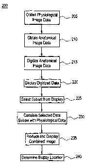
35 U.S.C.
119(e) to U.S. Provisional Application Serial No. 60/425,288, entitled "Method
and
Apparatus for Comparing and Correlating PET and X-ray Images", filed November
12, 2002,
the contents of which are incorporated by reference herein.
Field of the Invention
[0002] The present invention relates to a method and an apparatus for
determining a
biopsy location in a body part, and more particularly a method and an
apparatus for
correlating image data obtained from at least two separate devices to
determine a biopsy
location in a body part.
Description of the Related Art
[0003] Increasing the number of medical imaging studies that apply to a single
feature
or to several features can increase the diagnostic confidence of the physician
interpreting the
studies. Diagnostic confidence is increased further if the image sets are
correlated; i.e., the
CA 02499663 2005-03-21
WO 2004/027713 PCT/US2003/029246
spatial coordinate systems of the image sets are identical. For display
purposes, once the
spatial coordinate systems are shared, it is often helpful to display the
images in a single
window. In the past, such "correlative image displays" have been implemented
by using
gray-scale for one image set (i.e., x-ray) and a color scale for the second
set. Alternatively,
one image set uses hue and the other intensity. Aside from increasing
diagnostic confidence,
correlating images can be useful if each image set has a different intrinsic
utility. For
example, an imaging modality such as x-ray imaging has high spatial resolution
and is
therefore often better for guiding interventions, because the spatial
resolution allows the user
to avoid important anatomic structures of interest (e.g., major blood
vessels). Another
imaging modality (e.g., positron emission tomography, or "PET") is useful for
providing
biochemical and/or physiological information about structures in the human
body.
[0004] It is l~nov~m in the literature that PET and x-ray images can be
combined. For
example, see I. Weinberg et al., "Combining X-Ray and Functional Mammography
Images",
Radiology 1997, pp. 205-261. As another example, see I. Weinberg et al.,
"linplementing
PET-Guided Biopsy: Integrating Functional Imaging Data with Digital X-Ray
Mammography Cameras", Proceedings of SPIE Volume 4319, Medical Imaging 2001:
Visualization, Display, and Image-Guided Procedures, published May 2001.
SUMMARY OF THE INVENTION
[0005] In one aspect, the invention provides a system for determining a biopsy
location in a body part. The system includes a first device configured to
obtain digital
physiological image data about the body part, a second device configured to
obtain second
image data about the body part, a monitor configured to display the second
image data, a
signal processing module that includes an analog-to-digital converter
configured to digitize
2
CA 02499663 2005-03-21
WO 2004/027713 PCT/US2003/029246
the second image data, a memory configured to store the digital physiological
image data and
the digitized second image data, and a correlator coupled to the memory and
configured to
correlate the digital physiological image data with the digitized second image
data and to
produce a combined image as a result of the correlation. A determination of a
biopsy
location is made on the basis of the combined image, or on the basis of
features derived from
the two images. The first device may include a positron emission tomography
scanner
machine. The second device may include one of the group consisting of a
digital x-ray
machine, an x-ray mammography machine, an x-ray cranial axial tomography
machine, a
magnetic resonance imaging machine, and an ultrasound machine. The system may
also
include a localization device configured to select a preferred subset of the
second image data
based on the digital physiological image data obtained from the first device.
The localization
device may include a computer mouse. The first device may be configured to use
a
predetermined spatial coordinate system. The correlator may include a
transformer
configured to transform at least one of the digital physiological image data
and the digitized
second image data into the predetermined spatial coordinate system.
[0006] W another aspect, the invention provides a method for determining a
biopsy
location in a body part. The method includes the steps of obtaining
physiological image data
about the body part, obtaining independent second image data about the body
part,
correlating the second image data with the physiological image data, producing
a combined
set of image data based on the correlating, and determining a biopsy location
based on the
combined set of image data. The second image data may include anatomical image
data, and
the step of obtaining second image data may be performed by using one of the
group
consisting of a digital x-ray machine, an x-ray mammography machine, an x-ray
cranial axial
tomography machine, a magnetic resonance imaging machine, and an ultrasound
machine.
3
CA 02499663 2005-03-21
WO 2004/027713 PCT/US2003/029246
The step of obtaining physiological image data may be performed by using a
positron
emission tomography scanner machine. The obtained physiological image data may
be in
digital form, and the method may further include the step of digitizing the
obtained second
image data. The method may further include the step of selecting a preferred
subset of the
obtained second image data based on the obtained digital physiological image
data. The step
of selecting a preferred subset may be performed by using a computer mouse.
[0007] lii yet another aspect, the invention provides an apparatus and a
method for
coupling a non-networked device to a computer network by capturing an output
signal from
the non-networked device, digitizing the captured signal, and processing the
digitized signal
with a computer for presentation and transmission over the computer network.
The non-
networked device may include a monitor that is configured to display the
output signal to be
captured. In an alternative embodiment, the invention provides an apparatus
and a method
for coupling a first device to a second device by capturing an output signal
from the first
device, digitizing the captured signal, and sending the digitized signal to
the second device.
The first device may include a monitor that is configured to display the
output signal from the
first device.
BRIEF DESCRIPTION OF THE DRAWINGS
[0008] Figure 1 is a block diagram of an apparatus for correlating image data
according to a preferred embodiment of the present invention.
[0009] Figure 2 is a flow chart that illustrates a method of correlating image
data
according to a preferred embodiment of the present invention.
4
CA 02499663 2005-03-21
WO 2004/027713 PCT/US2003/029246
DETAILED DESCRIPTION OF THE INVENTION
[0010] The present invention is a method and apparatus for obtaining x-ray or
image
sets for correlation with a physiological imaging set. The invention allows a
device used for
physiological imaging (hereinafter referred to as a "first device") to "grab"
images from any
of a variety of other devices (hereinafter referred to a "second device"),
without substantially
modifying the underlying software or hardware processes in any such second
device. This
feature is very important, because substantial modification of medical devices
may affect the
validation of such devices by medical regulations (e.g., according to the U.S.
Food and Drug
Administration). An example of a first device is a positron emission
tomography ("PET")
scanner machine. Examples of second devices include digital x-ray machines, x-
ray
mammography machines, x-ray cranial axial tomography (CT) machines, magnetic
resonance
imaging (MRI) machines, ultrasound machines, or any other medical imaging
device that
provides an image to a computer monitor.
[0011] The method involves the use of "frame grabber" circuitry to capture an
image
from an output port of a second device, and then to present that captured
image to the first
device. The frame grabber circuitry includes a signal splitting module the
also sends the
signal containing the captured image to a monitor coupled to the second
device. In the
process of presenting the captured image to the first device, amplification
and/or duplication
of the output signal may be performed, as well as other image or signal
processing functions.
The frame grabber circuitry includes an analog-to-digital converter to convert
the monitor
output into a digital signal that can be manipulated. The captured image from
the second
device can be manipulated via mathematical algorithms (e.g., affine
transformations) in
software or via digital signal processing (e.g., firmware) so as to share a
common spatial
CA 02499663 2005-03-21
WO 2004/027713 PCT/US2003/029246
coordinate system with the images or data collected with the first device.
Alternatively, the
image from the first device may be similarly manipulated to share a common
spatial
coordinate system with the images or data collected with the second device.
The manipulated
data from the second device can be displayed with data from the first device
to form a fused
image.
[0012] An exemplary conventional method of capturing images from a second
device
and sending the captured images to a printer is known in the art, and is used
by Codonics,
Inc. to print images from many imaging devices. In one embodiment, the present
invention
provides the advantage of a method of capturing images from a second device in
order to
combine image sets from the second device with image sets obtained using a
first device.
The invention includes the use of pointing and/or localization devices (e.g.,
a mouse) which
are coupled to the second device. Because the images shown on the second
device include
markers (e.g., cursors) as to the position of these pointing devices, the
capture of images from
the second device (and by reference, the capture of said cursors) represents a
feedback loop
by which the user can adjust the position of the pointing device with respect
to features that
are evident in either or both of the first device image and the second device
image to select
one or more spatial locations. Thus, a location for biopsy can be determined
using either an
image from the first device or one or more combined images from the first
device and the
second device. The determined biopsy location can then be shown to the user of
the second
device by having the user click or otherwise manipulate the mouse of the
second device and
showing or otherwise signaling the location of the second device's mouse
cursor with respect
to the image from the first device and/or one or more of the combined images.
6
CA 02499663 2005-03-21
WO 2004/027713 PCT/US2003/029246
[0013] Referring to Figure 1, a block diagram of a preferred embodiment of the
invention includes a first device 105 and a second device 110. The second
device 110
includes a localizer 115, such as a mouse, that can specify locations on
images obtained by
the second device 110 (and, with the aid of correlation, on images obtained
with the first
device 105); a CPU 117; a signal splitter 120; and a monitor 125. The frame-
grabber
circuitry 130 is coupled to both the first device 105 and the second device
110, and includes a
data digitizer, such as an analog-to-digital converter (ADC). The circuitry
130 may also
include a signal amplification functionality and/or other digital signal
processing
functionalities. The frame-grabber circuitry 130 captures the image obtained
by the second
device 110 as displayed on the monitor 125, digitizes the captured image, and
provides the
digitized image to the first device 105. The first device 105 includes an
acquisition section
135 for obtaining an image (e.g., using physiological imaging as is commonly
obtained with
radiotracer imaging); a memory section 140 for holding the digital image data
corresponding
to both devices 105 and 110; and a correlative section 145 for combining the
image data and
indicating the determined biopsy location to the user. A monitor 150 may be
used to display
the combined image data to the user.
[0014] Refernng to Figure 2, a flow chart 200 illustrates a method for
determining a
biopsy location in a body part according to a preferred embodiment of the
present invention.
At the first step 205, image data about the body part is obtained using a
first device.
Preferably, the image data is digital and contains physiological information
about the body
part. The first device may be a positron emission tomography scanner machine.
At the
second step 210, second image data about the body part is obtained using a
second device.
Preferably, the second image data is anatomical image data that is
transmittable to a video
monitor. The second device may be one of a digital x-ray machine, an x-ray
mammography
7
CA 02499663 2005-03-21
WO 2004/027713 PCT/US2003/029246
machine, an x-ray cranial axial tomography machine, a magnetic resonance
imaging machine,
and an ultrasound machine. At the next step 215, the video signal from the
second image
data is captured via digitization (e.g., by an analog-to-digital converter),
and at step 220, the
captured digitized second image data is displayed on a monitor.
[0015] At step 225, a user selects a preferred subset of the captured
digitized second
image data. For example, the user may be able to use a computer mouse to
select a specific
axea on the moW for display. Then, at step 230, the preferred subset of the
captured digitized
image data (said data containing anatomical or other information about the
body part) is
correlated with the digital image data from the first device (said data
containing physiological
information about the body part). At step 235, a combined set of image data is
produced on
the basis of the correlation. Finally, at step 240, the user determines a
biopsy location based
on the combined set of image data. For example, the combined set of image data
may be
displayed to the user on a monitor coupled to the first device, and the user
may then make a
visual determination of the biopsy location.' The display of the combined
image may also use
a spatial coordinate system to enable the user to be precise in the
determination of the biopsy
location.
[0016] Alternatively, image data from the first and second devices may be
presented
in combination to the user without direct combination of the image data sets.
For example,
data from the second image may be processed with feature extraction software
in order to
generate locations of features of interest that axe then superimposed on the
first image data
display. In one exemplary application, the location of a mouse cursor in the
second image
may be extracted, and the extracted location may be displayed as a cursor
superimposed on
the first image.
CA 02499663 2005-03-21
WO 2004/027713 PCT/US2003/029246
(0017] In another embodiment, a system and a method for coupling a non-
networked
device to a computer network are provided. The system is configured to captw-e
an output
signal from the non-networked device, digitize the captured signal, and
process the digitized
signal with a computer for presentation and transmission over the computer
networlc. The
non-networked device may include a monitor that is configured to display the
output signal to
be captured. An exemplary computer network may comprise a picture and
archiving and
communications system (PACS). W an alternative embodiment, a system and a
method for
coupling a first device to a second device are provided. The system is
configured to capture
an output signal from the first device, digitize the captured signal, and send
the digitized
signal to the second device. The first device may include a monitor that is
configured to
display the output signal from the first device.
[0018] While the present invention has been described with respect to what is
presently considered to be the preferred embodiment, it is to be understood
that the invention
is not limited to the disclosed embodiments. To the contrary, the invention is
intended to
cover various modifications and equivalent arrangements included within the
spirit and scope
of the appended claims. The scope of the following claims is to be accorded
the broadest
interpretation so as to encompass all such modifications and equivalent
structures and
functions.
[0019] The contents of each of the following publications are hereby
incorporated by
reference:
1) I. Weinberg et al., "Combining X-Ray and Functional Mammography Images",
Radiology
1997, pp. 205-261.
9
CA 02499663 2005-03-21
WO 2004/027713 PCT/US2003/029246
2) I. Weinberg et al., "Implementing PET-Guided Biopsy: Integrating Functional
Imaging
Data with Digital X-Ray Mammography Cameras", Proceedings of SPIE Volume 4319,
Medical Imaging 2001: Visualization, Display, and Image-Guided Procedures, May
2001.
3) http~//www.codonics.com/tech/saindex.htm (undated).
4) PEM-2400 User Manual, Appendix A to U.S. Provisional Patent Application No.
60/425,288, filed November 12, 2002.
5) PEM-2400 Software Instructions Printout, Appendix B to U.S. Provisional
Patent
Application No. 60/425,288, filed November 12, 2002.
