Note : Les descriptions sont présentées dans la langue officielle dans laquelle elles ont été soumises.
CA 02500054 2005-03-23
WO 2004/030515 PCT/US2003/029509
DEVICE FOR PROVIDING AUTOMATIC STITCHING OF AN INCISION
CROSS REFERENCE TO RELATED APPLICATIONS
This application is a Continuation-Tn-Part of U.S. Application Serial No.
10/261,429, filed September 30, 2002, the entire contents of which is
incorporated herein
by reference.
BACKGROUND OF THE INVENTION
1. Field of the Invention
The present invention relates generally to a device for providing automatic
stitching of an incision, and more particularly, to a device for providing
access to a hollow
organ as well as automatic stitching on an incision in the hollow organ.
2. Prior Art
Surgery may be performed using open-chest techtvques while the heart is
under cardioplegic arrest and circulation is maintained by cardiopulmonary
bypass. Using
such techniques, a gross thoracotomy is created in order to gain access to the
heart and
great vessels, facilitating clamping and cannulation of the aorta for inducing
cardioplegic
arrest, and allowing instruments to be introduced into the chest cavity and
into the heart to
perform a surgical repair. The necessity of stopping the heart significantly
heightens the
risks attendant such procedures, particularly the risks of causing ischemic
damage to the
heart muscle, and of causing strobe or other injury due to circulatory emboli
produced by
aortic clamping and vascular cannulation.
A number of endovascular approaches for use in procedures in which the heart
is
2s arrested have been developed in the prior art. These approaches attempt to
allow
intracardiac access using catheters introduced transluminally from peripheral
vessels into
the heart. However, these devices suffer from many problems including a lack
of control
and precise positionability from the proximal ends of the highly flexible and
elongated
devices, the significant size constraints of peripheral vessels, and the
inability to position
the devices in all potentially diseased sites within the heart.
A number of minimally invasive or endoscopic access devices for use in beating
heart procedures have also been developed in the prior art. These endoscopic
devices are
CA 02500054 2005-03-23
WO 2004/030515 PCT/US2003/029509
used to gain intracardiac access to the heart. Such devices are disclosed in
U.S. Patent Nos.
6,079,414 to Roth and 5,829,447 to Stevens et al., which are hereby
incorporated by
reference. However, such devices generally have a substantially long axial
bore into which
instruments are passed. The long length of the bore restricts the manipulative
capability of
the instruments passed through the bore into an interior of the heart. For
example, a distal
end of the instrument mainly moves in an axial direction and cannot stray very
much from
a central axis in the axial direction. Furthermore, the instruments must be
very straight in
order to traverse the long length of the bore, thus, curved instruments cannot
be utilized
with the endoscopic access devices of the prior art. Lastly, because such
endoscopic
access devices are directed to the heart wall under observation of a viewing
device, they
cannot be directly secured to the heart wall to maintain a tight seal against
blood flow from
the heart.
Furthermore, stitching of the incision made to provide aceess to hollow organs
(as
well as stitching of wounds and stitching to repair damaged portions of
tissue) often
require special skills on the part of the surgeon, are not uniform or
reliable, and can be
time consuming and therefore costly.
SUMMARY OF THE INVENTION
Therefore it is an object of the present invention to provide an access device
that
overcomes the disadvantages of the prior art.
Accordingly, an automatic suturing device is provided. The automatic suturing
device comprising: a body for insertion into an opening in tissue; a plurality
of hooks
movably disposed in the body between retracted and extended positions; a
suture holder
having sutures disposed therein, the suture holder having means for engaging a
portion of
the hooks when in the retracted position and for attaching the sutures to a
portion of the
plurality of hooks; and actuation means for actuating the plurality of hooks
from the
retracted position to the extended position and for embedding the exposed
plurality of
hooks into the tissue surrounding the opening.
Preferably, the device further comprises means for providing access into a
hollow
organ through the opening. The means for providing access preferably
comprises: the
body having a bore for passage of at least a distal portion of an instrument
into an interior
-2-
CA 02500054 2005-03-23
WO 2004/030515 PCT/US2003/029509
of the hollow organ; and a valve disposed in the bore for allowilig passage of
the
instrument and substantially preventing a fluid in the interior of the hollow
organ from
leaking outside the hollow organ.
The body preferably comprises first and second body portions movable relative
to
each other, wherein the actuation means comprises: rotatable actuation means
for exposing
the plurality of hooks upon rotation of one of the first and second body
portions relative to
the other of the first or second body portions; and translatable actuation
means for
embedding the exposed plurality of hooks into the wall upon translation of one
of tile first
and second body portions relative to the other of the first or second body
portions. In
which case, the automatic suturing device preferably further comprises a fluid
seal
between the first and second body portions.
Preferably, the suture holder is separately formed from the body and inserted
on a
distal portion of the body.
In a first version, the means for engaging a portion of the hooks when in the
retracted position and for attaching the sutures to a portion of the plurality
of hooks
comprises an adhering means for adhering the sutures to a portion of the
plurality of
hooks. Preferably, the adhering means comprises: the suture holder having a
longitudinal
channel for holding the sutures therein; and the suture holder having two or
more collet
assemblies each of which correspond to a frayed end of the sutures, each of
the collet
2o assemblies having a collapsible collet having a glue chamber containing a
dose of glue
disposed in an internal channel and means for collapsing the collapsible
collet radially into
the internal channel; wherein the at least a portion of the plurality of hooks
and the frayed
ends of the sutures are disposed in the interior channel and wherein each of
the collapsible
collets are collapsed to compress the frayed ends of the sutures and dose of
glue against a
portion of the hooks disposed in the interior channel to adhere the sutures to
the hooks.
In a second version, the means for engaging a portion of the hooks when in the
retracted position and for attaching the sutures to a portion of the plurality
of hooks
comprises a fastening means for mechanically fastening the sutures to a
portion of the
plurality of hooks. Preferably, the fastening means comprises: the suture
holder having a
longitudinal channel for holding the sutures therein; at least a portion of
the plurality of
hoolcs having one or more projections; and the suture holder having two or
more collet
-3-
CA 02500054 2005-03-23
WO 2004/030515 PCT/US2003/029509
assemblies each of which correspond to a frayed end of the sutures, each of
the collet
assemblies having a collapsible collet having an internal channel and means
for collapsing
the collapsible collet radially into the internal channel; wherein the at
least a portion of the
plurality of hoofs and the frayed ends of the sutures are disposed in the
interior channel
and wherein each of the collapsible collets are collapsed to compress the
projections
against a corresponding hoolc to capture the frayed ends of the sutures
against a portion of
the hoofs disposed in the interior channel to fasten the sutures to the hoofs.
Also provided is an automatic suturing device comprising: an access device for
providing access into a hollow organ during an open surgical procedure, the
access device
to comprising: a body having a distal portion for insertion into an opening in
a wall of the
hollow organ, the body further having a bore for passage of at least a distal
portion of an
instnunent into an interior of the hollow organ; a valve disposed in the bore
for allowing
passage of the instrument and substantially preventing a fluid in the interior
of the hollow
organ from leaking outside the hollow organ; a plurality of hoolcs movably
disposed in the
body between retracted and extended positions; and actuation means for
actuating the
plurality of pins from the retracted position to an extended position and for
embedding the
exposed plurality of hooks into the wall to secure the body to the wall; and
the automatic
suturing device further comprises a suture holder having an internal bore
disposed on the
distal portion of the body, the suture holder having sutures disposed therein
and means for
2o engaging a portion of the plurality of hoolcs when in the retracted
position and for
attaching the sutures to a portion of the plurality of hooks.
Preferably, the body comprises first and second body portions movable relative
to
each other and wherein the actuation means comprises: rotatable actuation
means for
exposing the plurality of hooks upon rotation of one of the first and second
body portions
relative to the other of the first or second body portions; and translatable
actuation meaazs
for embedding the exposed plurality of hooks into the wall upon translation of
one of the
first and second body portions relative to the other of the first or second
body portions. In
which case, the automatic suturing device preferably further comprises a fluid
seal
between the first and second body portions.
3o Preferably, the body has a low-profile length in an axial direction of the
bore to
increase a manipulative capability of the instrument through the bore.
Preferably, the
-4-
CA 02500054 2005-03-23
WO 2004/030515 PCT/US2003/029509
length of the body in the axial direction of the bore is substantially within
a range of 1.5T
to ST, where T is a thickness of the wall.
In a first version, the means for engaging a portion of the hoolcs when in the
retracted position and for attaching the sutures to a portion of the plurality
of hooks
comprises an adhering means for adhering the sutures to a portion of the
plurality of
hooks. Preferably, the adhering means comprises: the suture holder having a
first
longitudinal chamiel for holding the sutures therein; the suture holder having
a second
longitudinal channel for holding a glue therein; and a linlcing channel for
linl~ing at least a
portion of the first and second longitudinal channels and corresponding to at
least a portion
i0 of the plurality of hooks when in tile retracted position; wherein the at
Ieast a portion of the
plurality of hooks are disposed in the linking channel and in communication
with both the
sutures and glue in the respective first and second longitudinal channels when
in the
retracted position to adhere at least a portion of a suture to at least a
portion of each of the
plurality of hooks. Preferably, the suture holder has an internal bore and the
first and
second longitudinal channels are disposed on an inner surface of the internal
bore.
Furthermore, the linking channel is preferably disposed on a distal surface of
the suture
holder.
In a second version, the means for engaging a portion of the hooks when in the
retracted position and for attaching the sutures to a portion of the plurality
of hooks
2o comprises a fastening means for mechanically fastening the sutures to a
portion of the
plurality of hooks. Preferably, the fastening means comprises: the suture
holder having a
longitudinal channel for holding the sutures therein; at least a portion of
the plurality of
hooks having one or more projections; and the suture holder having two or more
collet
assemblies each of which correspond to a frayed end of the sutures, each of
the collet
assemblies having a collapsible collet having an internal channel and means
for collapsing
the collapsible collet radially into the internal channel; wherein the at
least a portion of the
plurality of hooks and the frayed ends of the sutures are disposed in the
interior channel
and wherein each of the collapsible collets are collapsed to compress the
projections
against a corresponding hook to capture the frayed ends of the sutures against
a portion of
3o the hooks disposed in the interior channel to fasten the sutures to the
hooks.
Still provided is a method for automatically stitching an opening in tissue.
The
-5-
CA 02500054 2005-03-23
WO 2004/030515 PCT/US2003/029509
method comprising: inserting a portion of a device into the opening; extending
a plurality
of hoofs from the device and through the tissue surrounding the opening;
inserting at least
a portion of each of the plurality of hooks baclc into the device; attaching
the at least a
portion of each of the plurality of hooks to a suture; withdrawing the
plurality of hooks and
attached suture from the tissue surrounding the opening and through the
opening; severing
the sutures from the at least portion of each of the plurality of hooks; and
pulling the
sutures to close the opening. The method preferably further comprises tying
the sutures
together after closing the opening.
The attaching preferably comprises adhering the at least a portion of each of
the
l0 plurality of hooks to the sutures. Alternatively, the attaching comprises
mechanically
fastening the at least a portion of each of the plurality of hooks to the
sutures.
Still yet provided is a method for providing access into an interior of a
hollow
organ for manipulation of an instnunent therein. The method comprising:
providing
access to the hollow organ; making an opening in a wall of the hollow organ;
inserting a
body of an access device in the opening; securing the body to the wall;
passing at least a
distal portion of an instrument through a bore in the access device to an
interior of the
hollow organ; substantially preventing a fluid in the interior of the hollow
organ from
leaking outside the hollow organ; removing the access device from the opening;
and
automatically closing the hole in the wall of the internal organ upon removal
of the access
2o device from the opening.
Preferably, the automatically closing comprises: inserting a portion of the
access
device into the opening; extending a plurality of hooks from the access device
and through
the tissue surrounding the opening; inserting at least a portion of each of
the plurality of
hooks back into the access device; attaching the at least a portion of each of
the plurality of
hooks to a suture; withdrawing the plurality of hoolcs and attached suture
from the tissue
surrounding the opening and through the opening; severing the sutures from the
at least
portion of each of the plurality of hooks; and pulling the sutures to close
the opening.
Preferably, the method further comprises tying the sutures together after
closing the
opeiung.
-6-
CA 02500054 2005-03-23
WO 2004/030515 PCT/US2003/029509
BRIEF DESCRTPTION OF THE DRAWINGS
These and other features, aspects, and advantages of the apparatus and methods
of
the present invention will become better understood with regard to the
following
description, appended claims, and accompanying drawings where:
Figure 1 illustrates an isometric view of a first preferred implementation of
an
intracardiac access device having an expandable balloon.
Figure 2A illustrates a sectional view of the intracardiac access device of
Figure 1
as taken along line 2-2 therein in which the access device is inserted into an
opening in a
heart wall and the expandable balloon is in a relaxed state.
to Figure 2B illustrates the sectional view of Figure 2A in which the
expandable
balloon is in an expanded state.
Figure 3A illustrates the sectional view of Figure 2B having a straight
instrument
passed therethrough.
Figure 3B illustrates the sectional view of Figure 2B having a curved
instrument
15 passed therethrough.
Figure 4 illustrates an isometric view of a second preferred implementation of
an
intracardiac access device having a plurality of hooks, the hooks being shown
in an
exposed position.
Figure 5 illustrates a sectional view of the access device of Figure 4 as
taken along
20 line 5-5 in Figure 4.
Figure 6 illustrates an isometric view of the access device of Figure 6 with
the
plurality of hooks being rotated while in an extended position.
Figure 7 illustrates a sectional view of the access device of Figure 4 with
the
plurality of hooks being in an extended position.
25 Figure 8 illustrates an isometric view of the access device of Figure 7
with the
plurality of hooks in the unexposed position.
Figure 9 illustrates m isometric view of a spacer for use with the access
device to
lock the same with the hooks in the exposed position.
Figure 10 illustrates a side view of a preferred implementation of one of the
3o plurality of hooks for use with the access device of Figure 4.
Figure 11 illustrates a preferred implementation of a valve for use with the
access
CA 02500054 2005-03-23
WO 2004/030515 PCT/US2003/029509
device of Figure 4.
Figure 12 illustrates a perspective exploded view of the access device
substantially
similar to that of Figure 4 used together with a suture holder to provide an
automatic
stitching of an incision.
Figure 13 is a sectional view of the exploded view of the access device and
suture
holder of Figure 12.
Figure 14 is a perspective view of the access device and suture holder of
Figure 12
with the suture holder being loaded onto the access device.
Figure 15 is a perspective view of the access device and suture holder of
Figure 14
to having a distal portion being inserted into an incision in tissue.
Figure 16 is a sectional view of the access device and suture holder of Figuxe
15
with the hooks in an extended position.
Figure 17 is a sectional view of the access device and suture holder of Figure
1 S
with the hooks retracted into the tissue surrounding the incision and with the
sharp pointed
15 ends of the hooks being engaged with the suture holder.
Figure 18 is a sectional view of the access device and suture holder of Figure
1 S
with each of the hoolcs being in an extended position and having a suture
retained thereon.
Figure 19 is a perspective view from the distal end of the access device in
which
the upturned portions of the hooks having the thread retained thereon are
entering their
20 respective second longitudinal chazmels.
Figure 20 is a perspective view of the access device and suture holder with
the
upturned portions of the hoofs being entered into the second longitudinal
channels.
Figure 21 is a perspective view of the access device and suture holder being
removed from the incision.
25 Figure 22 is a sectional view of the access device and suture holder of
Figure 21.
Figure 23 is a sectional view of the access device and suture holder after the
suture
holder is separated from the access device and the sutures are separated from
the sharp
pointed ends of the hooks.
Figure 24 illustrates a top view of an alternative suture holder of the
present
30 invention.
Figure 25 illustrates a sectional view of Figure 24 as taken along line 25-25
of
_8_
CA 02500054 2005-03-23
WO 2004/030515 PCT/US2003/029509
Figure 24, showing the collet assemblies in an open position.
Figure 26 illustrates the sectional view of Figure 25 with the collet
assemblies in a
closed position.
Figures 27a and 27b illustrate alternative hoofs for use with the alternative
suture
holder of Figure 24.
DETAILED DESCRIPTION OF THE PREFERRED EMBODIMENT
Although this invention is applicable to numerous and various types of
procedures
and providing access to various hollow organs, it has been found particularly
useful in the
environment of providing intracardiac access in a beating heart open chest
procedure.
Therefore, without limiting the applicability of the invention to providing
intracardiac
access in a beating heart open chest procedure, the invention will be
described in such
environment.
Referring now to Figures 1, 2A, and 2B there is shown a first preferred
implementation of an intracardiac access device having an expandable balloon,
the first
preferred implementation of the access device being generally referred to by
reference
numeral 100. The access device 100 provides access into a hollow organ 102,
such as the
heart, during an open surgical procedure. The access device has a body 104
that is inserted
into an opening or incision 106 in a wall 108 of the hollow organ 102. The
body 104 is
preferably cylindrical in shape and is typically fabricated from a medical
grade
thermoplastic and can be fabricated from airy methods known in the art, such
as
conventional machining or injection molding. The body 104 has a bore 120 sized
to
permit at least a distal portion of an instrument (not shown) to pass through
the bore 110
and into an interior of the hollow organ 102. The bore extends in an axial
direction A
from an exterior of the hollow organ 102 to an interior of the hollow organ
102.
A valve 112 is disposed in the bore 110 of the body 104 for allowing passage
of the
instrument while substantially preventing a fluid in the interior of the
hollow organ 102
from leaking outside the hollow organ 102. Preferably, the valve 112 is what
is commonly
referred to in the art as a duckbill valve. The duckbill valve 112 is
fabricated from an
elastomer, such as silicone, and has a cylindrical portion 116 and a tapered
portion 118.
The tapered portion 118 terminates in a slit 120. The slit 120 is normally
closed to provide
CA 02500054 2005-03-23
WO 2004/030515 PCT/US2003/029509
a seal and is configured to conform to a shape of an instrument passed through
the slit 120
to provide a seal around the instrument. The ducl~bill valve 112 further has a
stepped
portion 122 that xests on a corresponding shoulder I24 of the body 104. The
ducl~bill
valve 112 can be press fit into the body or retained therein by way of a
medical grade
adhesive. Alternatively, a flange (not shown) can be used to capture a portion
of the
ducl~bill valve 112. Although, ducl~bill valves are preferred, other types of
valves lcnown
in the art can be used without limiting the scope or spirit of the present
invention, such as a
flexible znembrane (not shown) having a small expandable aperture.
The access device 100 also has securing means for securing the body I04 to the
l0 wall 108 of the hollow organ 102. The securing means fixes the body 104 to
the wall 108
such that it is not in danger of coming off or falling into the interior of
the internal organ
102. Preferably, the securing means also provides a seal between the opening
106 and the
body 104 of the access device 100. In a first preferred implementation, the
securing means
comprises a balloon canfiguration. In such a configuration, a lip 126, which
is preferably
cylindrical, is disposed on a proximal portion 104a of the body 104. The lip
126 is
preferably integrally formed with the body 104, but znay also be formed
separately and
attached to the body 104 by any means Icnown in the art, such as by ultrasonic
welding,
thermal welding, or with a medical grade adhesive.
A balloon I28 is disposed on a distal portion 104b of the body 104. The
balloon is
shown in a deflated or relaxed position in Figure 2A. The relaxed position of
the balloon
128 may be due to the Iacl~ of a fluid, such as saline or air, therein, or by
applying a
vacuum to the balloon. A conduit 130 is preferably formed in the body for
supplying the
fluid from a fluid source (not shown) or applying a vacuum from a vacuum
source (not
shown) to the balloon for expansion or contraction, respectively, thereof. A
port 132 is
preferably provided in fluid communication with the conduit 130 to facilitate
connection
of the fluid or vacuum source to the conduit 130. Preferably, the fluid and
vacuum source
comprise a syringe (not shown) and the port 132 comprises a self sealing
needle port as is
l~nown in the az~t. Figure 2B shows the balloon 128 in an expanded position in
which the
wall 108 of the hollow organ 102 is captured between the lip 126 and the
balloon 128.
Although not shown, it is preferred that the wall I08 be compressed slightly
upon the
expansion of the balloon 128.
-10-
CA 02500054 2005-03-23
WO 2004/030515 PCT/US2003/029509
Referring now to Figures 3A and 3B, the body 104 has a low-profile length L in
the
axial direction A of the bore 1 IO to increase a manipulative capability of
the instrument
134 through the bore 110. Preferably, the length L of the body I04 in the
axial direction A
of the bore 110 is substantially within a range of 1.5T to 5T, where T is a
thickness of the
wall I08. For example, the thickness for a typical heat wall varies between
approximately
3-7 mm and the length L of the body 104 is in the range of 4.5 mm to 35 mm,
most
preferably about 10-I5 mm.
As clearly seen in Figure 3A, the low-profile length L of the body 104 as
compared
to the thickness T of the wall I08 allows an instrument 134 to be manipulated
at a greater
l0 angle ~/ with respect to a central axis of the bore than the endoscopic
access devices of the
prior art. Furthermore, as clearly shown in Figure 3B, the low-profile length
L of the body
104 as compared to the thickness T of the wall 108 allows insertion of a
curved instrument
having a radius R, which is not possible with the endoscopic access devices of
the prior
art.
Referring now to Figures 4 and 5, there is illustrated a second preferred
implementation of an access device of the present invention, the second
preferred access
device being generally referred to by reference numeral 200. Access device 200
also
preferably has a low-profile shape as discussed above with regard to the first
preferred
implementation and has the same advantages as discussed above with regard to
Figures 3A
and 3B. Access device 200 includes a body 202 having first and second body
portions
204, 206, respectively. The first and second body portions 204, 206 are
fabricated from
any medical grade material, such as stainless steel or a polymer. The first
body portion
204 includes a flange 208 and a cylinder portion 210. The first body portion
204 further
has a bore 212 that accommodates a valve 214. Referring now to Figure 1 l, the
valve 214
is preferably a duckbill or slit valve fabricated from a medically approved
elastomer, such
as silicone. The valve 214 has a flange 2I6 which fits within a corresponding
stepped
groove 218 in the bore 212 of the first body portion 204. The valve 214 also
has a
cylindrical body portion 220 that fits within the bore 212 of the first body
portion 204.
The valve 214 has a slit 222 on a conical nose 224 thereof to sealingly
accommodate an
instrument inserted through the access device 200. The valve 214 is retained
in the bore
212 by any means known in the art such as by adhesive or press-fit. The valve
214,
-11-
CA 02500054 2005-03-23
WO 2004/030515 PCT/US2003/029509
although shown disposed in the first body portion 204 may also be disposed in
the second
body portion 206 and although shown and described as a discrete part may be
integrally
foz~ned with either of the first and second body portions 204, 206.
Referring baclc to Figures 4 and 5, the second body portion 206 has a bore
226, at
least a portion of which acconnnodates the cylinder portion 210 of the first
body portion
204 such that it is free to both rotate and translate within the bore 226 of
the second body
portion 206. The second body portion 206 further has at least one shoulder or
flange 228
on an exterior surface thereof. A seal, such as aaz o-ring 230 is provided to
seal a fluid path
between the first and second body portions 204, 206. The second body portion
206 further
i0 has a plurality of first longitudinal channels 232 corresponding to each of
a plurality of
hoofs 234 disposed circumferentially about the second body portion 206. Each
of the
plurality of hoofs 234 have at least a portion thereof which is slidingly
disposed in a
corresponding first longitudinal channel 232. The second body portion also
includes a
plurality of second longitudinal channels 236 for housing an upturned portion
238 of the
hoofs 234 when the hoofs 234 are in an unexposed position.
Referring now to Figure 10, one of the plurality of hoofs 234 is shown
therein.
The hoofs 234 are fabricated from a medically approved metallic material, such
as
stainless steel and have a sharp pointed end 240 at the end of the upturned
porfiion 238. At
a proximal end of the hoolc is a tuned-in portion 242 that engages with and is
retained in
pOrt1o11S Of the first body portion 204, such as in corresponding
circumferential slots 244 in
the bore 212 of the cylinder portion 210. At the distal end of the hoofs 234
is the upturned
portion 238. A straight portion 246 connects the in-turned 242 and upturned
238 portions
with a curved portion 248 at a transition between the straight portion 246 and
the upturned
portion 238. At least a portion of the straight portion 246 is slidingly
disposed in a
corresponding first longitudinal channel 232.
Referring now to Figures 5-8, an operation of the access device 200 of the
second
preferred implementation will be described. The access device 200 is securely
positioned
in an incision 106 in a wall 108 of a hollow organ 102, such as the heart. The
incision is
made by any methods known in the art and may be a slit or a punched hole after
access is
provided to the hollow organ, such as by a gross thoracotomy. The wall 108 is
shown in
Figure 5, but omitted from Figures 6-8 for the sake of clarity. Referring f
rst to Figure 8,
-12-
CA 02500054 2005-03-23
WO 2004/030515 PCT/US2003/029509
the upturned portions 238 of the hooks 234 are disposed in corresponding
second
longitudinal channels 236 such that the sharp pointed ends 240 are unexposed.
The access
device is inserted into the incision 106 while the hooks 234 are in the
unexposed position
as shown in Figure 8. While the upturned portions 238 are shown as being
disposed in the
second longitudinal channels 236 in the unexposed position, they can
alternatively be
disposed in corresponding cut-outs (not shown) on the exterior of the second
body portion
206.
Referring next to Figure 6, the first body portion 204 is translated relative
to the
second body portion 206 in the direction of arrow A to extend the upturned
portions 238
l0 from the second longitudinal channels 236. Referring now to Figure 7, the
first body
portion 204 is then rotated in the direction of arrow B about a central axis C
to turn the
upturned portions 238 90 degrees and expose the sharp pointed ends 240. When
the first
body portion 204 is rotated, the hooks 234 are rotated by an interference with
the in-turned
portions 242 of the hooks 234 and a wall of the corresponding slots 244. Once
the hooks
234 are both extended and exposed as shown in Figure 6, the first body portion
204 is
translated in the direction of arrow D (opposite to the direction of arrow A)
to embed the
upturned portions 238 into the wall 108 of the hollow orgaa~ 102
circumferentially about
the incision 106, as shown in Figure 5. The access device 200 is then secured
to the wall
108 by sandwiching the wall 302 between the step or flange 228 and the curved
portions
248 of the hooks 234. Referring now to Figures 5 and 9, while the hooks 234
are
embedded into the wall 108, a locking clip 250 is disposed in a gap 252
between the flange
208 of the first body portion 204 and the second body portion 206 to prevent
any
translation of the fixst body portion 204 in the direction of arrow A. The
thicl~ness t of the
locking clip 250 substantially conforms to a thickness t of the clip. The
loclcing clip 250 is
preferably fabricated from a medically approved polymer and has fingers 254
which
elastically deform to fit within the gap 252. The loclcing clip 250 fuxther
has a pull 256 for
facilitating handling and inserting and removing the locking clip 250 into and
from the gap
252. Locl~ing clip 250 may have a tether attached to it on one end and to a
point outside
the operative field on another end to prevent locking clip 250 from
inadvertently being left
within the patient when the procedure is complete. Alternatively, the locl~ing
clip 250 may
be tethered to the access device 200 itself. While the access device 200 is
secured and
-13-
CA 02500054 2005-03-23
WO 2004/030515 PCT/US2003/029509
locked to the wall 108, surgical instruments (not shown) are inserted through
the valve
such that the worlcing ends thereof are inserted into an interior of the
hollow organ for
performing a necessary surgical procedure.
After completion of the surgical procedure, the access device 200 is removed
and
the incision 106 is closed. To remove the access device 200 from the incision
106, the clip
250 is removed and the first body portion 204 is translated in the direction
of arrow A to
dislodge the upturned portions 238 of the hooks 234 from the wall 108. The
first body
portion 204 is then rotated in a direction opposite to that of arrow B about
the central axis
C to rotate the hooks 90 degrees such that the sharp pointed ends 240 are
aligned with the
l0 second longitudinal channels 236. The first body portion 204 is then
translated in the
direction of arrow D to return the up-turned portions 238 of the hoolcs 234 to
the
unexposed positions in the second longitudinal channels 236. The access device
200 is
then removed from the incision 106 and the incision 106 is closed by any means
known in
the art, such as with sutures or surgical glue.
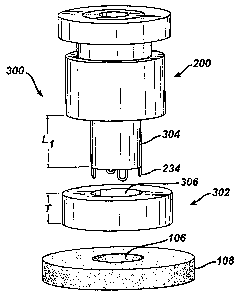
Referring now to Figures 12 and 13 there is illustrated an automatic stitching
device, referred to generally by reference numeral 300. Preferably, the
automatic stitching
device 300 comprises the access device 200 substantially similar to that
described above
used in combination with a suture holder 302 to provide an automatic stitching
capability
to the access device 200 for automatically stitching the incision 106 of the
hollow organ
102 after completion of a surgical procedure. Although the automatic stitching
device 300
is described in combination with the access device 200, those skilled in the
art will
appreciate that the same can be used without the features of the access device
200 that
facilitate use thexewith with surgical instruments. For example, the access
device 200 can
be configured without the bores 212, 226, and/or valve 214. Furthermore,
although
described as a separate piece, those spilled in the art will appreciate that
the suture holder
302 may be integrally formed with the access device 200. Additionally, the
incision 106 is
described by way of example only as being in the wall 108 of a hollow organ
102 as
described above. Those spilled in the art will appreciate that the automatic
stitching
device can be used to automatically stitch any incision, wound, or damaged
tissue, and can
3o also be used to join two tissues together such as an anastomodic device or
in a valve repair
or replacement. Lastly, the access device 200 is described as having a
cylindrical distal
-14-
CA 02500054 2005-03-23
WO 2004/030515 PCT/US2003/029509
portion 304 for insertion into the incision 106, however, those skilled in the
art will further
appreciate that the distal portion 304 can be provided in many different
shapes fox use with
different shaped incisions. For example, the distal portion 304 may by oval
shaped for use
with a linear incision.
The suture holder 302 is preferably disk-shaped and has a bore 306 for
acceptance
of the distal portion 304 of the access device 200. The suture holder 302 has
a thicl~ness T
smaller than the length Ll of the distal portion 304 of the access device such
that when the
suture holder 302 is inserted onto the distal portion 304 of the access device
(as shown in
Figure 14), a portion LZ of the distal portion 304 protrudes from the suture
holder 302.
l0 The suture holder 302 has two internal grooves 308, 310 about a periphery
of the bore 306.
A proximal one of the internal grooves 310 holds sutures (one each for each of
the hooks
234). A distal one of the internal grooves 308 holds a glue for, as will be
described below,
gluing an end of the suture onto each of the sharp pointed ends 240 of the
hooks 234. The
glue is preferably a two-part medically approved pressure sensitive high
viscosity epoxy
15 wherein each of the two parts are separated in the groove by a membrane.
Examples of
glues for use With the suture holder 302 are a medical cyanoacrylate glue or
Vitralit
medical grade adhesive. Alternatively, two grooves can be provided to hold the
glue, one
for each of the two parts of the epoxy. The sutua-e holder 302 also has a
linl~ing groove
312 on a distal surface 314 of the suture holder 302 for accepting the
upturned portions
2o 238 of the hooks 234 and for linking the two internal groves 208, 210.
Alternatively, the
linking groove 3I2 can be individual holes corresponding to each of the
upturned portions
238 of the hooks 234. The suture holder is preferably a disposable device
where the glue
and sutures are loaded into their respective internal grooves 210, 208 and
once used, it is
discarded. However, those skilled in the art will appreciate that it can also
be reusable
25 where the glue and sutures are loaded into their respective internal
grooves 210, 208 prior
to each procedure.
Referring slow to Figures I4-23, the operation of the automatic stitching
device 300
will be described. As discussed above, the operation of the automatic
stitching device 300
will be described with regard to the access device 200 described above.
Referring
30 specifically to Figure 14, the access device is operated to have the
upturned portions 238
of the hoolcs 234 inserted into their corresponding second longitudinal
channels 236, as
-15-
CA 02500054 2005-03-23
WO 2004/030515 PCT/US2003/029509
described above, by rotating the first body poxtion 204 relative to the second
body portion
206 and translating the first body portion 204 relative to the second body
portion 206 in
the direction of arrow D. The distal portion 304 of the access device 200 is
then inserted
into the bore 306 of the suture holder 302 such that a portion 316 protrudes
therefrom a
distance L2. Referring now to Figure 15, portion 316 is inserted into the
incision 106.
Referring now to Figure 16, the hooks are then extended, as described above,
by
translating the first body portion 204 relative to the second body portion 206
in the
direction of arrow A. The hooks 234 are further rotated 90 degrees, as
described above, by
rotating the first body portion 204 relative to the second body portion 206 in
the direction
io of arrow B. As shown in Figure 17, the hooks 234 are then retracted, as
described above,
to embed the upturned portions 238 of the hooks 234 in the tissue wall 108 to
secure the
access device 200 to the tissue wall 108. The access device 200 can then be
used, if
necessary, in combination with the locking clip 250 to perform a surgical
procedure, as
described above, by inserting an manipulating surgical instruments through the
valve 2I4
and bores 212, 226.
When the hoofs 234 axe retracted, each of the sharp pointed ends 240 further
enter
the linking channel 312 to engage a portion of a suture 318 and glue 320
disposed in the
internal grooves 308, 310. If necessary, the sharp pointed ends 240 further
puncture the
membrane separating the two parts of the epoxy. Thus, while a procedure is
being
performed, the sutures in one of the internal channels 310 are adhered to each
of the sharp
pointed ends 240 of the hooks 234. Preferably, a locating means, such as a
lcey (not
shown) in the bore 306 and a corresponding keyway (not shown) is provided to
orient the
suture holder 302 in a predetermined position with respect to the sharp
pointed ends 240 of
the hooks 234 such that an end of a suture can be located in the linking
channel 312 at the
location of the sharp pointed ends 240. In this way, the sharp pointed ends
240 would
pierce the membrane between parts of the epoxy glue 320 and then contact the
suture 318
end to adhere the same to the sharp pointed ends 240 of the hooks 234.
Although, the
suture holder 302 described above is preferred, those skilled in the art will
appreciate that
such is given by way of example ouy and not to limit the scope or spirit of
the present
invention. Many configurations of the suture holder are possible, such as a
disk having
sutures with looped ends, where the looped ends correspond to each of a hole
or linking
-l6-
CA 02500054 2005-03-23
WO 2004/030515 PCT/US2003/029509
channel. In such a configuration, each of the upturned portions 238 of the
hooks 234
would have a downwardly facing slit. In operation, on the upstrolce through
the linking
channel, the hooks 234 would displace the loop ends from the hole and pass
through the
hole, however, on the down stroke, the loop end would be captured in the slit
and be
retained therein.
Referring now to Figure 18, after the procedure has been completed and/or
after
the sutures 318 have been retained on each of the hooks 234, the hooks 234 are
again
extended, as discussed above. As shown in Figure 18, as the hooks 234 are
extended, they
withdrawn the suture 318 from the longitudinal channel 310 and pull the
sutures 318
to through the tissue wall 108 in an area surrounding the incision 106. As
shown in Figure
19, the hooks 234 are rotated 90 degrees, as discussed above, to correspond
with their
respective second longitudinal channels 236. As shown in Figure 20, the
upturned
portions 238 of the hooks 234 are then retracted into the second longitudinal
channels 236
along with a corresponding portion of suture 318. It is preferred that the
suture 318 be
tightly retained in the second longitudinal channels 236, and as such, the
second
longitudinal channels 236 are sized closely to that of the upturned portions
238.
Refernng now to Figures 21 and 22, the automatic stitching device 300 is then
removed from the incision 106 which in turn continues to withdraw suture 318
from the
longitudinal channel 310 and the suture holder 302 is removed from the distal
portion 304
of the access device 200. As shown in Figure 23, the sutures 318 are then cut
free of the
suture holder 302 and/or the access device. At this point, the sutures 318 are
looped
through the tissue wall 108 surrounding the incision I06 and can be pulled
tightly to close
the incision 106 and tied. Furthermore, the sutures can alternatively be
anchored in the
device such that the removal of the device itself pulls the sutures and closes
the incision.
Those skilled in the art will appreciate that the preferred implementation of
the
automatic stitching device 300 described above simplifies the stitching of
incisions (or
wounds or damaged portions of tissue) and results in a reliable, and uniform
stitch that is
quickly made and does not require special shills on the part of the surgeon.
Furthermore,
when used in combination with the access device 200, it provides a single
device that
3o provides access, secures to an area surrounding an incision in the tissue,
and automatically
closes and stitches the incision upon withdrawal of the device. As discussed
above, the
-17-
CA 02500054 2005-03-23
WO 2004/030515 PCT/US2003/029509
automatic stitching device 300 can also be used to create an anastomosis
between vessels
or to repair a damaged heart valve.
Referring now to Figures 24-26 there is illustrated an alternative embodiment
of
the suture holder of the present invention, the alternative suture holder
being referred to by
reference numeral 400. Although shown separately for the sake of clarity, the
alternative
suture holder 400 is intended to be used with the access device 200
substantially as shown
and described above with regard to suture holder 302. That is, the distal
portion 304 of the
access device 300 is disposed in a bore 402 of the alternative suture holder
400 similarly to
that described above with regard to suture holder 302. As also discussed
above, the access
l0 device 300 and suture holder 400 may be integrally formed. The alternative
suture holder
has a channel 404 for holding one or more sutures 406. The sutures 406
preferably have
frayed ends 408 corresponding to the hoolcs 234 of the access device 300. As
discussed
above, the alternative suture holder 400 and the access device 300 have
locating means,
such as a key and corresponding lceyway (not shown) for locating the frayed
ends 408 of
the sutures 406 disposed in the suture holder 400 with the hooks 234 of the
access device
300.
The alternative suture holder 400 has a main body portion 410 and an annular
ring
412 rotatably disposed in a groove 414 in the main body 410. Preferably the
main body
410 has upper and lower halves 410a, 410b, which when assembled, define the
groove 414
and allow easy assembly of the ring 412 to the main body 410. The azmular ring
412 has a
ring gear 416 on an inner surface of the annular ring 412.
The alternative suture holder 400 has a plurality of collet assemblies 418
disposed
in a circular pattern about the bore 402. Although four such collet assemblies
418 are
shown in Figure 24, two or more are necessary to perform the auto-stitching of
tissue as
described above. Each of the collet assemblies 418 includes an inner collet
420 having
three or more slits 422 and an internal channel 423 in which is disposed the
frayed ends
408 of the sutures 406. A lower portion of the inner collets 420 has a tapered
surface 424.
The collet assemblies 418 further have an idler 426 having a geared surface
428
meshingly mating with the inner-geared surface of the ring gear 416 and an
inner threaded
surface 430. A sliding nut 432 is disposed in each of the collet assemblies
418 and having
an inner bore disposed over the tapered surface 424. The sliding nut 432 has
an outer
-la-
CA 02500054 2005-03-23
WO 2004/030515 PCT/US2003/029509
threaded surface 434 in mating relationship with the inner threaded surface
430 of the idler
426. Finally, each collet assembly 418 has a glue chamber 436 disposed in the
internal
channel 423. The glue chambers 436 each have a dose of glue, as described
above,
disposed within a cavity in the chamber 436.
Similarly to that described above with regard to the access device 300 and
suture
holder 302, the access device 300 is disposed in the bore 402 of the
alternative suture
holder 400 and the distal portion 304 of the access device 300 is inserted
into an incision
or other opening in tissue to be sutured. The hooks 234 are deployed from the
access
device 300, pierce the tissue, and are accommodated in the internal channels
423. As
to discussed above, the locating means (not shown) preferably locates each of
the hooks 234
to correspond with one of the collet assemblies 418, although more hooks 234
can be
provided which do not correspond to collet assemblies 418 or additional collet
assemblies
418 can be provided for each of the additional hoolcs 234.
As the hoofs 234 penetrate the internal channels 423 of the collet assemblies
418,
15 the sharp pointed ends 240 of the hooks pierce the glue chambers 436 to
coat the sharp
pointed ends 240 of the hooks 234 with a dose of glue. At this point, the
inner bore of the
sliding nuts 432 are engaged with a lowered end of the tapered surface 424 of
the inner
collets 420 as is shown in Figure 25. The annular ring 412 is then rotated
which in turn
rotates the idlers 426 meshingly mated thereto by way of the ring gear 416 and
geared
2o surface 428. As the idlers 426 rotate, the sliding nuts 432 move upward
such that their
inner bores further engage and push a corresponding tapered surface 424 due to
the
engagement of the inner threaded surface 430 of the idler and the outer
threaded surface
434 of the sliding nuts 432. As the inner bores of the sliding nuts 432 engage
the tapered
surface 424 the inner collets 420 close about the slits 422 to compress the
frayed ends 408
25 of the sutures 406 against the sharp pointed ends 240 of the hooks 234 and
the glue
disposed thereon as shown in Figure 26. After the glue has dried, thus
adhering the sutures
406 to the hooks 234, the access device is removed and the tissue opening is
sutured as
described above.
Alternatively, the frayed ends 408 of the sutures 406 can be pre-coated with
30 pressure sensitive glue, eliminating the need for a glue chamber 436. In
such an
alternative configuration, the radial pressure from the collet assemblies 418
will serve to
-19-
CA 02500054 2005-03-23
WO 2004/030515 PCT/US2003/029509
attach the suture 406 to the hook 234.
Referring now to Figures 27a and.27b, there are shown alternative hoolcs,
referred
to generally by reference numerals S00 and 550, respectively. The alternative
hooks 500,
SSO are similarly configured to the hooks described above with the exception
of the sharp
pointed ends 502, 552 which are illustrated in Figures 27a and 27b,
respectively. The sharp
pointed ends 502, 552 include means for mechanically capturing and, swaging
the suture
406 to the hook 500, 550. 'The alternative hoolcs 500, 550 can be used
together with the
alternative suture holder 400 to swage the frayed ends 408 of the suture 406
to the sharp
pointed ends 502, S52 of the hooks 500, 550.
l0 The alternative hooks 500, SSO replace the hooks 234 in the access device
300 and
are used as described above to pierce the tissue surrounding a tissue opening
and which are
acconunodated in the internal channels 423 of the collet assemblies 418.
However, as the
annular ring 412 is rotated, the collet assemblies 418 act to mechanically
compress the
sharp pointed ends 502, 552 of the hooks 500, 5S0 to thereby capture the
frayed ends 408
of the suture 406. In the first alternative configuration, shown in Figure
27a, the sharp
pointed end 502 includes at least one projection 504 forming an opening 506.
As the
collet assemblies 418 compress the proj ection, the suture 406 or frayed ends
408 thereof,
are captured between the projections 504 and the sharp pointed ends 502 of the
hook S00
in the opening 506, thereby swaging the suture 406 to the hook 500 to provide
a
2o mechanical bond between the sutures 406 and hoolc 500. In the second
alternative
configuration, shown in Figure 27b, an opening S54 is formed in the sharp
pointed end
552, preferably in the shape of a diamond.' As the collet assemblies 418
compress the
diamond shaped sharp pointed end 552, the suture 406 or frayed ends 408
thereof, are
captured in the opening 554, thereby swaging the suture 406 to the hook 550 to
provide a
mechanical bond between the sutures 406 and hook 550.
The glue chamber 436 may also be used with the alternative hooks 500, 550 to
both
glue and swage the frayed ends 408 of the sutures 406 to the sharp pointed
ends 502, 552
of the hooks 500, 550.
While there has been shown and described what is considered to be preferred
3o embodiments of the invention, it will, of course, be understood that
various modifications
and changes in form or detail could readily be made without departing from the
spirit of
-20-
CA 02500054 2005-03-23
WO 2004/030515 PCT/US2003/029509
the invention. It is therefore intended that the invention be not limited to
the exact forms
described and illustrated, but should be constructed to cover all
modifications that may fall
within the scope of the appended claims.
-21-