Note : Les descriptions sont présentées dans la langue officielle dans laquelle elles ont été soumises.
CA 02504948 2005-05-03
WO 2004/051267 PCT/US2003/037384
LOADING AND EJECTION SYSTEMS FOR
BIOLOGICAL GROWTH PLATE SCANNER
FIELD
[0001] The invention relates to techniques for analysis of biological growth
plates to
detect and enumerate bacteria or other biological agents in food samples,
laboratory
samples, and the like.
BACKGROUND
[0002] Biological safety is a paramount concern in modern society. Testing for
biological contamination in foods or other materials has become an important,
and
sometimes mandatory requirement for developers and distributors of food
products.
Biological testing is also used to identify bacteria or other agents in
laboratory samples
such as blood samples taken from medical patients, laboratory samples
developed for
experimental purposes, and other types of biological samples. Various
techniques and
devices can be utilized to improve biological testing and to streamline and
standardize
the biological testing process.
[0003] In particular, a wide variety of biological growth plates have been
developed.
As one example, biological growth plates have been developed by 3M Company
(hereafter "3M") of St. Paul, Minnesota. Biological growth plates are sold by
3M
under the trade name PETRIFILM plates. Biological growth plates can be
utilized to
facilitate the rapid growth and detection of bacteria or other biological
agents
commonly associated with food contamination, including, for example, aerobic
bacteria, E. coli, coliform, enterobacteriaceae, yeast, mold, Staphylococcus
aur-eus,
Listeria, Campylobacter,. The use of PETRIFILM plates, or other growth media,
can
simplify bacterial testing of food samples.
[0004] Biological growth plates can be used to enumerate or identify the
presence of
bacteria so that corrective measures can be performed (in the case of food
testing) or
proper diagnosis can be made (in the case of medical use). In other
applications,
biological growth plates may be used to rapidly grow bacteria or other
biological agents
in laboratory samples, e.g., for experimental purposes.
[0005] Biological scanners refer to devices used to scan or count bacterial
colonies,
or the amount of a particular biological agent on a biological growth plate.
For
CA 02504948 2005-05-03
WO 2004/051267 PCT/US2003/037384
example, a food sample or laboratory sample can be placed on a biological
growth
plate, and then the plate can be inserted into an incubation chamber. After
incubation,
the biological growth plate can be placed into the biological scanner for
automated
detection and enumeration of bacterial growth. In other words, biological
scanners
automate the detection and enumeration of bacteria or other biological agents
on a
biological growth plate, and thereby improve the biological testing process by
reducing
human error.
SUMMARY
[0006] In general, the invention is directed to a biological scanner for
biological
growth plates. A biological growth plate is inserted into the biological
scanner. Upon
insertion of the biological growth plate, the biological scanner generates an
image of
the plate and performs an analysis of the image. For example, the amount of
biological
agents that appear in the image, such as a number of bacteria colonies, can be
counted
or otherwise determined using image processing and analysis routines performed
by the
biological scanner. In this manner, the biological scanner automates the
analysis of
biological growth plates.
[0007] The biological scanner may incorporate an automated loading mechanism
and
an automated ejection mechanism to facilitate handling and analysis of
biological
growth plates by the scanner. The automated loading mechanism may be
configured to
draw the growth plate into the scanner and place the growth plate in a
scanning
position. In addition, the biological scanner may include a multiple
orientation
mounting platform that permits the scanner to be selectively placed in
different
positions for convenience and space requirements. The mounting platform may
cooperate with the ejection mechanism to permit selection of the position of
an ejection
port for exit of the biological growth plate following analysis.
[0008] In one embodiment, the invention provides a biological scanner for
biological
growth plates comprising a drawer that opens to receive a biological growth
plate and
closes to move the plate into the scanner. The biological scanner may also
include an
apparatus, such as a clamp, pincer, securing lever, or the like, to
temporarily hold the
plate at a location inside the scanner when the drawer is subsequently closed
following
the movement of the plate into the scanner. The biological scanner may also
include a
CA 02504948 2005-05-03
WO 2004/051267 PCT/US2003/037384
conveyor to remove the plate from the scanner following release of the plate
by the
apparatus when the drawer is subsequently opened. For example, the drawer may
form
part of a scanner unit of the biological scanner, and the conveyor may be
housed in a
mounting platform of the scanner.
[0009] In another embodiment, the invention provides a biological scanner for
biological growth plates comprising a scanner unit and a mounting platform.
The
scanner unit receives a biological growth plate to be scanned, and the
mounting
platform ejects the biological growth plate after the plate has been scanned.
The
scanner unit and the mounting platform can be configured to allow the scanner
unit and
the mounting platform to be coupled to one another in a plurality of different
possible
positions relative to one another. By way of example, the scanner unit may
house an
imaging device and a processor. In addition, the scanner unit may include a
drawer that
opens to receive the plate and closes to move the plate into the scanner unit.
The
mounting platform may include a conveyor to eject the plate from a slot in the
mounting platform after the plate has been scanned.
[0010] In an additional embodiment, the invention provides a biological
scanner for
biological growth plates comprising a drawer that opens to receive a
biological growth
plate and closes to move the plate into the scanner. The drawer may include a
platform
on which the plate rests, and one or more levers to elevate and lower the
platform. In
addition the biological scanner may include a platen inside the scanner,
wherein upon
closing the drawer, the levers) elevate the platform so that the plate is
positioned
adjacent the platen.
[0011] In an added embodiment, the invention provides a biological scanner for
scanning biological growth plates, the scanner comprising a scanner unit that
receives a
biological growth plate to be scanned, and a mounting platform that ejects the
biological growth plate after the plate has been scanned, wherein the scanner
unit is
positionable on the mounting platform in a plurality of different possible
positions.
[0012] In another embodiment, the invention provides a biological scanner for
scanning biological growth plates, the scanner comprising a scanner unit that
scans
biological growth plates, and a platform to support the scanner unit, wherein
one of the
platform and the scanner unit delivers operating power to the other of the
platform and
CA 02504948 2005-05-03
WO 2004/051267 PCT/US2003/037384
the scanner unit and the scanner unit is positionable on the mounting platform
in
different positions.
[0013) The invention may provide a number of advantages. For example, the
invention may ensure that a biological growth plate can be inserted into a
biological
scanner, properly positioned within the scanner, imaged or otherwise scanned
to
identify or enumerate amounts of biological agents, and then ejected from the
biological scanner in an automated fashion. In particular, the configurations
described
herein can automate the insertion and positioning of biological growth plates
in a
manner that ensures that reliable imaging can occur, thereby improving the
integrity of
automated scanning of such biological growth plates. Automation of the
ejection of the
plate from the biological scanner can also simplify the process for a user.
Furthermore,
the ability to select the position of the scanner unit relative to a mounting
platform can
allow the biological scanner to be conveniently placed in different laboratory
environments that have different layouts or space limitations, e.g., while
consistently
providing operating power from the mounting platform to the biological scanner
unit or
from the biological scanner unit to the mounting platform in each position.
[0014) Additional details of these and other embodiments are set forth in the
accompanying drawings and the description below. Other features, objects and
advantages will become apparent from the description and drawings, and from
the
claims.
BRIEF DESCRIPTION OF DRAWINGS
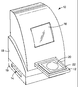
[0015) FIG. 1 is a perspective view of a biological scanner in accordance with
one
embodiment of the invention.
[0016) FIG. 2 is another perspective view of an exemplary biological scanner.
[0017) FIGS. 3 and 4 are top views of an exemplary growth plate.
[0018] FIG. 5 is a conceptual block diagram illustrating exemplary internal
components of a biological scanner.
[0019) FIGS. 6A-6C are cross-sectional side views collectively illustrating
operation
of a loading mechanism for loading a~biological grovVfh plate into a
biological scanner.
[0020) FIGS. 7A-7C are additional cross-sectional side views illustrating the
operation of a loading mechanism and an ejection mechanism.
CA 02504948 2005-05-03
WO 2004/051267 PCT/US2003/037384
[0021] FIGS. 8A and 8B are additional perspective views of a biological
scanner in
accordance with another embodiment of the invention.
[0022] FIGS. 9A and 9B illustrate an exemplary electrical coupling
configuration for
a mounting platform and scanning unit of a biological scanner.
[0023] FIGS. l0A and lOB illustrate another exemplary electrical coupling
configuration for a mounting platform and scanning unit of a biological
scanner.
DETAILED DESCRIPTION
[0024] The invention is directed to a biological scanner for biological growth
plates.
A biological growth plate can be presented to the biological scanner, which
generates
an image of the plate and may perform an analysis of the image to detect
biological
growth.
[0025] In particular, the scanner may enumerate or otherwise quantify an
amount of
biological agents that appear in the image, such as a number of bacteria
colonies. In
this manner, the biological scanner automates the analysis of biological
growth plates,
thereby improving such analysis and reducing the possibility of human error.
[0026] In addition, the biological scanner may incorporate an automated
loading and
ejection system that facilitates handling of biological growth plates, as well
as a
multiple-position mounting platform that enables the biological scanner to
occupy
different orientations for ease of placement and use in a variety of
laboratory
environments presenting different space limitations and layout
characteristics.
[0027] The invention may be useful with a variety of biological growth plates.
For
example, the invention may be useful with different plate-like devices for
growing
biological agents to enable detection and/or enumeration of the agents, such
as thin-
film culture plate devices, Petri dish culture plate devices, and the like.
Therefore, the
term "biological growth plate" will be used broadly herein to refer to a
medium suitable
for growth of biological agents to permit detection and enumeration of the
agents by a
scanner. In some embodiments, the biological growth plate can be housed in a
cassette
that supports multiple plates, e.g., as described in U.S. Patent No. 5,573,950
to Graessle
et al.
[0028] FIG. 1 is a perspective view of a biological scanner 10 in accordance
with one
embodiment of the invention. As illustrated, biological scanner 10 includes a
drawer
CA 02504948 2005-05-03
WO 2004/051267 PCT/US2003/037384
12 that receives a biological growth plate, and moves the growth plate into
biological
scanner 10 for scanning and analysis. Biological scanner 10 also includes an
ejection
slot 14 through which the growth plate can be ejected following analysis of
the
biological growth plate. Biological scanner 10 may also include other
features, such as
a display screen 16 to display the progress or results of analysis of the
biological
growth plate to a user. Alternatively or additionally, display screen 16 may
present to a
user an image of the plate inserted into biological scanner 10 via drawer 12.
In some
embodiments, the displayed image may be optically magnified or digitally
scaled
upward.
[0029] As further shown in FIG. 1, biological scanner 10 may have a two-part
design.
In particular, biological scanner 10 may have a scanner unit 18 and a mounting
platform 19. Scanner unit 18 is mounted on mounting platform 19 and, as will
be
explained, may occupy multiple orientations relative to the mounting platform.
In the
example of FIG. l, scanner unit 18 includes a drawer 12, which extends outward
from
the scanner unit 18 to receive a biological growth plate and retracts into the
scanner
unit 18 to place the biological growth plate into scanner 10 for analysis.
[0030] Scanner unit 18 also houses an imaging device for scanning the
biological
growth plate and generating an image of an inserted plate. In addition,
scanner unit 18
may house a processor that performs analysis of the scanned image, e.g., in
order to
determine the level of biological agents in the plate. For example, upon
insertion of the
biological growth plate via drawer 1'2, the plate may be positioned adjacent
to a platen
which is also housed within scanner unit 18. An image of the growth plate can
be
captured when the plate is positioned within scanner unit 18, e.g., adjacent
to a platen.
Then, when the drawer 12 is subsequently opened, the plate drops downward into
the
mounting platform 19 for ejection via ejection slot 14 .
[0031] Mounting platform 19 houses a conveyor that ejects a plate from
biological
scanner 10 via ejection slot 14. After a biological growth plate is inserted
into drawer
12, moved into scanner unit 18, and scanned, the biological growth plate drops
into
mounting platform 19, where a horizontal conveyor, such as a moving belt,
ejects the
plate via slot 14. A display screen 16 on scanner unit 18 of scanner 10 may
display
results of analysis of a plate being scanned.
CA 02504948 2005-05-03
WO 2004/051267 PCT/US2003/037384
[0032] FIG. 2 is another perspective view of biological scanner 10. As shown
in
FIG. 2, drawer 12 extends outward from biological scanner 10 to receive a
biological
growth plate 20. As illustrated, drawer 12 may include a platform 22 on which
plate 20
rests, and a set of cam levers 24, which facilitate the precise positioning of
plate 20
within scanner 10. Upon placement of biological growth plate 20 on platform
22,
drawer 12 retracts into scanner unit 18 to place the biological growth plate
in a
scanning position, i.e., a position at which the biological growth plate ~is
optically
scanned.
[0033] FIGS. 3 and 4 are top views of an exemplary growth plate 20. By way of
example, a suitable growth plate 20 may comprise biological growth plates sold
by 3M
under the trade name PETRIFILM plates. Alternatively, biological growth plate
20
may comprise other biological growth plates for growing particular bacteria or
other
biological agents. Biological growth plates 20, for example, may facilitate
the rapid
growth and detection of bacteria or other biological agents including, for
example,
aerobic bacteria, E. coli, coliform, enterobacteriaceae, yeast, mold,
Staphylococcus
aureus, Listeria, Campylobacter and the like. The use of PETRIFILM plates, or
other
growth plates, can simplify bacterial testing of food samples. Moreover, as
outlined
herein, biological scanner 10 can further simplify such testing by automating
the
process of scanning results on a biological growth plate 20, e.g., by counting
bacterial
colonies on an image of the plate.
[0034] As shown in FIG. 3, a biological growth plate 20A defines a growth area
30.
Optionally, growth area 30 may be a flat surface or a recessed well. A
determination of
whether a given sample being tested in plate 20A is acceptable, in terms of
bacterial
colony counts, may depend on the number of bacterial colonies per unit area.
Accordingly, scanner 10 quantifies the amount of bacterial colonies per unit
area on
plate 20A, and may compare the amount, or "count," to a threshold. The surface
of
biological growth plate 20A may contain one or more growth enhancing agents
designed to facilitate the rapid growth of one or more types of bacteria or
other
biological agents.
[0035] After placing a sample of the material being tested (typically in
liquid form)
on the surface of biological growth plate 20A, plate 20A can be inserted into
an
incubation chamber (not shown). In the incubation chamber, bacterial colonies
or other
CA 02504948 2005-05-03
WO 2004/051267 PCT/US2003/037384
biological agents being grown by growth plate 20 manifest themselves, as shown
in
biological growth plate 20B of FIG. 4: The colonies (represented by the
various dots
on biological growth plate 20B) may appear in different colors on plate 20B,
which can
facilitate and improve automated detection by scanner 10.
[0036] In some cases, a biological growth plate 20 may include identification
(ID)
markings to identify the type of biological agent being grown and tested by
the given
plate, or to identify the manufacturerof the plate for quality assurance
purposes.
Moreover, the detection and interpretation of the ID markings may be automated
within
biological scanner 10. For example, a processor within biological scanner 10
may
implement different image processing routines or counting algorithms to count
different
types of bacterial colonies grown on different types of growth plates, as
identified by
the ID markings.
[0037] FIG. 5 is a conceptual block diagram illustrating internal operation of
biological scanner 10. As illustrated in FIG. 5, a biological growth plate 20
is
positioned within biological scanner 10 on platform 22 of drawer 12 (not shown
in FIG.
5). More specifically, inside scanner unit 18 of biological scanner 10,
platform 22
elevates biological growth plate 20 to position the growth plate adjacent to
platen 52.
In other words, inside scanner unit 18 of biological scanner 10, biological
growth plate
20 is sandwiched between platform 22 and platen 52. Platen 52 may define a
focal
plane for radiation emitted by an imaging device 54 to scan biological growth
plate 20.
Accordingly, platen 52 is optically transparent, permitting transmission of
the radiation
to the surface of growth plate 20. In some cases, platform 22 may comprise a
first
platen and platen 52 may comprise a second platen within biological scanner
10. In
that case, it would be desirable to position the plate between the two
platens, e.g., in a
sandwich-like configuration, to facilitate illumination on both sides of the
plate during
imaging.
[0038] Imaging device 54 generates an image of biological growth plate 20 by
scanning radiation across growth plate 20 and capturing reflected or
transmitted
radiation to form an image. In some embodiments, imaging device 54 may be
formed
by a collection of discrete components, including illurriinatiori-subsystem
and an image
capture subsystem. The illumination subsystem may take the form of a variety
of
radiation sources such as lamps, light emitting diodes, and the like. The
image capture
CA 02504948 2005-05-03
WO 2004/051267 PCT/US2003/037384
subsystem may take the form of a line or area camera that receives reflected
or
transmitted radiation.
[0039] A processor 56 controls the illumination and image capture processes,
and
processes captured imagery to identify or enumerate the amount of biological
agents in
plate 20 based on the generated image. For example, imaging device 54 may
comprise
a camera that generates one or more digital images of biological growth plate
20 and
provides the digital images to processor 56 for analysis. Processor 56
generates results,
such as a bacterial colony count, and presents the results to a user, e.g., by
driving
display screen 16 (FIG. 1) to display the results. In addition, processor 56
may identify
ID markings on plate 20, and select an appropriate image processing routine
and
biological analysis algorithm based on the type of plate being used. For
example, the
counting of bacterial colonies or other biological agents may be performed
differently
for different types of plates, e.g., plates used to grow different types of
bacteria.
[0040] In one exemplary embodiment, platform 22 comprises a first platen that
provides back illumination to biological 'growth plate 20 via a three color
illumination
system, which may incorporate red, green and blue (RGB) illumination LEDs. In
that
case, the RGB LEDs may provide side illumination to platform 22 and thereby
provide
back illumination to a biological growth plate 20 that rests on platform 22.
In addition,
similar RGB illumination LEDs may be used to provide top illumination of
platen 52.
Front illumination can be delivered to biological growth plate 20 via platen
52. Thus,
platform 22 and platen 52 may collectively form an illumination chamber used
to
provide front and back illumination to biological growth plate 20.
[0041] Upon illumination, imaging device 54 captures one or more images of
biological growth plate 20 and provides the images) to processor 56 for
analysis. In
one example, imaging device 54 comprises a monochromatic imaging device that
captures monochromatic images of biological growth plate 20. For example,
biological
growth plate 20 may be illuminated by one or more red LEDs, at which time
imaging
device 54 generates a first image. Then, biological growth plate 20 may be
illuminated
by one or more green LEDs, at which time imaging device 54 generates a second
image. Finally, biological growth plate 20 may be illuminated by one or more
blue
LEDs, at which time imaging device 54 generates a third image.
CA 02504948 2005-05-03
WO 2004/051267 PCT/US2003/037384
[0042] Processor 56 receives the three different monochromatic images and then
performs analysis on the individual images in order to generate a bacterial
colony
count. The use of a monochromatic imaging device 54 to generate one or more
separate monochromatic images may improve image resolution for each color, and
at
the same time, can reduce implementation costs associated with imaging device
54. The
different images can be combined by processor 56 for viewing or analysis
purposes.
Alternatively, or in addition, processor 56 may analyze individual images
obtained
during illumination with individual colors.
[0043] In some embodiments, scanner 10 may process images of different
biological
growth plates 20 according to different image processing profiles. The image
processing profiles may be selected based on user input or identification of
the type of
biological growth plate 20 presented to scanner 10. The image processing
profile may
specify particular image capture conditions, such as illumination intensities,
exposure
durations, and colors, for capturing images of particular plate types. Thus,
the scanner
may apply different image capture conditions, including different illumination
conditions, in processing images of different biological growth plates 20.
[0044] As an illustration, some types of biological growth plates 20 may
require
illumination with a particular color, intensity and duration. In addition,
some biological
growth plates 20 may require only front or back illumination, but not both.
For
example, an aerobic count plate may require only front illumination as well as
illumination by only a single color such as red. Alternatively, an E.
coli/Coliform plate
may require only back illumination and a combination of red and blue
illumination.
Similarly, particular intensity levels and durations may be appropriate. For
these
reasons, illumination may be controlled in response to image capture
conditions
specified by an image processing profile.
[0045] After plate 20 has been scanned by biological scanner 10, platform 22
moves
to release plate 20 onto conveyor 58, which is housed in mounting platform 19.
In
particular, drawer 12 (not shown in FIG. 5) re-opens to withdraw platform 22
from the
scanner unit 18 of biological scanner 10. At that point, however, plate 20 may
be
temporarily held in place, e.g., by a clamp, pincer~-securing lever, or other
apparatus as
outlined in greater detail below. Consequently, the subsequent movement of
platform
22 from scanner unit 18 does not move plate 20 from its location adjacent
platen 52.
CA 02504948 2005-05-03
WO 2004/051267 PCT/US2003/037384
11
Instead, once platform 22 has been moved, the apparatus (not shown in FIG. 5)
temporally holding plate 20 adjacent platen 52 can release plate 20 to fall
onto
conveyor 58 of mounting platform 19. Conveyor 58 ejects the biological growth
plate
20 from mounting platform 19 of biological scanner 10 via slot 14 (FIG. 1).
[0046] FIGS. 6A-6C are cross-sectional side views collectively illustrating
the
movement of a drawer 12 into the biological scanner 10, causing elevation of a
biological growth plate 20 into a desired location within biological scanner
10. In
particular, drawer 12 moves laterally outward from scanner unit 18 to open and
thereby
receive a biological plate 20 placed on platform 22 by a user. Drawer 12 then
retracts
into scanner unit 18 to place biological growth plate 20 in a scanning
position. A
motor or other suitable mechanical control mechanism can be used to actuate
drawer 12
for the lateral movement which opens and closes drawer 12. To open drawer 12,
a user
may push inward against the drawer or depress an eject button (not shown) on
scanner
unit 18. Similarly, to close drawer 12, the user may again push inward against
the
drawer or depress the eject button. In each case, drawer 12 may be coupled to
a switch
that senses the force applied inward against the drawer and toward scanner
unit 18 by
the user. A motor or other mechanical control mechanism may be responsive to
the
switch to automatically open and close the door.
[0047] Drawer 12 includes a platform 22 mounted on a sliding cartridge 62 via
levers
24A and 24B or another suitable attachment mechanism. Sliding cartridge 62, in
turn,
may attach to the motor (not shown) that causes the lateral movement of drawer
12. As
an example, the motor may drive sliding cartridge 62 via a variety of
mechanical
transmissions, such as a lead screw or pulley arrangement. A spring 64, or the
like, can
be used to add a spring bias to platform 22. As illustrated in FIGS. 6A and
6B, drawer
12 moves laterally into biological scanner 10. As shown in FIG. 6B, this
lateral
movement (illustrated by the arrow) causes a leading edge 63 of platform 22 to
abut
stop 66.
[0048] Once platform 22 abuts stop 66, additional lateral movement of platform
22 is
impeded, such that relative movement between sliding cartridge 62 and platform
22
causes platform 22 to elevate. More specifically; -additional lateral
movement' of
sliding cartridge 62 (from the position illustrated in FIG. 6B to that
illustrated in 6C)
causes levers 24 to pivot and thereby elevate platform 22 to a location
adjacent platen
CA 02504948 2005-05-03
WO 2004/051267 PCT/US2003/037384
12
52. In other words, the elevation of platform 22 places plate 20 at a desired
location
within scanner 10, i.e., adjacent platen 52, where an image of plate 20 can be
taken for
analysis.
[0049] Levers 24 may be connected approximately near the four corners of
platform,
or a greater or fewer number of levers may be used. In any case, levers 24 can
be
configured to raise and lower in only one lateral direction so that once
platform 22 is
completely elevated, the lateral movement of sliding cartridge 62 is
inhibited.
Additionally, once platform 22 has elevated biological growth plate 20 to this
desired
location, plate spring 64 may flex to further bias plate 20 against platen 52.
At this
point, one or more images of biological growth plate 20 can be scanned and
processed,
and then used for analysis to determine the amount of biological agents grown
on plate
20.
[0050] FIGS. 7A-7C are additional exemplary cross-sectional side views
illustrating
the movement of a drawer 12 into the biological scanner 10 and the subsequent
ejection
of drawer 12 from biological scanner 10. As illustrated in FIGS. 7A-7C,
biological
scanner 10 includes an apparatus 72 that temporarily holds a plate 20C at the
location
adjacent platen 52. As shown in FIG. 7A, drawer 12 moves laterally into the
scanner to
move plate 20C to a location where it can be imaged. FIG. 7B shows biological
scanner 10 with the drawer 12 moved into the scanner unit 18 to position plate
20C. In
that case, platform 22 has elevated plate 20C to the desired location where
imaging can
occur, i.e., adjacent platen 52.
[0051] In the example of FIGS. 7A-7C, apparatus 72 comprises a clamp, pincer,
securing lever, or the like, to temporally hold plate 20C at the location
adjacent platen
52. Accordingly, when drawer 12 is subsequently opened (as illustrated in FIG.
7C),
apparatus 72 temporarily holds plate 20C at that same location, preventing the
plate
from being withdrawn from scanner unit 18 with drawer 12. In other words, when
drawer 12 is subsequently opened, plate 20C does not remain on platform 22.
Instead,
apparatus 72 temporarily holds plate 20C at the location adjacent platen 52
when the
drawer is subsequently opened. Then, once drawer 12 has opened a sufficient
amount,
vacating an area beneath growth plate 20C, apparatus 72 releases plate 20C,
which falls
onto a motor driven conveyor 58 housed within mounting platforml9 of
biological
scanner 10.
CA 02504948 2005-05-03
WO 2004/051267 PCT/US2003/037384
13
[0052] Conveyor 58 moves biological growth plate 20C to eject it from
biological
scanner 10 via ejection slot 14 in mounting platform 19. Upon projection of
drawer 12
outward from scanner unit 18 and ejection of biological growth plate 20C from
ejection
slot 14, another biological growth plate 20D can be inserted onto platform 22
of drawer
12 for movement into biological scanner 10. In this manner, another biological
growth
plate 20 can be inserted into biological scanner 10, properly positioned
within the
scanner unit 18, imaged or otherwise scanned to identify or enumerate amounts
of
biological agents, and then ejected from biological scanner 10. The
configuration of
scanner 10 described herein, automates the insertion, positioning, and
ejection of
biological growth plates in a manner that ensures that reliable imaging can
occur and
promotes user convenience, thereby improving the integrity of automated
scanning of
such biological growth plates.
[0053] FIGS. 8A and 8B are additional perspective views of a biological
scanner 10
in accordance with another embodiment of the invention. Again, biological
scanner 10
includes a drawer 12 that can receive.a biological growth plate, and move the
plate into
biological scanner 10. Biological scanner 10 also includes ejection slot 14
through
which the plate 20 can be ejected following analysis. For example, following
analysis
of the biological growth plate 20 inside scanner unit 18, the plate 20 may be
released,
as outlined herein, to fall through hole 85 and into mounting platform 19. A
conveyor
(not shown in FIGS. 8A and 8B) housed in mounting platforml9 can then eject
the
plate 20 via ejection slot 14. Biological scanner 10 may also include other
features,
such as a display screen 16 to,display the analysis of the biological plate to
a user, as
further shown in FIGS. 8A and 8B.
[0054] In the embodiment illustrated in FIGS. 8A and 8B, scanner unit 18 and
mounting platform 19 of biological scanner 10 are detachable from one another.
Furthermore, scanner unit 18 and mounting platform 19 of biological scanner 10
are re-
configurable, in that scanner unit 18 and mounting platform 19 can be rotated
relative
to one another. In other words, scanner unit 18 and mounting platform 19 can
be
attached to one another in one of a plurality of different positions.
Accordingly, drawer
12 of scanner unit 18 and the ejection slot 14 of mounting platform 19 can be
positioned along a common side of biological scanner 10 (as illustrated in
FIG. 8A), or
alternatively, drawer 12 of scanner unit 18 and the ejection slot 14 of
mounting
CA 02504948 2005-05-03
WO 2004/051267 PCT/US2003/037384
14
platform 19 can be positioned such that they are not on a common side of
biological
scanner 10 (as illustrated in FIG. 8B). This re-configurability can allow
biological
scanner 10 to be placed in different laboratory environments that have
different space
limitations or layout concerns.
[0055] In order to facilitate the attachment of scanner unit 18 to mounting
platform
19 in different relative positions, scanner unit 18 may include pins 82 that
mate with
sockets 84 formed in mounting platform 19. Alternatively, mounting platform 19
may
include the pins 82 and scanner unit 18 may be formed with the sockets 84. The
term
"socket," as used herein, may refer to a variety of hole, receptacle or other
female
terminal structures capable of engagement with a pin. The term "pin," as used
herein,
may refer to a variety of peg, pin, plug, protrusion or other male terminal
structures
capable of engagement with a socket. Pins and sockets 82, 84 form electrical
interfaces
for communication of power between mounting platform 19 and scanner unit 18.
For
example, scanner unit 18 may provide operating power to mounting platform 19.
Alternatively, mounting platform 19 may provide operating power to scanner
unit 18
via pins and sockets 82, 84. In some embodiments, pins and sockets 82, 84 also
may
communicate control or status signals, e.g., to control operation of the
conveyor in
mounting platform 19. In addition to providing electrical connections, pins
and sockets
82, 84 may provide mechanical positioning, alignment and registration. between
scanner unit 18 and mounting platform 19.
[0056] Other cooperative engagement hardware may be provided in place of pins
and
sockets. In any case, the distance between any two sockets 84 along a common
side of
mounting platform 19 and the distance between any two pins 18 along a common
side
of scanner unit 18 is substantially the same. Accordingly, pins 82A and 82B of
scanner
unit 18 can mate with sockets 84A and 84B of mounting platform 19 when the
scanner
unit and mounting platform are coupled to one another in a first position as
illustrated
in FIG. 8A, or alternatively, pins 82A and 82B of scanner unit 18 can mate
with sockets
84B and 84C of mounting platform 19 when the scanner unit and mounting
platform
are coupled to one another in a second position as illustrated in FIG. 8B. The
mounting
arrangement depicted in FIGS. 8A and 8B permits scanner unit 18 to be rotated
three-
hundred sixty degrees (ninety degrees between adjacent positions) between four
different positions.
CA 02504948 2005-05-03
WO 2004/051267 PCT/US2003/037384
[0057] In some embodiments, scanner unit 18 and mounting platform 19 may have
separate power supplies, e.g., separate batteries or separate AC power cords.
Alternatively, a single power supply or power cord may be used. In the latter
case, one
or more pins 82 and one or more sockets 84 may include electrical connectors
that
electrically couple scanner unit 18 to mounting platform 19 and facilitate the
transfer of
electrical current between the scanner unit 18 and mounting platform 19.
Moreover, in
some embodiments, control signals may be transferred between scanner unit 18
and
mounting platform 19, e.g., to control on/off switching of the conveyor within
mounting platform 19 when a plate is to be ejected. Alternatively, the
conveyor within
mounting platform 19 may run substantially all the time when power is
supplied,
without being controlled by on/off switching.
[0058] In one example, one or more of the pins 82 may be "active" in the sense
that
they provide both an electrical interface to facilitate the transfer of
electrical current,
and a signal transfer interface to facilitate the transfer of control signals.
Each of
sockets 84 may be configured to couple with either the active pin or one of
the passive
pins that do not include electrical interfaces. In this manner, current and
control signals
can be transferred between scanner unit 18 and mounting platform 19 regardless
of the
relative positioning of the portions 18, 19. Many other types of electrical
and control
signal connections could also be used, including, for example, internal or
external
cords, wires, or the like.
[0059] FIGS. 9A and 9B illustrate an exemplary electrical coupling
configuration for
a mounting platform and scanning unit of a biological scanner. In particular,
FIG. 9A
provides a top view of mounting platform 19 and FIG. 9B provides a bottom view
of
scanner unit 18. A diode rectifier bridge 86 and other electrical coupling
hardware is
represented functionally in FIG. 9A to depict an exemplary electrical coupling
configuration. In operation, as scanner unit 18 is placed on mounting platform
19,
sockets or pins 84A-84D engage reciprocal sockets or pins 82A-82D on the
mounting
platform. For purposes of illustration, it will be assumed that scanner unit
18 includes
sockets 84 and mounting platform 19 includes pins 82.
[0060] Pins 82 engage sockets 84 upon placement of scanriei unit 18 on
mounting
platform 19. In some embodiments, sockets 84 may define either spring-loaded
electrical contacts for electrically conductive engagement with pins 82 or
insulative
CA 02504948 2005-05-03
WO 2004/051267 PCT/US2003/037384
16
surfaces to electrically insulate pins 82. In the example of FIG. 9B, scanner
unit
includes a first socket 84A having an electrical contact, a second socket 84B
having an
insulating surface, a third socket 84C having an insulating surface, and a
fourth socket
84D having an electrical contact. Accordingly, two pins 82 from mounting
platform 19
are electrically coupled to sockets 84A, 84D, depending on the orientation of
scanner
unit 18 relative to the mounting platform. In particular, either pin 82A or
82B is
coupled to one of sockets 84A or 84D, which are mounted on diagonally opposing
corners of scanner unit 10. Similarly, either pin 82C or 82D is coupled to one
of
sockets 84A or 84D. In this manner, one of pins 82A, 82B is electrically
coupled and
the other is electrically insulated, while one of pins 82C, 82D is
electrically coupled
and the other is electrically insulated. Insulating surfaces in sockets 84B,
84C serve to
prevent shorting of the two unused pins 82 to the housing of scanner 10 or
other
surfaces.
[0061] Diode rectifier bridge 86 serves to deliver a steady power supply from
mounting platform 19 to electronics within scanner unit 18, or vice versa,
regardless of
the orientation of the mounting platform and scanner unit. As shown in FIG.
9A,
opposing terminals 87, 89 of diode rectifier bridge 86 are coupled across a
motor 91
housed within mounting platform 19. Motor 91 drives a conveyor within mounting
platform 19. Terminal 93 of diode rectifier bridge 86 is coupled to pins 82A,
82B, one
of which is coupled to a conductive socket 84A, 84D in scanner unit 18.
Terminal 95
of diode rectifier bridge 86 is coupled to pins 82C, 82D, one of which is
coupled to a
conductive socket 84A, 84D in scanner unit 18. In this manner, conductive
sockets
84A, 84D receive a constant source of power from mounting platform 19
regardless of
the orientation of scanner unit 18 relative to the mounting platform.
Alternatively,
conductive pins 82C, 82D receive a constant source of power from scanner unit
18.
[0062] FIGS. l0A and lOB illustrate another exemplary electrical coupling
configuration for mounting platform 19 and scanning unit 18 of biological
scanner 10.
In the example of FIGS. l0A and lOB, conductive sockets 84A, 84B in scanner
unit 18
are disposed adjacent one another, i.e., on adjacent corners of the scanner
unit. To
support supply of power from mounting platform 19 to scantier uriif 18, or
vice versa,
regardless of the orientation of scanner unit 18, the mounting platform
includes an
alternative arrangement for diode rectifier bridge 88 and pins 82.
CA 02504948 2005-05-03
WO 2004/051267 PCT/US2003/037384
17
[0063] As shown in FIG. 10A, terminals 97, 99 of diode rectifier bridge 88 are
coupled across a motor 101. Also, pins 82A and 82D are electrically coupled to
one
another, and to terminal 103 of diode rectifier bridge 88. Similarly, pins 82B
and 82C
are electrically coupled to one another, and to terminal 105 of diode
rectifier bridge 88.
In operation, either pin 82A or 82D is coupled to one of sockets 84A or 84D,
which are
mounted on adjacent corners of scanner unit 10. Similarly, either pin 82B or
82C is
coupled to one of sockets 84A or 84b. In this manner, one of pins 82A, 82D is
electrically coupled and the other is electrically insulated, while one of
pins 82B, 82C is
electrically coupled and the other is electrically insulated. Again, the
arrangement
shown in FIGS. l0A and lOB serves to provide a continuous source of power from
mounting platform 19 to scanner unit 18, or vice versa, without regard to the
relative
positioning of the scanner unit and the mounting platform.
[0064] In general, scanner unit 18 and mounting platform 19 of scanner 10
provide
multiple possible positions. More particularly, scanner unit 18 and mounting
platform
19 may permit selective relative positioning that support continued supply'of
power,
e.g., without the need to disconnect cables or actuate switches. Instead, the
user may
conveniently reposition scanner unit 18 and mounting platform 19 to obtain
desired
plate loading and ejection orientations, which may be a function of a
laboratory
environment and applicable space limitations.
[0065] A number of embodiments of a biological scanner have been described.
For
example, techniques and structures have been described for automating the
insertion of
biological growth plates into a scanner and the ejection of biological growth
plates
from the scanner. In particular, the insertion and ejection techniques
described herein
can ensure that reliable imaging can occur within the biological scanner.
Also, a re-
configurability feature for a biological scanner has been described in which a
scanner
unit of the biological scanner can be attached to a mounting platform of the
scanner in
one of a plurality of possible configurations. This re-configurability feature
can allow
the biological scanner to be placed in different laboratory environments that
have
different space limitations or concerns.
[0066] Nevertheless, various modifications may be -made without departing from
the
spirit and scope of the invention. For example, one or more features described
herein
may be used with or without other described features. Moreover, several
features
CA 02504948 2005-05-03
WO 2004/051267 PCT/US2003/037384
18
described herein may be used in a biological scanner that simply generates a
high
quality image of the biological growth plate, and presents the high quality
image to a
user for analysis. In that case, a processor used to count bacterial colonies
may be
eliminated in favor of a less complicated processor that simply presents
images to a
user. In other words, the processor may simply drive a display such as display
16 (FIG.
1 ) to present a high quality image of the plate to a user so that the user
can analyze the
image and identify or enumerate the number of bacterial colonies. These and
other
embodiments are within the scope of the following claims.