Note : Les descriptions sont présentées dans la langue officielle dans laquelle elles ont été soumises.
CA 02505125 2005-05-05
WO 2004/043233 PCT/US2003/035690
ASSAY FOR AUTOANTIBODIES TO FOLATE RECEPTORS
FIELD OF THE INVENTION
This invention relates to autoantibodies to folate receptors (FRs) on the
surface of cells. In particular, the present invention relates to an assay for
detecting
the autoantibodies to the FRs in mammals, particularly.humans. The invention
also
relates to the prevention of diseases, disorders or conditions associated with
impaired
, cellular uptake of folate as a consequence of the presence of autoantibodies
to the
FRs, which causes folate-sensitive abnormalities, such as birth defects (e.g.,
a neural
tube defect, i.e., NTD), infertility, spontaneous abortion, male sterility,
unsuccessful in
vitro fertilization, neurologic disorders, and impaired intestinal absorption
of folic
acid. The present invention provides a, method of diagnosing such folate-
sensitive
abnormalities. The present invention further relates to 'an assay for
detecting
autoantibodies to the FRs in women and provides diagnostic screening for women
at
risk of a pregnancy with fetal complications, such as a NTD.
BACKGROUND OF THE INVENTION
Folic acid is essential for normal embryonic development because it
participates in one-carbon metabolism for the synthesis of nucleic acids and
amino
acids required by highly proliferative embryonic cells (Lucock, Mol. Genet.
Metab.
71:121 (2000)). Maternal nutrition, especially with regard to folate intake
for the
prevention of neural tube defects (NTDs), has been the focus of much attention
for the
past four decades. The early studies linking folate intake to the prevention
of NTDs
(Hibbard and Smithells, Obstet. Gynaecol. Br. Commonwealth 71:529 (1964);
Hibbard and Smithells, Lancet l, 1254(1965); Sinithall et al., Arch. Dis.
Child 51:944
(1976)) have been confirmed by recent randomized controlled studies. These
studies
show that women who were given periconceptional folate supplements had about a
70% reduction in the.occurrence and recurrence of NTDs (Laurence et al., Br.
Med. J..
(Clip. Res. Ed.) 282:1509 (l 981); MRC Vitamin Study Research Group, Lancet
338:131(1991); Czeizel and Dudas, N. Engl. J. Med. 327:1832 (1992)).
CA 02505125 2005-05-05
WO 2004/043233 PCT/US2003/035690
However, most mothers who give birth to babies having NTDs and/or
other birth anomalies, do not exhibit signs of clinical folate deficiency.
Therefore,
extensive research has been in progress to identify genetic defects that
impair cellular
metabolism or uptake of folate. The effect of these genetic defects can be
corrected
by pharmacologic intake of folic acid (Kirke et al., Lancet 348:1037 (1996)).
Pharmacologic intake of folic acid raises the plasma concentration of the
vitamin and
provides sufficient mother-to-fetus transport of folate to bypass impaired
folate uptake
and/or intracellular metabolism, thereby reducing the occurrence of folate-
sensitive
congenital abnormalities. Thus far, a number of candidate genes encoding some
of
the enzymes related to folate metabolic pathways have been identified in
association
with NTDs. These genes, however, account for only a small number of birth
defects
(van der Put et al., Exp. Biol. Med. (Maywood), 226:243 (2001)).
Decreased folate uptake by maternal and/or fetal placental cells and
embryonic cells in the presence of normal or a low-level blood folate, may be
caused
by quantitative or functional defects in the membrane proteins required for
the uptake
of folate. No genetic abnormalities that alter expressions of these membrane
proteins
have been unambiyously identified (De Marco el al., Am. J. Med. Genet. 95:216
(2000); Barber et al., Am. J. Med. Genet. 76:310 (1998)).
Cellular uptake of folate is mediated via two distinct pathways: the
reduced folate carrier (RFC) (Henderson, Auhu. Rev. Nuts. 10:319 (1990)),
which is
an integral transmembrane protein that is present in most cells, and the
folate receptor
(FR), which is anchored to the plasma membrane of cells by a
glycosylphosphatidylinositol (GPI) adduct that internalizes folate by
endocytosis of
the folate-receptor complex (Antony, Ahhu. Rev. Nutr. 16:501 (1996)). There
are
three isoforms of the FR (a,, (3, y), which are expressed at different levels
in tissues
and have different affinities to folate. Cellular uptake of folate depends on
the
expression levels of each folate receptor isoform (Ross et al., Caracey~
73:2432 (1994)).
The contributions of the FRs and the RFC to the cellular uptake of
folate during embryogenesis were not appreciated until Piedrahita et al.
demonstrated
that the ortholog of the human FRoc in the mouse (Folbp 1 ) is essential for
embryonic
organogenesis while the mouse Folbp2, which is the ortholog of the human FR~i,
appears to have no function in embryogenesis (Piedrahita, et al., Nat. Genet.
23: 228
2
CA 02505125 2005-05-05
WO 2004/043233 PCT/US2003/035690
(1999)). Nullizygous Folbpl knockout mouse embryos (Folbpl -/-) had
significant
congenital malformations and none survived beyond gestation day 10, while
nullizygous Folbp2 -l , heterozygous Folbpl +/- or Folbp2 +/- embryos, showed
no
difference in development and viability as compared to wild type embryos
(Piedrahita,
et al.). Knockout of the RFC gene also proved lethal to embryos (Zhao, et al.,
J. Biol.
Claem. 276:10224 (2001)). Heterozygous RFC dams that were given folic acid
produced normal full term nullizygous RFC (-/-) offspring. These studies
indicated
that both RFC and FR pathways for folate uptake are essential for fetal
development.
The ~ Ra is expressed in huyan placental syncytiotrophoblasts. High
concentrations of both FRa and FR(3 isoforms are found in maternal placental
tissue
(Prasad et al., Biochim. Biophys. Acta. 1223:71 (1994)). The essential
function of the
FRs in human embryogenesis is to ensure cellular uptake of folate. It has been
reported that mother-to-fetus transfer of folate is mediated via the FRa,
(Clark et al.,
Hum. Repr~od. Update 7:501 (2001)). This prompted studies of the FRa gene as a
candidate gene responsible for a folate sensitive birth defect, such as a
neural tube
defect. However, no consistent nucleotide polyrnorphisms or mutations that
affect
expression of the FR gene, or the function of the FR protein, have been
identified
(Barber et al.; De Marco et al.) that could account for the occurrence rate of
NTDs.
Instead, only a small fraction of women with a NTD-complicated pregnancy were
shown to have a polymorphism in the gene encoding folate-dependent enzymes,
such
as methylene-tetrahydrofolate reductase (MTHFR) (Christensen et al., Am. J.
Med.
Genet. 84(2):151-57 (1999)).
Since genetic studies have not provided evidence that a mutation of
relevant genes (encoding enzymes or FRs) are a significant cause of congenital
dysmorphogenesis, it is, therefore, possible that NTDs and other folate-
sensitive
abnormalities could be autoimmune disorders. Autoantibodies to several
proteins
have been associated with infertility, miscarriages and fetal abnormalities
(Coulam,
Early Pregnancy 4:19 (2000); Clark et al., Hum. Reprod. Update 7:501 (2001)).
Several previous studies demonstrated that antibodies raised in a rabbit to
kidney,
heart muscle, testes, placenta and other rat tissues, caused dose-dependent
congenital
defects and embryonic resorptions when administered to pregnant rats (Brent et
al.,
3
CA 02505125 2005-05-05
WO 2004/043233 PCT/US2003/035690
Pnoc. Soc. Exp. Bid. Med. 106:523 (1961); Barrow and Taylor, J. Exp. Zool.
176:41(1971); Brent, Proc. Soc. Exp. Biol. Med. 125:1024 (1967)). The
mechanism
by which these anomalies occurred was not established. But the administered
antibodies were concentrated on the yolk sac, suggesting that the antibodies
interfered
with delivery of nutrients to the embryo (Slotnick and Brent, J. Immunol.
96:606
(1966)). Thus, it was speculated that antibodies which block the folate
binding sites
on the FRs (da Costa and Rothenberg, Biochim. Biophys. Acta. 1292:23 (1996))
could
interfere with the cellular uptake of folate. Such interference would then
impair
intracellular folate homeostasis that is essential for normal embryogenesis
and fetal
development.
There is evidence that women who have had a spontaneous or induced
abortion, or a later miscarriage, have an increased risk of a fetal NTD
complication in
a subsequent pregnancy (Evans, Bait. Med. J. 1: 975, (1979); Carmi et al., Am.
J. Med.
Genet. 51: 93, (1994); Cuckle, Prenat. Diagn. 3: 287, (1983)).
The cause of NTD(s) is multifactorial and includes chemotherapeutic
drugs, especially the antifolates (Hernandez-Diaz et al., NEngl JMed. 343:1608-
14,
(2000)), anti-epileptic drugs (Dansky et al., Neur°ology 42: 32-42
(1992))
chromosomal abnormalities (Seller, C'lin Dysrnorphol. 4:202-07 (1995)),
environmental (Finnell et al., Ann NYAcad Sci. 919:261-77 (2000)) and genetic
factors (De Marco and Moroni, Am JMed Genet. 95: 216-23 (2000)). Studies that
have shown a reduction of about 70% in the occurrence of NTDs with folic acid
supplementation beginning at the time of conception (MRC Vitamin Study
Research
Group, supra) provide evidence that folate circumvents either an impaired
intracellular folate-dependent enzymatic pathway, or an inhibitor of the
cellular uptake
of folate. There is, however, no evidence for diminished function of an
enzymatic
pathway that could account for a 70% decrease in the occurrence of NTD with
the
folate supplementation. It is also not known whether the folate-sensitive
disorders are
due to interference of folate uptake by autoantibodies to the FRs. Therefore,
a woman
starting a pregnancy does not know whether grain or pharmacologic folate
supplements would help to prevent congenital defects, such as a neural tube
defect.
The present invention relates to the discovery that folate-sensitive
disorders or conditions, such as infertility, spontaneous abortion,
unsuccessful in vitro
4
CA 02505125 2005-05-05
WO 2004/043233 PCT/US2003/035690
fertilization, or birth defects, are due to interference of folate uptake by
an
autoantibody against the folate receptor. The present invention provides a
reliable
assay to detect autoantibodies to folate receptors in a marmnal, especially in
a human.
SUMMARY OF THE INVENTION
The present invention is directed to the discovery that folate-sensitive
abnormalities are caused by the presence of autoantibodies to the folate
receptors
(FRs) in a subject's body fluids, such as serum. Such folate-sensitive
abnormalities
include, but are not limited to, neural tube defects (NTDs), infertility,
spontaneous
abortion, male sterility, unsuccessful in vitro fertilization following
implantation in
the uterus, neurologic disorders, e.g., dementia, or impaired intestinal
absorption of
folic acid.
One aspect of the present invention is directed to the identification of
autoantibodies to the FRs on the surface of a cell, which blocks folate uptake
and
results in intracellular folate deficiency and therefore affects intracellular
metabolism.
Accordingly, a method for detecting the presence of autoantibodies to folate
receptors
in a biological sample from a subject is provided by the present invention.
Another aspect of the present invention is directed to a method for
detecting the presence of blocking autoantibodies to folate receptors in a
biological
sample from a subject.
Still another aspect of the present invention is directed to a test kit for
detecting autoantibodies to FRs in a biological sample from a subject.
Yet another aspect of the present invention is directed to a test kit for
detecting blocking autoantibodies to FRs in a biological sample from a
subject.
In one aspect, the present invention provides a method for diagnosing a
folate-sensitive abnormality or disorder in a subject at risk of the
abnormality or
disorder by detecting the presence of autoantibodies to FRs in a biological
sample
from the subject.
In another aspect, the present invention provides a method for
screening a woman at risk for having a neural tube defect-complicated
pregnancy by
detecting the presence of maternal autoantibodies to the FRs in a biological
sample
from the woman.
5
CA 02505125 2005-05-05
WO 2004/043233 PCT/US2003/035690
In still another aspect, the present invention provides a method for the
prevention of folate-sensitive abnormalities or disorders.
BRIEF DESCRIPTION OF THE FIGURES
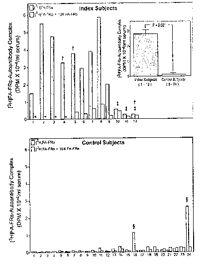
Figure 1 Autoantibodies to Folate Receptors in the Serum from Index Subjects
and Control Subjects shows the results of a binding assay for detecting
autoantibodies to the
FRs in serum from women with a prior NTD pregnancy or a current NTD pregnancy,
and
women that have no history of a NTD. Blue bars : Incubation of serum from
Index and
Control Subjects with [3H]folic-folate receptors. Orange bars: Incubation as
above with a 10
fold excess of unlabeled folic acid-folate receptors that competes out the
binding of [3H]folic-
folate receptors to the autoantibodies. The inset depicts the mean ~ SEM of
the values
obtained for the Index and Control Subjects. Control Subjects 1-4 were
nulligravid and were
excluded from the statistical analysis for the lack of a recognized pregnancy.
The P-value was
determined using Student t-test. Figure lA: * indicates insufficient serum
available for this
determination. ~- subjects were pregnant at the time of blood sampling; $
Subjects 10, 11 and
12 had a NTD-complicated pregnancy and lack the autoantibody to the folate
receptors.
Figure 1B: Subjects 1-4 were nulligravid, 5-16 had previous pregnancies
without NTD
complications, 17-24 were pregnant at the time the blood was sampled.
~ Serum from Control Subjects 16 and 24 contained autoantibodies to the folate
receptors.
Figure 2 Ovarian follicle from a rat showing expression of the FRs on the
oocyte and the granulosa cells: shows that FRs are expressed on the granulosa
cells that
surround the ovum in the ovarian follicle of a rat. Antibodies to the rat FRs
(purified from rat
placenta) were produced in a rabbit, and used to localize the FRs present on
the ovary of a rat.
The brown/orange color identifies the FRs. Oocyte, a; granulosa cells, b.
Figure 3 The Oviduct of a rat (fallopian tube in a human) showing expression
of the FRs on the epithelial lining and on two embryos: shows that epithelial
cells lining the
oviduct of a rat (fallopian tubes in human beings) express the FRs to which
the autoantibodies
can bind, thereby interfering with the advancement of the fertilized ovum
(that has progressed
to a blastocyst stage) to the uterus The autoantibodies can block the uptake
of folate by the
epithelial cells and this contributes to infertility by interfering with the
intracellular
metabolism that requires folate. Antibodies to the FRs present on reproductive
tissue of the
rat. The brown/orange color identifies the FRs.
6
CA 02505125 2005-05-05
WO 2004/043233 PCT/US2003/035690
Figure 4 The uterus of a rat showing expression of the FRs on the endometrial
lining: shows that the folate receptors are highly expressed on the
endometrial lining of a rat
uterus. Antibodies to the rat FRs (purified from rat placenta) were produced
in a rabbit, and
used to localize the FRs present on the endometrium of the uterus of the rat.
The
brown/orange color identifies the FRs.
Figure 5 FRs expression on the epididymis of a rat: shows that the folate
receptors are highly expressed on the epithelial cells of the epididymis of a
rat. Antibodies to
the rat FRs purified from rat placenta were produced in a rabbit, and used to
localize the FRs
present on the epididymis of a rat. The brown/orange color identifies the FRs.
Figure 6 Blocking of [3H]FA Binding to FRs on Placental membranes, KB
cells and ED27 cells by Serum Containing Blocking Autoantibodies to FRs: shows
the
ability of the autoantibodies to FRs to block the binding of folate to human
placental
membranes and to two human cultured cell lines (ED27 cells and KB cells). The
number in
each bar indicates the percent blocking by the autoantibodies of [3H]FA
binding to the apo-
FRs on the cell membranes at 4°C. The methodology is described in
Example 6.
Figure 7 Effect of Isolated Autoantibodies to the FRs on the Cellular Uptake
of
[3H]FA by KB cells: shows the ability of the autoantibodies to FRs to block
the uptake of
folate by cultured KB cells. Autoantibodies to FRs were isolated from the
serum of two
Subjects as described in Example 7. KB cells were pre-incubated overnight at
37°C with this
isolated fraction of serum from either an Index Subject (contains FRs
autoantibodies) ( o ), or
a Control Subject (lacks FRs autoantibodies)( ~ ). Following this incubation,
the uptake of
[3H]FA by the KB cells was determined. A control incubation lacking the
isolated fraction of
serum is shown ( ~ ). The I bars represent the SEM.
Figure 8 Determination of the Binding Affinity of the Autoantibodies for the
Folate Receptors: shows the graphic determination of the binding affinity
constant (K~) of the
autoantibodies to FRs from the serum of five subjects. Placental membranes
with apo-folate
receptors were prepared as described in Methods and incubated overnight at
4°C with the
serum containing autoantibodies. [3H]folic acid was then added and the
fraction bound to the
folate receptors was subtracted from the total folate binding capacity of the
folate receptors to
derive the pmoles of receptors blocked (B) per liter by the autoantibodies.
The ratio of the
autoantibody-blocked receptor to the free apo-receptor (B/F) was used for the
Scatchard
analysis, to compute the apparent association constant (Ka) which is shown in
the inset.
7
CA 02505125 2005-05-05
WO 2004/043233 PCT/US2003/035690
Figure 9 shows the principles and the methodology for detecting the
presence of blocking autoantibodies in the serum to the FRs.
Figure 10 Expression of FRs on brain ventricular surface and choroid
plexus: shows the FRs that are expressed on the choroid plexus of the rat
brain.
Immunohistochemistry of brain tissue using a rabbit polyclonal antiserum to
FRs.
Normal Rabbit serum (NRS) served as the negative control. The brown color
indicates
the localization of the rabbit antibodies on the FRs. ventricular surface
epithelium, vs;
choroid plexus, cp.
Figure 11 Expression of FRs on small intestine: shows the FRs that are
expressed on the intestinal mucosa of the rat. Immunostaining of the rat
duodenum
(A), jejunum (B) and ileum (C) with the rabbit serum containing antibodies to
the rat
FRs. Observe that the intensity of the immunostaining is most evident on the
surface
of the cells in the duodenum and jejunum (as indicated by the arrows) and
substantially reduced on the surface of the ileum indicating diminished
expression of
the FRs in this region of the small intestine. Folates are absorbed
predominantly in the
proximal small intestine (duodenum and jejunum).
DETAILED DESCRIPTION OF THE INVENTION
The present invention is directed to the discovery that folate-sensitive
abnormalities, such as neural tube defects (NTDs), infertility, spontaneous
abortion,
unsuccessful ih vitr°o fertilization, neurologic disorders (e.g.,
dementia), or impaired
intestinal folate absorption, are caused by the presence of the autoantibodies
to the
folate receptors (FRs) in a subject's body fluids, such as serum.
By "test group" or "index group" is meant a group of women, each of
whom had a previous pregnancy, or is currently enduring a pregnancy
complicated by
abnormal fetal development of the central nervous system; or previously gave
birth to
a baby having congenital abnormalities. By "control group" is meant women who
previously had normal pregnancies; women who have never given birth to a baby
having a NTD; women who have never been pregnant (nulligravidas); or women who
have never given birth previously (nulliparous).
By "diagnosing", "diagnosis", "detecting" or "screening" is meant an
act or process of identifying or determining the presence, nature and cause of
a folate-
sensitive abnormality or disorder, through evaluation of patient history,
examination
8
CA 02505125 2005-05-05
WO 2004/043233 PCT/US2003/035690
and identification ofthe presence, in the serum or other body fluids of a
subject, of
autoantibodies against the folate receptors.
By "subject" is meant any mammalian subject, such as a human. A
preferred subject is a woman who previously gave birth to an infant having a
NTD, a
woman who was pregnant with a conceptus having a NTD, a woman who had a
spontaneous or induced abortion, or a later miscarriage. Another preferred
subject is
an egg-donor (i.e., female) or a sperm-donor (i.e., male) for an ih vitro
fertilization
procedure. By "index subject" is meant a subject in an index group or test
group.
By "control subject" is meant a subject in a control group. Without
intending to be bound by any particular theory, it is believed that women who
had an
induced abortion or a miscarriage may have developed autoantibodies to the FRs
but
not any of the folate-sensitive developmental abnormalities. Therefore, such
women
are also considered as control subjects in the present invention.
By "risk" is meant the frequency or possibility of contracting,
developing or having a folate-sensitive abnormality or disorder, such as
infertility,
spontaneous abortion, unsuccessful in vitro fertilization following
implantation in the
uterus, neurologic disorders (e.g., dementia), or impaired intestinal folate
absorption.
"Risk" in accordance with the present invention also connotes giving birth to
a baby
having congenital birth defects, such as NTDs, or enduring a pregnancy with a
conceptus having congenital birth defects.
By "prevention" or "prevent" is meant that the risk of having an
abnormality or disorder can be predicted or determined in sufficient time so
as to keep
the disorder or abnormality from occurring or significantly reduce the risk of
having
the abnormality or disorder.
By "biological sample" is meant a clinical sample for testing taken
from any tissue of a mammal, preferably, body fluid from a mammal, more
preferably, serum from a human. By "control sample" is meant a biological
sample
taken from a subject that is the same or homologous species as the subject to
be
assayed for autoantibodies and is known to have normal biological state, e.g.,
without
detectable autoantibodies against folate receptors. A control sample includes
a
sample taken from a control subject.
9
CA 02505125 2005-05-05
WO 2004/043233 PCT/US2003/035690
An "antibody" refers to an immunoglobulin of any class or subclass, a
portion thereof or an active fragment thereof, wherein an active fragment of
an
antibody retains its specific binding capability. As used herein, an
"autoantibody"
refers to an antibody, e.g., an IgG antibody, in a subject that is directed
against
components of the subject's own body. An "autoimmune disorder" refers to a
disorder or condition that a subject's immune system mistakenly attacks and
leads to
the destruction of the subject's own body cells and/or tissues. An
"autoantibody to the
folate receptors (FRs)" refers to any autoantibody that is directed against
any isoform
or peptide sequence of the FRs, including the a and (3 isoforms of the FRs. In
the
present invention, autoantibodies against FR(s) are also termed anti-FRs
autoantibodies.
The "cell membrane folate receptors (FRs)" or "cell surface folate
receptors (FRs)" refers to any folate receptors (FRs) on the surface/membrane
of a
cell; "circulating folate receptors (FRs)" refers to any isoform of a folate
receptor or its
antigenic components) that circulates in the body fluid of a subject. By "apo-
FRs" is
meant any folate receptor without the ligand, i.e., folic acid, bound to it.
By "folate binding capacity" used herein is meant quantified amount of
the folic acid bound to the FRs on the membrane or matrix per unit volume,
e.g., per
milliliter, of the membrane or matrix.
By ";ell" used herein can be any cell of a tissue culture cell line or any
cell within a specific tissue/organ that binds and internalizes folate, e.g.,
the granulosa
cells that surround the ovum in the ovarian follicle of a mammal, the
epithelial cells
lining the oviduct of a mammal (i.e., fallopian tubes in humans), cells of the
endometrial lining of a mammalian uterus, cells of the mammalian placenta, the
cells
of choroid plexus of a mammalian brain, the cells of intestinal mucosa of a
mammal,
or any mammalian cultured cell line, e.g., I~B cells.
A "blocking autoantibody" refers to an autoantibody that binds with its
antigenic components) and blocks the function of the antigen, which in this
instance,
blocks folate binding to the cell membrane FRs and the subsequent folate
uptake by
the cells.
By "folate supplement" is meant folic acid or folinic acid administered
to a subject in order to overcome intracellular folate deficiency,
particularly because of
CA 02505125 2005-05-05
WO 2004/043233 PCT/US2003/035690
blockage of the folate uptake mechanism by autoantibodies to folate receptors.
By
"pharmacologic amount" is meant an amount much greater than normal to overcome
the deficiency or disorder, e.g., a pharmacologic amount of folate supplement
in the
present invention can be referred to at least 0.8 mg of folic acid daily but
not more
than 4 mg daily to an adult woman.
"Label," "labeled" or "detectably labeled" refers to incorporation of a
detectable marker, for example, by incorporation of a radioactively labeled
compound
or moieties attached to a compound or polypeptide, such as biotin, which can
be
detected by the binding of a second moiety, such as labeled avidin. Various
methods
of labeling polypeptide, nucleic acids, carbohydrates, and other biological or
organic
molecules are known in the art. Such labels can have a variety of readouts,
such as
radioactivity, fluorescence, color, chemiluminescence or other readouts known
in the
art or later developed. The readouts can be based on enzymatic activity, such
as beta-
galactosidase, beta-lactamase, horseradish peroxidase, alkaline phosphatase,
luciferase; radioisotopes such as 3H, 14C, 3sS, izsl or lsy; fluorescent
proteins, such as
green fluorescent proteins (GFP); or other fluorescent labels, such as FITC,
rhodamine, and lanthanides. Where appropriate, these labels can be the product
of the
expression of reporter genes, as that term is understood in the art.
"Treating" or "treatment" as used herein means to ameliorate, suppress,
mitigate or eliminate the clinical symptoms after the onset (i.e., clinical
manifestation)
of a disease state. An effective or successful treatment provides a clinically
observable improvement.
A "specific binding member" refers to a member of a group of two or
more moieties that can specifically bind with each other rather than becoming
non-
specifically associated with each other, such as by precipitation. Examples of
specific
binding members include, but are not limited to, antigen-antibody, receptor-
ligand and
nucleic acid-nucleic acid pairs.
"Specific," "specifically," "specifically bind" or a "specific binding
reaction" in the context of the binding of a first specific binding member
with at least
one other specific binding member refers to binding that is preferential and
not non-
11
CA 02505125 2005-05-05
WO 2004/043233 PCT/US2003/035690
specific. Preferably, a specific binding reaction is unique for the specific
binding
members, but that need not be the case.
"Detestably bind" refers to the specific binding of one specific binding
member with at least one other specific binding member that can be detected.
For
example, one specific binding member can be detestably labeled such that the
detectable presence of the label indicates a specific binding event. The
detection
limits of such detectable binding are related to the detectable label used and
the
detection method or device used. "Detestably label" refers to detestably
binding of a
label.
A "tissue" refers to a collection of cells as known in the art. A
"culture" of cells is a collection of cells as known in the art and can be a
clonal
population of cells or a mixed population of cells.
A "sample" includes any physical sample that includes a cell or a cell
extract from a cell, a tissue, a biopsy sample, a tissue extract, for example.
A sample
can be from a biological source such as a subject or animal or a portion
thereof, or
from a cell culture. Samples from a biological source can be from a normal or
abnormal organism (such as an organism suffering from a condition or disease
state,
such as a NTD) or portion thereof and can be from any fluid, tissue or organ,
including healthy or abnormal (such as diseased) body fluids, tissues or
organs.
The present invention is directed to the identification of autoantibodies
to folate receptors (FRs) on the surface of a cell, or any FRs isoform
thereof, in a
biological sample from a subject, e.g., serum of a woman. Without intending to
be
bound to a particular mechanism, it is believed that the autoantibody blocks
folate
uptake and results in intracellular folate deficiency and therefore affects
intracellular
metabolism.
One embodiment of the present invention is directed to a method for
detecting the presence of autoantibodies to folate receptors (FRs) or anti-FRs
autoantibodies in a biological sample of a subject comprising:
a. acidifying the biological sample to a pH about 3.0 to pH about 5.0,
preferably pH about 3.5, whereby the anti-FRs autoantibodies and
endogenous folate are dissociated from the endogenous FRs, which are in
12
CA 02505125 2005-05-05
WO 2004/043233 PCT/US2003/035690
circulation following their release from cell membranes in vivo, under an
acidic condition so that apo-FRs in the biological sample are generated,
b. removing the dissociated endogenous folate, preferably, by adsorption of
the dissociated folate to dextran or hemoglobin-coated charcoal,
c. subsequent incubating the biological sample with labeled folic acid (FA) at
a pH about 8.0 to pH about 8.9, preferably pH about 8.6, to permit the
formation of labeled FRs by the binding of the labeled FA to the apo-FRs
under the basic pH condition present in the biological sample,
d. incubating the biological sample from Step c with labeled purified FRs,
whereby these additional FRs are added to the biological sample because
the endogenous FRs concentration may be low and thereby not sufficient
to detect all the autoantibodies and whereby at the basic pH, the
autoantibodies that dissociated from the endogenous FRs in Step a bind to
either the low concentration of labeled endogenous FRs, i.e., labeled FRs
from Step c, or to the additional labeled purified FRs, and
e. detecting and quantifying the formation of an immune complex between
the anti-FR autoantibodies present in the biological sample and the labeled
FR, either purified or endogenous FRs,, whereby the immune complexes
can be separated for detection and quantification by precipitating the
immune complex by ammonium sulfate, sodium sulfate, alcohol, or
polyethylene glycol with the addition of carrier IgG, or by adding,
incubating and precipitating with an immunoglobulin-binding agent, e.g.,
an anti-IgG and /or anti-IgM antibody which can also be detestably
labeled, preferably, by adding to the reaction with a protein-A membrane
suspension, e.g., a Staphylococcus protein-A membrane suspension,
incubating at a low temperature for a period of time sufficient to bind all
the IgG (including the autoantibodies in the form of antoantibody-FRs
immune complex) to the protein A membrane, e.g., at 4°C for 10 minutes,
and whereby the presence of the immune complex is indicative that the
subject has anti-FRs autoantibodies.
According to the present invention, an example of the above method
comprises following steps:
13
CA 02505125 2005-05-05
WO 2004/043233 PCT/US2003/035690
a. acidify the biological sample of a subject to an acidic pH,
preferably, to about pH 3.5,
b. remove dissociated folate from the acidified sample, e.g., by
adding dextran-coated charcoal to adsorb the dissociated folate
in the acidified sample,
c. purify solubilized FRs, preferably non-aggregated or
monomeric FRs, from cell membranes of a mammal which is
the same species as the subject; and dissociate the endogenous
folate bound t~ the solublilized FRs protein by acid treatment of
the sample, followed by charcoal treatment, raising the pH to
7.4, followed by the binding of the resulting apo-FRs to a folic
acid affinity matrix. The resulting purified FRs are eluted from
the affinity matrix at an acidic pH, preferably pH 3.5, and the
neutralized to pH about 7.4.
d. incubate the purified FRs from Step c with folic acid (FA)
which is labeled in some way that can be visualized or detected
by an assay, e.g., labeled with radioactive [3H], in a neutral pH,
preferably, pH 7.4, to generate the labeled FA-FRs antigen
complex, e.g., [3H]FA-FRs. A sufficiently labeled FRs, e.g.,
10-20% excess labeled FA over the concentration of the FRs, is
added so that all purified FRs in the solution are coupled with
at least one labeled FA and there is free or unbound excess
labeled FA in the solution,
e. adjust the solution from Step d to a basic pH, preferably, to pH
8.9, more preferably, with 0.2 M veronal at pH 8.9,
f. divide solution of Step a equally into a first test tube and a
second test tube, and add a 10 to 20 fold greater concentration
of unlabeled FA-FRs to the second test tube,
g. add an equal volume of the sample from Step b to the first and
second tubes of Step f, and incubate the mixture for a sufficient
time at a low temperature, e.g., for 24 hours at 4°C, so that the
autoantibodies to the FRs from the biological sample bind
14
CA 02505125 2005-05-05
WO 2004/043233 PCT/US2003/035690
preferentially to the higher concentration of the labeled FA-FRs
than to lower concentration of the soluble FRs in the biological
sample. If the binding of the labeled FA-FRs to the
autoantibody is specific, this binding is competed out by the
excess unlabeled FA-FRs contained in the second tube,
h. separate the resulting labeled FA-FRs from labeled FA-FRs-
autoantibody complex after the incubation in Step g, e.g., by
adding to the reaction a protein-A membrane suspension,
preferably, a Staphylococcus protein-A membrane suspension,
and incubating at a low temperature for a period of time
sufficient to bind all the IgG to the protein A membrane, e.g., at
4°C for 10 minutes, or, alternatively, by adding an anti-IgG or
IgM antibody, e.g., an antibody raised in a rabbit or goat to the
human IgG ( or IgM ) in ammonium sulfate, sodium sulfate,
50% ethanol, polyethylene glycol,
i. centrifuge the resulting products of step h at a speed and for a
period of time, e.g., at 6000 RPM for 3 min, sufficient to
precipitate the labeled FA-FRs-autoantibody complex in step h,
e.g. the complex bound by the protein A or the anti-IgG (or
IgM) antibody,
j. remove the supernatant fraction and wash the pellets 3 times
with a washing solution, e.g., 0.01 M sodium phosphate buffer,
pH 7.4, containing 0.05 % Triton X-100,
k. suspend the washed pellet in scintillation cocktail and
determine the radioactivity present using a scintillation counter,
1. compare the quantity of the label present in the pellets from the
first test tube and the second test tube. If the label, e.g., the
radioactivity, of the pellet from the first test tube is significantly
greater than the label of the pellet from the second test tube, it
indicates that the biological sample being tested contains the
autoantibodies to the FRs. A quantitative estimate of the
autoantibody titer is determined by the amount of labeled
CA 02505125 2005-05-05
WO 2004/043233 PCT/US2003/035690
receptor bound. This would be the molar equivalent of the
labeled FA bound to the FRs by the methods described in
Example 2.
Another embodiment of the present invention is directed to a method
for detecting the presence of an autoantibody that blocks the binding of
folate to FRs,
i.e., a blocking autoantibody, in a biological sample from a subject
comprising:
a. obtaining a FRs-bound matrix, e.g., preparing placental
membranes by homogenizing human placenta in three volumes
of buffer, pelleting the membranes by centrifugation, followed
by three washes in the same buffer,
b. dissociating folate bound to the FRs on the matrix by acidifying
said matrix at a pH about 3.0 to pH about 5.0, preferably, pH
about 3.5, to generate the apo-FRs on the matrix,
c. removing the dissociated folate from Step b, e.g., by washing
the matrix in an acid buffer,
d. resusperiding the matrix at a pH about 7.0 to pH about 8.6,
preferably, pH about 8.6,
e. determining the folate binding capacity per unit volume, e.g.,
by adding to a portion of the matrix an amount of labeled folic
acid, washing to removing free labeled acid, and quantifying
the amount of the folic acid bound to the FRs on the matrix per
unit volume, e.g., per milliliter, of the matrix,
f. removing free folate from the biological sample, e.g., by
acidifying the biological sample and treating the acidified
biological sample with dextran or hemoglobin-coated charcoal,
g. obtaining a control sample, whereby free folate in the control
sample is removed by acidifying the control sample and treating
the control sample with dextran or hemoglobin-coated charcoal,
h. incubating suspended matrix from Step d with said biological
sample from Step f, in a buffer of pH about 8.6,
i. incubating suspended matrix from Step d and with said control
sample from Step g, in a buffer of pH about 8.6,
16
CA 02505125 2005-05-05
WO 2004/043233 PCT/US2003/035690
washing said matrix from Step h and Step i, e.g., with cold
buffer and determining the folic acid binding capacity of the
membrane suspension for both biological samples,
k. incubating said matrix from Step j with labeled folic acid,
1. determining and quantifying the labeled folic acid binding
capacity of the matrix from Step h and to the matrix from Step
i, whereby a reduction of the labeled folic acid binding to the
matrix in Step h when compared to the labeled folic acid
binding to said matrix from Step i indicates the presence of
autoantibodies that block the binding of folate to FRs in the
subj ect.
In a preferred embodiment, the FRs in the present invention are
detectably labeled, as describe above.
According to the present invention, the method for identifying the
autoantibodies to the FRs employs purified FRs, preferably non-aggregated or
monomeric FRs, e.g., prepared from the membrane proteins isolated from
mammalian
placenta, such as human placenta. See e.g., Example 2. The solubilized
placental
FRs serve as the reagent antigens after dissociating the endogenous folate and
purifying the FRs by coupling endogenous folic acid to a matrix, such as
Sepharose
6B (Sadasivan et al., BioclZitn. Bioph. Acta. 925:36-47 (1957)). The FRs are
eluted
from the matrix at an acidic pH, preferably pH 3.5, and neutralized to pH
about 7.4.
The prepared FRs can be used either in a radioactive assay or a non-
radioactive assay,
such as ELISA.
In accordance, a particular embodiment of the present invention is
directed to a method for identification of autoantibodies to the folate
receptors (FRs)
in a subject's body fluids, e.g., serum of a woman by ELISA assay. The
contemplated
method comprises coating the wells of the ELISA plates with purified folate
receptor
protein from Step c above; adding treated sample prepared as described in Step
a-b
above to the neutralizing buffer contained in the well; after an incubation
period,
washing the wells with neutral buffer and then adding a secondary biotinylated
anti-
human IgG antibody to the wells; after an incubation period, washing the wells
again
with the same washing buffer and then adding the avidin-biotin-alkaline
phosphatase
17
CA 02505125 2005-05-05
WO 2004/043233 PCT/US2003/035690
(or peroxidase) complex; and after an additional incubation, adding the
chromogenic
substrate (p-nitrophenyl-phosphate); the intensity of the color developed is
quantified
by reading the absorbance at 405-420 nm in the microtitration plate reader.
This
technique can also be used to assay for IgM autoantibodies to the folate
receptors
utilizing a secondary biotinylated anti-human IgM antibody.
In another particular embodiment, the present invention is directed to a
method for identification of autoantibodies to the folate receptors (FRs) in
serum of a
woman by the binding of radiolabeled FRs. According to the present invention,
the
serum from subjects to be tested for the autoantibodies is acidified with an
acid or
acidic reagent to adjust the pH of the serum to about 3.5. Examples of such
acid or
acidic reagents include glycine-HCl or any other acid buffer. At the acidic
pH,
soluble FRs in the serum are dissociated from the circulating autoantibodies.
The
acidic pH also dissociates endogenous folate from any serum receptor. The
dissociated folate is then removed from the serum by techniques known to
skilled
artisans, such as by adding dextran-coated charcoal to bind the dissociated
folate. The
acidified serum is mixed with [3H]FA-FRs, which consists of purified FRs,
prepared
as described above with an excess amount, preferably about 10-20%, more
preferably
about 20%, of radioactive labeled folic acid ([3H]FA). The [3H]FA-FRs are in a
solution at a basic pH before being mixed with serum, preferably in a solution
at a pH
of about 8.9. More preferably, the [3H]FA-FRs are in a 0.2 M veronal solution
at a
pH of about 8.9. When the prepared FRs and the acidified serum are mixed, the
resulting pH is 8.6 and the autoantibodies to the FRs in the subject's serum
bind
preferentially to the higher concentration of the radiolabeled FRs than to the
lower
concentration of the soluble FRs in the serum. The autoantibody-FRs immune
complex is adsorbed to Staphylococcus protein A membranes. The membranes are
washed 3 times and then suspended in the scintillation cocktail. The
radioactivity is
detected in a scintillation counter. The radioactivity in the assayed sample
is
compared to the radioactivity in a control (the second test tube as described
above),
which contains [3H]FA-FRs complex with an excess of unlabelled FA-FRs complex.
The unlabelled FA-FRs complex in the control sample is preferably at least at
a
concentration greater than 10 times of that of the [3H]FA-FR complex. If there
are no
autoantibodies to the FRs in the serum, the protein A in both of the first and
second
18
CA 02505125 2005-05-05
WO 2004/043233 PCT/US2003/035690
test tubes will only bind to other non-autoantibody-to-FR IgGs or antibodies
in the
serum. The non-autoantibody-to-FRs, IgGs or antibodies will not be labeled
specifically with [3H]FA-FRs and thereby [3H]FA-FRs will be washed away.
Accordingly, no radioactivity can be detected in either the first or the
second test tube.
If there are autoantibodies to the FRs in the serum, the protein A in the
first test tube
will bind these autoantibodies specifically and such autoantibodies will still
bind to
the protein A after washing, while the [3H]FA-FRs in the form of autoantibody-
[3H]FA-FRs complex in the second test tube will be competed out by the
excessive
amount of unlabeled FA-FRs. Accordingly, the pellet in the second tube will be
protein A bound with autoantibody-FA-FRs and therefore a very low base level
radioactivity is detected. Therefore, if the radioactivity from the protein A
membrane
from the test sample (the first test tube) is greater, preferably five times
greater or
more, than the radioactivity from the protein A membrane of the control sample
(the
second test tube), it is concluded that autoantibodies to FRs are present in
the subject.
The presence of autoantibodies to FRs indicates that the subject is at risk of
a
pregnancy with fetal complications or at risk of folate-sensitive
abnormalities, e.g.,
giving birth to a baby with congenital birth defects, such as NTDs. An example
of the
assay methodology using the radioactive labeled-FRs is illustrated in Example
3
below.
Still another embodiment of the present invention is directed to a test
kit for detecting autoantibodies to FRs in a biological sample from a subject
comprising purified FRs from a human or homologous species,preferably non-
aggregated FRs, reagents for treating(e.g. acidifying) the biological samples
from
subjects, labeled folic acid, and at least one indicator which detects a
complex of the
purified FRs and anti-FR(s) autoantibodies. A positive result indicates the
presence
of the autoantibody to the FRs in a subject, thereby establishing an increased
risk for
the subject having infertility, spontaneous abortion, male sterility,
unsuccessful in
vitro fertilization procedure, neurologic disorders, or impaired absorption of
folic
acid, or having a pregnancy with fetal complications, such as NTDs.
By "test kit" is meant a package for commercial sale, containing
materials needed for an assay.
19
CA 02505125 2005-05-05
WO 2004/043233 PCT/US2003/035690
Yet another embodiment of the present invention is directed to a test
kit for detecting autoantibodies to FRs that block the binding of folate by
the FRs in a
biological sample from a subject comprising apo-FRs from a human or homologous
species, reagents for treating the biological samples from subjects, labeled
folic acid,
and at least one indicator which detects the apo-FRs remaining in the
reaction. The
contemplated components and principles of the methodology of this test kit is
illustrated in Figure 9. The apo-FRs can be purified
glycosylphosphatidylinositol
(GPI)-FRs bound to a matrix (membrane, or via hydrocarbon chain or other
hydrophobic matrix, such as human placental membrane) or FRs covalently
coupled
to a matrix. Labeled folic acid (FA) refers to FA coupled to a carrier, e.g.,
enzyme or
radioactive label, or fluorescent marker, or biotin. The reduction in bound
FAs
obtained, which are coupled to a carrier, compared to the control incubation
which is
conducted in the absence of the biological sample to be tested or in the
presence of a
control sample indicates the presence of blocking autoantibodies to the FRs
and
provides the titer of the blocking autoantibodies to the FRs. See Figure 9A
and Figure
9B.
Accordingly, the test kitsthe present invention provided herein can also
determine the titer of blocking autoantibodies. The test kits of the present
invention
can be also employed to determine the apparent association constant (Ka) of
the
blocking autoantibodies to said FRs.
According to the present invention, the biologic effect of the
autoantibodies to the FRs on the cellular uptake of folate is a function of
two
parameters: the titer of the autoantibodies in the body fluids, and the
affinity (i.e.,
association constant, Ka) of the autoantibody for binding to the FRs on the
cell
membranes. If the autoantibodies have a high affinity constant (Ka of 109 to
10'0
L/mole) and the titer of the autoantibodies is high (i.e., sufficient to bind
to all the
FRs), this will block the cellular uptake of folate resulting in intracellular
folate
deficiency. Inorder to prevent the action of this antibody scenario, one has
to
administer a very largeamount of pharmacologic folic acid. If the apparent Ka
for the
binding of the autoantibodies to the FRs is lower (e.g., 106-10' L/mol), even
if the titer
is sufficient to bind to all the FRs on the cell membranes, a much lower
concentration
CA 02505125 2005-05-05
WO 2004/043233 PCT/US2003/035690
of folate may be sufficient to compete with the autoantibodies for binding to
the FRs
and
therefore prevent intracellular folate deficiency. Thus, the different
combinations of
autoantibo dy titer and the apparent I~ of the autoantibodies for binding to
the FRs can
be predictive of the biologic effect of the autoantibodies on intracellular
folate
metabolism. A third factor can also occur: If there is a very high titer of
the
autoantibodies there can be an acute immunological reaction that can cause
tissue
damage without folate deficiency.
In a particular embodiment of the present invention, the FRs in the test
kits are bound to a matrix, preferably a hydrophobic matrix, more preferably
placental
membrane containing FRs from a human or homologous species.
According to the present invention, the indicator in the test kits is
selected from the group consisting of enzyme, radioactive label, fluorescent
marker,
or biotin complexed with avidin.
In one embodiment, the present invention is directed to a method for
diagnosing a folate-sensitive abnormality or disorder in a subject at risk of
the
abnormality or disorder comprising the detection of the presence of
autoantibodies to
FRs in a biological sample according to the methods described above.
In another embodiment, the present invention is directed to a method
for screening a woman at risk for having a neural tube defect-complicated
pregnancy
comprising, detecting the presence of maternal autoantibodies to the FRs in a
biological sample from a woman according to the methods described above. The
method comprises identification of autoantibodies to the FRs in the woman's
serum
using the methods described above, e.g., either by radioactivity assay or by
ELISA
assay. The detection or identification of autoantibodies to FRs can be used to
avoid
the risk of having a pregnancy with fetal complications or the risk of giving
birth to a
baby having congenital abnormalities, such as NTDs.
According to the present invention, the cause of folate-sensitive
congenital central nervous system defects, such as NTDs, has been identified.
Specifically, the inventors have discovered that maternal autoantibodies to
the FRs
increase the likelihood for giving birth to an infant having a central nervous
system
defect, such as a NTD.
21
CA 02505125 2005-05-05
WO 2004/043233 PCT/US2003/035690
For example, in accordance with the present invention, the serum from
a woman, who either previously gave birth to an infant having a central
nervous
system defect or was pregnant with a fetus having a NTD; or had a spontaneous
or
induced abortion, or a later miscarnage, can be analyzed for autoantibodies to
the
FRs, using an assay as described above. According to the present invention,
identification of autoantibodies to the FRs indicates that the woman should
receive a
prescription for folic acid or folinic acid prior to the start of the next
pregnancy to
assure that the intake of the vitamin occurs at the time of conception.
According to the present invention, the inventors have discovered that
an antiserum to the FRs in the rat can induce embryonic and fetal
abnormalities.
According to the present invention, it has been discovered that folate-
sensitive
congenital central nervous system defects, such as NTD, in embryos, result
from
autoimmune disorders. Without intending to be bound by a specific mechanism,
folate-sensitive congenital abnormalities are caused by autoantibodies to the
mother's
folate receptors on the reproductive tissue and on the embryo that interfere
with the
cellular folate uptake and thus affect maternal-to-fetal transport of folate
necessary for
normal embryogenesis.
According to the present invention, an assay or a method as described
in the present invention for identification or detection of autoantibodies to
folate
receptors in the serum of a subject provides a strong indication of the risk a
woman
has for giving birth to an infant having a teratogenic abnormality, for
example, a
NTD. The subject can be any mammalian subject, particularly a human, more
particularly, a woman who either previously gave birth to an infant having a
NTD or
was pregnant with a fetus having a NTD; or a woman who had a spontaneous or
induced abortion, or a later miscarriage. The biological sample to be assayed
is serum
or plasma. Without wishing to be bound by a particular mechanism, it is
believed that
the autoantibodies detected in the present invention interfere or block the
binding of
folate to its receptor, thereby inhibiting the uptake of folate by the early
embryo. As a
consequence, the inhibition leads to a NTD, or other folate-sensitive
congenital birth
defects.
In accordance, by examining the serum, using an assay or method as
described above, for autoantibodies to the FRs in women who had either a
previous or
22
CA 02505125 2005-05-05
WO 2004/043233 PCT/US2003/035690
current pregnancy complicated by abnormal development of the central nervous
system, or women who gave birth to a baby having congenital abnormalities,
such as
NTDs, the inventors have identified such autoantibodies to the FRs in women
who
gave birth to infants with a spinal cord abnormality. For example, in one
assay, the
serum from 9 (Subject #1-9 in Figure 1) out of 12 women (75%) of a test group
had
the autoantibody to the FRs in their serum. The group consisted of 12 women,
each
of whom either had a previous or was undergoing a pregnancy complicated by
abnormal development of the central nervous system. In contrast, only 2 women
(Subject #16 and 24 in Figure 1) in the control group had autoantibodies to
the FRs in
their serum, but did not have a NTD complicated pregnancy.
Another embodiment of the present invention provides methods for
detection of the autoantibodies to FRs, which can be useful in diagnosing
infertility,
spontaneous abortion, unsuccessful irr. vitro fertilization, neurologic
disorders (e.g.,
dementia) or impaired intestinal folate absorption, particularly due to
abnormal
metabolism or uptake of folate, in a mammal subject, preferably in a human
subject.
In a particular embodiment, the present invention is directed to a
method of diagnosing a subject having a risk of infertility, spontaneous
abortion or
male sterility, by detecting in serum of the subject the autoantibodies to the
FRs.
According to the present invention, FRs are expressed on the
granulosa cells that surround the ovum in the ovarian follicle of a mammal,
such as a
rat or a human (Figure 2). The binding of autoantibodies to the FRs on the
granulosa
cells impedes the release of the mature ovum into the oviduct (i.e., Fallopian
tubes)
and interferes with fertilization. The autoantibodies to the FRs can also
prevent
fertilization of the ovum by sperm reaching the ampullae of the fallopian
tubes and
thereby resulting in infertility. According to the present invention, the
epithelial cells
lining the fallopian tubes also express the FRs (Figure 3) to which the
autoantibodies
to the FRs can bind, thereby interfering with the advancement of the
fertilized ovum
(that has pragressed to a blastocyst stage) that is entering the uterus. The
autoantibodies to FRs can also block the uptake of folate by the epithelial
cells and
thereby contribute to infertility by interfering with the function of the
fallopian tubes.
Localization of the FRs on human fallopian tubes has been shown by Weitman et
al.
Cahces° Res 52:6708 (1992).
23
CA 02505125 2005-05-05
WO 2004/043233 PCT/US2003/035690
According to the present invention, the folate receptors are also
expressed on the epithelial lining of the rat uterus (Figure 4). Similar
expression of
the FRs has been shown on human endometrial tissue (Weitman et al.). The
autoantibodies to the FRs can bind to these folate receptors thereby blocking
folate
uptake by these cells. The resulting folate deficiency can impair implantation
of the
blastocyst thus resulting in infertility or spontaneous abortion.
According to the present invention, the folate receptors are also
expressed on the epithelial cells of the epididymis of the rat (Figure 5), and
on rat
sperm cells (not shown). Weitman et al. have shown similar expression of the
FRs on
the epithelium of the human epididymis. Binding of the autoantibodies to the
FRs can
interfere with maturization of sperm cells, resulting in male sterility. The
autoantibodies to the FRs can bind to these FRs, thereby blocking folate
uptake by the
cells. The resulting intracellular folate deficiency impairs the function of
the
epididyrnis thus contributing to infertility.
In another particular embodiment, the present invention is directed to a
method of diagnosing a subject at risk for experiencing an unsuccessful ih
vitro
fertilization procedure, by detecting the autoantibodies to the FRs in the
subject.
According to the present invention, the FRs are expressed on the
epithelial layer of the endometrium. In accordance with the present invention,
serum
autoantibodies to the FRs can impede viability of the early embryo by
preventing the
uptake of folate.
In an ira vitro fertilization procedure, ova are harvested from the
ovarian follicles by laparoscopic or transvaginal surgery. Several ova are
selected and
transferred to a petri dish for fertilization with the sperm. After about 2-3
days in
culture, formed blastocysts are implanted into the uterine endometrium.
Accordingly,
if in vitro fertilization is planned, the donors of both the egg and the sperm
should be
tested for the autoantibodies to the FRs in their body fluids, preferably
serum, by
employing an assay or a method for identification or diagnosis of the
autoantibodies to
the FRs, as described in the present invention.
In still another particular embodiment, the present invention is directed
to a method of diagnosing a subject at risk for neurologic disorders, e.g.,
dementia, by
detecting the autoantibodies to the FRs in the body fluids of the subject.
24
CA 02505125 2005-05-05
WO 2004/043233 PCT/US2003/035690
According to the present invention, autoantibodies that block FRs can
impair cellular uptake of folate. Since FRs are present on the choroid plexus
(see
Figure 10), autoantibodies that block FRs can cause folate-sensitive
neurological
disorders, such as dementia, in men and women.
In yet another particular embodiment, the present invention is directed
to a method of diagnosing a subject at risk for impaired folate absorption, by
detecting
the autoantibodies to the FRs in the body fluids of the subject, by employing
an assay
or a method for identification or diagnosis of the autoantibodies to the FRs,
as
described in the present invention.
In addition, the presence of the FRs on the intestinal mucosa (see
Figure 11) demonstrates that autoantibodies that block the FRs can impair
folic acid
absorption.
In a further embodiment, the present invention is directed to a method
for the prevention of folate-sensitive abnormalities or disorders in a subject
including,
but not limited to, neural tube defects (NTDs), infertility, spontaneous
abortion, male
sterility, unsuccessful ih vitro fertilization, neurologic disorders and
impaired folate
absorption, comprising:
a. detecting the presence of autoantibodies to FRs in a biological
sample from the subject according to the methods described
above, and
b. administering pharmacologic folate supplements to the subject.
Thus, the present invention provides that the risk of having folate-
sensitive abnormali~ies or disorders can be prevented or significantly reduced
by
pharmacologic folate supplementation.
According to the present invention, resorption of rat embryos induced
by an antiserum to the FRs can be prevented by administration of folinic acid.
For
example, the inventors have discovered that administering an antiserum against
the
rat's FRs, such as antiserum generated in rabbits against the rat's FRs and
injected to
a pregnant rat, can induce resorption of early embryos and induce
abnormalities that
affect the brain and other organs, resulting in developmental defects during
embryogenesis and fetal maturation (da Costa et al., Bif~th Defects Researcda,
Pa~tA,
67(10)837, (2003)).
CA 02505125 2005-05-05
WO 2004/043233 PCT/US2003/035690
The present inventors have determined that it is the anti-FRs antibodies
that cause the embryonic resorptions and malformations in a subject, such as a
rat.
According to the present invention, purified FRs, e.g. FRa and FR(3, from the
mammalian placenta, e.g., the rat placenta, when coupled to a folate affinity
matrix
adsorb the specific anti-FRs antibodies from the antiserum. The specific
antibodies to
the FRs are removed by the folate affinity matrix to which the FRs has been
attached
(da Costa and Rothenberg). When a large dose of this adsorbed antiserum is
administered to a gestational mammal, such as a rat, no resorption or
structural
abnormalities occur in the embryos that are examined on a later date.
Thus, blocking folate uptake by the antibody to the FRs causes
resorptions and malformations of an embryo. According to the present
invention,
such resorptions and malformations of an embryo can be prevented by
pretreatment of
a subject with pharmacologic folate, such as folinic acid. For example,
gestational
rats were pretreated with a subcutaneous injection of folinic acid in an
amount, for
example, about 12 mg/kg in 3 divided doses. The pretreatment with folinic acid
started one hour before the administration of a large dose of the antiserum,
e.g., about
0.3 ml, with the folinic acid administered again on the following day. "A
large dose"
of an antiserum used herein refers to a dose that can consistently cause 100%
resorption of an embryo within 48 hours. When gestational rats are
administered with
the folinic acid as indicated above, the embryos appear normal when examined 2
days
later. However, when a larger dose of the antiserum, e.g., about 0.5 ml, is
administered, folinic acid does not prevent injury to the embryos which are
all
resorbed in a short period of time, for example, 48 hours after antiserum
administration.
According to the present invention, autoantibodies that block FRs can
impair cellular uptake of folate. For example, Figure 6 demonstrates the
ability of the
autoantibody to the FRs to block the binding of folate to folate-receptors on
two
human cell lines and on human placental membranes. In addition, Figure 7 shows
the
ability of autoantibodies to the FRs, isolated from the serum of a test
subject, to block
the uptake of folate at 37°C by I~B cells (a human cell line) in
culture.
Accordingly, by administering an effective amount of pharmacologic
folate to women having autoantibodies to the FRs, the risk for pregnancy with
fetal
26
CA 02505125 2005-05-05
WO 2004/043233 PCT/US2003/035690
complications, such as NTD, is significantly reduced in these women. It has
been
shown that supplementation of grain products with folic acid only reduces NTD
occurrence by approximately 19%, while supplementation by daily ingestion of
0.8 - 4
mg of folic acid at the time of conception can reduce the occurrence rate of
NTD by
about 72%. According to the present invention, without intending to be bound
by a
particular theory, the different outcomes of these two approaches are believed
to result
from the lower amount of folate present in grain as compared to pharmacologic
folate
supplementation. It is further believed that folate-enriched grain does not
contain a
sufficient quantity of folic acid, whereas pharmacologic folate provides an
amount
sufficient to prevent the NTD at an early stage of embryogenesis.
Thus, a particular embodiment of the present invention provides a
method that can detect the risk of a NTD pregnancy so that the disorder can be
prevented or the risk can be significantly reduced by administering sufficient
amounts
of pharmacologic folate to the subject in need thereof.
Without intending to be bound by a particular theory, it is believed that
the discovery that the serum from 9 of 12 women with a NTD complication
contained
the autoantibodies to the FRs provides substantial evidence that the
autoantibodies to
the FRs impairs embryogenesis by blocking folate uptake. It is also believed
that the
percentage of women who have autoantibodies in the test group (9/12 or 75%)
corresponds to previous studies that showed a decrease of approximately 70% in
the
occurrence of NTD pregnancies with the start of daily supplementation with
folic acid
at the time of conception. It was reported that supplementation of grain
products with
folic acid only reduces NTD occurrence by approximately 19% (Honein et al,
JAMA,
285(23): 2981 (2001)), while supplementation of 0.8-4 mg of folic acid at the
time of
conception can reduce the occurrence rate of NTD by about 72%. It is believed
that
the different outcomes of these two approaches result from the lower amount of
folate
present in grain as compared to pharmacologic folate supplementation. It is
believed
that approximately 70% of NTD occurrences are due to interference of folate
uptake
by the autoantibodies to the FRs, while approximately 30% of NTD occurrences
are
not folate-responsive and may be the result of other well recognized causes,
such as
chemotherapeutic drugs, especially the antifolates, anti-epileptic drugs,
chromosomal
abnormalities, and environmental or genetic factors.
27
CA 02505125 2005-05-05
WO 2004/043233 PCT/US2003/035690
In accordance with the present invention, a woman who is identified as
having the autoantibodies to the FRs in body fluids has an increased risk of
having a
pregnancy with a NTD fetus, or giving birth to a baby with folate-sensitive
birth
defects, such as NTDs. The woman should therefore take a pharmacologic amount
of
folate supplements when beginning a pregnancy to prevent dysmorphogenesis. A
subject, whose body fluids have no detectable autoantibodies to the FRs, may
still
have some risk due to factors not related with the autoantibodies to the FRs.
While
large oral doses of folic acid can achieve striking reduction in the
occurrence of birth
defects, such as NTDs, grain fortified with folic acid can only achieve low-
levels of
reduction of the occurrence of NTDs. However, a woman beginning a pregnancy
often does not take pharmacologic folate supplements, and thereby may
necessarily
increase the risk of dysmorphogenesis. Therefore, the present invention, for
the first
time, permits the identification of women who have an increased risk of
dysmorphogenesis and therefore should take pharmacologic folate supplements,
if the
autoantibodies to the FRs is detected in their serum. Accordingly, a
particular
embodiment of the present invention provides a precise method for identifying
a
woman who requires folic acid supplementation thereby provides a clear guide
for
one who is planning a pregnancy as to whether or not she should take a
pharmacologic folate supplement to prevent the risk of having a folate-
sensitive
abnormality or disorder. In accordance, a particular embodiment of the
presentation
provides a precise method for identifying a woman who requires folic acid
supplementation. The method comprises identification or detecting
autoantibodies to
the FRs in a woman by employing an assay or a method described in the present
invention, and notifying or alerting the woman having such autoantibodies that
she
should take folate supplements to avoid folate-sensitive abnormalities or
disorders.
The advantage of tl:e method thereby provides a clear guide for one who starts
a
pregnancy as to whether or not she should take a pharmacologic folate
supplement to
prevent the risk of having a folate-sensitive abnormality or disorder. The
assay for
detecting the autoantibodies to the FRs in the serum of a woman who begins a
3Q pregnancy, by employing a method described above, is also encompassed by
the
present invention.
28
CA 02505125 2005-05-05
WO 2004/043233 PCT/US2003/035690
A particular embodiment of the present invention is directed to the
prevention of folate-sensitive abnormalities, such as infertility, spontaneous
abortion,
male sterility, unsuccessful in vitro fertilization, neurologic disorders, or
impaired
folate absorption, by supplementing the diet of a subject having the risk for
such
abnormalities with an increased amount of folic acid or folinic acid,
preferably, about
0.8 mg to about 4 mg daily.
According to the present invention, some of the risk of an unsuccessful
iya vitro fertilization procedure can be prevented. For example, if the
autoantibodies to
the FRs are found in the prospective female donor, she should start taking
supplemental folic acid at the time the procedure is initiated to ensure
adequate folate
for the fertilized ovum at implantation. If the autoantibodies are found in
both the
prospective female and male donors, both parties should start taking
supplemental
folic acid at the time the procedure is initiated to ensure adequate folate
for the
fertilized ovum at implantation.
Yet another embodiment of the present invention is directed to the
determination of the titer of the circulating autoantibodies and the
association constant
(Kay for the binding of the FRs by the autoantibody (see Figure 8)
According to the present invention, without intending to be limited to
any specific theory, it is believed that folate-sensitive congenital
abnormalities, such
as NTDs, are caused by autoantibodies to the FRs in the body of a pregnant
mammal,
such as an animal or a human. It is further believed that this effect is
autoantibody-
titer-dependent.
By "association constant" or "Ka" is meant an autoantibody's affinity
for an antigen, preferably the FRs. Ka is expressed quantitatively. According
to the
present invention, a high value for Ka (e.g., 101° L/mole) indicates a
high affinity of
the autoantibody for the FRs. In such an instance, it will be necessary to
provide one
having a determined high value Ka with a higher dose of folic acid (or folinic
acid)
supplementation, e.g., to a woman at the time of conception, to circumvent the
blocking of the folate binding sites on the membrane FRs by the autoantibodies
to the
FRs. For example, in accordance with the present invention, a daily intake of
4 mg of
folic acid can raise plasma folate concentration sufficiently to provide
cellular folate
by diffusion or by dissociating the autoantibodies bound to the FRs.
Conversely, a
29
CA 02505125 2005-05-05
WO 2004/043233 PCT/US2003/035690
low Ka value (e.g., 106 L/mole) for the binding of the autoantibodies to the
FRs would
not cause a disease or disorder because folate has a higher affinity for the
membrane
folate receptors. Raising the plasma folate concentration with a smaller daily
dose of
folic acid (e.g., 1 mg/day) can displace the autoantibodies to the FRs from
the folate
receptors.
According to the present invention, a method for determining the Ka
for the interaction of a binding protein (such as an antibody) and a ligand
(such as an
antigen, e.g., a folate receptor) is described in Example 6. The determination
of the
Ka for the blocking autoantibodies present in the serum of three of the index
subjects
who had NTD-complicated pregnancies and two of the control subjects is shown
in
Figure 8.
Accordingly, a high titer autoantibody with high Ka, e.g., 101°
L/mole
or higher, can induce severe immune reactions which can injure a conceptus and
cannot be prevented by supplemental folinic acid or folic acid. In contrast, a
low titer
autoantibody with a lower Ka, e.g., 106 L/mole or lower, would be folate-
responsive
and supplemental folinic acid or folic acid can be effective in preventing
NTDs and/or
other abnormalities, as described above. See also Example 1.
Thus, in a particular embodiment, the present invention is directed to
the prevention of folate-sensitive abnormalities, for example, by
supplementing with
folic acid the diet of a woman having a risk of a pregnancy complicated by an
abnormality such as a NTD, or by informing the woman and her obstetrician that
the
fetus be closely monitored with ultrasonography. According to the present
invention,
such ameliorative steps should be taken if the Ka of the detected blocking
autoantibody to the FRs is equal to or more than 109 L/mole.
In a further embodiment, the present invention provides a test kit for
detecting autoantibodies to the FRs in body fluids, e.g., serum, of a subject.
By "test
kit" is meant a package for commercial sale, containing materials needed for
an assay.
The test kit of the present invention comprises purified FRs described above,
preferably non-aggregated FRs, reagents for treating (e.g. acidifying) serum
samples
from a subject, and at least one indicator which will detect a complex of
autoantibodies to the FRs from the subject's serum and a purified FRs. A
positive
result indicates the presence of the autoantibodies to the FRs in a subject,
thereby
CA 02505125 2005-05-05
WO 2004/043233 PCT/US2003/035690
establishing an increased risk for the subject having infertility, spontaneous
abortion,
male sterility, unsuccessful iya vitro fertilization procedure, neurologic
disorders, or
impaired intestinal absorption of folic acid, or having a pregnancy with fetal
complications, such as NTDs.
Another test kit containing materials needed for detecting the presence
of blocking autoantibodies in serum to the FRs, and determining the Ka of
these
autoantibodies, is also contemplated by the present invention. The
contemplated
components and principles of the methodology of this test kit is illustrated
in Figure
9. This test kit comprises purified glycosylphosphatidylinositol (GPI)-FRs
bound to a
matrix (membrane, or via hydrocarbon chain or other hydrophobic matrix) or FRs
covalently coupled to a matrix, folic acid (FA) coupled to a carrier (e.g.
enzyme or
radioactive label, or fluorescent marker, or biotin), and at least one
indicator which
will be used to detect the presence of the blocking autoantibodies in the
subjects
serum. The reduction in bound FAs obtained, which are coupled to a carrier,
compared to the control incubation which is conducted in the absence of serum
containing autoantibodies to the FRs indicates the presence of and provides
the titer
of the blocking autoantibodies to the FRs.
All the publications mentioned in the present disclosure are
incorporated herein by reference. The terms and expressions which have been
employed in the present disclosure are used as terms of description and not of
limitation, and there is no intention in the use of such terms and expressions
of
excluding any equivalents of the features shown and described, or portions
thereof, it
being recognized that various modifications are possible within the scope of
the
invention.
The present invention is further illustrated by the following specific
examples which are not intended in any way to limit the scope of the
invention.
31
CA 02505125 2005-05-05
WO 2004/043233 PCT/US2003/035690
Example 1
Effects of administering an antiserum to FRs to pregnant rats
In the study, (da Costa et al., Birth Defects Research, Pas t A,
67(10)837, (2003)), titrated volumes of an antiserum raised in a rabbit to the
rat FRs
were injected into the peritoneal cavity of gestation day 8 rats and the
embryos
examined at a different time periods. Gestation day 8 rats were chosen because
this is
the period of marked growth and differentiation (i.e. organogenesis).
Gestation day 8
is also the period of neurulation when the neural folds are forming but have
not begun
to fuse. The antiserum, which has a high titer for the binding of the FRs (1
ml of
antiserum immunoprecipitates 12 ug of FRs) was administered in doses of 0.1 -
1 ml.
The administration of 0.3 -1 ml of the antiserum caused complete resorption of
all the
embryonic implants by gestation day 10; 0.27 ml and 0.25 ml of this antiserum
induced resorption of approximately 50% of the embryos. The surviving embryos
examined on gestation day 15 showed growth and developmental defects. Some of
the embryos developed abnormalities of the central nervous system that
resulted in
internal hydrocephalus; abnormal cardiac and palate development were also
observed.
When smaller doses of the same antiserum, e.g. 0.1-0.2 ml, was administered to
gestation day 8 rats, no identifiable abnormalities were observed in the
fetuses
examined on gestation day 17. There was no embryonic resorption or
developmental
abnormalities in control rats given 1 ml of normal rabbit serum (NRS).
To establish that the embryonic resorptions and malformations were
caused specifically by the antibodies to the FR(s), purified FRa and FR(3
isoforms
from the rat placenta were coupled to the folate affinity matrix to adsorb the
specific
anti-FR antibodies from the antiserum. When about 0.4 ml and about 0.3 ml of
the
adsorbed antiserum was administered to gestation day 8 rats, no resorption or
structural abnormalities occurred in any of the embryos examined on gestation
day 20.
To determine whether the cause of the resorptions and malformations
of the embryos were caused by blocking folate uptake by the antibodies to the
FRs,
rats on gestation day 8 were pretreated with subcutaneous injection of folinic
acid (12
mg/kg ) in 3 divided doses starting one hour before the administration of 0.3
ml of the
antiserum (a dose that consistently caused 100% resorption of embryos within
48
hours), and again on the following day. The embryos examined 2 days later
appeared
32
CA 02505125 2005-05-05
WO 2004/043233 PCT/US2003/035690
normal after administration of folinic acid. However, when 0.5 ml of the
antiserum
was administered, the administration of folinic acid did not prevent injury to
the
embryos which were all resorbed by day 10. Microscopic examination of the
embryonic tissue revealed evidence of inflammation indicating that an intense
irreversible immunologic reaction injured the embryo. However, when
dexamethasone, a steroid with anti-inflammatory properties, was administered
before
the administration of antiserum, the injury to the embryos was prevented.
33
CA 02505125 2005-05-05
WO 2004/043233 PCT/US2003/035690
Example 2
Purification of folate receptors from human placenta
1. Materials and Methods
[3H]folic acid (FA) with a specific activity of 27.6 Ci/mmol was
purchased from Moravek Biochemicals (Brea, CA). It was stored at -80°C
after
determining its purity to be > 95% by ZnS04 precipitation.
Phenylmethylsulfonyl
fluoride (PMSF), Trasylol and Norit A Charcoal were purchased from Sigma (St.
Louis, MO). Ethylenediaminetetraacetic acid (EDTA ) was obtained from Fischer
Scientific. Triton X-100, and the complete scintillation cocktail, Cytoscint,
were
purchased from ICN Biochemicals. The Dc Protein assay kit, sodium dodecyl
sulfate
(SDS) and acrylamide vVere purchased from Bio-Rad (Richmond, VA).
The concentration of the folate compounds was determined from their
published extinction coefficients. The protein concentration was determined
with the
Dc Bio-Rad Protein Assay kit, which uses the modified Biuret method.
2. Purification of the FRs from Human placentas
Human placentas were obtained from the Obstetrics Department of the
hospital following a delivery. One hundred grams of the human placenta was
homogenized in 300 ml of O.O1M sodium phosphate buffer, pH 7.4 containing 1.0
mM PMSF and 10 xnM EDTA, using a polytron homogenizes. The resulting
suspension was centrifuged at 40,000 x g for 1 hour at 4°C. The pellet
was washed
three times by resuspension in the above buffer followed by centrifugation.
The
washed membrane pellet was suspended in the solubilization buffer (0.01 M
sodium
phosphate buffer, pH 7.4 containing 1.0 mM PMSF, 10 mM EDTA and 1% Triton X
100), and the proteins solubilized at 37°C for 2 hours. Following
solubilization, the
suspension was centrifuged at 40,000 x g for 1 hour at 4°C. The
supernate pH was
lowered to 3.5 with 1 N HCl and 4% dextran-coated charcoal was then added to
adsorb the released endogenous folate. The charcoal was removed by
centrifugation
and the pH of the supernate that contains the apo-FRs was raised to 7.4 with
the
addition of 1 N NaOH. This preparation was then mixed at 25°C for 1
hour with 1 ml
of a folate affinity matrix that was prepared by coupling FA to epoxy-
activated
Sepharose 6B as previously described in an article by Sadasivan et al.,
Biochim.
34
CA 02505125 2005-05-05
WO 2004/043233 PCT/US2003/035690
Bioph. Acta. 925:36-47 (1987). The affinity matrix was then pelleted at 3000
rpm for
min and washed with the following buffers: 1) 50 ml x 3 with 0.01 M sodium
phosphate buffer, pH 7.4 containing 1 mM PMSF, 10 mM EDTA and 0.1 % Triton X-
100, 2) 50 ml X 3 with O.1M sodium phosphate buffer, pH 7.4 containing 1 mM
5 PMSF, 10 mM EDTA and 0.1% Triton X-100, 3) 50 ml X 3 with 0.01 M sodium
phosphate buffer, pH 7.4 containing 1 mM PMSF, 10 mM EDTA, 0.1 % Triton X-100
and 1 M NaCI, and 4) 50 ml X 3 with O.O1M sodium phosphate buffer, pH 7.4
containing 1 mM PMSF, 10 mM EDTA and 0.1% Triton X-100.
The FRs were dissociated from the affinity matrix by incubating it for
10 minutes in 1 ml of 0.1 M glycine buffer pH 3.0, containing 1 mM PMSF, 10 mM
EDTA and 0.1 % Triton X-100. The matrix was pelleted by centrifugation and the
supernatant fraction was neutralized with 1 ml of 0.2 M veronal.
This process of acid elution of the FRs from the affinity matrix was
repeated three times and the folate binding capacity of each eluate was
determined
using [3H]FA as previously described (Luhrs et al., Ar°ch. Biochem.
Biophys. 250:94-
105 (1986)). The purity of the FRs in this preparation was determined by SDS
(10%)
polyacrylamide gel electrophoresis (PAGE) and the gel was stained for protein
with
Coomassie brilliant blue. The purity of the FRs preparation was also
determined by
Western blotting using an antiserum generated in a rabbit immunized with the
purified FRs.
3. Generating non-aggregated FRs
Non-aggregated FRs were generated by removing the
glycosylphosphatidylinositol (GPI) adduct from the protein. The enzyme,
glycosylphosphatidylinositol specific phospholipase C, hydrolyzes the GPI
adduct
releasing the FR protein from the cell membrane (Other enzymes such as
alkaline
phosphatase and phospholipase D may also be used for this purpose). The FR was
then isolated by binding to folic acid coupled to a matrix. After thorough
washing of
the matrix, the FR protein was released by acidification. The matrix was
removed by
centrifugation, and the pH of the supernatant fraction was raised to 7.4. This
preparation of the FRs does not aggregate and can be used to coat the wells of
the
ELISA plates for assaying serum for the autoantibodies to the FRs.
CA 02505125 2005-05-05
WO 2004/043233 PCT/US2003/035690
Example 3
Procedure for identifying autoantibodies in the serum to the folate receptors
FRs
The rationale and steps in this procedure are summarized below:
1. Serum to be tested for autoantibodies against the FRs was acidified to pH
3.5 with
0.1 M glycine-HCl at room temperature to dissociate the endogenous folate from
soluble FRs in the serum. This acidification also dissociated immunoreactive
soluble
FRs in the serum from the autoantibodies.
2. The dissociated folate was removed from the solution by adsorption to
dextran
(molecular weight 60,000 - 90,000) coated charcoal.
3. The FRs purified from the cell membranes isolated from human placenta ( as
described in Example 2 ) were incubated with [3H]FA in 0.1 M sodium phosphate
buffer, pH 7.4, to generate the [3H]FA-FRs radiolabeled antigen. Sufficient
[3H]FA
was added to provide a 10-20% excess over the FRs concentration.
4. To identify the autoantibodies in the serum to the FRs, the acidified serum
( from
step 1) was added to 0.2 M veronal, pH 8.9, containing the [3H]FA-FRs in one
test
tube; and to a second test tube, a 10 to 20 fold greater concentration of
unlabeled
FA-FRs was added with the [3H]FA-FRs. The pH of the reaction was approximately
8.6 and the samples were incubated for 24 hours at 4°C. At this pH, the
excess free
[3H]FA rapidly binds to any soluble apo-FRs in the serum which then binds to
the
autoantibodies. If the complexing of the radiolabeled FRs with the
autoantibodies was
specific, this binding would be competed out by the excess unlabeled FA-FRs
contained in the second tube.
5. After the overnight incubation, a Staphylococcus protein-A membrane
suspension
sufficient to bind all the IgG was added to the reaction and incubated at
4°C for 10
minutes.
6. The samples were centrifuged at 6000 RPM for 3 min to pellet the protein-A
with
the bound IgG.
7. The supernatant Fraction was removed and the pelleted membranes were washed
3
times with 0.01 M sodium phosphate buffer, pH 7.4, containing 0.05 % Triton X-
100.
36
CA 02505125 2005-05-05
WO 2004/043233 PCT/US2003/ 035690
8. The washed pellet was suspended in a scintillation cocktail or mixture and
the
radioactivity was measured in a scintillation counter.
9. The radioactivity in the [3H]FA-FRs sample that was significantly greater
than the
assay containing the unlabeled folic acid bound to the FRs indicated that the
serum
being tested contained the autoantibodies. A quantitative estimate of the
autoantibody
titer was determined by the molar amount of radiolabeled folate bound to the
receptor.
Result
Autoantibodies to the FRs were detected in 9 (Subject #1-#9) out of 12 women
in the test group. Autoantibodies were identified in serum of 2 women (Subject
#16
and #24) in the control group one of whom was pregnant. No autoantibody was
identified in serum from the remaining 22 women in the control group, 18 of
whom had
a total of 20 normal pregnancies, and 4 were nulligravidas. See Figure 1.
37
CA 02505125 2005-05-05
WO 2004/043233 PCT/US2003/035690
Example 4
Alternative methods for detection of autoantibody-FRs complex.
Alternative methods to the Protein-A membranes to separate the free
[3H]FA-FRs antigen from the autoantibody-bound antigen include selective
precipitation of the immune complex with ammonium sulfate, sodium sulfate,
50°10
ethanol, polyethylene glycol and an antibody raised in a rabbit or goat to the
human
IgG ( or IgM ).
38
CA 02505125 2005-05-05
WO 2004/043233 PCT/US2003/035690
Example 5
ELISA Assay Protocol
A Test kit to facilitate convenient analysis of biological samples for
detecting the
autoantibodies using the antigens (FRs) of the subject invention. Such kits
can utilize
recombinant or synthetic peptides, and the associated methods for ELISA and
RIA
technologies to detect autoantibodies that are already established.
For example, for the ELISA assay, the kit could contain the following
components:
1. One or more of the FRs antigens of this invention;
2. Enzyme (e.g., peroxidase or alkaline phosphatase);
3. Conjugated anti-human immunoglobulin (animal:goat, rabbit etc.);
4. Positive and negative controls.
The above kit could include variations such as 96 well plates, colorimetric
reagents,
ELISA readers, blocking reagents, and wash buffers.
The kit described above may be modified to include any appropriate laboratory
supplies. In addition to using immunoprecipitation techniques, the subject
invention
can be practiced utilizing any other procedures that could detect FRs
autoantibodies.
The principles and experimental methods of these procedures are well known to
those
skilled in the art. The assays can use natural or recombinant FRs which bind
to the
autoantibodies. Both whole cell and cell lysates may also be used to detect
the FRs
autoantibodies. The amino acid sequence of the FRs may be used to ascertain
immunologically reactive epitopes which will react with the autoantibodies.
These
sequences can then be produced using recombinant peptides.
Purified protein or lysate of the cells producing the protein could also be
used
for the assays. In addition, it should be understood that the examples and
embodiment
described herein are for illustrative purposes only, and that various
modifications or
changes will be suggested to persons in the art and are to be included within
the spirit
and purview of this application and the scope of the appended claims.
39
CA 02505125 2005-05-05
WO 2004/043233 PCT/US2003/035690
Example 6
Determining the presence of blocking autoantibodies in serum to FRs.
The effect of the autoantibodies on the binding of folate to FRs was
determined using placental membranes and two cultured cell lines, ED27 cells
derived
from first trimester human placenta and KB cells derived from a human
epidermoid
carcinoma. The placental membranes were prepared by homogenizing human
placenta
in three volumes of 0.01 M sodium phosphate buffer, pelleting the membranes by
centrifugation at 3000 x g followed by three washes in the same buffer. The
membranes were then suspended in 0.1 M acetic acid for 5 minutes to dissociate
the
folate bound to the FRs, washed three times to remove the dissociated folate,
and then
resuspended in O.O1M sodium phosphate buffer, pH 7.4, and the folate binding
capacity per unit volume determined by the binding of [3 H]FA.
The placental membranes were used to determine the effect of the
autoantibodies on the binding of [3 H]FA to the FRs by incubating 200 ~,l of a
1
suspension of the membranes with charcoal-treated test serum overnight at
4°C. The
following day, the membranes were washed with ice cold PBS and [3H]FA (125 pg)
in
1 milliliter of PBS was added and the suspension incubated for 30 minutes at
4° C.
The membranes were then washed with cold buffer, solubilized with 1 N NaOH and
the radioactivity determined.
The ED27 and KB cells were each plated in triplicate at a density of
20,000 cells in 1.83 cma wells containing folate deficient Dulbecco's Minimum
Essential Medium with 10% fetal calf serum and the test serum, both of which
were
treated with charcoal to remove free endogenous folate, and incubated
overnight at
37°C. The following day, the temperature of the wells was lowered to
4°C and the
cells washed three Mmes with ice cold Hanks Balanced Salt Solution (HBSS).
[3H]FA
(125 pg) in 1 milliliter of HBSS at 4°C was then added to the wells and
the incubation
continued for 30 minutes, followed by three washes with 1 milliliter of cold
HBSS.
The cells were then lysed with 500 ~,l of 1 N NaOH and the radioactivity
determined.
Results of this assay is shown in Figure 6.
40
CA 02505125 2005-05-05
WO 2004/043233 PCT/US2003/035690
Example 7
Determining the presence of autoantibodies to FRs
that block the uptake of folate by cells in culture
For the folate binding and cellular uptake studies with the I~B cells, the
autoantibodies were isolated from the serum of a Test Subject using placental
membranes that served as an affinity matrix to bind the autoantibodies to the
FRs.
Serum from a Control Subject lacking autoantibodies to the FRs was similarly
treated.
Ten milliliters of each serum sample was incubated with placental membranes (1
milliliter packed volume) overnight at 4°C and then washed extensively
with cold
HBSS to remove the unbound components in the serum. The autoantibodies bound
to
FRs on the membranes were eluted by suspending the preparation for 5 minutes
in 0.1
M acetic acid containing 0.1 % bovine serum albumin, and this was repeated two
additional times. The fractions were pooled and dialyzed against HBSS at
4°C
overnight. The final preparation was concentrated by vacuum dialysis overnight
at
4°C.
The I~B cells (20,000) were plated in duplicate in 3.5 cm2 culture
dishes and incubated overnight at 37°C with the isolated
autoantibodies. The control
serum was similarly processed and incubated with the I~B cells. As an
additional
control, the I~B cells were incubated in medium lacking the isolated fraction
of serum.
The following morning, the cells were washed with HBSS and fresh medium
containing [3H]FA (125 pg) was added to the wells and the incubation continued
for
15, 30 and 60 minutes at 37°C, with a duplicate set incubated at
4° C. The cells were
then washed with HBSS at 4° C, lysed with 500 O1 of 1 N NaOH and the
radioactivity
determined. The difference in the cell associated radioactivity at 4° C
and 37° C
represents the cellular uptake of the[3H]FA. The results are shown in Figure
7.
41
CA 02505125 2005-05-05
WO 2004/043233 PCT/US2003/035690
Example 8
Determining the association constant (Ka) for the binding of
the f3H1 Folic acid-FR complex by the blocking antibody to the FRs
There are several methods for determining the Ka for the interaction of
a binding protein (such as an antibody) and a ligand (such as an antigen). A
Scatchard
plot (Scatchard, A~zra. N. Y. Acad. Sci. 51: 660, (1949)) is one of a number
of arithmetic
methods to determine the Ka value for the interaction of a protein and ligand.
Berson
and Yalow used this method to determine the Ka for binding of insulin by
antibodies
generated to this hormone (Berson and Yalow, J. Clira. Iyavest. 38: 1996,
(1959)).
Placental membranes with apo-FRs were prepared by homogenizing
human placenta in three volumes of 0.01 M sodium phosphate buffer, pelleting
the
membranes by centrifugation at 3000 x g followed by three washes in the same
buffer.
The membranes were then suspended in 0.1 M acetic acid for 5 minutes to
dissociate
the folate bound to the FRs, washed three times to remove the dissociated
folate, and
then resuspended in 0.01 M sodium phosphate buffer, pH 7.4, and the folate
binding
capacity per unit volume determined by the binding of [3 H]FA. This membrane
preparation was incubated overnight at 4°C with the autoantibodies t~
FRs from the
serum of a subject. [3H]FA was then added and the fraction bound to the FRs
was
subtracted from the total folate binding capacity of the FRs to derive the
pmoles of
FRs blocked per liter by the autoantibodies. The ratio of the autoantibody-
blocked
receptor to the free apo-receptor was used for the Scatchard analysis, which
is shown
in the inset of Figure 8.
42