Note : Les descriptions sont présentées dans la langue officielle dans laquelle elles ont été soumises.
CA 02509655 1995-O1-17
I
CERVICAL TISSUE SAMPLING DEVICE
This is a divisional application of Canadian Patent
Application Serial No. 2,210,310 filed on January 17, 1995.
Technical Field
Microscopic screening of slides of sampled cervical
tissue and mucous has proven to be a highly effective method
of reducing the mortality rate of women from cervical
cancer. The American Cancer Society recommends that women
obtain the so-called Pap-Smear test not less than annually.
Unfortunately, many women do not participate in annual
screening for a variety of reasons, including the personally
invasive nature of the procedure, the high cost of an office
visit to a gynecologist, distrust of the accuracy of the
IS test, and time lost from work or other activities to visit a
physician's office to be tested. Studies indicate that
effective annual screening should reduce the incidence and
mortality of invasive cervical cancer by ninety percent. It
should be understood that the expression "the invention" and
2o the like encompasses the subject matter of both the parent
and the divisional applications.
Disclosure Of Invention
In order to address these concerns, the present
invention provides a cervical tissue sampling device which
25 allows females to obtain in the privacy of the individual's
home a viable specimen including endocervical, cervical, and
vaginal cavity cells for laboratory testing. The inventive
cervical tissue sampling device allows women to collect
tissue samples at home for transmission to a laboratory by
30 mail or other means and includes a cylindrical barrel having
an open circular front end and an open circular rear end
terminating in a radially extending irregularly shaped
finger grip flange. A plunger assembly slidably received
CA 02509655 1995-O1-17
2
within the barrel includes a circular brush and surrounding
circular sponge for collecting cervical tissue and mucous.
The brush and sponge collection assembly is detachably
secured to the plunger shaft by a quick release connection.
In use, the barrel is inserted by a female into her vagina
with the sponge and brush disposed in a retracted condition
within the barrel 12. After insertion, the plunger is moved
to an extended condition and rotated to collect tissue and
mucous samples on the brush and sponge. After the samples
have been collected, the sponge and brush are detached from
the plunger shaft and mailed in a sealed container to a
laboratory for analysis.
The present invention also provides a cervical tissue
sampling device comprising a barrel, a plunger disposed for
reciprocal sliding movement in the barrel, at least one
radially outwardly extending projection on the plunger, at
least one internal ridge in the barrel dimensioned and
disposed for engagement with the projection on the plunger
to substantially prevent inadvertent extension or retraction
of the plunger relative to the barrel and to provide a
tactile indication to a user of plunger retraction, and a
collecting member for collecting tissue secured to a distal
end of the plunger.
These and various other advantages and features of
novelty which characterize the invention are pointed out
with particularly in the claims annexed hereto and forming a
part hereof. However, for a better understanding of the
invention, its advantages, and the objects obtained by its
use, reference should be made to the drawings which form a
further part hereof, and to the accompanying descriptive
matter, in which there is illustrated and described a
preferred embodiment of the invention.
Brief Description Of The Drawings
CA 02509655 1995-O1-17
WO 96122053 PGTlUS95I00610
3
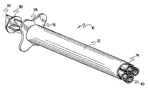
Figure 1 is a front perspective view
illustrating the cervical tissue sampling device of the
present invention in an assembled, retracted condition.
Figure 2 is a rear perspective view
illustrating the cervical tissue sampling device of the
present invention in an assembled, retracted condition.
Figure 3 is a rear perspective view
illustrating the plunger assembly of the cervical tissue
sampling device of the present invention.
Figure 4 is a front end elevational view
illustrating the cervical tissue sampling device of the
present invention in a retracted condition.
Figure 5 is a rear end elevational view
illustrating the cervical tissue sampling device of the
present invention.
Figure 6 is a side elevational view
illustrating the cervical tissue sampling device of the
present invention in a retracted condition.
Figure 7 is a top plan view illustrating the
cervical tissue sampling device of the present invention
in a retracted condition.
Figure 8 is an exploded perspective view
illustrating the component parts of the cervical tissue
sampling device of the present invention.
Figure 9 is a rear perspective view
illustrating the cervical tissue sampling device of the
present invention in an extended condition.
CA 02509655 1995-O1-17
wo 9snZOS3 rcrius9sroo6 i o
4
Figure 10 is a diagrammatic view illustrating
the manner of use of the cervical tissue sampling device
of the present invention.
Hest Mode ~o~, ar ingot The Invention
Referring now to the drawings, wherein like
reference numerals designate corresponding structure
throughout the views, and referring in particular to
Figures 1, 2, and 8, the cervical tissue sampling device
according to a first preferred embodiment of the
10 invention includes an elongated substantially rigid,
substantially cylindrical barrel 12 possessing a
circular open front end 14 and terminating at a rear
open circular end 28 within a circular recess 29 in an
irregularly shaped radially extending finger grip flange
16_ The barrel 12 and flange 16 are preferably formed
from #PE206'7 low-density polyethylene, available from
Branchcomb Industries, Sapulpa, Oklahoma. As shown in
Figures 4, 5, and 8, the finger grip flange 16 possesses
convex arcuately curved corner projecting portions 20,
22, 24, and 26 spaced around the periphery of the flange
16. These corner projections are separated by
respective concave arcuate recesses 21, 23, 25, and 27.
As may be appreciated with reference to Figure 2, the
barrel 12 may be integrally molded with the flange 16,
or alternatively may be assembled from separate
components by press fitting the rear circular open end
28 of the barrel 12 into a conforming aperture formed
centrally in the flange 16. Suitable adhesives may also
CA 02509655 1995-O1-17
wo ~rzZOSS pcr~s9sroo6io
be employed to effect securement of the flange 16 to the
barrel 12. The barrel 12, as depicted in Figure 8,
possesses internal axially spaced annular ridges 30 and
32 formed internally within the barrel 12 adjacent
5 flange 16 for the purpose of retaining a plunger
assembly therein, in a manner to be described
subsequently in greater detail.
With reference to Figures 3 and 8, the cervical
tissue sampling device l0 according to the present
invention includes a plunger assembly 33 terminating at
a distal end in a circular brush 34 preferably formed by
twisting nylon bristles between strands of stainless
steel wire 36. A suitable brush is made from type 304
stainless steel wire, 0.02 inches in diameter, mil spec
MS209956, and type 612 natural level nylon, 0.005 inches
in diameter, FDA# 21CFR177-1500, and is available from
Gordon Brush Company, Los Angeles, California. The
initially straight brush is subsequently deformed into
the generally circular illustrated configuration, with
a stem portion 38 of the stainless steel wire 36
extending rearwardly through a central aperture of a
circular sponge 40. A suitable sponge is a 2 inch
diameter circle of 0.0625 inch thick open-cell cellulose
sponge, # 1935, with a 0.25 inch central hole, available
from Lundell Manufacturing Corporation, Minneapolis,
Minnesota. The circular brush 34 has a diameter of
about 3.5 centimeters, while the surrounding sponge 40
has a diameter of about 5.0 centimeters. A typical
CA 02509655 1995-O1-17
WO 96121053 PCT/US95100610
6
cervix has a diameter of 5 . 0 to 5 . 5 centimeters . The
stem portion 38 of the stainless steel wire 36 is press
fit within a central bore 46 formed in a radially
enlarged circular end flange 42 of a stem 44. The stem
44 is inserted through a sleeve 50 terminating in a
second radially enlarged circular end flange 48.
Accordingly, the end flange 42 forms an abutment surface
for the rear face of the sponge 40 and serves to press
the sponge 40 against the rear face of the circular
brush 34. The flange 42 also serves as a stop
restricting forward axial movement of sleeve 5o due to
flange 42 having a greater outer diameter than the inner
diameters of flange 48 and sleeve 50. The stem 44
terminates at an axially inward end in a semi-
cylindrical connector tab end portion 52. A pin 54
extends transversely to the axis of the stem 44 from a
central location on the flat interior surface of
connector tab portion 52. A second sleeve 56 which
includes a radially enlarged terminal circular flange 58
is dimensioned for a relatively tight fitting, sliding,
frictional engagement over the stem 44 and also over a
plunger shaft 64. As shown in Figure 8, the plunger
shaft 64 terminates in a second semi-cylindrical
connector tab end portion 60 provided with a
transversely extending aperture 62 dimensioned for
frictional engagement with the pin 54 of tab end portion
52 of stem 44. Accordingly, it may now be understood
that a selectively detachable connection is formed by
CA 02509655 1995-O1-17
PVO 96112053 PCT/US9SI006I0
7
stem 44 and plunger shaft 64, such that the stem 44 and
attached brush 34 and sponge 40 may be removed as
desired by sliding sleeve 56 rearwardly along shaft
portion 64 until engaged tab end portions 52 and 60 are
exposed. The pin 54 may then be manually disengaged
from aperture 62, to complete detachment of stem 44 from
shaft 64.
The plunger shaft 64 is formed by four axially
extending ribs 66, 68, 70, and 72 disposed at ninety
degree circumferential increments. A supporting cross
formed by intersecting strut members 74, 76, 78, and 80,
which form radially outwardly extending projections at
a medial position of shaft 64, serves to maintain the
plunger shaft 64 approximately centered within the
barrel 12 in an assembled condition. The supporting
cross cooperates, in the manner of a detent, with
internal ridges 30 and 32 within barrel 12 (Figure 8) to
prevent inadvertent extension or retraction of the
plunger assembly 33. A rear end portion of the plunger
shaft 64 terminates in radially enlarged portions 82,
84, 86 and 88 integrally molded with a cylindrical end
plug 90. An axially projecting gripping flange 92
extends transversely from an outer end face of plug 90
to provide a manual grasping surface to enable a user to
extend, retract, and rotate the plunger assembly 33
relative to the barrel 12. The various components of
the plunger assembly 33 are preferably formed from
CA 02509655 1995-O1-17
wo 9saZOS3 pcrn~s9s~oo6io
s
#PP14B12A co-polymer polypropylene, available from
Branchcomb Industries, Sapulpa, Oklahoma.
In the manner of use of the cervical tissue
sampling device according to the present invention, a
female lies in a generally reclined position as
illustrated in Figure 10 and inserts the barrel 12 into
the vagina V. It should be noted that the barrel 12 of
the cervical tissue sampling device 10 is shown in a
partially inserted position in Figure 10. In practice;
the barrel 12 is further inserted such that the circular
open front end 14 of barrel 12 contacts the face of the
cervix C, thereby allowing slight pressure to be applied
to the face of the cervix C. In so doing, the os (or
opening) of the cervix C is substantially axially
aligned with the barrel 12 and when the plunger assembly
33 is extended, as described below, the bristles of the
brush 34 are allowed to enter the os in order to obtain
endocervical cells for analysis.
With the plunger assembly 33 disposed in the
retracted condition depicted in Figures 1 and 2, the
supporting cross formed by struts 74, 76, 78, and 80 is
disposed between ribs 30 and 32 (Figure 8). In this
condition, the circular sponge 40 is in a folded
orientation within barrel 12, as shown in Figures 1, 2,
4, 6, and 7. The retracted condition of the sponge 40
is such that the sponge 40 is substantially located
within the radial boundary of barrel 12 to facilitate
insertion. After insertion of the barrel 12, the female
CA 02509655 1995-O1-17
WO 96/22053 PCTIUS95/00610
9
extends the plunger assembly 33 with the aid of finger
gripping flange 16, plug 90, and integral gripping
flange 92. The extension of the plunger assembly 33
causes the supporting cross to snap past rib 32 (Figure
8) due to the resilient nature of the plastic material
forming struts 74, 76, 78, and 80. The plug 90 then is
received in recess 29 (Figure 2) in flange 16, limiting
further extension of the plunger assembly 33. The
recess 29 forms a journal bearing surface for
rotationally mounting the plug 90 and attached plunger
shaft 64 in an axially central orientation to facilitate
sample collection. The sponge 40 then, due to its
natural resiliency, springs to the open orientation
shown in Figure 9, exposing the circular brush 34
illustrated in Figure 8. The exposed brush 34 and
sponge 40 then collect endocervical, cervical cap, and
vaginal wall tissue and mucous samples from the vagina
V and cervix C, facilitated by manual rotation of the
plunger assembly 33 by the user via gripping flange 92.
After sampling is complete, the female retracts
the plunger assembly 33 to its starting position, aided
by the tactile indication of retraction provided by the
snapping of the support cross past rib 32, protecting
the collected specimen on the brush 34 and sponge 40
within the barrel 12. The female then withdraws the
device 10 by grasping flange 16, removes the plunger
assembly 33 from the barrel 12, and detaches the axially
outer end portion of the plunger assembly 33 including
CA 02509655 1995-O1-17
WO 96IZ2053 PCTlUS95/006I0
brush 34 and sponge 40, utilizing the detachable
connection formed by connector tabs 52 and 60 shown in
Figure 8.
It should be noted that the sponge 40 not
5 only functions as a specimen collection agent, but also
as a cloak of protection for the brush 34 and the
specimen sample thereon. As the plunger assembly 33 is
being retracted, the sponge 40 folds back to its
position shown in Figures 1 and 2, thereby
10 encapsulating and protecting said brush and sample.
The sampling device 10 may be sold as a kit
including a suitable lubricant gel to facilitate
insertion, along with a small sealable container in
~ which to mail or otherwise transmit detached outer end
portion of the plunger assembly 33 including the sponge
40 and brush 34 to a laboratory for analysis.
The device 10 is extremely inexpensive and
simple to use. Thus, the device may be utilized by
women in the privacy of their own homes to collect
tissue samples for analysis.
It is to be understood, however, that even
though numerous characteristics and advantages of the
present invention have been set forth in the foregoing
description, together with details of the structure and
function of the invention, the disclosure is
illustrative only, and changes may be made in detail,
especially in matters of materials, shape, size and
arrangement of parts, within the principles of the
CA 02509655 1995-O1-17
WO 96122053 PCT/US95/00610
11
invention to the full extent indicated by the broad
general meaning of the terms in which the appended
claims are expressed.
