Note : Les descriptions sont présentées dans la langue officielle dans laquelle elles ont été soumises.
CA 02514857 2005-07-28
WO 2004/069035 PCT/US2004/002971
System and Method for Rapid Placement of Chest Tubes
Inventors
Philip J. Simpson
3185 Pioneer Place
Escondido, CA 92025-7662
David G. Matsuura
859 Summersong Court
Encinitas, CA 92024
Walter Dean Gillespie
1327 Pacific Beach Drive #11
San Diego, CA 92109
Chris K. Salvino
401 E. Ontario, Apt. 4406
Chicago, IL 60611
Jim Trinchera
935 Hygeia Ave.
Leucadia, CA 92024
CA 02514857 2011-01-14
WO 2004/069035 PCT/US2004/902971
2
Field of the Invention
[0001] The present invention relates to methods and apparatus for
performing a rapid tube thoracostomy, and more particularly relates to
methods and apparatus for performing a rapid tube thoracostomy using
conformable tubes and cannula.
20 Background of the Invention
[0004] A trocar generally comprises an obturator and a cannula. The
obturator has a pyramid-shaped piercing tip at one end, and moves the
piercing tip into tissue to form a hole to provide access to a body cavity or
a
target tissue. The cannula is located around the obturator. The cannula is
inserted into the body cavity together with the obturator through the hole
formed by the piercing tip. Such a trocar, therefore, forms a pathway in the
inside of the cannula for inserting an endoscope or a surgical tool into the
body cavity, by extracting or withdrawing the obturator from the cannula,
which is inserted into the body cavity. Known methods of sealing the tissue to
30 the cannula include the use of sutures and/or adhesive tape in order to
maintain the position of the cannula and provide a fluid and air tight seal.
However, this method fails to provide adequate barrier or an appropriate seal
for fluid and/or gases. Therefore what is needed is a system and method for
CA 02514857 2005-07-28
WO 2004/069035 PCT/US2004/002971
3
providing an air and fluid tight seal without the use of. sutures and/or
adhesive
tape.
Summary of the Invention
[0005] The present invention provides methods and systems for safely and
easily performing a rapid tube thoracostomy. Tube thoracostomy is a method
for allowing the sterile drainage of fluid or air from the pleural space
utilizing a
semi-rigid drainage tube. In at least some implementations, the present
invention also minimizes the need for exposed sharp instruments such as
scalpels.
io [0006] More particularly, the present invention provides a chest tube
installation system which includes, in an exemplary embodiment, a chest tube
insertion device, a diametrically compliant cannula, a chest tube and a chest
tube pneumo seal/wound dressing. The chest tube insertion device utilizes a
cutter such as that disclosed in the Related Application, referenced above,
and incorporated herein by reference.
Brief Description of the Figures
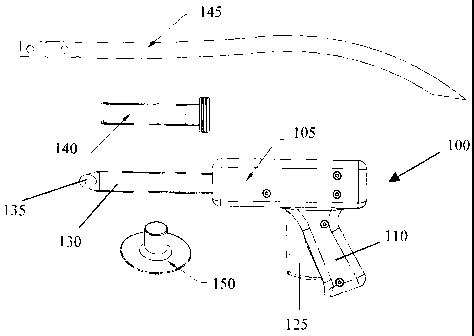
[0007] Figure 1 illustrates in perspective view generally an insertion gun, a
20 cannula and chest tube in accordance with one aspect of the present
invention.
[0008] Figure 2 illustrates, in side elevation view, in addition to the system
of Figure 1, a seal in accordance with one aspect of the present invention.
[0009] Figure 3 illustrates the gun of Figure 1 applied to an area of tissue
characteristic of a patient needing aid, with the cannula on the probe of the
gun.
[0010] Figure 4 illustrates the gun of Figure 1 inserted partly into the
patient after appropriate cuts have been made.
[0011] Figure 5 illustrates the gun removed from the patient with the
30 cannula remaining inserted into the patient.
[0012] Figure 6 illustrates the insertion of the chest tube through the
cannula of Figure 5.
CA 02514857 2005-07-28
WO 2004/069035 PCT/US2004/002971
4
[0013] Figure 7 illustrates the chest tube inserted.into the patient through
the cannula.
[0014] Figure 8 illustrates the removal of the compliant cannula over the
chest tube.
[0015] Figure 9 illustrates the application of a sealing element over the
chest tube.
[0016] Figure 10 illustrates the sealing of the wound with the seal over the
chest tube, thus completing the chest tube installation.
[0017] Figures 11 A-11 B show in cut-away view one implementation of the
to gun of Figure 1, with Figure 11 B providing a detail view of the cutting
tip of the
gun.
[0018]. Figure 12 shows in cut-away view certain details of the
implementation of Figure 11 A.
[0019] Figure .13 illustrates in cut-away view the gun of Figure 11 A with the
trigger at approximately mid-point.
[0020] Figure 14 illustrates in more detail an implementation of the gun of
Figure 13.
[0021] Figure 15A illustrates the implementation of the gun of Figure 1 1A
with the trigger fully retracted.
20 [0022] Figure 15B illustrates in detail cut-away view the cutting tip of
Figure 15A.
[0023] Figures 16A-16D illustrate the eccentric nature of the cutting tip of
the gun.
[0024] Figures 17A-17D illustrate cross-sectional view of various chest
tubes, both in compressed and uncompressed views.
[0025] Figures 18A-18E illustrate various details of some examples of
chest tubes in accordance with the present invention.
[0026] Figures 19A-19B illustrate perspective and cross-sectional side
views of a seal in accordance with the invention.
30 [0027] Figure 20 illustrates an adhesive method for attachment of the
chest tube assembly to a patient.
CA 02514857 2005-07-28
WO 2004/069035 PCT/US2004/002971
[0028] Figures 21 A-21 C illustrate, respectively, a side elevation view of a
snap lock fitting for attachment of the chest tube to a patient, a cross-
sectional side view, and a perspective view of the snap lock fitting itself.
[0029] Figures 22A-22B illustrate in cross-sectional side view the
attachment of the snap lock fitting of Figures 21 A-21 C to a patient.
Detailed Description of the Invention
[0030] Referring first to Figures 1 and 2, a system for chest tube insertion
in accordance with the present invention is shown generally. A chest tube
io insertion device 100, which may also be thought of as a cutting and
insertion
gun, includes a housing 105, a handle 110 with a trigger 125, a probe tip 130
having a cutting tip 135 at the distal end thereof, a cannula 140, a chest
tube
145 and a seal 150. The cutting tip 135 may be of the type described in the
Related Application.
[0031] Referring next to Figures 3-10, the process for inserting a chest
tube according to the present invention can be better appreciated. The chest
tube insertion procedure using this system is safer, faster, and easier than
other known methods. Although not a required part of the present, invention,
clinicians considering insertion of a chest tube typically include the steps
of
20 selecting a site, preparing the patient and then draping the area, followed
by
anesthetizing the site. The anesthesia can be of any acceptable type; one
typical approach is 5 to 15 ml of 1 % lidocaine delivered through a 'syringe
and
small gauge needle.
[0032] Following the foregoing preliminary steps, the chest tube insertion
procedure in accordance with the present invention proceeds as follows:
[0033] 1. Make an incision through skin to the pleural space with the
chest tube insertion device, or gun, 100. Start by placing a cannula
140 over the probe tip or shaft 130 of the device 100 until the cutting
tip 135 extends beyond the cannula 140.
30 [0034] 2. Place the device 100 against the patient's target tissue 170 as
shown in Figure 3. Visually aligning the shaft 130 of the device in the
desired direction. Firmly press the distal tip 135 into the skin and
actuate the trigger 125. Actuation of the trigger will initiate a cutting
CA 02514857 2005-07-28
WO 2004/069035 PCT/US2004/002971
6
event, creating an incision 175, as shown in Figure 4. Each cutting
event will cut approximately 1 mm in depth as long as the cutting tip is
maintained appropriately against the tissue 170. As the device 100
cuts through skin, the device 100 may be used as a blunt dissection
device, and may be thought of as a blunt tip obturator.
[0035] 3. Once the tip of the probe 130 extends into the pleural cavity in
the desired amount, the cannula will also extend into the cavity as
shown in Figure 4. Withdrawal of the gun 100 from the incision 175
can be achieved while leaving the cannula 140 in place within the
patient as shown in Figure 5.
[0036] 4. As shown best in Figure 6, introduce the distal tip 600 of the
chest tube 145 into the cannula 140.
[0037] 5. Advance the tube 145 until all of the transverse drain holes 700
of the chest tube 145 are within the pleural space, as shown in Figure
7.
[0038] 6. Withdraw the cannula 140 over the chest tube 145 while holding
the chest tube 145 in place, best shown in Figure 8.
[0039] Then, as is typical of thoracostomies, the clinician will typically
take
the additional steps of suturing the skin on both sides of the chest tube and
tying the tube in place with the tag ends of the suture; applying sterile
petroleum gel over the incision to create an airtight seal and cutting notches
in sterile gauze to fit around the chest tube, followed by securing the gauze
and tube in place using a suitable surgical tape.
[0040] From the foregoing, a method of rapidly placing a chest tube
according to the invention can be appreciated. However, the chest tube
insertion device 100 may be implemented in any of a variety of designs, just
as the cutting tip 135 may be implemented in a variety of ways as discussed
in the Related Application. Several of these implementations are described in
connection with Figures 11A through 15.
[0041] Referring next to Figures 11 A-11 B and 12, a first implementation of
the chest tube insertion device shown generally at 100 may be better
appreciated. An eccentrically mounted circular blade 1 is housed inside a
bearing block 2 and is attached to the bearing block 2 by an axle 6. The
CA 02514857 2005-07-28
WO 2004/069035 PCT/US2004/002971
7
blade 1 need not be an eccentrically mounted circular blade, but may instead
be any of the forms shown in the Related Application. The bearing block 2 is
located at the distal end of a shaft or probe 3, which is similar to the probe
130 of Figure 1. The blade 1 is also connected to the distal end of an input
rod. 4 by a pivot pin 5. A compression spring 8, is constrained at a
compressed height 10, by a flange 7 at the proximal end of the input rod 4
and a wall 9 inside the mechanism housing. The force from the compression
spring 8, translates through the input rod 4 to the eccentrically mounted
blade
1 through the pivot pin 5. The force acting on the pivot pin 5 results in a
to moment about the axle 6 which keeps the blade 1 recessed inside the
bearing block 2 until the operator initiates a cutting event.
[0042] To initiate a cutting event, the operator moves an input lever or
trigger 14, which is similar to the trigger 125 of Figure 1, from a stationary
position 15, as shown in Figures 11 A-11 B and the more detailed view of
Figure 12, through an intermediate position 16 as shown in Figure 13 to a
final position 17 shown in figure 15. As the input lever 14 is moved, it
pivots
about a lever axle 18. The angular rotation of the lever 14 is translated
through one segment of circular gear teeth 19 mounted concentric to the lever
axle 18, to a meshing segment of circular gear teeth 20 attached to a cam 21.
20 The cam 21 is allowed to rotate about a shaft 22 as it is motivated to do
so by
motion of the gear teeth 20.
[0043] The cam 21 profile is exaggerated through a pair of elongated
members 23 which contact a momentum storage mass 25 through a
matching pair of latches 26 which are attached to the mass 25 by pins 28.
The latches 26 are able to rotate about the pins 28 that connect them to the
mass 25. The mass 25 is constrained to move only longitudinally on an axis
co-linear with the shaft 3. Angular cam motion is translated to distal-to-
proximal linear motion of the mass 25 as the cam 21 rotates from a stationary
position 24 shown in Figure 11 A to a final position 29 as shown in Figure 15.
30 In the particular implementation shown, the mass 25 is moved rearward or
toward the proximal end of the device 100.
[0044] Distal-to-proximal linear motion of the mass 25 causes the angled
outer surfaces of the matching pair of latches 26 to encounter stationary
CA 02514857 2005-07-28
WO 2004/069035 PCT/US2004/002971
8
protrusions 27. As detailed in figure 14, the protrusions 27 act on the angled
outer surfaces of the latches 26 so that the latches 26 rotate about their
pivot
pins 28 until they no longer contact the cam members 23. Simultaneously,
the mass 25 compresses a spring 11 from its free length 12 as depicted in
Figure 11, as it travels proximally, to a compressed length 13 as depicted in
Figure 13. Release of the latches 26 from the cam members, enable the
resultant force created by compression of the spring 11 to act on the
momentum storage mass 25 thereby accelerating it in a proximal to distal
direction. Potential energy stored in the spring 11 at the compressed length
l0 13 is converted to kinetic energy as the mass 25 is accelerated.
[0045] The proximal to distal motion of the mass 25 causes its distal most
face 30 to strike the proximal end of the rod 4. The mass 25 and the rod 4
continue with a proximal to distal motion until the flange 7 strikes a travel
limiting structure 32. The proximal to distal motion of the rod 4 acts on the
blade 1 such that it rotates about the axle 6, which attaches the blade 1 to
the
bearing block 2. As the mass 25 and the rod 4 travel proximal to distally, the
blade 1 rotates about the axle 6, which connects the blade 1 to the bearing
block 2. Depicted in Figures 16A-D, as the blade 1 rotates, it emerges from
the bearing block 2 from a stationary position to a fully exposed position
when
20 the rod 4 encounters the travel limiting structure 32.
[0046] As the momentum storage mass 25 travels in a proximal to distal
direction it compresses a spring 36 constrained between a flange 39 and
stationary internal structure 40. Force stored in the spring 36 created by
compressing the spring 36 to a pre-loaded height 38 acts on the mass 25
once its proximal to distal motion has been halted by the travel limiting
structure 32. The force generated by the spring 36 causes the mass 25 to
move in a distal to proximal direction until equilibrium is achieved. With the
mass 25 reset, the compression spring 8 that is in contact with the proximal
end of the rod 4 acts on the rod 4 to move the rod 4 in a distal to proximal
3o direction thereby recessing the blade 1 to a safe position inside the
bearing
block 2.
[0047] The operator resets the mechanism after initiating a cutting event
by releasing the input lever 14. The input lever 14 then returns to the
CA 02514857 2005-07-28
WO 2004/069035 PCT/US2004/002971
9
stationary position 15 by, means of a spring (not shown) in contact with the
input lever 14, causing the input lever 14 to rotate about the lever. axle 18.
Motion of the lever 14 causes the cam 21 to move to its stationary position
24. As the elongated members 23 move to their stationary positions the
latches 26 attached to the mass 25 are acted on by an extension spring 41 to
return the latches 26 to their position 42. The mechanism is now reset and
ready for another operator initiated cutting event.
[0048] Optionally, an additional user control may be incorporated into the
device 100 to hold the cutting element fully extended when actuated. This
io alternative control allows the clinician to optionally use the device as a
sharp
trocar as well.
[0049] One aspect of the chest tube insertion device is that the shaft 130
is, in an least some embodiments, substantially ovate in cross section. This
allows for passage of a larger bore transversely compliant cannula to be
inserted between the ribs without dilating the rib cage. At room temperature,
standard chest tubes are fairly diametrically compliant, across the transverse
axis of the tube, and become much more compliant at body temperature.
Therefore, standard chest tubes can be passed through a substantially ovate
cannula. Thus, a larger chest tube can be inserted with significantly less
pain
20 to the patient. Optionally, the cannula may be pre-formed with an ovate
cross
section of a less compliant material.
[0050] Another aspect of the device is the use of preferentially compliant
chest tubes. Standard chest tubes are formed with uniform wall thickness.
These tubes are formed with walls heavy enough to prevent kinking across
the transverse axis of the tube due to longitudinal bending loads. When using
a substantially ovate cannula, the load required to pass the circular chest
tube
through the cannula, can be substantially reduced through the use of a tube
that is preferentially more diametrically compliant, across the transverse
axis,
than standard tubes.
30 [0051] In some embodiments of the invention, it is desirable to use a
generally thinner walled tube, which may for example be formed by extrusion,
with walls formed with a multiplicity of longitudinal ribs, scallops or
splines as
shown in Figures 17A-17D. The ribs may be on the inside or outside of the
CA 02514857 2011-01-14
tube, and may take any of a wide variety of shapes. The space between the ribs
form
flexure zones, thus allowing the tube to be more diametrically compliant than
a standard
tube. Another purpose of the ribs is to provide adequate section modulus to
prevent
transverse kinking of the tube when bent under normal loading conditions, such
as shown
by the equations set for the below and shown in Figures 18A-18E. Figures 18A
and 18D
show a standard tube with standard wall thickness 1803. Figures 18B and 18E
show a
ribbed tube with reduced wall thickness portion 1804. Figure 18C illustrates
the cantilever
moment load 1801 applied to a rib with height 1802. Furthermore, design of
interior ribs
could provide patent lumens, to prevent complete shut off, even when exposed
to extreme
loading conditions. Additionally the ribs add sufficient section to provide
adequate
resistance to tensile loads.
Cantilever-moment load
Modulussection section 12
deflection. Modulus Elastic-Modulus' section
Moment,= 2
Length
[0052] Another innovative aspect of the some embodiments of the system of the
invention is the use of an elastomeric pneumo-seal, such as shown at 150 in
Figure 2 and
better seen in Figures 19A-19B. The term pneumo-seal is used herein to refer
to a seal
that acts as a barrier to prevents the flow of fluids and gases. The pneumo-
seal 150 can
be formed of a compliant material with upper flange 1900 and lower flange
1905, which
may be filled with air/liquid to conform and seal to the tube and incision.
The pneumo-seal
150 may have pressure sensitive adhesive attachment areas 1910 as shown in
Figures
and 21 to adhere to and seal to the patient and the chest tube. Figure 20
shows the
annular pressure sensitive adhesive attachment area 1910 being used to secure
the seal
to the patient, and the adhesive patches 1915 are for grasping the tube. The
patches
1915 fold up to contact the tube as indicated by the arrows. The pressure
sensitive seal
or barrier formed by the pneumo-seal is accomplished without stitches or
sutures. The
pneumo-seal may be pre-loaded and reside on the cannula 140 or may be used as
a
separate device. Figures 21A-21C illustrate, respectively, a side elevation
view of a snap
CA 02514857 2011-01-14
11
lock fitting for attachment of the chest tube patient, a cross-sectional side-
view, and a
perspective view of the snap lock fitting itself. The snap lock fitting 2110
is shown in
Figure 21 B. Peel-away adhesive cover 2120 is shown in Figure 21 C.
[0053] Alternatively, the pneumo-seal device 150 may have a fitting 2200 that
mates
to a matching fitting integrally formed with the chest tube or, as shown in
Figure 22B,
provided as a snap-lock fitting added to the chest tube. Fitting 2200
comprises a
mechanical interlock 2240 formed as a grasping taper. Tube 2230 conforms to
the
grasping taper. The purpose of the fitting is to mechanically attach the tube
to the patient
patch and to form an air-tight seal.
[0054] Alternatively, mechanical sealing ribs (Figure 22A may also replace the
adhesive contacting tube. Chest tube 2210 conforms to mechanical sealing ribs
2220.
The basic configuration of the pneumo-seal 150 can also be used for other
cannulations
into the body such as central venous lines and other drainage tubes.
Optionally, the
device may be molded from a transparent material, such as Pebax, to allow
visualization
of the wound through the device. The advantages of the pneumo-seal device
include that
it is faster than cutting bandages, requires no use of scissors, is faster
than suturing or
tying, requires no needles, does not require petroleum gel to form seal thus
allowing use
of standard wound dressing tapes.
[0055] It will thus be appreciated that a new and novel method of chest tube
insertion
has been disclosed, as well as a new and novel chest tube insertion system and
components thereof. Among the advantages offered by one or more of
implementations
of the invention are a controlled depth of cut,
a retractable blade offering increasing user and patient safety, greater
safety
for the clinician. Having fully disclosed a variety of implementations of the
present
invention, it will be appreciated by those skilled in the art that numerous
alternatives and
equivalents exist which do not materially alter the invention described
herein. Therefore,
the invention is not intended to be limited by the foregoing description, but
instead only by
the appended claims.