Note : Les descriptions sont présentées dans la langue officielle dans laquelle elles ont été soumises.
CA 02515767 2005-08-11
WO 2004/071346 PCT/US2004/004109
DEVICE FOR FUSING TWO BONE SEGMENTS
Cross-Reference to Related Applications
This application claims the benefit of U.S. Provisional Application No.
60!446,963
filed on February 12, 2003. U.S. Provisional Application No. 60/446,963 is
herein
incorporated by reference for all legitimate purposes. This application also
claims the
benefit of U.S. Patent Application Ser. No. (Attorney Docket No. 31132.41),
entitled
"Instrument and Method for Milling a Path Into Bone" (Inventor: Lukas
Eisermann) filed
on January 7, 2004. U.S. Patent Application Ser. No. (Attorney Docket No.
31132.41) is
herein incorporated by reference for all legitimate purposes. This application
is also
related to U.S. Patent Application Serial No. 10/430,473, which is hexein
incorpoxated by
reference for all legitimate purposes.
Background
The present disclosure relates generally to the field of orthopedics and
spinal
surgery, and in some embodiments, the present disclosure relates to fusion-
promoting
prosthetic devices for insertion into an intervertebral disc space.
In the treatment of diseases, injuries or malformations affecting spinal
motion segments,
and especially those affecting disc tissue, it has long been known to remove
some or all of
a degenerated, ruptured or otherwise failing disc. In cases involving
intervertebral disc
tissue that has been removed or is otherwise absent from a spinal motion
segment,
corrective measures are taken to ensure the proper spacing of the vertebrae
formerly
separated by the removed disc tissue. In some instances, fusion-promoting
prosthetic
devices, such as fusion cages and the like, are inserted into the disc space
to maintain the
structural integrity of the spinal column.
Anterior plating is often used in conjunction with fusion devices to
supplement the
stability provided by such fusion devices. However, in some instances,
anterior plating is
inappropriate for use due to the presence of vascular structure, which impedes
the
implantation and positioning of the anterior plating.
CA 02515767 2005-08-11
WO 2004/071346 PCT/US2004/004109
2
Therefore, what is needed is an implantable fusion device which eliminates, or
at
least reduces, the need for supplemental plating external of the
intervertebral space.
Summary
A fusion-promoting prosthetic device for insertion into an intervertebral
space is
described. The prosthetic device includes a sagitally-extending plate having
caudal and
cephalad edges, the caudal edge being adapted for complete insertion within a
first
vertebral body and the cephalad edge being adapted for complete insertion
within a second
vertebral body adjacent to the first vertebral body. The prosthetic device
further includes a
first transverse plate connected to the sagitally-extending plate, and a
second transverse
plate connected to the sagitally-extending plate, the first and second
transverse plates
being adapted for complete insertion within the intervertebral space.
A fusion-promoting, spinal plating assembly is described. The plating assembly
includes
a first plate adapted to engage a first vertebral body and a second vertebral
body, and at
least one additional plate connected to the first plate, the at least one
additional plate
extending transversely to the first plate, wherein the at least one additional
plate is adapted
to be inserted within an intervertebral space.
A method for promoting :Fusion in an intervertebral space defined between
first and
second vertebral bodies is. described. The method includes providing a
prosthetic device
having a first plate adapted to engage each of the first and second vertebral
bodies, and a
pair of additional plates connected to the first plate, the additional pair of
plates extending
in a direction transverse to the first plate. The method further includes
inserting the
prosthetic device into the intervertebral space such that a first edge of the
first plate is
completely inserted within the first vertebral body, a second edge of the
first plate is
completely inserted within the second vertebral body, and each of the
additional pair of
plates are completely disposed within the intervertebral space.
Brief Description of the Drawings
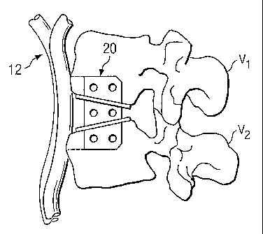
Fig. 1 is a lateral view of a pair of adjacent vertebral bodies.
Fig. 2 is a lateral view of a prosthetic device for insertion between the
adjacent
vertebral bodies of Fig. 1.
CA 02515767 2005-08-11
WO 2004/071346 PCT/US2004/004109
Fig. 3a is a longitudinal view of the prosthetic device of Fig. 2.
Fig. 3b is a longitudinal view of an alternative prosthetic device.
Fig. 4a is a longitudinal view of a pair of the verterbral bodies of Fig. 1
having
longitudinally-formed slots for receiving the prosthetic device of Fig. 2.
Fig. 4b is a lateral view of the prosthetic device of Fig. 2 shown
longitudinally
disposed between the vertebral endplates of Fig. 1.
Fig. Sa is a lateral view of a pair of verterbral bodies having laterally-
formed slots
for receiving a prosthetic device.
Fig. Sb is a lateral view of an alternative prosthetic device shown laterally
disposed
between the vertebral bodies of Fig. Sa.
Description
For the purposes of promoting an understanding of the principles of the
disclosure,
reference will now be made to the embodiments, or examples, illustrated in the
drawings
and specific language will be used to describe the same. It will nevertheless
be understood
that no limitation of the scope of the disclosure is thereby intended. Any
alterations and
further modifications in the described embodiments, and any further
applications of the
principles of the disclosure as described herein are contemplated as would
normally occur
to one skilled in the art to which this disclosure relates.
Referring now to Fig. 1, shown therein is a lateral view of a portion of a
spinal
column 10, illustrating a pair of adjacent upper and lower vertebrae V1 and
V2,
respectively, separated by an intervertebral space S created by the removal of
a natural
intervertebral disc. The illustration of two vertebrae is only intended as an
example.
Another example would be a sacrum and one vertebrae. Vascular structure 12,
such as,
for example, the aortic artery and associated segmental arteries, is shown
disposed
anteriorly adjacent to the upper and lower vertebrae Vl, V2. As can be
appreciated, it is
not desirable to impart pressure to, or otherwise contact, the vascular
structure 12 during
insertion and upon implantation of a prosthetic device.
Refernng now to Figs. 2 and 3a, a prosthetic device for insertion into the
space S
(Fig. 1) is generally referred to by reference numeral 20. In one embodiment,
the device
20 includes a sagittally-extending support plate 22 and a pair of additional
support plates
CA 02515767 2005-08-11
WO 2004/071346 PCT/US2004/004109
4
24, 26 integrally formed with and extending generally transverse to the
sagittal plate. It is
understood that the transverse plates 24, 26 may alternatively be removably
connected to
the sagittal plate 22 such that the prosthetic device 20 is generally modular
in nature.
Although the sagittal plate 22 and the transverse plates 24, 26 of the
prosthetic device 20
rnay be formed from a wide variety of materials, in one embodiment of the
disclosure, the
sagittal plate 22 and the transverse plates 24, 26 are formed of a cobalt-
chrome-
molybdenum metallic alloy (ASTM F-799 or F-75). However, in alternative
embodiments
of the disclosure, the sagittal plate 22 and the transverse plates 24, 26 may
be formed of
other materials such as titanium or stainless steel, a polymeric material such
as
polyethylene, or any other biocompatible material that would be apparent to
one of
ordinary skill in the art.
The sagittal plate 22 is adapted to engage the upper and lower vertebrae Vl,
V2,
respectively, and in the present example, the sagittal plate 22 extends in a
plane
substantially parallel to the sagittal plane (represented by axis Y lying in
the sagittal plane
in Fig. 1) when engaged with the upper and lower vertebrae. The sagittal plate
22 includes
an anterior edge 30, a posterior edge 32, a caudal edge 34 and a cephalad edge
36. It is
understood that reference to anatomical directions in this specification such
as sagittal,
anterior, posterior, caudal and cephalad is for purposes of descriptive
clarity only, and is
not intended to limit the prosthetic device 20 to having a specific
orientation relative to
such anatomical directions.
The caudal and cephalad edges 34, 36 of the sagittal plate 22 are formed as
keel-
like structures, which aid in the insertion of the prosthetic device 20 into
the intervertebral
space S. For example, in one embodiment, the caudal edge 34 of the sagittal
plate 22 is
beveled at the posterior portion thereof to provide a sharp, forward edge 38
with which to
pierce the vertebral body V2 upon insertion of the prosthetic device 20. In a
like manner,
the cephalad edge 36 of the sagittal plate 22 is beveled at the posterior
portion thereof to
provide a sharp, forward edge 39 with which to pierce the vertebral body V 1
upon
insertion of the prosthetic device 20.
It should be understood that other shapes and orientations of the caudal and
cephalad edges 34, 36 are also contemplated. For example, the caudal and
cephalad edges
34, 36 may be angled along the entire surface thereof to aid in the
circumvention of
CA 02515767 2005-08-11
WO 2004/071346 PCT/US2004/004109
vascular structure 12, or other obstacles, that may be in place during
insertion of the
prosthetic device 20. Also, the caudal and cephalad edges 34, 36 may be
angled, tapered,
or configured in some other shape to facilitate the functional demands of
insertion. In still
another embodiment, such as the one depicted in Fig. 3b, the caudal and
cephalad edges
S 34, 36 may be configured as having winged portions, including a transverse
portion 34a,
36a extending across each edge 34, 36, respectively.
Referring again to Figs. 2 and 3a, the sagittal plate 22 is adapted to promote
fusion
between the vertebral bodies Vl, V2 (Fig. 1), and as such, in one embodiment,
a plurality
of openings 40 are defined through the sagittal plate to promote fusion
therethrough. It
should be understood that any number of openings 40 may be defined through the
sagittal
plate 22, including a single opening or two or more openings. It should also
be understood
that the openings 40 need not necessarily extend entirely through the sagittal
plate 22, but
may alternatively extend partially therethrough. It should further be
understood that the
sagittal plate 22 need not necessarily define any openings 40 extending either
partially or
entirely therethrough. Additionally, although the openings 40 are illustrated
as having a
circular configuration, it should be understood that other sizes and
configurations of the
openings 40 are also contemplated.
To further promote fusion, the sagittal plate 22 is preferably coated with a
bone-
growth promoting substance, such as, for example, a hydroxyapatite coating
formed of
calcium phosphate. Additionally, the sagittal plate 22 may be roughened prior
to being
coated with the bone-growth promoting substance to further enhance bone on-
growth.
Such surface roughening may be accomplished by way of, for example, acid
etching,
knurling, application of a bead coating, or other methods of roughening that
would occur
to one of ordinary skill in the art.
The sagittal plate 22 may include one or more notches (not shown) or other
types
of indicia for receiving or engaging with a corresponding portion of a
surgical instrument
(not shown) to aid in the manipulation and insertion of the prosthetic device
20 within the
intervertebral space S (Fig. 1) between the adjacent vertebral bodies Vl, V2
(Fig. 1) . The
surgical instrument (not shown) is preferably configured to release the
sagittal plate 22
once properly positioned between the adjacent vertebrae. One example of a
surgical
CA 02515767 2005-08-11
WO 2004/071346 PCT/US2004/004109
6
instrument that can be used to insert the prosthetic device 20 is described in
co-pending
application U.S. Serial No. 10/430,473.
The transverse plates 24, 26 are adapted to engage the vertebral bodies V 1,
V2 via
a pair of bearing surfaces 42, 44, respectively. In the present example, the
transverse
S plates 24, 26 angle towards one another in the posterior direction to
accommodate an
angular relationship ~ defned between the upper and lower vertebrae Vl, V2. As
can be
appreciated, the angular relationship between the vertebral bodies Vl, V2 will
vary
depending on the particular region of the spine such as the thoracic and
lumbar regions.
Moreover, a variety of conditions can contribute to a variety of more
pronounced angular
relationships between the vertebral bodies V1, V2, such as lordosis, kyphosis,
etc., and
therefore, the transverse plates 24, 26 may extend across the sagittal plate
22 at a variety
of angles relative to the sagittal plate, including at right angles.
As with the sagittal plate 22, the transverse plates 24, 26 are adapted to
promote
fusion between the vertebral bodies Vl, V2, and as such, a plurality of
openings SO are
1 S defined through each of the transverse plates to promote fusion
therethrough. It should be
understood that any number of openings SO may be defined through the
transverse plates
24, 26, including a single opening or two or more openings. It should also be
understood
that the openings SO need not necessarily extend entirely through the
transverse plates 24,
26, but may alternatively extend partially therethrough. It should further be
understood
that the transverse plates 24, 26 need not necessarily define any openings 50
extending
either partially or entirely therethrough. Additionally, although the openings
SO are
illustrated as having a cixcular configuration, it should be understood that
other sizes and
configurations of the openings SO are also contemplated.
To further promote fusion, the transvexse plates 24, 26 are preferably coated
with a
2S bone-growth promoting substance, such as, for example, a hydroxyapatite
coating formed
of calcium, phosphate. Additionally, the transverse plates 24, 26 may be
roughened priox
to being coated with the bone-growth promoting substance to further enhance
bone on-
growth. Such surface roughening may be accomplished by way of, for example,
acid
etching, knurling, application of a bead coating, or other methods of
roughening that
would occur to one of ordinary skill in the art.
CA 02515767 2005-08-11
WO 2004/071346 PCT/US2004/004109
Referring to Figs. 4a and 4b, the prosthetic device 20 (Figs. 2 and 3) may be
inserted into the space S between the vertebrae V1, V2 from a variety of
approaches. For
example, in operation, to accommodate insertion of the prosthetic device 20,
the vertebral
bodies Vl, V2 can be prepared to accept the prosthetic device therebetween
from an offset
longitudinal, or anterior-oblique, approach. Specifically, elongate openings
or slots 60, 62
may be formed in the vertebral endplates of the upper and lower vertebrae V1,
V2,
respectively, at a predetermined width and to a predetermined depth. The slots
60, 62 can
be substantially aligned with each other to accommodate the sagittal plate 22,
and more
specifically, to accommodate the caudal and cephalad edges 34, 36 defined on
the sagittal
plate. In one embodiment, the elongate slots 60, 62 are rectangular-shaped and
are formed
by chiseling or curetting. However, other methods of forming the slots 60, 62
are also
contemplated as would occur to one of ordinary skill in the art, such as, for
example, by
drilling or reaming. Furthermore, for some embodiments of the prosthetic
device 20, the
caudal and cephalad edges 34, 36 can form their own corresponding slots by
engagement
and impaction of the beveled edges 38, 39 with the vertebrae V1, V2, and thus
no
preformed slots are necessary.
As is readily apparent from Fig. 4b, upon insertion into the intervertebral
space S
defined between the vertebrae V1, V2, the prosthetic device 20 is completely
disposed
within the intervertebral space S such that no portion of the prosthetic
device extends
beyond the anterior or posterior portion of the vertebrae V 1, V2. Moreover,
no plating
external of the intervertebral space S is required upon insertion of the
prosthetic device 20,
which is advantageous in avoiding the problems associated with contacting the
vascular
structure 12. In addition, the disposition of the prosthetic device 20 in the
intervertebral
space S results in a relatively large graft area between the vertebrae V1, V2,
the graft area
being defined, in one embodiment, by that portion of the intervertebral space
S not
occupied by the prosthetic device 20. As such, bone grafts (not shown) may be
inserted
into the graft area between the vertebrae V 1, V2 to encourage fusion between
the
vertebrae. Moreover, although not required, supplemental screws (not shown)
may be
impacted into the vertebrae V1, V2 to provide additional support. Such screws,
however,
can be press-fit completely into the vertebrae V1, V2 such that no portion of
the screws
extends beyond the anterior or posterior portion of the vertebrae V l, V2.
CA 02515767 2005-08-11
WO 2004/071346 PCT/US2004/004109
Referring now to Figs. Sa and Sb, in another embodiment, a prosthetic device
70
may be laterally inserted into an intervertebral space S' defined between an
upper vertebra
V 1' and a lower vertebra V2'. In this embodiment, the upper and lower
vertebrae V 1', V2'
are substantially parallel to one another, and as such, the prosthetic device
70 includes a
pair of transverse plates 72, 74, which are substantially parallel to one
another and are
substantially perpendicular to an associated sagittal plate 76.
To accommodate insertion of the prosthetic device 70, the vertebral bodies
Vl',
V2' can be prepared to accept the prosthetic device therebetween from the
lateral approach
by laterally forming elongate openings or slots 78, 80 in the vertebral
endplates of the
upper and lower vertebrae V1', V2', respectively, at a predetermined width and
to a
predetermined depth. The slots 78, 80 can be substantially aligned with each
other to
accommodate the sagittal plate 76. In one embodiment, the elongate slots 78,
80 are
rectangular-shaped and are formed by chiseling or curetting. However, other
methods of
forming the slots 78, 80 are also contemplated as would occur to one of
ordinary skill in
the art, such as, for example, by drilling or reaming. Furthermore, as
described above with
respect to the prosthetic device 20, the prosthetic device 70 may be
configured to form
their own corresponding slots by engagement and impaction with the vertebrae
Vl, V2,
and thus no preformed slots axe necessary.
The present disclosure has been described relative to several preferred
embodiments. Improvements or modifications that become apparent to persons of
ordinary skill in the art after reading this disclosure are deemed within the
spirit and scope
of the application. For example, although described with respect to
circumventing
vascular structure 12, it is understood that the above-described prosthetic
device 20 may
be desirable for use in scenarios where vascular structure 12 is not present.
Moreover,
although described with respect to longitudinal and lateral insertion, it is
understood that
the prosthetic device 20 may be inserted into the intervertebral space S from
a variety of
other approaches such as the transforaminal approach. Accordingly, it is
understood that
several modifications, changes and substitutions are intended in the foregoing
disclosure
and, in some instances, some features of the disclosure will be employed
without a
corresponding use of other features. It is also understood that all spatial
references, such
as "longitudinal," "lateral," and "transverse," are for illustrative purposes
only and can be
CA 02515767 2005-08-11
WO 2004/071346 PCT/US2004/004109
varied within the scope of the disclosure. Accordingly, it is appropriate that
the appended
claims be construed broadly and in a manner consistent with the scope of the
disclosure.