Note : Les descriptions sont présentées dans la langue officielle dans laquelle elles ont été soumises.
CA 02515920 2005-08-12
WO 2004/071331 PCT/EP2004/001306
1
CORRECTIVE ELEMENT FOR THE ARTICULATION BETWEEN THE
FEMUR AND THE PELVIS
Technical Field
The present invention relates to a corrective element or implant for the
articulation between the femur and the pelvis.
Background Art
As is known, when problems in the articulation between the femur and
the pelvis arise, currently the head of the femur is extracted from the
acetabular seat and a prosthesis is inserted, by way of the most disparate
technologies, recreating in practice the seat for accommodating the head of
the femur.
This approach is particularly traumatic, since dislocation of the head of
the femur causes considerable problems and further entails performing long
and complex surgery.
Disclosure of the Invention
The aim of the invention is to eliminate the drawbacks noted above, by
providing a corrective element for the articulation between the femur and
the pelvis that allows to restore correct articulation without having to
dislocate the head of the femur.
Within this aim, an object of the invention is to provide a corrective
element that can be positioned in situ rapidly and easily, allowing to use a
surgical method that is not invasive and reduces all the negative side effects
linked to conventional surgery.
Another obj ect of the present invention is to provide a corrective
element for the articulation between the femur and the pelvis that thanks to
its particular constructive characteristics is capable of giving the greatest
assurances of reliability and safety in use.
Another object of the present invention is to provide a corrective
element for the articulation between the femur and the pelvis that can be
obtained easily starting from commonly commercially available elements
CA 02515920 2005-08-12
WO 2004/071331 PCT/EP2004/001306
2
and materials and is advantageously competitive from a merely economical
standpoint.
This aim and these and other objects that will become better apparent
hereinafter are achieved by a corrective element for the articulation between
the femur and the pelvis, according to the invention, characterized in that it
comprises a contoured body that can be inserted, without dislocation of the
head of the femur, in the iliac acetabular region after incision of the
articular
capsule, said contoured body having a convex smooth outer surface that
reproduces the acetabular shape and a concave smooth inner surface that
forms the seat for accommodating said femur head.
This implant remains free and is not fixed by primary or secondary
fixation. Its shape utilizes the load resultant and self centers and is kept
stable by the contact surfaces (head-femur-acetabulum) by muscle tension
and by the articular capsule.
Brief description of the Drawings
Further characteristics and advantages of the invention will become
better apparent from the description of a preferred but not exclusive
embodiment of a corrective element for the articulation between the femur
and the pelvis, illustrated by way of nonlimiting example in the
accompanying drawings, wherein:
Figure 1 is a schematic perspective view of the corrective element
according to the invention;
Figure 2 is a sectional perspective view of the corrective element, taken
from the other side;
Figure 3 is a schematic side view of the step for the movement of the
muscle bundles at the articulation between the femur and the pelvis;
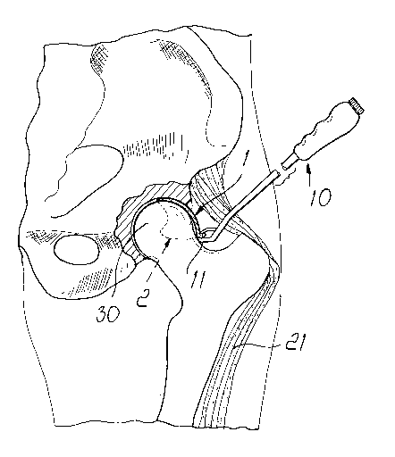
Figure 4 is a view of the initial step of insertion of the contoured body
between the head of the femur and the acetabulum;
Figure 5 is a schematic view of the contoured body after its insertion;
Figure 6 is an enlarged-scale view of the contoured body arranged in the
CA 02515920 2005-08-12
WO 2004/071331 ' PCT/EP2004/001306
3
acetabulum and of the repositioned muscle bundles;
Figure 7 is a plan view of the corrective element according to a further
embodiment;
Figure 8 is an elevation view of the corrective element of Figure 7 ;
Figure 9 is a sectional view of Figure 7 taken along a median plane;
Figure 10 is a plan view of the corrective element according to still
another embodiment;
Figure 11 is a sectional view of Figure 10 taken along a median plane.
Ways of carrying out the Invention
With reference to the figures, the corrective element for the articulation
between the femur and the pelvis, according to the invention, comprises a
contoured body l, which is preferably made of steel and has a shape that
approximate a spherical portion.
The contoured body 1 has a beveled insertion edge 2, in which the
thickness decreases to zero, and has, in a central portion, a central recess 3
for the reason explained hereinafter.
On the opposite side, the body 1 has a pushing and positioning edge 4,
provided with multiple holes 5 for manipulation by means of a suitable
instrument or lever 10 that facilitates insertion operations.
The instrument 10 has a push plate 11 that is provided with coupling
pins 12 that enter the holes 5 located on the pushing edge 4, and there is
also a complementary jaw 14, which is actuated by a suitable lever
mechanism and engages the edge 4 on the opposite side, so as to practically
clamp the edge 4 on the instrument 10 in order to allow to apply the force
required for insertion. The edge 4 lies substantially at right angles to the
surface of the body 1.
In practical use, in order to insert the corrective element, first the
articular capsule is cut and optionally an osteotomy of the upper part 20 of
the greater trochanter is performed, moving the muscle bundles, generally
designated by the reference numeral 21.
CA 02515920 2005-08-12
WO 2004/071331 PCT/EP2004/001306
4
This exposes the iliac acetabular region, and after cutting into the
articular capsule, as shown schematically in Figure 4, it is possible to begin
the insertion of the contoured body, which in practice is fitted over the head
of the femur, designated by the reference numeral 30, and is inserted in the
acetabulum.
As insertion continues, the contoured body 1 is arranged so that it lies in
close contact with the iliac acetabular region, since the contoured body has
an outer surface that is shaped substantially complementarily to the iliac
acetabular region and has an internal surface that forms a seat for
accommodating the head of the femur 30.
This coupling in practice achieves self positioning and self centering of
the corrective element in the acetabulum, accordingly achieving the
possibility to restore the correct articulation without having to first
dislocate
the head of the femur.
Once positioning of the contoured body has been performed, the muscle
bundles are repositioned and any portion of the greater trochanter that had
been osteotomized is reconnected by using conventional surgical pins 40.
Another surgical technique can also be carried out, i.e. without greater
trochanter osteotomy and only with posterior or anterior capsulotomy.
With reference to Figures 7, 8 and 9 an outer spherical surface 22 and an
inner spherical surface 23 of the contoured (shell-like) spacer 1 terminate in
an
approximately equatorial plane 15 in correspondence with a pushing and
positioning edge 4, which extends in the plane 15 only over a part of the
periphery. If referred to a pole axis 24 of the pushing and positioning edge,
the
same extends through an angle (3 with a value from 130° to 180°.
For the
overall rigidity it is even more suitable for the value of j3 to be above
150°.
Clear of the pushing and positioning edge 4 the outer and the inner spherical
surfaces 23 and 22 are connected by an insertion edge 2, which in relation to
the positioning edge 4 constitutes a central recess 3 around the pole region.
In
plan view (figure 7) it can be seen that the spherical surfaces 22 and 23,
when
CA 02515920 2005-08-12
WO 2004/071331 PCT/EP2004/001306
related to the pole axis 24, describe an angle a larger than 180°. The
angle a
can exceed 210°. In the bisector of the angle ~i the contoured body or
spacer 1
has a plane of symmetry 25, which divides the spacer into a left half and a
right
half. The pole axis 24 forms, as seen in the plan view, simultaneously a
center
5 7 for the largest radius R2 of the inner spherical face 23. With respect to
the
pole axis 24 the recess 3 possesses a minimum radius R3, which, in comparison
to the largest internal radius, corresponds to a percentage of 27% to 37% ,In
the left and right halves the insertion edge 2 extends a distance d past the
pole
axis 24 in the form of projecting ears 6, the distance d corresponding to 25%
to 30% of the maximum inner radius R2 of the inner spherical surface 23.
If referred to the pole axis the pushing and positioning edge 4 has an
outer radius R1 which corresponds to 120 to 140% of the radius R2. The
positioning edge 4 can have a thickness 16 from 1 to 5 mm. The two ears 6
are, as shown in figure 7, smoothly rounded. The actual edge between the
inner and the outer spherical surfaces has a radius of at least 0.5 mm. The
wall
thickness 17 becomes smaller between the inner and outer spherical surfaces
23 and 22. In the remaining portion the wall thickness 17 may have values
from 1 to 3.5 mm.
As shown in figure 8 the insertion edge 2, when considered in a
projection perpendicular to the plane 25 of symmetry, extends away from the
pushing and positioning edge 4 along a straight line 19, extending at an angle
'y
from the plane 15. The angle ~y can be from 35 to 50°. If the angle y
is selected
to be from 40° to 45°, insertion of the implant is still
possible and furthermore
a relatively large support surface is formed with the ears 6.
In accordance with the natural shape of a femur head it is advantageous
for the inner spherical face 23 to have a flattened area in the eventual
working
direction. One form of such a flattened area is illustrated in figure 9. In
the
plane of the drawing two centers 9 are shown, which are arranged at a small
distance s apart and which respectively define (starting with the outer
border),
based on a radius R4 the outline of the inner spherical face 23. In the median
CA 02515920 2005-08-12
WO 2004/071331 PCT/EP2004/001306
6
portion, which is characterized by an angle 8 the outline is continued, on the
basis of a larger radius R5, to bridge over the distance s, which may amount
to
1 to 3 mm. The angle 8 can lie from 40 to 70°. It is important that the
transition from one curvature to the other curvature takes place continuously
so
that there is no irregularity in the curvature. Another possibility of
providing a
flattened area in the eventual working direction would be to provide smaller
radiuses of curvature toward the pole. The spherical surface would then
correspond to a section, cut in a very weak ellipsoid.
If it is assumed that the pushing and positioning edge 4 limits
movements in relation to the acetabulum, then at least the inner spherical
surface 23 should have a roughness of less than 0.1 Vim.
The embodiment of figures 10 and 11, which in its structure corresponds
to the embodiment of figures 7, ~ and 9, includes as a further feature a bead
1 ~
projecting in the middle part of the central recess 3 toward the pole axis 24,
and such bead may constitute a further security means to prevent accidental
slipping out of place from the acetabulum. This could be an advantage in the
case of a spacer 1 of a rubber-like material.
An application of the implant is in one case conceivable for elderly
patients with local damage of the femur head cartilage or of the acetabulum
cartilage. The implant would practically bridge over the defective area. A
further application is merely as a placekeeper with the purpose of reducing
pain. Elderly patients, who owing to the risk of a thrombosis, cannot be
subjected to a major operation like the complete replacement of the hip joint
could - more particularly if tied to a wheel chair - be freed of part of their
pain,
since the operation would rather be considered to be a minor one.
The selection of the material for the implant is therefore not limited at
the outset. Rigid shells of a physiologically compatible metal alloy are
conceivable, which have a low roughness Ra of less than 0.1 ~.m on their load
bearing surfaces 23 and 22. The implant may also consist of a somewhat
elastic physiologically compatible material. In the case of a merely
CA 02515920 2005-08-12
WO 2004/071331 PCT/EP2004/001306
7
placekeeper function with small movements without a load elastic, rubber-like
but dimensionally stable plastics are conceivable.
Moreover plastics in the form of a hydrogel could be employed to
provide inserts with a small wall thickness.
Coating of the load bearing surfaces 23 and 22 with a physiologically
compatible anti-friction layer with the body is conceivable as well.
A further possibility is to endow the load bearing surfaces with a
porosity like that of the natural meniscus in order to favor colonization with
the
own body cells.
From the above description it is therefore evident that the invention
achieves the intended aim and objects, and in particular the fact is stressed
that ~a contoured body is provided which has, in its front portion, a hollow
that allows to preserve the round ligament and its vascularization,
accordingly maintaining optimum conditions for its integration in the
articulation without removing functional connections.
The contoured body can be manufactured in different sizes, depending
on the anatomy of the patient, and can have various thickness, depending on
the defect to be corrected; one should bear in mind that the inner and outer
surfaces of the contoured body must be smooth, so as to allow its insertion
without particular traumas, and that the beveled penetration edge must have
a limited thickness both to facilitate its insertion and to avoid producing a
dangerous discontinuity in the seat for accommodating the head of the
femur.
The invention thus conceived is susceptible of numerous modifications
and variations, all of which are within the scope of the appended claims.
All the details may further be replaced with other technically equivalent
elements.
In practice, the materials used, so long as they are compatible with the
specific use, as well as the contingent shapes and dimensions, may be any
according to requirements.
CA 02515920 2005-08-12
WO 2004/071331 PCT/EP2004/001306
The disclosures in Italian Patent Application No. MI2003A000274 from
which this application claims priority are incorporated herein by reference.