Note : Les descriptions sont présentées dans la langue officielle dans laquelle elles ont été soumises.
CA 02519412 2005-09-16
SPECIFICATION
COMBTNED USE OF G-CSF AND FACTORS HAVING ANGIOGENIC AC'Y'XOI~T
'~ECHNZC~1.L FIELD
The present ~.nvent~.on relates to a remedy for
ischemio disease, ~..e., a composition for treating.ischemic
disease, which comprises granulocyte colony-stimulating
factor (G-CSF) and a factor having an az~giogenic action as
active ingredients.
io
BACKC124UND .A.RT
The present invention is an invention concerned with
remedies for isohemio disease. One typical ischemic
disease. obstructive arteriosclerosis, will be described
~.5 first .
Obstructive arteriosclerosis a.s a disease in which an
arteriosclerotic (atherosclerotic) lesion results in
deposition of an atheromatous substance mainly consisting
of fats on the endarterium, to arouse occlusion or stenosis
20 of a major truncal artery in the extremity, especially in
the lower limb, thereby causing an isch,emic disorder in ~.ts
periphery. Ch.nical symptoms of this disease are
c~.assified as coldness or numbness, intermittent
claudication, rest pain, and ulcer/necrosis. In Japan,
25 patients writh obstructive arteriosclerosis are estimated to
number about 100.000 (Yusuke Tada: Biomedicine &
Therapeutics, Vol. 31, 289-292; 1997). The number of
patients with this disease is expected to increase because.
- 1 -
CA 02519412 2005-09-16
of the increase in the elderly population and the
westernization of diets.
Therapies of obstructive arteriosclerosis include
kinesitherapy or exercise therapy, phaz~nacotherapy, and
revascularization, which are selected depending on symptoms
or the patient's condition. Other measures, now under
consideration, for avoiding a resection of a severely
ischemic l3.mb are angiogenie therapies (gene therapy, bone
marrow autotransplantation, etc.) for promoting
angiogenesis. These therapies are currently achieving some
success in the treatment of obstructive arteriosclerosis,
but the respective therapies involve the following problems.
In some mild cases, the distance of walking has
increased in exercise therapy. However, the effect of this
therapy is difficult tv predict. Moreover, patients are
not satisfied with the increase in the walking distance, if
any, and 30% of them are reported to have requested
revascularization (Takashi Ohta: Japan Medical Journal,
Vol. 3935, 25-29. 1999). Thus, at present, this therapy is
not a very effective form of treatment .
In pharmacotherapy, antiplatelet agents are mainly
prescribed, but they merely prevent an aggravation of
symptoms. Microcirculation improving agents and oxygen
transport ~.mproving agents, which have recently been
developed aggressively, are only expected to be ind5.cated
for mild cases. Nowadays, there are no radical remedies
available for obstructive arteriosclerosis.
Revascularization, on the other hand, is currently
- Z -
CA 02519412 2005-09-16
the most effective tk~erapy, which involves percuta~neous
az~gioplasty or a bypass operation depend~.ng on the
condition of the patient or the location or extent of the
lesion. However, these surgical operations are so
extensive that they pose problems, such as surgery-
associated complications or death, and a poor prognosis for
a long life.
Gene therapy using angiogenic factor is aa.med at
correcting ischemia by deveJ.oping collateral circulation
channels. Examples of known az~gipgen~.c factors are .
vascular endothelial growth factor (VEGF), epidermal growth
factor (EGF), hepatocyte growth factor (HGF), and
fibroblast growth factor (FGF). zn Japan, clinical studies
using human HGF are under way. A method, tnihich involves
its intramuscular injection into the lower limb muscle
using a plasmid carrying HGF gene, has been invest~.gated in
patients with severely ischemic limbs, and expectations are
growring for its efficacy. However, this therapy is still
at the experimental stage, and evaluations of its safety
and efficacy have not been fully carried out. Thus, gene
therapy has not become popular.
Intramuscular transplantation of autologous bone
marrow cells, wh~.ch has recently attracted attemtivn, is a
therapy xz~ which bone marrow cells are transplanted into
the muscle near the diseased part, whereafter they are
differentiated into vascular endothel3.a3. cells to dorm
blood vessels, thereby treating the diseased part. Bone
marrow autot~cansplantation has no adverse effects on the
- 3 -
CA 02519412 2005-09-16
immune system, and has been recognizEd to present
differentiation of bone marrow cells into endothelial cells
or increase the number of blood vessels in animal models.
Although its efficacy will have to be evaluated in an
increased number of patients, this therapy is expected to
become a promising one, because it can treat severe cases.
However, the bone marrow is taken under general anesthesia
in a clinical setting, so the heavy burden imposed on the
patient and medical staff in taking the bone marrow may
present problems.
Recent studies have shown that hematopoietic stem
cells, which can differentiate into vascular endothelial
cells, are present not only in the bone marrow, but also in
the peripheral blood, and they take part in angiogenesis
(Qun Shi et al., ~laod vo1.~92, 362-367, 1998; Takayuki
Asahara et al., Circulation Research vol. 85, 221-228,
1999; Marie Peiahev et al.. Blood vol. 95, 952-958, 2000).
(The hematopoietic stem yells are called "precursor Cells
for endothelial cells" from the viewpoint of the function
of differentiating into endothelial cells. However, these
cells are originally derived from hematopoietic stem cells.
Thus, the term "hematopvietic stem cells" is used herein in
accordance with the concept that they are a cell population
capable of becoming endothelial cells.) Hence,
hematopoietic stem cells in the peripheral blood are taken
and transplanted into the muscle close to the diseased part,
whereby treatment of obstructive arteriosclerosis can be
expected. This procedure is advantageous in that the
- 4 -
CA 02519412 2005-09-16
burden imposed on the patient and medical staff at the time
of taking peripheral blood stem cells is less than that
during transplantation of stem cells present in the bone
marrow. No~nally, however, the frequency of existence of
hematopoietic stem cells in the peripheral blood ~.s
extremely low. Thus, it is highly quEStionable whether a
necessarx and adequate amount of hematopoietic stem cells
for the treatment of obstructive arteriosclerosis can be
obtained.
Human G-CSF is a hematopOietia factor disaavered as a
differentiation/growth factor for progenitor cells of the
granuloeytio lineage. It is clinically applied as a remedy
for neutropenia following bore marrow traz~splaz~tation or
cancer ohemotherapy, because it facilitates neutrophilic
hematopoiesis in vivo. In addition to this action, human
G-CSF acts on hematopoietic stem cells to stimulate their
proliferation and differentiation, and also acts to
mobilize hematopoietic stem cells present in the bone
marrow into the peripheral blood. ,Actually, based on the
latter action, transplantation of the peripheral blood
hematopoietic stem cells mab5.lized by human G-CSF, i.e.
peripheral blood stem cell transplantation, is perfoz~a~ed in
the cl~.n~.cal setting, with the aim of accelerating
hematopoietic recovez~y in cancer patients after a.ntens~.ve
chemotherapy. This hematopoietic stem cell mobilizing
action of G-CSF is fa~c more potent than that of GM-CSF,
also a hematopoietic factor for the granuloaytic lineage.
In terms of few side effects as well, G-CSF has superiority
..
CA 02519412 2005-09-16
over GM-CSF.
HGF is a protein which is produced by various
mesenchymal cells and targets many epithelial cells,
neurons, endothelial cells, and some mesenahymal cells.
HGF is known to have cEll motility promoting activity and
epithelial morphogeaesis (luminal structure, etc.) inducing
activity, in addition to cell proli~eratxon promoting
activity. Since HGF functions as an organ regenerating
factor for promoting the regeneration of the kidney, the
lung and the digestive tract, as well as the lzver, in
adults, it is expected to be a remedy for organ disease.
DrSC~OSU12E OF THE INVENTION
In patients with obstructive arteriosclerosis,
administration of G-CSF prior to treatment with
intramuscular transplantation of bone marrow cells can be
expected to incrEase the frequency of hematopoietic stem
cells in the bone marrow. Thus, the number of bone marrow
punctures for collecting bone marrow cells can be reduced,
and the burden on the patient can be reduced. On this
occasion, the burden on the patient and the medical staff
can be further reduced by obtaining hematopoietic stem
cells for transplantation from the peripheral blood.
Furthermore, hematopoietic stem cells in the peripheral
blood have been shown to contribute to vasculogenesis, so
that the increase of hematopoietic stem cells in the
peripheral blood induced by the administration of G-CSF is
speculated to promote vasculogenesis. Hence, the mere
- 6 -
CA 02519412 2005-09-16
administration of G-CSF to patients can be expected to
treat obstructive arteriosclerosis. This treatment for
obstructive arteriosclerosis by the administrative of G-CSF
will clearly reduce the burden on the patient and the
medical staff markedly in that it obviates the need for
collection and transplantation of hematopoietic stem cells.
Besides, the combined use of G-CSF and gene therapy
using angiogenic factor is expected to enhance the
therapeutic effect. That is, G-CSF is caused to act on
hematopoietic stem cells, stimulating their proliferation
and differentiation. Also, hematvpoietic stem cells in the
bone marrow are mobilized into the peripheral blood to
promote vasoulogenesis. At the same time, angiogenesxs is
promoted by HGF or FGA. Effective utilization of these
different actions can be predicted fio show an additzve or
synergistic angiogenic effect_
Treatment for obstructive arteriosclerosis using
G-CSF can be expected to take effect in severe cases, and
will be of great benefit to patients. If this treatment is
combined with treatment with an angiogenic factor which
promotes differentiation and growth of vascular endothelial
precursor cells, such as vascular endothelial growth factor
(VEGF),'epidermal growth factor (EGF), hepatocyte growth
factor (HGF) or fibroblast growth factor (FGF), or with the
gene therapy of these factors, the therapeutic effect of
that treatment is expected to be augmented further. Tn
this case, these factors or their genes can be administered
to patients, for example, at sites near the disoased part.
CA 02519412 2005-09-16
Similarly, G-CSF 3s expectEd to show an increased
therapeutic effect, when combined with agents clinically
used as drug therapies for obstructive arteriosclerosis,
such as ant~.platelet agents, vasodilators, microcirculatiox~
improvers, ant~.coagu~.ants and antilipamic agents.
As a result of the foregoing analyses, the inventors
of the present invention have found that G-CSF, whEn
administered in combination with HGF or FGf as an
angiogenio factor, produces a particularly significant
improving effect on ischemic blood vessels, and thereby
have accomplished the present invention.
Thus, the present invention provides a remedy for
ischemic disease, which comprises G-CSF and HGf or FGf as
active ingredients.
~5 Furthermoz~e, the remedy of the present invention is
applicable as a remedy for the following diseases, similar
ischemic diseases: trauma, refection reaction during
transplantation, ischemic cerebrovascular disorder (such as
apoplexy or cerebra. ~.z~farction), isohemia renal disease,
ischemic pulmonary disease, infection-related ischemic
disease, ischemic disease o~ l~.mbs, and isChpmic heart
disease (such as ischemic cardiomyopathy, myocardial
infarction or ischemic heart failure). That is, the
present invention provides remedies for these diseases,
which contain G-CSF and HGF ox FGF as active ingredients.
The present ~.nvention also provides a remedy for
obstructive arteriosclerosis.
The present invention further provides an agent for
_ g _
CA 02519412 2005-09-16
revascularization or muscle regenaratian.
BRIEF DESCRIPTION OF bRAWINGS
Figure 1 is a view showing the effects of
administration of physiological saline (control), HGF,
G-CSF and HGF~G~CSF, respectively, on the lower limb muscle
weight ratio (~) in mice with an ischemia left paw.
Figure 2 is a view showing the effects of
administration of physiological saline (control), HGF,
.G-CSF and HGF+G-CSF, respectively, on the lower limb blood
flow ratio (%) in mice with an ischemic left paw.
Figure 3 is a view showing the effects of
administration of physiological saline (control), HGF,
G~CSF and HGF+G-CSF, respectively, on the blood flow rate
~5 in mice with an ischemic left paw. The red portion
represents the highest flow rate, followed by the yellow,
green and blue portions in decreasing order.
Figure 4A presents fluorescence photomicrographs
showing the effects of administration of physiological
saline (control: HGF~, G-CSF-), HGF plasmid (HGF+, G-CSF-),
G-CSF (HG~-, G-CSF+) and G-CSC+HGF plasmid (HGF+, G-CSF+),
respectively. on the ratio of GFP~positive cell numbers in
mine with an ischemic left paw. GFP-positive cells are
indicated in green and nuclei are indicated in blue.
Figure 4B is a graph showing scores for GFP-positive cell
numbers 'calculated from the photomicrographs.
Figure 5A presents fluorescence photamioragraphs
showing the effects of administration of physiological
- 9 -
CA 02519412 2005-09-16
saline (control: HGF-, G-CSF-), HGF plasmid (HGF+, G-CSF-).
G-CSF.(HGF-. G-CSf+) and G-CSF+HGF plasmid (HGF+, G-CSF+),
respectively, on the ratio of vWF-positive cell z:umbers in
mice with an ischem~.c left paw. vWF-positive cells are
indicated in red and nuclei. are indicated in blue. Figure
5B is a graph showing scores for vWF-positive cell numbers
calculated from the photomicrographs.
Figure 6A shows fluorescence photomicrographs of
GFP-pos~.t~.ve cells (green), vascular endothelial cells
(red) and nuclei (blue), as well as a merged image thereof.
Figure 6H shows fluorescence photomicrographs of GFP-
positive cells (green), vascular smooth muscle cells (red)
and nuclei. (blue), as wall as a merged image thereof.
Figure 7A shows fluorescence photomicrographs of
GFP-positive cells (green), skeletal muscle cells (red) and
nuclei (blue), as well as a merged image thereof. Figure
7B shows merged images of serial sections.
Figure 8 is a view showing the effects of
administration of physiological saline (a: next day after
surgery, b: 4 weeks after treatment), HGF plasmid (c: 4
weeks after treatmEnt), G-CSF (d: 4 weeks after treatment)
and G-CSF+HGF plasmid (e: 4 weeks after treatment),
respectively, on the lower ~.imb blood flow rate in mice
with an ischemic left paw. Tk~.e asterisk "*~ denotes p<0.05
(vs. physiological saline treatment group) and the plus
sign °+" denotes p<0.01 (vs. physiological saline treatment
group). LikewisE, the number mark "#~ denotes p<0.05 (vs.
HGF plasmid group) and the section mark °~" denotes p<0.05
- 10 -
CA 02519412 2005-09-16
(vs. G-CSF group).
Figure 9 is.a graph showing scores for the degree of
lower limb damage 4 weeks after treatment. YTh~.te indicates
no zzecrosxs, gray xz~d~.cates toe necrosis, and black
indicates limb necrosis.
Figure 10 is a view show~.ng the effects of
administration of physiological saline, G--CSF, HGF plasmid,
FGF, G-CSF+HGF plasmid and G-CSF-~FGF, respectively, on the
lower limb blood flow z~ate after 4 weeks in mice with an
ischemic left pave. The asterisk "*" denotes p<0.05 (vs.
physiological saline treatment group) and the dagger "t"
denotes p<0.01 (vs. physiological saline treatment group).
Likewise, the n~unber mark "~" denotes p<0.05 (vs. G-CSF
group), the section mark "5" denotes p<0.05 (vs. HGF
plasmid group), and the paragraph mark "~[" denotes p~O.Ob
(vs. FGF group).
Figure 11 is a graph showing scores for the degree of
lower limb damage 4 weeks after treatment. White indicates
no necrosis, gray indicates toe necrosis, and black
ind~.cates limb necrosis.
BEST MODE FOR CARRYING OUT THE INVENTION
The present invention relates to a remedy for
ischemic disease, Which comprises G-CSF and IiGF ar FGF.
As used herein, the term "ischemic disease" refers to
a disease associated with local anemia caused by an organic
disturbance in blood supply (e. g., arteriostenosis).
Examples of ischemic diseases include trauma, rejection
- 11
CA 02519412 2005-09-16
reaction during transplantation, zschemic cerebrovascular
disorder (such as apoplexy or cerebral infarction),
a.schemic renal disease, ischemic pulmonary disease,
infection--related ischemic disease, ischemic disease of
limbs, and .fSChAmiC heart disease (such as ischemic
cardiomyopathy, myocardial infarction or ischemic heart
failure).
The present i.nvez~tion also relates to an agent ~oz'
revascularization or muscle regeneration, which comprises
G-CSF and HGF or FGF.
Human G-CSF is a known protein composed of 174 amino
acid residues.
When G-CSf is used as the active ingredient of the
remedy for ischemic disease according to the present
invention, any type of G-CSF can be used, but highly
purified G-CSF is preferred. Specific examples of G-CSF
include mammalian G-CSF, especially human G-CSF, or G-CSF
having substantially the same biological activity as
mammalian G-CSF. The origin of G-CSF is not la.mited, and
both naturally occurring G~CSF as well as G-CSF obtained by
genetic recombination can be used. Tha G-CSF obtained by
genetic recombination may be that having the same amino
acid sequence as naturally occurring G-CSF (e. g., JP 1990-
5395, JP 19$7-2364$$ A), or that having this amino acid
sequence subjected to deletion, substitution and/or
addition of one or more amino acids, and having the same
biological activity as naturally occurring G-CSF. Foz~
example, a polypeptide functionally comparable to G-CSF can
- 12 -
CA 02519412 2005-09-16
be prepared by appropriately introducing a mutation into
the amine acid sequence o~.G-CSF by use of such a method as
site-directed mutagenesis (Gotoh, T. et al. (1995) Gene 152,
271-275; Zoller, M.J. and Smith, M. (1983) Methods Enzymol.
100, 468-500; Kramer, W. et al. (1984) Nucleic Acids Res.
12, 9441-9456: Kramer, W. and Fritz H.J. (1987) Methods
Enzymol. 154, 350-367; Kunkel, T.A. (1985) Proc. Natl. Acad.
Sci. USA, 82, 488-492; Kunkel (1988) Methods Enzymol. 85,
2763-2766). The mutation of an amino acid can occur in the
natural world. xt is already known that a polypept~.de
having a certain~am~.no acid sequence modified by deletion
and/or addition of one or more amino acid residues and/or
substitution of an amino acid for the other amino acid
retains the biological activity, of the original polypeptide
(Mark, D.F. et al., Proc. Natl. Acad. Sci. USA (1984) 81,
5662-5666; Zoller, M.I,. & Smith; M. Nucleic Acids Research
(1982) 10, 6487-6500: Wang, A. et a1., Sc~.ence (1984) 22~,
1431-1433; Dalbadie-McFarland, G. et al., Proc. Natl. Acad.
Sci. USA (1982) 79, 6409-6413).
Hence, a polypeptide comprising an amino acid
sequence which has one or more amino acid mutations in
G-CSF sequence, and being functionally equivalent to G-CSF,
can also be used as the remedy for ischemic disease of the
present invention. The zr.umber of amino acid mutations in
such a polypeptide are normally within 30 amino acids.
preferably within 15 amino acids, more preferably within 5
F~mino acids (for example, within 3 amino acids).
In the substitution mutant, substitution of an amino
- 13 -
CA 02519412 2005-09-16
acid for the other amino acid which conserves the mature of
the amino aoid side cha3.n is desirable. As the amino acid
which conserves the nature of the amino acid side chain,
there can be named, for example, hydrophobic amino acids (A,
I, L, M, F, P, W, Y, V), hydrophilic amino acids (R, D, N,
C, E, Q, G. H, K. S. T), amino acids having an aliphatic
side chain (G, A, V, L, I, P), amino acids having a
hydroxyl group-containing side chain (S, T, Y), am~.no acids
having a sulfur atom-containsng s~.de chain (C, M), amino
IO acids having a carboxylic acid- or an amide-containing side
chain (D, N, S, Q), amino acids having a base-containing
side chain (R, K,.H), and amino acids having an aromatic--
containing side chain (H, F, Y, W) (the symbols in the
parentheses represent one-letter abbreviations for the
1.5 corresponding amino acids).
Polypeptides in which a plurality o~ ami.no acid
residues are added to the amino acid sequence of G-CSF
include fusion polypeptides with G-CSF.. Sucks fusion
poZypeptides are polypeptides produced by fusion between
20 G-CSF and other polypeptide, and can also be used in the
present invention. A fusion polypeptide can be prepared by,
for example, l~.gat~.ng DNA coding for G-CSF with DNA coding
for another polypeptide in-frame, transferring the
resulting construct into a suitable expression vector, and
25 expressing the insert in a suitable host. Other
polypepti.de to be fused to G-CSF is not limited as long as
the resulting fusion polypeptide retains biological
activity comparable to that of G-CSF.
- 14 -
CA 02519412 2005-09-16
Numerous reports are already present on G-CSF
derivatives with the amino acid sequence of G-CSF changed,
and thus these knornrn G-CSF derivatives can be used (for
example. USP 5,581.476, USP 5,214,132, USp 5,362,853 and
USP 4,904,584).
Moreover, chemically modified G-CSF can be used.
Examples of the chemically modified G-CSF include G-CSF
subjected to conformational change, addition or deletion of
the sugar chain, and G-CSF to which a compound such as
polyethylene glycol has been bound (for example, USP
5,824,778, USP 5',824,784, WO 96/11953, WO 95/21629, WO
94/20069, USP 5,218,092, JP 1992-164098 A).
G-CSF in the present invention may be produced by any
method. For example, it is possible to use G-CSF prepared
by culturing a human tumor cell line, followed by
extraction, isolation and purification by various methods,
or G~CSF prepared by causing Escherichia coli; yeast:
mammalian sells, such as Chinese hamster ovary sells (CHO
cells), C127 cells, COS cells, myeloma cells or BHK cells;
or insect cells_to perform production by genetic
engineering techniques, followed by extraction, isolation
and purification by various methods (for example, JP 1989-
44200, JP 1990-5395, JP 1987-129298 A, JP 1987-132899 A,
JP 1987-236488 A and JP 1989-85098 A).
The method.for producing this G-CSF may be any method
which can give the product defined above. Concretely. the
G~CSF is produced using G-CSF-producing tumor, G-CSF-
producing hybridoma, or a transformed host Which has been
- 15 _
CA 02519412 2005-09-16
granted a G-CSF-producing potential by genetic
recombination. Depending on the structure o~ G-CSF to be
produced, a changing operation or warzous modifying
operations are appropriately applied at a suitable stage of
the production process. If the G-CSF is to be produced by
genetic recombination, any routinely used host can be
employed, such as Escherichia coli or animal cells.
In the present invention, G-CSF may be administered.
either as a protein or in the form of a gene coding for
G-CSF. as in gene therapy.
HGF is a known heterodiznerie protein comprising a
69 kDa a chain and a 34 kDa ~ chain.
The mode of administration of HGF is not limited, and
HGF may be administered as a protein, but it is preferred
to administer a gene coding for HGF, as in gene therapy.
The gene coding for HGF is generally administered, for
example, as an expression vector containing an expression
cassette. The vector is not limited, and a non-virus
vector may be used, or a virus vector may be used (e. g.
Supplementary Volume of Experimental Medicine,
"Experimental Methods for Gene Transfer and Expression
Analysis," YODOSHA, 1997; Supplementary Volume of
Experimental Medicine, °Basic xechniques for Gene Therapy°,
YODOSHA, 1996). Examples of the vector include a plasmid
vector, a virus vector, a phage vector, a cosmid vector and
a YAC vector. The expression vector normally includes a
regulatory element, such as a promoter, and an antibiotic-
resistance gene.
- 16 -
CA 02519412 2005-09-16
Any methods are available for gene transfer, and
include, for example, calcium phosphate transfection,
lipofeotion, a method using a liposome, the naked-DNA
method, receptor-mediated gene transfer, a method using a
gene gun, DEAF-dextran transfection, and a method using a
cap~.llazy tube. in the present in~rention, the gene may be
directly transferred into a body, or after gene transfer
into cells taken up from the body, the cElls may be
returned into the body.
Since many reports have been issued on HGF and HGF
expression vectors (HGF expression plasm~.ds), those skilled
in the art can appropriately select and administer them
(e. g. Nakamura, T., Nishiaawa, T., Hagiya, M. et a.1. Nature
19$9, 342, 440-443: Hayashi, S., Morishita~, R.. Higaki. J.
et al. Biochem Biophys Res Commun 1996, 220, 539-545;
Morishita, R., Sakalci, M., Yamamoto, K. et al. Circulation,
2002, 105, 1491-1496). The administration of the gene
encoding HGF can be performed by a method known to those
skilled in the art (for example, WO 01/32220, WO 01/26694,
WO 97/07824, WO 01/21214),
When HGF is ac'iministered as a protein, any type of
HGF can be used, but highly purified HGF is preferred.
Specific examples of HGF zn,cl.ude mammalian HGF, especially
human HGF, or HGF having substantially the same biological
acti~crity a$ mammalian HGF. The origin of HGF is not
limited, and naturally occurring HGF and HGF 'obtained by
genetic recombination can be used. The HGF obtained by
genetic recombination may be that having the same amino
- 17
CA 02519412 2005-09-16
acid sequence as naturally occurring HGF (e. g., GenBank
Accession Nos.: M73239, M73240, M29145, L02931 and M6071.8),
or that having this amino acid sequence subjected to
deletion, substitution and/or addit~.on of one or more amino
acids, and having the same biological activity as naturally
occur~ciz~g HGF. For example, a polypeptide functionally
comparable to HGF can be prepared by appropriately
introducing a mutat~.on into the amino acid sequence of HGF
by use of such a method as s~.te-directed mutagenesis (Gotoh,
T. et al., 1995, Gene 152, 27J.-275; Zoller, M.J, and Smith,
M., 1983, Methods Enzymol. 100, 468-500: Rramer, W. et al.,
1984, Nucleic Acids rtes. 12, 9441-9456; Kramer, W. and
Fritz, H.J., 1987 Methods Enzymol. 254, 350-367; Kunkel,
T.A., 1985, Proc. l~atJ.. Acad. Sci. USA 82, 488-492; Kunkel
(1988) Methods Enzymol. 85, 2763-2766). The mutation of an
amino acid can occur iz~ the natural wrorld. It is already
knornrn that a polypeptide having a certain amino acid
sequence modified by delet~.on and/or addition of one or
more amino acid residues and/or substitution, o~ az~ amino
acid far the other amino acid retains the biological
activity of the original polypeptide (Mark, D.F. et al.,
Proc. Natl. Acad. Sci. USA 81, 1984. 5662 5666; Zoller, M.L.
& Smith, M., Nucleic Acids Res. 10, 1982, 6487-6500: Wang,
A. et al., Science 224, 1984, 1431-1433; Dalbadi.e-MoFarHand,
G. et al., Proa. Natl. Acad. Scx. USA 79, 1982. 6409-6413).
Hence, a polypeptide comprising an amino acid
sequence which has one oz' more amino acid mutation in HGF
sequence, and being functionally equivalent to HGF, can
18 -
CA 02519412 2005-09-16
also be used as a remedy for ischemic disease of the
pz~esent invention. The number of amino acid mutations in
such a polypept3.de ~.s normally within 30 amino acids,
preferably within 15 amino acids, more preferably within 5
amino acids (for example, within 3 amino acids).
In the substxtutxon mutants of HGF, substitution of
as amino acid for the other amino acid which conserves the
nature of the amino acid side chain is desirable, as in the
case of G~CSF. Polypeptides in which a plurality of amino
acid residues is added to the amino acid sequence of HGF
include fusion polypeptides with HGF. Such fusion
polypeptides are polypeptides produced by fusion between
HGF and other polypeptide, and can also be used in the
present invention. A fusion polypeptide can be prepared by,
y5 for example, ligating DNA coding for HGF with DNA coding
for another polypeptide in-frame, transferring the
resulting construct into a suitable e~cpress~.on vector, and
exp~ress~.ng the insert in a suitable host. Other
polypeptide to be fused to HCF is not limited as long as
the fusion polypeptide retains biological activity
comparable to that of HGF_
The gene coding for the HGF of the present invention
includes a gene coding for such a polypeptide funct~.onal.ly
equivalent to the HGF.
FGF encompasses acidic FGF (FGF1, 140 amino acids)
and basic FGF (FGF2, 155 to 157 amino acids). Other
functionallx similar members include FGF3 (int-2), FGF4
(hst-Z), FGF5. FGFf (hst--2), FGF7 (~CCF: keratinocyte growth
_ 19 -
CA 02519412 2005-09-16
factor), FGF8 (AIGF: androgen-induced growth factor) and
FGF9 (G,F~,f: G~ia-activating factor) . As used herein, the
term "FGF" is intended to include all of them.
The mode of administration of FGF 3.s not limited, and
FGF may be administered either as a protein or in the form
of a gene coding for FGF, as in gene therapy. The gene
coding for FGF is generally administered, for example, as
an expression vector containing an expression cassette.
The vector is not limited, and a non-virus vector may be
used, or a virus vector may be used (e. g. Supplementary
Volume of Experimental Medicine, "Experimental Methods for
Gene Transfor and Expression Analysis," YODOSHA, 2997;
supplementary Volume of Experimental Medicine, "Basic
Techniques foz~ Gene Therapy", YODOSHA, 1996). Examples of
the vector include a plasmid vector, a virus vector, a
phage vector, a cosmid vector and a YAC vector. The
expression vector normally inoludes a regulatozy element,
such as a promoter, and an antibiotic-resistance gene.
any methods are available for gene transfer, and
include, for example, calcium phosphate transfectxor~,
lipofect~.on, a method using a liposome, the naked-DNA
method, receptor-mediated gene transfer, a method using a
gene gun, DEAF-dextran transfection, and a method using a
capillary tube. In the present invention, the gent may be
directly transferred into a body, or affier gene transfer
into cells taken up f~com the body, the cells may be
returned into the body.
Since many reports have been issued on FGF and FGF
-- 20 -
CA 02519412 2005-09-16
expression vECtors (FGF expression plasmids), those skilled
in the art can appropriately select and administez~ them
(e. g.. Kurokawa, T et al., FEES ~ett. 213, 189-194. (1987),
GenBank Access3.on Nos.: J04513 and M27968). The
adm~,nistration of the gene encod~.ng FGF can be performed by
a method known to those skilled in the art (for example,
Jejurikar S et aZ., Journal of Surgical Research, 67(2),
13 7146, (1997), Reynolds P N et al., Tumor Targeting, 3(3),
156-168, (1998), Ruffinl. F et al., Gene Therapy, 8(26),
1207-1213, (2001)).
When FGF is administered as a protein, any type of
FGF can be used, but highly purified FGF is preferred.
Specific examples of FGF inc7.ude mammalian FGF, especially
human FGF, or FGF having substantially the same biological
activity as mammalian FGF. The origin of FGF is not
limited, and naturally occurring FGF and FGF obtained by
genetic recombination can be used. The FGF obtained by
genetic recombination may be that having the same amino
acid sequence as naturally occurring FGF (e. g., FEES Lett.
213: 189-194, 1987. GenBank Accession Nos.: 404513 and
M27968), or that having this amino acid sequence sub,~ected
to deletion, substitution and/or addition of one or more
amino acids, and having the same biological activity as
natural~.y occurring FGF. For example, a polypeptide
functionally comparable to FGF can be prepared by
appropriately introducing a mutation into the amino acid
sequence of FGF~ by use of sucks a method as site-directed
mutagenesis (Gotoh, T. et al., 7.995, Gene 152, 271-275;
- 21 -
CA 02519412 2005-09-16
Zoiler, M.J. and Smith, M.. 1983, Methods Enzymol. 100,
468-500; Kramer, W. et al., 1984, Nucleic Acids Res. 12,
9441-9456; Kramer, ~1'. and Fritz, H.J., 1987 Methods Enzymol:
154, 350-367; Kunkel, T.A., 1985, Prop. Natl. Acad. Sci.
USA 82, 488-492; Kunke~ (1.988) Methods Enzymol. 85, 2763-
2766). The mutation of an amino acid can occur in the
natural world. It is already known that a pollrpept~.de
having a certain amino acid sequence modified by deletion
and/or add9.tion of one or more amino acid residues and/or
substitution of an am~.no acid for the other amino acid
retains the biological acti~cri.ty of the original polypeptide
(Mark, D.F. et al., Proc. Natl. Acad. Sci. USA 81, 1984,
5662 5666: Zoller, M.L. & Smith. M., Nucleic Acids Res. 10,
x.982, 6487-6500; Wang, A. et al., Science 224, 1984, 1431
1433; Dalbadie--McfarHand, G. et al., Proc. Natl. Road. Sci.
USA 79, 1982, 6409-6413).
Hence, a polypeptide compra.sir~g an amino ac~.d
sequence which has one or more amino acid mutations in FGF
sequence, az~d being functionally equivalent to FGF, can
also be used as the remedy for ischemic disease of the
present invention . The numbe5c of amino acid mutations ~.n
such a polypeptide are normally within 30 amino acids,
preferably within 15 amino acids, more preferably within 5
amino aczds (for example, within 3 amino acids).
In the substitution mutants of FGF, substitution of
an amino acid for the other amino ac~.d which conserves the
nature of the amino acid side chal.n is desirable, as in the
case of G-CSF. Polypeptides 3n which a plurality of amino
- x2 -
CA 02519412 2005-09-16
acid residues is addEd to the amino acid sequence of FGF
include fusion polypeptides with FGF. Such fusion
polypeptides are polypept~.des produced by fusion between
FGF and other polypeptide, and can also be used in the
present invention. A fusion polypeptide can be prepared by,
for example, ligating DNA coding for FGf with DNA coding
for another polypeptide in-frame, transferring the
resulting constz~uct into a suitabZe~expression vector, and
expressing the insert in a su~.tabie host. Other
~O polypeptide to be fused to FGF is not limited as long as
the fusion polypept~.de retains biological aati~rity
comparable to that of FGF.
The gene coding for the FGF of the present invention
includes a gene coding for such a polypeptide functionally
equ~.valent to the FGF.
Moreover, chemically modified HGF or FGF can be used
in the present invention. Examples of the chemically
modified HGF or FGF include ~IGF subjected to conformational
change, addition or deletion of the sugar chain, and HGF or
FGF to which a Compound such as polyethylene glycol has
been bound.
HGF or FGF used in the present invention may be
produced by any method. For example, it is poss3.ble to use
HGF or FGF prepared by culturing a human tumor cell line,
follornred by extraction, isolation and purification by
various methods, or HGF or FGF prepared by causing
Escherichia coli: yeast; mammalian cells, suoh as Chinese
hamster ovary cells (CHO cells), Clz7 cells, COS cells,
23 -
CA 02519412 2005-09-16
myeloma cells or BHK cells; or insect cells to perform
production by genetic engineering techniques, followed by
extraction, isolation and purification by various methods.
The method for producing this HGF or FGF may be any method
which can give the product defined above. Concretely, the
HGF or FGF is produced using a transformed host which has
been granted an HGF or FGF-producing potential by, for
example, gez~etxc recombination. Depending on the structure
of HGF or FGF to be produced, a changing operation or
various modifying operations are appropriately applied at a
suitable stage of the product~.on process. If the HGF or
FGF is to be produced by genet3.c recombination, any
routinely used host can be employed, such as Escherichia
oali or animal cells.
G~CSF, HGF and FGF are commercially available; it is
also possible to use these commercially a~crailable products.
The remedy for ischemic disease according to the
present invention can contain pl~aarmaceutical carriers and
vehicles necessary for assuming the form of a medicinal
pharmaceutical composition, and can further contain
stabilizers and adsorpt~.on preventing agents. Suitable
dosage forms can be selected, including injections (such as
subcutaneous injection, ~.ntradermal injection,
~.ntramuscular injection, intravenous injection and
intraperitoneal injection), depot preparat5.ons, transnasal
preparations, oral preparations (such as tablets, capsules,
granules, liquids. and solutions, and suspensions),
tz~anspulmonary preparations, transdermal preparations and
- 24
CA 02519412 2005-09-16
transmucosal prEparations. Tf desired, suitablE devices
can be used.
The remedy for ischemic disease according to the
present ~.nventian can incorporate, if desired depending on
the mode o~ i.ts administxatzon and its dosage form, a
suspending agent, a solution adjuvant. a stabilizer, a
tonicity agent, a preser~rati~cre, an adsorption preventing
agent, a surfactant, a diluent, an excipient, a pH
regulator, a soothing agent, a buffering agent, a sulfur-
containing reducing $gent and an antioxidant.
Examples of the suspending agent are methylcellulose,
polysorbate 80, hydroxyethylcellulose, acac~.a, tragacanth
powder, sodium carboxymethylcellulose and polyoxyethylene
sorbitan monolaurate.
Examples of the so~.ution adjuvant are polyoxyethylene
hydrogenated castor oil, polysorbate 80, nicotinamide,
poJ.yo~cyethylerxe soz-bitar~ monolaurate, macrogol and castor
oil fatty acid ethyl ester.
Examples of the stabilizer are dextran 40,
methylcellulose, gelatin, sodium sulfite and sodium ,
metasulfite.
Examples of the ton3.city agent are D-man,nitol and
sorbitol.
E~eamples of the preservative are methyl
p-hydroxybenzoate, ethyl p-hydroxpbenzoate, sorbic acid.
phenol, cresol and chlorocresol.
Examples of the adsorption preventing agent are human
serum albumin, lecithin, dextran. ethylene oxide-propylene
25 -
CA 02519412 2005-09-16
oxide copolymer. hydroxypropylcellulose, methy~.ce7.lulosa,
polyoxyethylene hydrogenated castor oil and polyethylene
glycol .~
Examples of the sulfur~containing agent are
N-acetylc~rste,ine, N-acetylhomocysteine. thioctic acid,
thzodiglycoi, thioethanolamine, thioglycerol, thiosorbitol,
thioglycollic acrd and its salts, sodium thiosulfate,
glutathione and those having a sulfhydryl group such as a
thioalkanoic acid having 1 to 7 carbon atoms.
Examples of the antioxidant are erythorbic acid.
d3.butylhydroxytoluer~e, butylhydroxyanisol, a-tocopherol,
tocopheryl acetate, L-ascorbic acid and its salts,
L-ascorbyl palmitate, L-asaarbyl stearate, sodium bisulfate,
sodium sulfite, triamyl gallate, prapyi gailate, and
~.5 chelating agents such as disod~.um
ethylenediaminetetraaoetate (EDTA), sodium pyrophosphate
and sodium metaphosphate.
Thp remedy for ischemic disease of the present
invention may further contain normally added ingredients,
such as inorganic salts, e.g., sodium chloride, potassium
chloride, calcium chloride, sodium phosphate,~potassium
phosphate and sod~.um bicarbonate; and organic salts. e.g..
sod~.um citrate, potassium citrate and sodium acetate.
The dose and the frequency of dosing of G-CSF
contained in the remedy for ischemic disease according to
the present ~.nvention can be determined in consideration of
the condition of the patient for cahom this remedy is
indicated. The dose is usually 0.1 to 500 ~g/kg/day,
- 26 -
CA 02519412 2005-09-16
preferably 1 to 50 ~.g/kg/day, per adult. As the frequency
of dosing, the remedy of the invention can be administered
once to three times a day, for J. to '7 days weekly. The
mode o~ administration preferably includes intravenous
administration, subcutaneous administration and
intz~amuscular administration.
When G-CSF gene is given, its dose per adult is
0.1 ~,g to 100 mg, preferably 0.001 to 10 mg. When HGF gena
is administered in the form of a liposome, its dose per
l0 adult is selected from the range of about 1 ~,g to about
4 mg, preferably the range of about 10 ~.g to about 400 ~Cg.
In the present invention, when HGF gene is
administered, choice is made of the mode of administration
and the site of administration that are suitable for the
disease and symptoms to be treated. The preferred site of
administration is the muscle. The preferred mode of
administration is the parenteral route.
The dose diffe~cs according to symptoms of the patient.
When HGF gene is g3.ven, its dose per adult is 0.1 wg to
100 mg, preferably 0.001 to 7.0 mg. When HGF gene is
administered in the form of a l3.posome, its dose per adult
is selected from the range of about 1 ~,g to about 4 mg,
preferably the range of about l0 ~.g to about 400 ~.g. The
frequency of dosing is selECted appropriately depending on
symptoms of the patient. Preferably, the remedy is
administered once in several days to several weeks, more
preferably once weekly, totaling a plurality of times,
further preferably a total of 8 times.
- 27 -
CA 02519412 2005-09-16
When HGF is administered as a protein, its dose and
frequency of dosing can be determined in consideration of
the condition of the patient for whom this remedy is
indicated. The dose is usually 0.1 tv 500 ~g/kg/day,
preferably 1 to 50 ~,g/kg/day, per adult. As the frequency
of dosing, the remedy can be administered once to three
times a day, for 1 to 7 days weekly. The mode of
adm3.nistration preferably includes intravenous
administration, subcutaneous administration and
intramuscuhar administration.
In the present invention, when FGF gene is
admzz~,istered, choice is made of the mode of administration
and the site of administration, that are suitable for the
disease az~d symptoms to be treated. The preferred site of
administration is the muscle. The preferred mode of
administration is the parentEral route.
The dose differs according to symptoms of the patient.
When FGF gene is given, its dose per adult is 0.1 ~,g to
100 mg, preferably 0.001 to 10 mg. When FGF gene is
administered in the form of a liposome, its dose per. adult
is selected from the range of about 1 ~,g to about 4 mg,
preferably the range of about 10 ~,g to about 400 ~.g. The
frequency of dosing is selected appropriately depending on
symptoms of the patient. Preferably, the remedy ~.s
administered once in several days to several weeks, more
preferably once weekly, totaling a plurality of times,
further preferab~.y a total of 8 times .
When FGF is administered as a protein, its dose and
- 28 -
CA 02519412 2005-09-16
frequency of dosing can be determined in consideration of
the cond~.tion of the patient for whom this remedy is
indicated. The dose is usually O.x to 500 ~g/kg/day,
preferably 1 to 50 ~,g/kg/day, per adult. As the frequency
of dosing, the remedy can be administered once to three
times a day, for 1 to 7 days weekly. The mode of
administration preferably includes intravenous
administration, subcutaneous administration and
intramuscular administration.
However, the present invention is not limited by the
doses of G--CSF and HGF or FGF. In the present xz~ve~nt~.on,
G-CSF and HGF or FGF can be prepared and administered as a
single preparation. Alternatively, they can be prepared
separately, and administered on different occasions.
By using the remedy for isahemic disease according to
the present invention, the number of hematopoietic stem
cel3.s can be increased. 'the collection of these
hematopoietic stem cells from the bone marrow or peripheral
blood and their bone marrow autotransplantation to the
patient himself or herself can contribute to vascuJ.ogenesis
in peripheral blood, treating ischemic disease. The
administration of the remedy accord.ing,to the present
invention also mobilizes hematopoietic stem cells into the
peripheral blood, thus making it possible to treat ischemic
disease, without collection or transplantation of
hematopoietic stem cells.
Moreover, the remedy of the present invention can be
comb~.ned with drugs hitherto used writh expectation of
29 -
CA 02519412 2005-09-16
effectiveness against isahem~.c disease, such as
ant~.p7.atelet agents, vasodilators, micrvcirculatioz~
improvers. anticoagulants and antxlipemic agents, and can
also be used in combination with gene therapy.
The present xnvent~.on will be described in more
detail with reference to Experiments (pharmacological
efficacy) arid Examples (preparation examples) , which ~.n zoo
way limit the present invention.
EXAMPLES
Experiment 1 (pharmacological efficacy)
(1) wild type mice (C57Bh/6, 8-10 weeks of age; Cr.LA,
Tokyo, Japan) were irradiated once with a lethal dose of
total body irradiation (850 cGy). BOnE marrow cells (5 x
106 call) were collected from GFP transgenic mice (C57BL/6,
10~~12 weeks of age) (Okabe et al., (1997) FEES. Lett. 407,
313-319) and transplanted into the wild type m~.ce through
their tail veins. At 2 months after transplantation, the
left femoral artery of each mouse was ligated at two
locations to prepare lower limb ischamia models. These
models were randomly divided into four groups, i.e., a
physiological saline treatment group, a G-CSF treatment
group, an HGF p~asm3.d treatment group and a G-CSF+HGF
plasmid treatment group (5 annuals per group). HGF plasmid
(Nakamura, T., Nishizawa, T., Hagiya, M. et al., Natures
1989, 342, 440-443; Hayashi, S., Morishita, R., Higaki, J.
et al., Biochem Biophys Res Commun 1996, 220, 539-545;
Morishita, R., Sakaki, M., Xamamoto. K. et al., Circulation
=
CA 02519412 2005-09-16
2002, 105, 1491-1496) was prepared using a plasmid
pur.ifiaation kit (manufactured by QIAGEN) in a.CCOrdance~
with the manufacturer's protocol. The physiological saline
treatment group and the G-CSF (recombinant human G-CSF
(Chugai Pharmaceutical Co., T~td., Japan)) (300 ~g/kg/day)
treatment group received subcutaneous administration for 10
days, beginning 24 hours after ligature. The HGF plasmid
treatment: group received admir~istrativn in a dose of 500
wg/animal by intramuscular injection performed 24 hours
after ligature, xhe G-CSF+HGF plasmid treatment group
received intramuscular injection of HGF (500 ~.g/animal)
24 hours after ligature and, ~.mmediate3.y afterwards,
received G-CSF treatment (300 ~,glkg/day) for 10 days.
The drawings shown the lower limb muscle Weight ratio
(Figure 1), the lower limb blood flow ratio (Figure 2), and
the typical blood flow rate (Figure 3), 4 weeks after
treatment, in each of the groups. The experimental data
are shown in Table I.
Table 1
Lower
limb
muscle
Body weight Left foot/rightLeft foot/right
weight (g)
(g)
foot muscle
foot Blocd
flow
Before After weight
ratio
expert- expert- Right Left (off) ratio C96>
foot foot
ments menu
Physiological
saline 20.2811.5219.08*1.180.9610.050.70x0.0872.1015.6887.8012.92
HGF plasmid21.48f0.7519.3310.230.8810.050.78.0388.44-6.1191.4312.34
G-CSF 19.04*1.00.18.26*0.830.90*0.030.7210,0779.95*6.4788.24*2.55
HGF plasmid20 19.52x0.550.92*0.040.8810.0795.27*4.2994.56x1.64
98x0
45
~- G-CSF .
.
- 31 -
CA 02519412 2005-09-16
The G-CSF treatment group and the HGF plasmid
treatment group showed a tendency tov~rard improvement over
the physiological saline treatment group. In the G-CSf+gGF
plasmid treatment group, compared with the other groups,
significant improvements were observed in the lower limb
muscle weight ratio, the lower limb blood flow ratio and
the blood flow rate, showing reduction of damage to the
lower limb.
The above results suggested the combination of HGF
and G--CSF to enhance a therapeutic effect as compared with
HGF or G-CSF administered alone.
(2) Next, for histologioal observation, wild type mice
were treated in the same manner as described above and then
anesthetized with ketamine (30 mg/kg) and xylazine (6
~.5 mg/kg). Their lower limb blood vessels were perfused with
P8S and faced by perfusion of 4~ paraformaldehyde in PSS.
Tschemic lower limb muscle was excised, embedded in OCT
compound (Miles Scientific, Naperville, x~. USA) and then
rapidly frozen in liquid nitrogen to prEpare sliced
sections. The frozen sections (6 ~.m) were washed with PBS
and stazned overnight at 4°C using antibodies. The
antibodies used for staining were: an anti-von Willebrand
factor (vWF) antibody (clone F8/86; DAKO) for vaseu~lar
endothelial cell staining; an a-smooth muscle actin
antibody (clone 7.A4; 5lgma Aldrich) for vascular smooth
muscle cell staining; and an az~ti-actznxn antibody (clone
EA-53: Sigma Aldrioh) for skeletal muscle cell staining.
The sections were then washed three times with PBS and
- 32 -
CA 02519412 2005-09-16
incubated at 4°C for 4 hours in the presEnce of TRTTC (DAKO,
Japan)-labeled secondary antibody (red). The nuclei were
stained in blue with TOTO-3 (Molecular Probes). The
3.mmunosta5.ned sections were observed under a aonfocal laser
scanning m~,.croscope (LSM510META; Carl ~eiss, Jena, Germany)
(Figures 4A and SA).
Images obtained ~cr~.th the confocal laser scanning
microscope were transferred to a computer and analyzed with
NIH image software. GFP-positive cell numbers (Figure 4B)
and vWF-positive cell numbers (Figure 5B) were calcu7.ated
relative to nucleated cell numbez~s per section of the
physiolog~.cal saline treatment group (~GF-, G-CSF-).
Further, Figure 6A shows a typical merged image of
GFP-positive cel~.s and vascular endothelial cells in serial
sections, Figure 6B shows a merged image of GFP-positive
cells and vascular smooth muscle cells, and Figure 7A shows
a merged image of GFP-positive cells and skeletal muscle
cells.
The immunostaini.ng of lower limb muscle indicated
that bone marrow cell~der3.ved GFP-positive cells were
d~.fferenti.ated into vascular smooth muscle cells and
vascular endothelial cells. Although revascularization was
enhanced even in the HGF plasmid treatment group as
compared with the physiological saline treatment group, the
G-CSF+HGF plasm5.d treatment group showed a s~rnergistic
effect on revasaularization as compared with the other
groups. The regenerated lower limb muscle derived from
bone marrow cells was also observed. These results suggest
- 33 -
CA 02519412 2005-09-16
that the combined therapeut3.a effect of G-CSF and HGF on
lower limb damage is due to regeneration of blood vessels
and/or lower limb muscle in ischemic limbs.
Experiment 2 (pharmacological efficacy)
The left femoral artery of nude mice (BALB/cA) was
ligated at two locations to prepare lower limb ischemia
models. These models were randomly divided into four
groups. i.e., a physa.ological saline group (20 anzmals), an
HGF plasmid group, a G-CSF group and a G~CSF+HGF plasmid
group (10 animals per group). In the same manner as shown
in Experiment l, the physiological saline treatment group
and the G-CSF (300 wg/kg/day) treatment group received
subautanevus administration for 10 days, beginning 24 hours
after ligature. The HGF plasmid group received
administration in a dose of 500 ~,g/an~nal by intramuscular
injection performed 2~1 hours postoperatively. The
G-CSF+HGF plasmid treatment group received intramuscular
injection of HGF (500 [~gtanimai) 24 hours postoperatively
and, immediately afterwards, received G-CSF treatment
(300 ~g/kg/day) for 10 days. Figure 8 shows the typical
blood flow rate, 4 weeks after treatment. in each of the
groups. The blood flow rate was expressed as mean t SEM,
and statistical significaz~ae betweezz mean values was
calculated by ANOVA. Comparisons were made by log-ran3t
test or non-paramatria F~.sher's multiple comparison test.
The asterisk "*" denotes p<0.05 (vs. physiological saline
treatment group) az~d the p~.us sign "+° denotes p<0.01 (vs.
- 34 -
CA 02519412 2005-09-16
physiological saline treatment group). Likewise, the
number mark °~#° denotes p~0.05 (vs. HGF plasmid group) and
the section mark ~~" denotes p<0.05 (vs. G-CSF group).
Moreover, the degree of lovrer limb damage 4 weeks
after treatment was scored for evaluation (Figure 9).
White indicates no necrosis, gray indicates toe necrosis,
and black indicates limb necrosis.
Although even the groups treated with G-CSF or HGF
plasmid alone showed a significant i.mprovemezzt iz~ the lower
limb blood flow over the physiological saline treatment
gz~oup, the G-CSF+HGF piasmid group showed a more
significant and synergistic effect on ~.xnpz~ov~,ng the lower
limb blood flow as compared with the other groups. This
effect had the same tendency as observed in the results of
~.5 the wild type mice (Experiment 1), but it was found to be
s~.gn~.fxcantly k~,i.gher in the nude mice than in the wild type
mice. i,ikewise, the lower limb damage was also
significantly reduced.
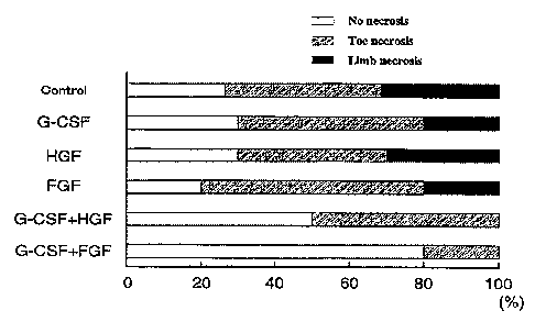
Exper3mant 3 (pharmacological. efficacy)
The left femoral artery of nude mice (BAr,B/cA) was
ligated at two locations to prepare lower limb ischemia
models. These models were randomly divided into six groups,
i.e., a physiological saline group (20 animals), a G~CSF
group, az~ HGF plasmid group, an FGF group, a G-C5F+HGF
plasmid group~and a G-CSF+FCf group (5 animals per group).
Tn the same manner as shown in Experiment 1, the
physiological saline treatment group and the G-CSF
- 35 -
CA 02519412 2005-09-16
(300 ~.g/kg/day) treatment group received subcutaneous
administration for 10 days, beginning 24 hours after
ligature. The HGF plasmid group received administration in
a dose of 500 ~.g/ani.mal by intramuscular injection
pErformed 24 hours postoperatively, The FGF group rece~.ved
intramuscular injection of FGF (500 ~,g/animal) (Trafermin:
Kaken Pharmaceutical Co., Ltd., Japan) 24 hours
postoperatively. The G-CSF+HGF plasmid treatment group
received intramuscular injeoti.on of HGF (500 ~,g/animal) 24
hours postoperatively and, immediately afterwards, rece~.ved
G-CSF treatment (300 ~g/kg/day) for 10 days. The G-CSF+FGF
treatment group received intramuscular injection of FGF
(500 wg/animal) 24 hours postoperatively and. immediately
afterwards, received G-CSF treatment (300 ~,g/7cg/day) for
7.0 days. F~.gure 10 shows the typical blood flow rate,
4 weeks after treatment, in each of the groups. The blood
flo~nr rate was exprESSed as mean ~ SEM. and statistical
significance between mean values was calculated by ANOVA.
Comparisons were made by log-rank test or non--parametric
Fisher's multiple compar~.son test. The asterisk "*"
denotes p~0.05 (vs. physiological saline treatment group)
and the~dagger "t" denotes p~0.01 (vs. physiological saline
treatment group). i.ikewise, the number mark "#° denotes
p<0.05 (vs. G-CSF group), the section mark "~" denotes
p<0_05 (vs_ HGF plasmid group) and the paragraph mark "1f°
denotes p<0.05 (vs. FGf group).
. Moreover, the degree of lower limb damage 4 weeks
after treatment was scored for evaluation (F~.gure X1.) .
36 -
CA 02519412 2005-09-16
White inda.Cates n0 necrosis, gray indicates toe necrpS~S,
and black Indicates limb necrosis.
Although even the groups treatEd wf.th G-CSF or FGF
alone showed a szgn~.fxcant improvement i,z~: the lower limb
blood flow over the physiological saline treatment group,
the G-CSF+FGF group showed a more sf.gnificant and
synergistic effect on xmpz~ova.ng the lower limb blood flow
as compared with the other groups. This effect had the
same tendency as abservad in the results of the wild type
mice (Experiment 1), but i.t was fouzzd to be significantly
higher in the nude mice than is the Wild type mice.
Likewise, the lower limb damage was also significantly
reduced.
Example 1 (preparation example)
Polysorbate 20 (Tween 20: polyoxyethylene sorbitan
monolaurate), a nonionzc surfactant, is added zz~ an amount
of 0.1 mg/ml to 50 ~g/ml of human G-CSF (10 mM phosphate
buffer, pI~ 7 . 0 ) , azzd the mixture is adjusted to an osmotic
pressure of 1 using NaCl. Then, the mixed solution is
sterilized by filtration through a membrane filter having a
pore size of 0.22 Vim. The resulting solution is charged
into a Sterilized Vial, whereafter the filled vial is
capped with a similarly sterilized rubber stopper and then
seamed with an aluminum cap to obtain a pharmaceutical
solution for i.njectzon. This preparation foz~ injection is
stored in a cold dark place at 10°C or lower.
37 -
CA 02519412 2005-09-16
Example 2 (preparation example)
Polysorbate 80 (Tween 80: polyoxyethylene sorbitan
monoo7.eate), a nonionic surfactant, xs added in an amount
of 0 . 1 mg/ml to 3.00 ~,g/ml of human G--CSF ( 10 mM phosphate
buffer, pH 7.0), and the mixture is adjusted to an osmotic
pressure of 1 using NaCI. Then, the mixed solution is
sterilized by filtration through a membrane filter having a
pore size of 0.22 Nm. The resulting solution is charged
into a sterilized vial, whereafter the filled vial is
capped with a similarly sterilized rubber stopper and then
seamed w~.th an aluminum cap to obtain a pharmaceutical
solution for injection. This preparation for injection is
stored in a cold dark place at 10°C or J.ower.
Example $ (preparation example)
Polysorbate 20 (TwEen 20: polyoxyethy~ene sorbitan
monolaurate), a nonionic surfactant, in,an amount of
0.1 mg/ml, 10 mg/ml of HAS and 50 mg/ml of mannitol are
added to 50,~.g/ml of human G-CSF (10 mM phosphate buffer.
pH 7.0), followed by dissolving the mixture. Then, the
solution is sterilized by filtration through a membrane
filter having a pore size of 0.22 Eun. The resulting
solution is charged into a sterilized vial, whereafter the
filled vial is half capped writh a similarly sterilized
z~ubber stopper and lyophilized to obtain a lyophilized
preparation for injection. This lyophilized preparation
for injection 3.s stored under temperature conditions at
room temperature or lower, and is dissolved, just before
- 38 -
CA 02519412 2005-09-16
use, with distilled water for injection.
INDUSTRIAL APPLICABILITY
The remedy for ischemic disease according to the
present invention, which contains G--CSF and HGF or FGF as
active ingredients, can be expected to show a therapeutic
effect in relatively severe cases of obstructive
arteriosclerosis, as demonstrated in Experiments 1 to 3.
This effect of G-CSF and UGF or FGF is inferred to be based
on the promotion of angiogenesis. Thus, this remedy can be
expected to be therapeutically effective against other
ischemic diseases, namely, trauma, rejection reaction
during transplantation, isahemic cerebrovascular disorder
(such as apoplexy or cerebral infarction), ischemic renal
disease, ischemic pulmonary disease, infection-related
ischemic disease, ischemic da.sease of 7.a.mbs, and ischemic
heart disease (such as ischemic cardiomyopathy, myocardial
infarction yr ischemic heaz~t failure). The therapies
according to the present invention are convenient, safe and
efficacious as compared with convent~.oz~al therapies.
- 39