Note : Les descriptions sont présentées dans la langue officielle dans laquelle elles ont été soumises.
CA 02522106 2005-10-12
1
SPECIFICATION
CELLULAR PREPARATION
TECHNICAL FIELD
The present invention relates to a cellular preparation
useful for pharmaceutical compositions for human beings or
animals. More particularly, the present invention relates to
a cellular preparation wherein cells capable of
producing/secreting biologically active factors such as
hormones and proteins useful for a living body, or cells capable
of detoxifying harmful substances can be retained stably for
a prolonged period of time, resulting in exhibiting efficacy
on prevention/treatment of endocrine/metabolic disease,
hemophilia, bone disease or cancer by its administration to
patients.
BACKGROUND ART
Bioartificial organs are a device intended to prevent or
treat diseases of patients by containing viable cells and
biotissues and supplying metabolic function needed to patients ,
more concretely, biologically active factors such as hormones
and proteins regulating the metabolic function in patients. As
compared with living donor organ transplantation having problems
of side effects of immunosuppressants, and demand and supply
of donors, bioartificial organs can protect cells by
immunoisolation membranes from the defense mechanism of a living
body, and are excellent in terms of their applicability not only
for allotransplantation but also for heterotransplantation
CA 02522106 2005-10-12
2
without immunosuppressants.
The bioartificial organ can deal with therapy to any kinds
of diseases by changing the kind of cells to be contained therein .
For example, a bioartificial pancreatic islet contains insulin
secreting cells, e.g. pancreatic islet cells, therein and is
employed for supplying insulin hormones secreted from the
pancreatic islet cells to a patient to normalize the blood sugar
level. A blood coagulation factor-production type
bioartificial organ contains liver cells producing the blood
coagulation factor therein and is used for treatment of
hemophilia causing blood coagulation disorder owing to
insufficiency of VIII factor or XI factor. Further, a growth
hormone production type bioartificial organ contains growth
hormone(hGH)-secreting cellstherein and isemployedfor medical
treatment of pituitary dwarfism or the like caused by
insufficient secretion of growth hormones. Also, addition of
cells secreting respectively parathyroid hormones and
erythropoietin to the bioartificial organ enables to treat for
other diseases such as hypoparathyroidism and anemia.
The bioartificial organ is formed in a variety of shapes.
Examples thereof are microcapsule shapes and macro-capsule
shapesencapsulating cellswith high molecular weight polymers.
They are characterized in that the cells contained therein are
protected from the defense mechanism of a living body by the
firmcrosslinkingstructureofthehighmolecularweightpolymers
and further the hormones or the like secreted from the cells
are supplied to the living body by utilizing the molecular
permeability of the high molecular weight polymers.
As a high molecular weight polymer to be used for a
CA 02522106 2005-10-12
3
microcapsule type artificial organ or the like , polyvinyl alcohol
(hereinafter, referred to as PVA) has been drawing attention
in recent years . PVA has been conventionally used in the field
of foods and medicines , and more concretely, it has been used
as surgical suture threads , containers to be brought into contact
with foods and the like. Also, a technique to use PVA for
artificial articular cartilage materials has been disclosed ( a . g .
Non-Patent Document No. l) and its safety has been evaluated
highly in clinical fields (e. g. Non-Patent Document No.2).
PVA becomes gel by chemical or physical treatment and is
formed in any shapes . As the chemical treatment , there may be
generally employed a method of adding glutaraldehyde (a
crosslinking agent) and hydrochloric acid (a catalyst) to an
aqueoussolution containing PVA(e.g.Non-Patent Document No.3),
and a method of applying an aqueous solution containing PVA to
a glass plate or the like and immersing the glass plate in an
aqueous Na2S04/KOH solution ( a . g . Non-Patent Document No . 4 ) or
the like. Also, the physical treatment used may include
generally a method of quenching an aqueous solution containing
PVA at -80°C for gelation. However, in the case of using PVA
as a high molecular weight polymer for microcapsule type
artificial organs, necessity of these chemical or physical
treatments becomes a problem.
Concretely, there is a method of producing a bioartificial
organ by chemically treating PVA to make a PVA gel, forming a
bag-like PVA gel membrane and placing cells into the bag . However,
with respect to this method, the cells may possible be damaged
by sealing treatment of the bag or crosslinking agents remaining
in the PVA gel membrane, or the cell necrosis takes place owing
CA 02522106 2005-10-12
4
to the coagulation of the cells in the bag, which inevitably
results in decrease of hormone secretion due to decreased number
of viable cells or reduction of the biological activity.
Since no chemical agent is used and cells can be encapsulated
dispersedly in PVA gel, the physical treatment is preferable
because of no risk of cell necrosis through coagulation or the
like, however, there is a possibility that cells may possibly
be broken at the time of quenching at -80°C.
It is desirable for cells contained in bioartificial organs
to stably supply a biologically active factor exhibiting
metabolic function needed for patients. Accordingly, although
PVA is a high molecular weight polymer suitable for such
microcapsule type bioartificial organ owing to its properties,
its application technique has not yet been established.
Therefore, any bioartificial organ just like the present
invention which utilizes the advantageous properties of PVA and
in which cells can be stably retained in PVA gel has never been
known yet.
Non-Patent Document No. 1:
Kobayashi M., et al., Preliminary study of polyvinyl
alcohol-hydrogel (PVA-H) artificial meniscus, Biomaterials,
2003, vol. 24, p639-647
Non-Patent Document No. 2:
C.C.Demerlis et. al, Review of the oral toxicity of PVA,
Food and Chemical Toxicology, 2003, vol. 41, p319-326
Non-Patent Document No. 3:
Krystyna Burczak et. al, Long-term in vivo performance and
biocompatibility of PVA hydrogel macrocapsulesfor hybrid-type
CA 02522106 2005-10-12
artificial pancreas, Biomaterials, 1996, vol. 17, p2351-2356
Non-Patent Document No. 4:
Tai-Horn Young et al., Assessment and modeling of PVA
bioartificial pancreas in vivo, Biomaterials, 2002, vol. 23,
5 p3495-3501
DISCLOSURE OF THE INVENTION
An object of the present invention is to provide a cellular
preparation useful for pharmaceutical compositions for human
beings and animals. More particularly, the present invention
aims to provide a cellular preparation wherein cells capable
of secreting biologically active factors such as hormones and
proteins useful for patients can be retained stably for a
prolonged period of time in PVA which is a proper material as
a high molecular weight polymer to be used for bioartificial
organs . , and a method for preparing such cellular preparation .
Further, another object of the present invention is to provide
a method for prevention and treatment of endocrine/metabolic
disease, hemophilia, bone disease or cancer by administration
of the cellular preparation to patients.
The inventors of the present invention have studied
extensively to accomplish the above-mentioned aimsand havefound
that the death ratio and the damage of cells in the PVA gel can
be reduced and the cells can be stably retained in the PVA by
previously treating PVA with a cell preservative, for example,
Euro-Collins solution, Cell Banker or UW solution, and then
mixing the PVA with cells to cause gelation of PVA. More
practically, it was found possible to obtain a cellular
preparation capable of suppressing decrease of the survival
~
CA 02522106 2005-10-12
6
ratio of cells in PVA and deterioration of secretion ability
of biologically active factors and further supplying hormones
or proteins stably for a long duration to patients by at first
producing a PVA-cell preservative mixed solution through
dissolution of powder PVA into a liquid cell preservative,
dispersing the cells in the resulting solution, quenching the
cells in the mixed solution and effecting gelation of the PVA.
The inventors of the present invention have made further studies
based on these findings and consequently have accomplished the
present invention.
That is, the invention relates to
(1) a cellular preparation comprising cells in polyvinyl
alcohol mixed with a cell preservative;
( 2 ) the cellular preparation as described in the above ( 1 ) ,
wherein the cell surface is further coated with an extracellular
matrix;
( 3 ) the cellular preparation as described in the above ( 1 )
or (2) further containing a growth factor;
(4) the cellular preparation as described in any one of
the above ( 1 ) to ( 3 ) , wherein the cells are one or two or more
kinds of cells selected from the group consisting of pancreatic
islet cells, pancreatic endocrine cells, hepatocytes,anterior
pituitary cells, growth hormone-secreting cells, osteocytes,
thyroid hormone-secreting cells, and parathyroid
hormone-secreting cells;
(5) the cellular preparation as described in any one of
the above ( 1 ) to ( 4 ) , wherein the cells are treated with a cell
preservative;
(6) the cellular preparation as described in any one of
CA 02522106 2005-10-12
7
the above ( 1 ) to ( 5 ) , wherein the cells are transformed cells ;
(7) the cellular preparation as described in any one of
the above ( 1 ) to ( 6 ) having a sheet-like, plate-like, rod-like,
tubular, or bead like shape;
(8) the cellular preparation as described in any one of
the above (1) to (7), wherein the cell preservative solution
is Euro-Collins solution, Cell Banker, or UW solution;
(9) the cellular preparation as described in any one of
the above (1) to (8), which is transplanted subcutaneously,
intramuscularly, or intraabdominally to mammals;
(10) a method for prevention and treatment of endocrine
or metabolic disease, comprising using the cellular preparation
according to any one of the above (1) to (9);
( 11 ) the method for prevention and treatment of endocrine
or metabolic disease as described in the above ( 10 ) , wherein
the endocrine metabolic disease is diabetes or pituitary
dwarfism;
( 12 ) a method for prevention and treatment of hemophilia,
which comprises using the cellular preparation according to any
one of the above (1) to (9);
( 13 ) a method for prevention and treatment of bone disease,
which comprises using the cellular preparation according to any
one of the above (1) to (9);
( 14 ) a method for prevention and treatment of cancer, which
comprises using the cellular preparation according to any one
of the above (1) to (9);
(15) a method for prevention and treatment of hepatic
insufficiency or inborn errors of metabolism, which comprises
using the cellular preparation according to any one of the above
CA 02522106 2005-10-12
g
(1) to (9);
( 16 ) a method for preparing a cellular preparation , which
comprises mixing a cell preservative with a polyvinyl alcohol,
and mixing cells with the polyvinyl alcohol, and gelling the
resulting cell-containing polyvinyl alcohol; and
(17) the cellular preparation as described in any one of
the above ( 1 ) to ( 9 ) , which is a medicine for human or animal
use.
BRIEF DESCRIPTION OF THE DRAWINGS
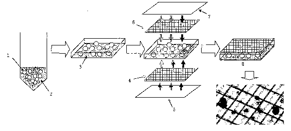
Fig. 1 is a schematic view showing a production method of
a sheet-like pancreatic islet cell preparation.
Fig. 2 is a graph showing test results of the recovery ratio
of viable cells of Production Example 1, Comparative Example
1, or free pancreatic islet cells after 1 day-culture.
Fig. 3 is a graph showing test results of the insulin content
in Production Example 1, Comparative Example 1, or free
pancreatic islet cells after 1 day-culture.
Fig. 4 is a graph showing test results of insulin secretion
with the lapse of time in Production Example 1, Comparative
Example 1, or pancreatic islet cells.
Fig. 5 is a microphotograph of pancreatic islet cells
contained in Production Example 1 or Comparative Example 1 by
observation with the lapse of time.
Fig. 6 is a graph showing test results of insulin secretion
of Production Example 2 or free pancreatic islet cells.
Fig . 7 is a graph showing the survival period of transplanted
mouse group and diabetic mouse group.
Fig. 8 is a graph showing the change in blood sugar level
CA 02522106 2005-10-12
9
of transplanted mouse group, diabetic mouse group, and normal
blood sugar mouse group.
Fig. 9 is a graph showing the change in body weight of
transplanted mouse group, diabetic mouse group, and normal blood
sugar mouse group.
Fig. 10 is a graph showing the change in BUN (blood urea
nitrogen) level of transplanted mouse group, diabetic mouse group,
and normal blood sugar mouse group.
Fig. 11 is a graph showing the change in creatinine level
of transplanted mouse group, diabetic mouse group, and normal
blood sugar mouse group.
Fig. 12 is a graph showing the change in 24-hours urine
of transplanted mouse group, diabetic mouse group, and normal
blood sugar mouse group.
Fig. 13 is a graph showing the change in 24-hours urine
glucose excretion of transplanted mouse group, diabetic mouse
group, and normal blood sugar mouse group.
Fig . 14 is a graph showing the change in urine ketone level
of transplanted mouse group, diabetic mouse group, and normal
blood sugar mouse group.
Fig. 15 is a graph showing the change in 24-hours urine
albumin excretion of transplanted mouse group, diabetic mouse
group, and normal blood sugar mouse group.
Fig. 16 is a graph showing the change in 1,5-AG level of
transplanted mouse group , diabetic mouse group , and normal blood
sugar mouse group.
In the above Fig., the reference numeral 1 represents a
pancreatic islet cells; 2 represents a PVA+EC solution; 3
represents PVA+EC gel; 4 and 6 represent mesh sheets; 5 and 7
CA 02522106 2005-10-12
represent glass plates ; and 8 represents a sheet-like pancreatic
islet cell-containing cellular preparation.
BEST MODE FOR CARRYING OUT THE INVENTION
5 The present invention is characterized in that a cellular
preparation contains PVA mixed with a cell preservative and
containscellsproducing/secreting biologically active factors
such as hormones and proteins useful for patients in the PVA.
The PVA to be used in the present invention is not particularly
10 limited without departing from the purpose of the invention and
is preferable to have purity enough to be used, for example,
corneal protectors/moisturizersin eye drops, medical materials
such as intestinal adhesion preventing membranes, and food
contact materials such as food wrapping films. It may also
include commercialized products, and freeze-dry products
obtained by producing polyvinyl acetate from vinyl acetate
followed by hydrolysis . In the case of producing the PVA, the
production method and the freeze-drying method may be carried
out according to the known conventional methods . The PVA has
preferably an average molecular weight of about 5 , 000 to 16 , 600
and a saponification degree of about 85~ or higher.
The PVA is preferably a fine granular freeze-dried product
which is easily suspended in a solution such as physiological
saline and phosphate buffers
The PVA mixed with a cell preservative means PVA containing
the cell preservative in the PVA. The cell preservative to be
used in the present invention is not particularly limited and
may include solutions to be employed generally for preservation
of animal cells and commercialized preservation solutions , for
CA 02522106 2005-10-12
11
example, Euro-Collins solution, Cell Banker (code 630-01601:
Wako Pure Chemical Industries , Ltd. Osaka, Japan ) and UW solution
(ViaSpan: Bristol-Myers Squibb Company, USA). They may be
commercialized ones or compositions prepared in laboratories .
In the case of preparation in laboratories, it is preferable
to carry out the preparation by adding and mixing each component
in distilled water and disinfecting the obtained solutions by
filtration with a filter.
For example, the composition of the Euro-Collins solution
is shown in the following Table 1.
Table 1
Component g/L
D-glucose 35.0
KZHP04 7 . 4
KHZP04 2 . 05
KC1 1.12
NaHC03 0 . 8 4
Nicotinamide 1.22
Also , the composition of the UW solution ( ViaSpan ) is shown
in the following Table 2.
Table 2
Component g/L
Pentafraction (note 1) 50
Lactobionic acid 35.83
Potassium dihydrogen phosphate 3.4
Magnesium sulfate 1.23
Raffinose 17.83
Adenosine 1.34
Allopurinol 0.136
Total glutathione 0.922
Potassium hydroxide 5.61
Sodium hydroxide Adjusted to pH 7.4
Water for injection I q.s.
Note 1: US Patent NO. 4,798,824
The PVA containing a cell preservative in the PVA can be
CA 02522106 2005-10-12
12
produced, for example, by mixing PVA with a cell preservative.
The resulting PVA containing the cell preservative is desirably
aseptic . The method of mixing PVA and a cell preservative is
not particularly limited, however, there can be, for example,
exemplified a method of suspending a commercialized granular
PVA in distilled water, etc. dissolving and sterilizing the
resulting PVA suspension by heating and disinfecting
(autoclaving or the like), and adding and mixing a cell
preservative to and with the obtained aseptic aqueous PVA
solution. The heating and disinfecting treatment and the
aseptic treatment themselves may be carried out according to
the known conventional methods.
Herein, PVA containing the cell preservative produced by
any means other than the above-mentioned mixing means may be
called as PVA treated with the cell preservative and therefore,
it should go without saying that PVA treated with the cell
preservative is also included within the scope of the present
invention.
The content of PVA in the mixed solution (hereinafter, called
as a PVA-cell preservative solution) obtained by mixing an
aqueous suspended PVA solution and a cell preservative is
generally about 2 to 5~ by weight, more preferably about 2.5
to 3.5~ by weight, and especially preferably about 2.5 to 3~
by weight based on the PVA-cell preservative solution. It is
because gelation of PVA is most easily caused in this
concentration range and also PVA gel with preferable strength
for encapsulating the cell in the cellular preparation of the
present invention. If the concentration of the cell
preservative to be used for preserving the cells is defined to
CA 02522106 2005-10-12
13
be 1 time, the concentration of the cell preservative in the
PVA-cell preservative solution is generally about 0.8 to 1 time,
more preferably about 0.9 to 1 time, and especially preferably
about 0.95 to 1 time as much. It is because if the concentration
is out of this range, the cells cannot sometimes survive stably.
Accordingly, in the present invention, it is preferable that
a concentrated cell preservative solution with a concentration
of about 2 to 10 times as high as that for normal cell preservative
solution is prepared or purchased, and then properly mixed with
the above-mentioned aqueous suspended PVA solution ( a . g . 10 times
high concentration cell preservative solution: aqueous PVA
suspended solution = 1 : 9 ) to obtain a PVA-cell preservative
solution containing the cell preservative in about 1 time
concentration.
The cellular preparation of the present invention may
further contain dimethyl sulfoxide (DMSO) and serum albumin for
protecting the cells, antibiotics, etc. for inhibiting
contamination of germ, and also vitamins such as nicotinamide
for cell activity retention. Further, in the present invention,
other additives which are generally allowed to add under
Pharmaceutical Affairs Law, e.g. an agent for proving sustained
release property, an isotonic agent, a pH adjustment agent, and
the like may be supplemented. The addition method of these other
additivesto the cellular preparation isnot particularly limited,
however, it is preferable to add them to the above-mentioned
PVA-cell preservative solution in aseptic manner at the time
of production of the solution. The contents thereof are
preferably within a range of not inhibiting the growth/survival
of the cells contained therein and/or the secretion of
CA 02522106 2005-10-12
14
biologically active factors by the cells, and without departing
from the scope and true sprit of the present invention.
The PVA treated with the cell preservative is produced in
aseptic manner as described above and therefore, the cellular
preparation of the present invention is seldom to be contaminated
with germ and possible to be preserved at room temperature ( about
to 35°C) for a long period of time.
The cells to be used for the present invention may include
pancreatic islet cells, pancreatic endocrine cells,hepatocyte,
10 anterior pituitary cells, growth hormone-secreting cells,
osteocytes, thyroid hormone-secreting cells, and parathyroid-
hormone-secreting cells. These cells are preferably derived
from a mammal such as human being, pig, rat, and mouse, and those
which produce/secret biologically active factors such as
15 hormones and proteins effective for patients . Selection of cell
types is preferably determined depending on the type of diseases
of patients receiving the cellular preparation. For example,
pancreatic islet cells or pancreatic endocrine cells, etc.,
producing insulin are preferable for diabetic patients, etc. ;
hepatocytes, etc., producing blood coagulation factors for
hemophiliac patients,etc.; anterior pituitary cells and growth
hormone-secreting cells, etc., producing growth hormones for
pituitary dwarfism patients , etc . ; and osteocytes producing bone
morphogenetic proteins (BMP) for patients suffering from bone
diseases such as bone fracture . Transformed cells capable of
producing a large quantity of tumor growth suppressing substances
by genetic recombination may be used for cancer patients.
Further, cells having functions of selectively metabolizing and
detoxifying harmful metabolites may be used for patients
CA 02522106 2005-10-12
IS
suffering from diseases which accumulates harmful metabolites
due to hepatic insufficiency, renal insufficiency, or inborn
errors of metabolism. In the case where a patient needs a
plurality of biologically active factors since he suffers from
a plurality of diseases , two or more kinds of the above-mentioned
cells may be combined. Biologically active factors efficacious
to the above-mentioned patients can be provided since the present
invention contains these cells. Accordingly, use of the
cellular preparation of the present invention makes it possible
to prevent or treat various diseases whose remediation is
possible, through supply of biologically active factors or
detoxification of harmful metabolites.
The above-mentioned cells to be employed in the present
invention may be an established cell line for laboratories or
cells isolated from tissues of a living body, however, they are
preferably differentiated, non-dividing cells since
differentiated, non-dividing cells are more capable of producing
/secretingtarget hormonesand proteins than non-differentiated,
dividing cells.
The isolation method of the cells from living tissues is
not particularly limited, and known conventional techniquesmay
be employed. For example, the known methods include a method
of extracting tissues by proper means , treating the tissues with
Dispase , EDTA, or the like and then with trypsin to finally isolate
single cells.
In the case where the cells used in the present invention
are pancreatic islet cells , isolation of the cells from living
tissues may be carried out by a separation method through the
known collagenase treatment , a . g . J . Adam Van Der Vliet et al . ,
CA 02522106 2005-10-12
16
Transplantation, 45(2), p493-495 (1988).
In the case where the cells to be employed in the present
invention are pancreatic endocrine cells , the isolation may be
carried out, for example, according to the method described in
Japanese Patent Application Laid-Open No. 2001-231548.
The isolated cells from the living tissues in the
above-mentioned manner are preferably free from pathogens such
as pathogenic virus.
It is preferable for the cells already established for
laboratories and single cells obtained from living tissues to
be cultured in a suitable media until they become confluent and
to be employed as cells for the present invention after passage
culture is repeated 2 or 3 times . For example, in the case where
the cells to be employed in the present invention are pancreatic
islet cells or pancreatic endocrine cells ( hereinafter, referred
to as pancreatic endocrine cells or the like ) , these cells are
preferably subjected to passage culture in a CMRL-1066 culture
medium supplemented with 10~ fetal bovine serum (hereinafter
abbreviated as FBS ) and nicotinamide ( Sigma , St . Louis , MO, USA ) .
The passage culture cells are again isolated as single cells
by the known method such as trypsin treatment or collagenase
treatment to be used as cells for the present invention.
The cells to be employed in the present invention may be
transformed cells into which gene codingpeptides of biologically
active factors such as hormones or proteins necessary for
patients to be administered are introduced. Use of the
transformed cells makes it possible to produce the target
biologically active factors efficiently in a large amount. The
base sequence of genes encoding peptides of such biologically
CA 02522106 2005-10-12
17
active factors has already been opened, and such genes include,
for example, genes available from American Type Culture
Collection ( ATCC ) or from commercially easily available sources ,
andsynthetic genesobtained by producing oligonucleotide probes
from the known sequences and carrying out the synthesis using
said probes by known conventional methods, for example, PCR
amplification method and DNA synthesis method. For example,
use of transformed cells containing a gene encoding tumor growth
suppression proteins makes it possible to use the cellular
preparation of the present invention for prevention and treatment
of cancers.
Although there is not particular limitation to host cells
into which a gene encoding the above-mentioned peptides of
biologically active factors , and known conventional host cells
to be used generally in the field of genetic recombination
technology may be employed. As the host cells other than the
above-mentioned pancreatic islet cells, pancreatic endocrine
cells, hepatocytes, anterior pituitary cells, growth
hormone-secreting cells, and osteocytes, there can be
exemplified monkey COS-7 cells, Vero cells, Chinese hamster CHO
cells ( hereinaf ter abbreviated as CHO cell ) , dhfr gene-defective
Chinese hamster CHO cells (hereinafter abbreviated as
CHO(dhfr-)cell), mouse BALB/3T3 cells, mouse L cells, mouse
AtT-20 cells, mouse C127 cells, mouse myeloma cells, rat GH3
cells, human HeLa cells, human FL cells, and the like.
The method for introducing the gene into the cells to be
used in the present invention is not particularly limited and
may be carried out by known conventional technique . For example ,
conventionally known methods of introducing the gene into
CA 02522106 2005-10-12
18
recombinant expression vectors such as plasmids or viruses , and
artificial vectors such as liposomes and microcapsules can be
exemplified. In the case of using recombinant vectors , examples
of applicable methods are competent cell method ( J . Mol . Biol . ,
53 , p154 ( 1970 ) ) , DEAE dextran method ( Science , 215 , p166 ( 1982 ) ) ,
In-vitro packaging method ( Proc . Natl . Acad. Sci . , USA, 72 , p581
(1975)), Virus vector method (Cell, 37, p1053 (1984)),
Microinjection method (Exp. Cell. Res., 153, p347 (1984)),
Electroporation method (Cytotechnology, 3, p133 (1990)),
Potassium phosphate method (Science, 221, p551 (1983)),
Lipofection method (Proc. Natl. Acad. Sci., USA, 84, p7413
(1987)), Protoplast method (Japanese Patent Application
Laid-Open No. 63-2483942, Gene, 17, p107, (1982), Molecular &
General Genetics, 168, plll (1979)).
Although there is no particular limitation on the vectors
for introducing their gene into the host cells, any expression
vectors so long as it can express desired gene in the cells into
which they are introduced and can produce efficiently the
peptides of biologically active factors maybe used. For example,
animal viruses such as bacteriophage , a . g . ~, phage , adenovirus ,
adeno-associated virus (AAV), retrovirus, poxvirus, herpes
virus, herpes simplex virus, lentivirus (HIV), Sendai virus,
Epstein-Barr virus (EBV), vaccinia virus, poliovirus, Sindbis
virus , SV 40 , and also pAl-11, pXT1, pRc/CMV, pRc/RSV, pcDNAI/Neo,
and the like can be exemplified.
The cells to be employed in the present invention are
preferably contained in the PVA by the following techniques.
Cells are added and mixed in the above produced PVA-cell
preservative solution. The cells to be added and mixed are
CA 02522106 2005-10-12
19
preferably treated previously with a cell preservative.
Practically, the cells are preferably suspended previously in
the cell preservative and recovered by centrifugation, and then
mixed with the PVA-cell preservative solution . The number of
the cells to be contained in the cellular preparation of the
present invention cannot be generalized since it depends on the
types and the extent of the diseases of patients to whom the
cellular preparation is administered, and it is therefore
preferable to determine the number of cells by a doctor. The
number of cells is preferably about 1x10' to 5x10' cells/ml in
the PVA-cell preservative solution. Adjustment of the number
of the cells within the range makes the cells dispersed evenly
in the PVA gel and the cells can stably survive for a long duration
without inhibition of supply of oxygen and nutrients to the cells
due to cell coagulation.
Also, in order to retain the cells to be employed in the
present invention in the stable condition for optimum supply
of the biologically active factors, the cellular preparation
of the present invention may further contain mucopolysaccharides
such as hyaluronic acid, chondroithin sulfuric acid, and
dermatanic acid; extracellular matrix containing one or more
substances such as erastin, collagen, and fibrin; and/or growth
factors such as hepatocyte growth factor (HGF), vascular
endothelial cell growth factor ( VEGF ) , human basic fibroblast
growth factor (bFGF), fibroblast growth factor (FGF),
platelet-derived growth factor (PDGF), insulin-like growth
factor (IGF), and growth hormone (GH). One or more kinds of
these growth factors may be contained. The method for adding
the extracellular matrix and/or growth factors in the present
CA 02522106 2005-10-12
invention may be a method involving, for example, immersing the
cells in a cell culture medium containing the extracellular
matrix and the growth factor for forming the extracellular matrix
on the surfaces of the cells before mixing the above PVA-cell
5 preservative solution with the cells , or a method of previously
adding the extracellular matrix and the growth factor in the
PVA-cell preservative solution, or the like. The content of
the above-mentioned extracellular matrix and/growth factor in
the cellular preparation of the present invention is not
10 particularly limited, however, it is within the range without
inhibition of the cell retention and the secretion function of
the biologically active factors and preferably in the range for
causing effects on prolongation of the cell survival period and
extension of the secretion period of the biologically active
15 factors .
The cellular preparation of the present invention can be
produced by mixing the PVA-cell preservative solution with the
cells, then cooling the cell mixed solution (hereinafter,
referredto ascell-containing PVA-cell preservativesolution),
20 and gelling the PVA . Cooling is preferably carried out by keeping
the solution in a housing at a temperature as ultra low as about
-20 to -80°C for about a half of a day to 3 days or in a housing
at a temperature as ultra low as about -80°C for about 18 to
hours.
25 In the present invention, since the cell preservative is
previously mixed with the PVA, even if the quenching is carried
out at the above-mentioned temperature, necrosis or damage of
the cells can be suppressed. Moreover, the cells can be contained
in the PVA gel in dispersed manner and coagulation of the cells
CA 02522106 2005-10-12
21
can be prevented.
Owing to the gelation of the PVA, the cellular preparation
of the present invention may be formed in various shapes such
as sheet , plate , board , rod, tube , or beads . For example , the
above-mentioned cell-containing PVA-cell preservativesolution
is applied to a glass plate and cooled together with the glass
plate to produce a cellular preparation in the form of a thin
sheet. The cellular preparation with a desired thickness can
be produced by properly adjusting the application dose of the
cell-containing PVA-cell preservative solution per unit surface
area of the glass plate and it is generally about 100 to 300
~,1/mmz .
The cellular preparation of the present invention may be
used in combination with a reinforcing material for reinforcement
and/or simplicity in the handling. For example, in the case
where the cellular preparation is formed in the form of a thin
sheet-like shape , to reinforce it and to simplify in its handling,
gelation is preferably carried out by fixing the cell-containing
PVA-cell preservative solution in a mesh sheet made of a resin
or the like. Specifically, in Fig. 1, for example, a mesh sheet
( 4 ) made of PET ( polyethylene terephthalate ) is put on a glass
plate (5) and the above-mentioned cell-containing PVA-cell
preservative solution is applied to the mesh sheet ( 4 ) . Another
mesh sheet ( 6 ) is put thereon and further another glass plate
(7) is put thereon to sandwich the cell-containing PVA-cell
preservative solution and the mesh sheets ( 4 ) and ( 6 ) with glass
plates (5) and (7). While being kept as it is, the assembled
body is cooled to about -20 to -80°C to form it into a sheet-like
gel. The above-mentioned mesh sheet is preferably immersed
CA 02522106 2005-10-12
22
previously in the PVA-cell preservative solution.
The above-mentioned reinforcing material, e.g. the mesh
sheets , is preferably materials which are safe to living body,
i.e.un-decomposable in vivo and excellent in biocompatibility,
such as PET, and the like,. It is because if the mesh sheets
in the cellular preparation of the present invention are
decomposed in vivo, the cellular preparation accretion to living
tissues takes place not only to make the cellular preparation
sometimes difficult to maintain the efficacy for a long duration
but also to make it harmful for living body. Of course, it is
no need to say that the reinforcing materials which are harmless
in a living body even they are decomposed in vivo are also usable
in the present invention.
The cellular preparation in the frozen state is preferably
administered to a living body after the cells in the frozen state
are activated. The activation of the cells in the frozen cellular
preparation is preferably carried out by thawing the cells and
culturing them again in a proper culture medium. To be more
specific, such activation is carried out by quickly immersing
the frozen state cellular preparation in a cell culture medium
of about 37°C, such as CMRL-1966, and culturing the thawed cells
again for about 24 hours in a new culture medium. In the case
where the cellular preparation contains DMSO, it is preferable
that the unfrozen cellular preparation is washed with a new cell
preservation solution, e.g. an UW solution, etc. and further
immersed in the UW solution at about 4°C for about 24 hours to
remove DMSO from gel. It is no need to say that the cellular
preparation which is thawed and made free from DMSO in the
above-mentioned manner is also included in the cellular
CA 02522106 2005-10-12
23
preparation of the present invention.
The administration manner of the cellular preparation
obtained by the above-mentioned method to a living body differs
depending on the type , the affected part , and severity of diseases
of patients to be administered, and it is therefore preferably
determined by a doctor. Hereinafter, a preferable
administration manner will be described.
The subjects to which the cellular preparation of the present
invention are administered include human being and also a mammal
other than human being, a . g . dog , cat , monkey, rabbit , mouse ,
and the like.
As described above, the cellular preparation of the present
invention can be administered in the form suitable to the
application site of a living body. Also, the cellular
preparation of the present invention may be transplanted to the
application site in a direct contact manner to the affected
tissues, and also it can be transplanted in the subcutaneous
tissue or the muscle with relatively slight invasion. For
example, the cellular preparation in tubular shape is cut into
small fragments and transplanted in the subcutaneous tissue by
using a surgical suture needle, or the cellular preparation can
be injected in the muscle and subcutaneous tissue by filling
an injector with the cellular preparation. The cellular
preparation is easy to be recovered from the administration site.
In the case where the cellular preparation is transplanted in
the subcutaneous tissue andmuscle, if the vascular distribution
in the transplantation site is sparse and supply of oxygen and
nutrients for growth and survival of the cells is supposed to
be difficult, transplantation may be carried out together with
CA 02522106 2005-10-12
24
known proper angiogenesis inducers. The angiogenesis inducers
may be added previously in the cellular preparation or may be
administered separately.
The dose of the biologically active factors such as hormones
and proteins to be supplied to a patient from the cellular
preparation of the present invention may properly be set by
determination by a doctor in consideration of the secretion of
the biologically active factors of the cells to be employed and
the change of the cell survival ratio during the transplantation.
For example, if a patient suffers from diabetes, the insulin
secretion of the cells to be used, a . g . pancreatic islet secretion
cells, is previously measured in vitro and depending on the
insulin secretion , the number of the cells , administration period,
and change of the survival ratio of the cells , the insulin supply
amount of the cellular preparation of the present invention can
be determined. Since the cellular preparation of the present
invention can stably preserve the cells contained therein for
a long duration, deterioration ratio of the secretion of the
biologically active factors with the lapse of time is low.
Accordingly, the present invention is suitable to supply the
biologically active factors needed for patients stably for a
long duration, resulting in decreased frequency of the
transplantation of the cellular preparation.
The secretion of the biologically active factors of the
cells to be used in the cellular preparation of the present
invent ion may quantitatively be determined according to the known
quantitative determination methods suitable for measurement of
such biologically active factors. For example, a quantitative
determination method of insulin in the case where the cells used
CA 02522106 2005-10-12
are pancreatic islet secretion cells will be described in detail
in the following Examples.
EXAMPLES
5 The invention will be illustrated more specifically by way
of Examples. However, it is not intended that the invention
be limited to Examples . In Examples , ~ means ~ by weight unless
otherwise specified.
10 Example 1
(Production Example 1) Production of sheet-like cellular
preparation containing pancreatic islet cells
(1) Preparation of pancreatic islet cells
Pancreatic islet cells were isolated from blister rats with
15 8 to 10 week-old and 300 to 350 g weight ( Shimadzu animal Co .
Ltd. Kyoto, Japan) by the following method.
At first, 10 ml of Hank's solution containing type I
collagenase ( Sigma, St . Louis , MO, USA, 350 U/mg ) and type XI
collagenase (Sigma, St. Louis, MO, USA, 2,200 U/mg) 800 U/ml
20 and 1500 U/ml , respectively, was injected to the pancreatic ducts
of rat extracted pancreas and subjected to enzyme treatment at
37°C for 15 to 20 minutes. Cooled Hank's solution was added
to stop the enzyme reaction . The pancreas tissues were dispersed
with a pipette and centrifuged to recover tissue pellets , and
25 then the tissue pellets were washed two times with Hank' s solution .
The centrifugation was carried out at 1, 000 rpm for 1 minute .
The tissue suspension was filtered through an 800 E.i,m size mesh
sheet . The filtrate was recovered in a 50 ml conical tube and
centrifuged ( 1, 500 rpm for 3 minutes ) to obtain tissue pellets .
CA 02522106 2005-10-12
26
The pellets were suspended in Hank's solution containing 27~
dextran ( Sigma, St . Louis , MO, USA, MW 70 . 000 ) and centrifuged
by discontinuousdensity-gradientcentrifugation. In thiscase,
the lowermost layer was controlled to be 27~ dextran ( density
1.094 g/ml), and the upper layer was made into two layers of
23~ dextran (density 1.081 g/ml) and 11~ dextran (density 1.041
g/ml). The discontinuous density-gradient centrifugation was
carried out at 400 rpm for 4 minutes and further at 1, 700 rpm
for 10 minutes. After centrifugation, the pancreatic islet
cells-containing fraction was sampled in the interface between
two uppermost layers with a Pustule pipette. The obtained
pancreatic islet cells were washed two times with CMRL-1066
culture medium (1,000 rpm for 3 minutes).
(2) Production of PVA-Euro-collins solution
PVA 3 g (manufactured by Gunse Co.: PVA-180H, Lot37435)
was suspended in distilled water 75 ml and then subjected to
heating dissolution and sterilization treatment through
autoclaving (121°C, 15 minutes) two times. Ten times
concentration of Euro-Collinsolution(hereinafter abbreviated
as EC solution) 10 ml, DMSO 5 ml, FBS 10 ml, and nicotinamide
0.122 g were aseptically added to and mixed with the obtained
aseptic aqueous PVA solution, thereby to obtain an aseptic 3~
PVA-EC solution 100 ml. The above-mentioned 10 times
concentration of EC solution was prepared by admixing the
components shown in Table 1 in distilled water 100 ml, and filtered
and disinfected through 0.22 hum mesh. Thereafter, it was
preserved at a room temperature.
(3) Cell Mixing
The pancreatic islet cells obtained in (1) were cultured
CA 02522106 2005-10-12
27
for 24 hours in a CMRL-1066 culture medium containing 10°s FBS
and nicotinamide 1. 22 g/L in a carbon dioxide gas incubator ( 5~
CO2, 37°C). The culture solution containing 1,000 pancreatic
islet cells was placed in a 1.5 ml centrifugation tube and
centrifuged at 800 rpm for 1 minute . Cell Banker ( code 630-01601:
Wako Pure Chemical Industries, Ltd. Osaka, Japan) 0.1 ml was
added to the obtained cell pellets and the cells were suspended,
and then kept still for 5 minutes. The cells were recovered
by centrifugation again. The recovered cells were suspended
in the aseptic 3~ PVA-EC solution prepared in ( 2 ) 100 ~ul to obtain
a pancreatic islet cells-containing PVA-EC solution.
(4) Formation of sheet
Two mesh sheets with 10 mm x 10 mm square made of PET
(manufactured by Gunse Co. : TNO 60 SS) were previously immersed
in an aseptic 3~ aqueous PVA solution for 5 minutes, and one
of the sheet ( 4 ) was put on a glass plate ( 5 ) . The pancreatic
islet cells-containing PVA-EC solution obtained in ( 3 ) was evenly
applied to the above-mentioned mesh sheet ( 4 ) , and another mesh
sheet ( 6 ) was put further thereon to obtain a three-layer sheet
composed of mesh sheet (4)/pancreatic islet cells-containing
PVA-EC solution/mesh sheet (6). Further another glass plate
( 7 ) was put thereon so as to obtain about 1 mm-thick three-layer
structure sheet by applying pressure, and PVA was converted to
a gel by keeping the assembled body at -80°C for 24 hours while
being sandwiched between two glass plates (see Fig. 1).
(5) Thawing of pancreatic islet cellular preparation in sheet
form and activation of cells
The frozensheet-like pancreatic islet cellular preparation
obtained in ( 4 ) was thawed quickly in CMRL-1066 culture medium
CA 02522106 2005-10-12
28
at 37°C. The thawed sheet-like pancreatic islet cellular
preparation was immersed in a UW solution 5 ml (ViaSpan:
Bristol-Myers Squibb Company, USA) for 5 minutes. This
procedure was repeated 3 times to remove DMSO contained in the
sheet. The sheet was further immersed in a UW solution 5 ml
at 4°C for 24 hours . The preparation after the immersion was
washed with CMRL-1066 culture medium twice and then cultured
in CMRL-1066 culture medium for 24 hours (37°C, 5~ COZ) to obtain
a pancreatic islet cellular preparation containing activated
cells ( Production Example 1 ) in a sheet from. Production Example
1 was employed for the following tests . An aseptic 3~ aqueous
PVA solution was prepared as described in ( 2 ) without using EC
solution 10 ml, DMSO 5 ml, FBS 10 ml, and nicotinamide 0.122
g and a pancreatic islet cellular preparation produced from
the solution in the form of sheet was employed as Comparative
Example 1.
(Test Example 1) Confirmation of cell stability in preparations
Cells of Production Example 1 and Comparative Example 1,
and free pancreatic islet cells (200 cells) were cultured
respectively in CMRL-1066 culture medium for 24 hours (5~ CO2,
37°C). The number of the viable cells in Production Example
1 and Comparative Example 1, and the number of free pancreatic
islet cells after 1-day culture were counted using a microscope
to calculate the recovery ratio ( ~ ) of the viable cells . The
recovery ratio was a relative value to 100 defined as the number
of the cells before the culture.
The results are shown in Fig. 2. The recovery ratio of the
viable cells in Production Example 1 was 82~, which was extremely
CA 02522106 2005-10-12
29
higher than that of Comparative Example 1 ( about 35~ ) , and the
value was higher than that of the free cells.
( Test Example 2 ) Confirmation of insulin content in preparations
Cells of Production Example 1, and Comparative Example 1,
and pancreatic isolated islet cells ( 200 cells ) were cultured
respectively in CMRL-1066 culture medium for one day ( 5~ C02,
37°C) . The respective insulin amounts after 1-day culture were
investigated. The cellsof Production Exampleland Comparative
Example 1, and the free pancreatic islet cells were recovered
in hydrochloric acid-EtOHand mashed respectivelywith a spatula,
and the gel and the cells in the preparation were stirred and
suspended in hydrochloric acid-EtOH with a vortex mixer ( -20°C,
24 hours). The insulin concentrations in the respective cell
suspensions were measured. Insulin was measured by using a
commercialized insulin measurement ELISA kit ( Lbis Insulin Kit ,
Shibayagi).
The results are shown in Fig. 3. The insulin content in
Production Example 1 was about 205 ng/ml which was extremely
higher than that of Comparative Example 1 ( about 20 ng/ml ) , and
the value was approximately the same as that of the free cells .
(Test Example 3) Confirmation of insulin secretion of
preparations and cell state
The cells of Production Example 1 and Comparative Example
1, and isolated islet cells ( 200 cells ) were cultured in CMRL-1066
culture medium (5~ CO2, 37°C) and recovered after 1 day, 7 days,
and 14 days . After the recovery, the recovered culture products
were subjected to a glucose reaction test . The glucose reaction
CA 02522106 2005-10-12
test was carried out as follows . At first the recovered cells
of Production Example 1 and Comparative Example 1, and free
pancreatic islet cells were cultured respectively in CMRL-1066
culture medium containing 3.3 mM glucose and 0.1~ bovine serum
5 albumin (BSA) for 1 hour (5~ CO2, 37°C) and then washed with
glucose-free CMRL-1066 culture medium. This procedure was
repeated again, and these cells were carried out in a CMRL-1066
culture medium containing 3.3 mM glucose for 1 hour and then
the insulin concentration in each supernatant solution was
10 measured. Next, culture was carried out in a CMRL-1066 culture
medium containing 16 . 7 mM glucose for 1 hour and then the insulin
concentration in each supernatant solution was measured.
The results are shown in Fig . 4 . In Fig . 4 , ( a ) shows the
amount ( concentration ) of insulin secretion after 1 day-culture ;
15 (b) shows the result after 7 days-culture; and (c) shows the
result after 14 days-culture . In the case of Comparative Example
1, no increase in the insulin secretion depending on the glucose
concentration was observed. On the contrary, in the case of
Production Example 1, increase in the insulin secretion
20 (concentration) depending on the glucose concentration was
observed after 1 day-culture, 7 days-culture, and 14 days-culture.
Moreover, in the case of free cells, no increase in the insulin
secretion depending on the glucose concentration was observed
after 7 days-culture, the increase was observed in the case of
25 Production Example 1 even after 14 days-culture.
As a result of observation of cells contained in Production
Example 1 and Comparative Example 1 by a microscope, the cells
in Comparative Example 1 were found to be already destructed
from the first day. On the other hand, no significant cell
CA 02522106 2005-10-12
31
destruction for the cells of Production Example 1 was observed,
and the cells were found to be relatively stable and were surviving
even after 14 days ( Fig . 5 ) . In Fig . 5 , ( a ) shows observation
images of the cells after 1 day-culture; (b) after 7 days-culture;
and (c) after 14 days-culture.
From the results , it was found that the islet cells stably
survived in Production Example 1 and maintained the activity
stably for a long duration and secreted insulin responding to
the increase of glucose concentration. Accordingly, it was
confirmed that previous addition of the EC solution to PVA
suppressed cell necrosis and damage through quenching at the
time of gelation of PVA, and as a result, it was proved that
a cellular preparation with a high survival ratio of pancreatic
islet cells contained in PVA and capable of retaining the insulin
secretion and the responsiveness to increase in glucose
concentration can be produced.
Example 2
(Production Example 2) Production of sheet-like cellular
preparation containing pancreatic islet cells
(1) Preparation of pancreatic islet cells
General anesthesia in blister rats were carried out through
diethyl ether inhalation and intraabdominal administration of
Nembutal 1 mg/kg. After abdominal section, common bile duct
was identified and clumped near the papilla of Vater by a
non-toothed forceps. A small opening was made in the joined
part of the cystic duct and a cannulation tube was inserted into
the common bile duct, and fastened and fixed, and type XI
collagenase ( Sigma, St . Louis , MO, USA) equivalent to 1200 to
CA 02522106 2005-10-12
32
1500 u/Ml was injected to swell the pancreas. Without damaging,
the whole pancreas was extracted, placed in a flask, immersed
in a thermostatic bath of 37°C and digested in about 18 minutes .
After digestion, the flask was shaken several times to destruct
the pancreas and washed three times with Hank's solution
containing 0.1~ bovine serum albumin. After the cells were
floated in the Hank' s solution, the solution was filtered through
an 850 Eun-diameter mesh filter to recover the contents.
The islet separation was carried out using specific gravity
gradation of Dextran 70. That is, the pancreatic islet cell
components from which the supernatant solution was removed were
stirred uniformly in Dextran with 1. 094 specific gravity + Hank' s
solution, and then Dextran with 1. 094 specific gravity was slowly
poured. and Dextran with 1. 081 specific gravity and Dextran with
1.041 specific gravity were slowly poured successively on the
cell layer to form 4 layers . The layered sample was centrifuged
at 400 rpm for 4 minutes and further at 1700 rpm for 13 minutes .
The islet separated between the first layer and the second layer
was recovered with a hand pick, washed and purified with a hand
pick. After washing 4 times, the pancreatic islet cells were
cultured overnight in an incubator under environments at 37°C,
5g CO2, 95~ air. The culture medium was CMRL 1066 + 10~
inactivated fetal bovine serum + antibiotic-antimycotic
solution (Gibco, BRL) + nicotinamide.
( 2 ) Preparation of pancreatic islet cellular preparation as sheet
form
Using the culture solution of the pancreatic islet cells
obtained in the above-mentioned manner, a sheet-like islet
cellular preparation ( Production Example 2 ) in which cells were
CA 02522106 2005-10-12
33
activated was obtained similarly as described in ( 2 ) , ( 3 ) , ( 4 ) ,
and (5) in Example 1.
(Test Example 4) Insulin secretion of preparations
The above obtained islet cellular preparation (Production
Example 2, after 1 day from the production) and free islet cells
were stimulated for each 1 hour by low concentration glucose
( 3 . 3 mM) and high concentration glucose ( 16 . 7 mM) in the same
manner as Test Example 3, and the amount of secreted insulin
was measured by ELISA.
The results are shown in Fig. 6. As can be seen from Fig.
6, the islet cellular preparation according to the present
invention was found to secrete insulin corresponding to the
glucose stimulation and was not inferior to the free islet cells
at the same period.
(Test Example 5) Transplantation test
(1) Production of diabetic mouse model
Streptozotocin 200 mg/Kg dissolved in a citric acid buffer
(pH 4. 5 ) was administered to C57BL/6 male mice of 8 to 10-week-old
by intraabdominal administration. One week after the
administration, blood sugar measurement was carried out three
times , and mice showing blood sugar 350 mg/dl or higher two or
more times were determined to be diabetic and used for the
experiment . Mice without streptozotocin were used as a normal
group of mice.
(2) Transplantation
The transplantation of the islet cellular preparation was
carried out as followings . That is , diabetic model mice ( after
CA 02522106 2005-10-12
34
1 week or longer from diabetes development) were undergone
abdominal section by median incision under ether anesthesia,
and the islet cellular preparation (Production Example 2)
obtained in Production Example 2 was kept in the abdomen and
the abdominal wall was closed in two layers . These mice were
defined as a transplanted mice group. Mice undergone
transplantation of the preparation containing no pancreatic
islet cells ( produced in the same manner as in Comparative Example
1) were defined as a diabetic mice group.
(3) Investigation of survival period
The survival period after the transplantation defined as
0 day was observed.
The results are shown in Fig. 7. It is apparent from Fig.
7 that a half or more of the diabetic mice group died within
4 weeks after the transplantation and no mouse could survive
up to 8 weeks during the passage observation period. On the
other hand, except two mice which showed continuously high blood
sugar state and finally died among the group of the mice with
the islet cells , others survived 8 weeks or longer ( survival
ratio up to 8 weeks: 0~ vs. 81.8; p < 0.001).
(4) Blood sugar/weight measurement
Both of the blood sugar and the weight were measured before
the transplantation and after 1 day, 3 days, 5 days, and 7 days
from the transplantation and thereafter measured once a week.
The blood sugar fluctuation of each group was shown in Fig. 8.
With respect to the transplanted group, many quickly showed
decrease of the blood sugar immediately after transplantation
to less than 300 mg/dl ( 13/16 ) and amajority of them were observed
to be normal, however it was gradually increased (the period
CA 02522106 2005-10-12
of keeping less than 300 mg/dl was 3 days to 7 weeks) . However,
the blood sugar increase was slight and the blood sugar level
was significantly improved (p < 0.0001) as compared with that
of the diabetic mouse group and the life recuperation up to 8
5 weeksfrom the transplantation was apparently improved. Among
9 mice observed up to 8 weeks , 6 mice were undergone extraction
of the islet cellular preparation and among them 5 mice showed
blood sugar increase again.
The average weight fluctuation of each group is shown in
10 Fig. 9. In the group of the transplanted mice, the weight was
decreased after surgical transplantation, and it is supposed
to be due to the poor dietary, and no significant difference
from the diabetic mice group was observed in the Repeated-Measure
Analysis of Variance carried out up to 3 weeks after the
15 transplantation. However, after 3 weeks from the surgical
transplantation, the weight was turned to increase, and as
compared with that in the first week when the average weight
was most decreased during the passage, significant increase was
observed after 3 weeks (p < 0.05) . Further, the average weight
20 significantly exceeded the average weight of the diabetic mice
group in 3 weeks and 4 weeks from the transplantation.
(5) Measurement of kidney function
BUN ( blood urea nitrogen ) and creatinine were measured once
a week before the transplantation and once a week after the
25 transplantation. The pancreatic isletcellular preparation was
extracted 8 weeks after the transplantation and thereafter the
measurement was carried out until 1 week after the extraction.
The fluctuation of BUN is shown in Fig. 10. The average
~ SE of BUN of mice with normal blood sugar measured before the
CA 02522106 2005-10-12
36
transplantation of the islet cellularpreparationwas 25 . 53 mg/dl
1. 804 ( 20 to 38 . 9 ) . Among the group of diabetic mice after
1 week from the administration of the streptozotocin, 41.3
showed BUN increase (24/58, diabetic mice group: 13/35 vs.
transplanted mice group: 11/23, BUN > 35 mg/dl). Among 14
diabetic mice in the diabetic mice group, 13 mice showed high
BUN value in the passage.
On the other hand, with respect to 16 transplanted mice
in the transplantation mice group, 9 mice showing the BUN increase
before the surgery, was found normalized after 1 week from the
transplantation. The BUN value in average decreased after 1
week from the transplantation were approximately normal value
and found significantly different from that of the diabetic mice
group (p = 0.002).
Further, among 6 mice undergone the extraction of the
pancreatic islet cellular preparation after 8 weeks from the
transplantation, the BUN value measurement was carried out for
4 mice in one week after the extraction and all showed increase
as compared with the value before extraction.
The change in creatinine level is shown in Fig. 11. The
normal value of creatinine was 0 . 351 mg/dl ~ 0 . 032 ( 0 .16 to 0 . 5 ) .
Almost a half of mice in the diabetic mice group showed as high
as 0.5 mg/dl 1 week after the streptozotocin administration and
all of the mice were found abnormal in the creatinine value 4
weeks after the administration. Further, after the creatinine
increase, all of the mice were dead in several weeks and no mouse
survived up to 10 weeks from the streptozotocin administration.
Although 6 mice in the transplanted mice group showed the increase
after implantation of the creatinine, the increase was slight
CA 02522106 2005-10-12
37
and mice couldsurvive during the observation passage. Although
the creatinine average value in the transplanted mice group
exceeded 0.5 mg/dl only 5 weeks after the transplantation, the
value was constantly around 0.4 mg/dl in any period and was
decreased significantly as compared with that of the diabetic
mice group (p = 0.0478) . Further, similarly to the case of BUN,
all mice showed increase again after the extraction of pancreatic
islet cells.
(6) Urine inspection
Urine was collected for 24 hours and the urine amount and
24-hour urine sugar excretion amount, 24-hour urine albumin
excretion amount, and urine ketone were measured. All of the
group of transplanted mice, the group of diabetic mice and the
group of normal blood sugar mice were fed in free drinking and
eating dietary environments for 24 hours, and the 24-hour urine
was collected. Urine sugar was measured by Fuji DRI-CHEM system
and the value calculated by multiplying the urine amount was
defined to be the urine sugar excretion amount. The 24-hour
urine albumin excretion amount was calculated by measuring the
urine albumin concentration by ELISA (Albuwell M (Exocill) ) and
multiplying the 24-hour urine amount. The urine ketone was
evaluated by using Urotape ( Eiken ) and evaluated by numeration
of the color tone.
The change of 24-hour urine amount is shown in Fig. 12.
The group of the transplanted mice did not show any significant
difference, though slightly lower, from the group of diabetic
mice.
The change of 24-hour urine sugar amount is shown in Fig.
13 . With respect to the urine sugar excretion amount , the group
CA 02522106 2005-10-12
38
of the transplanted mice showed slightly lower value and had
significant difference (p < 0.0452) to 3 weeks from the
transplantation.
The change of urine ketone amount is shown in Fig . 14 . With
respect to the urine ketone amount, both of the group of diabetic
mice and the group of the transplanted mice showed increase before
transplantation. In the group of the transplanted mice, the
value was normalized 1 week after the transplantation, and
thereafter, the value was decreased significantly as compared
with that of the group of the diabetic mice (p = 0.0294).
Further, the change of urine albumin amount is shown in
Fig. 15. The urine albumin amount was lower in the group of
the transplanted mice than in the group of diabetic mice, however
no significant difference was found.
(7) Measurement of 1,5-anhydro-D-glucitol (1,5-AG)
To investigate the grade of the diabetes , 1, 5-AG measurement
was carried out . The 1, 5 -AG is a polyol having extremely similar
structure to that of glucose and mainly taken from food and
absorbed and accumulated abundantly in a living body and
reabsorbed in the kidney. It has competitive action against
glucose, and in diabetes, the reabsorption of the urine sugar
AG is inhibited to increase excretion of AG in urine and decrease
AG in blood. Accordingly, 1,5-AG is very sensitively reacted
to the dynamism of glucose and therefore, 1, 5-AG can be a marker
reflecting real time condition of diabetes . Blood 25 ~,1 of the
group of transplanted mice, the group of the diabetic mice, and
the group of the mice with normal blood sugar was sampled from
the tail vein once a week after the transplantation and measured
by 1,5-AG kit for animal use (Nippon Kayaku Co., Ltd.)
,.
CA 02522106 2005-10-12
39
The results are shown in Fig. 16. As being made clear from
the drawing, as compared with the group of the normal blood sugar
mice, the group of diabetic mice showed apparently low 1.5-AG
concentration value, well reflecting the diabetic state. The
average value of the group of the transplanted mice was increased
within 1 week as compared with the value before the
transplantation. As compared with the value of the group of
the diabetic mice, significant increase was observed (p -
0.0203).
(8) Conclusion
As clearly shown from the above described transplantation
experiment, the pancreatic islet cellular preparation of the
present invention is effective for improving the diabetic state
and preventing the death from diabetes and diabetic renal
insufficiency by transplantation of the preparation. The
transplantation of the pancreatic islet cellular preparation
of the present invention is not only effective in treatment for
diabetes but also in prevention of diabetic complication.
INDUSTRIAL APPLICABILITY
The present invention can provide a cellular preparation
capable of stably and durably retaining cells having capabilities
of producing/secreting biologically active factors such as
hormones and proteins useful for patients in PVA, which is a
proper material as a high molecular weight polymer to be used
for bioartificial organs. Further, by administration of the
cellular preparationto patients,endocrine/metabolic disease,
hemophilia, systemic bone disease or cancer may be prevented
or treated. Since the pancreatic cellular preparation of the
CA 02522106 2005-10-12
present invention can retain cells stably for a long duration,
resulting in lessened frequency of transplantation and may be
formed in various shapes, it is possible to transplant such
cellular preparation by subcutaneous or intramuscular
5 injection.