Note : Les descriptions sont présentées dans la langue officielle dans laquelle elles ont été soumises.
CA 02529428 2005-12-13
WO 2004/112571 PCT/US2004/018548
LAPAROSCOPIC STONE SAFETY DEVICE AND METHOD
Field of the Invention
The present invention generally relates to medical equipment and, more
particularly, to a laparoscopic surgical instrument of the type used in
gallbladder and
biliary tract exploration and stone extraction procedures. This surgical
safety device
may reliably prevent migration of stones from the gallbladder, or the bile
duct
hepatobiliary tree and the common bile duct.
Background of the Invention
Many patients develop stones within their gallbladder. A diseased gallbladder
may contain dozens or several hundred stones and typically is removed by open
surgery or minimally invasive laparoscopic cholecystectomy. The gallbladder is
often cut or torn during the laparoscopic cholecystectomy procedure. As a
result,
stones may be spilled into the peritoneal cavity of the abdomen. This
undesirable
event may occur in approximately 10% to 40% of procedures performed. Without
the invention, unrecovered stones adjacent to the abdomen, liver, or other
vital
organs may thus be closed within the patent at the end of the surgical
operation,
and the subsequent location and removal of those stones is at best difficult
and
expensive. Unrecovered stones in the retrohepatic region may, for example,
create
delayed complications for the patient, including abscess or fistula formation
which is
hazardous to the patient's health and recovery. Multiple stones may be
displaced
from the gallbladder or bile duct, and may subsequently migrate into the space
behind the liver, which results in more of surgeon's time spent trying, often
unsuccessfully, to retrieve these extra biliary stones.
Spilled stones typically migrate to a location that is generally bordered by
the
common bile duct and portal vein, laterally by the chest wall and diaphragm,
superiorly by the liver, inferiorly by the hepatic flecture of the colon and
the C-loop
of.the duodenum, and posteriorly by the retroperitoneum and right kidney. A
CA 02529428 2005-12-13
WO 2004/112571 PCT/US2004/018548
surgeon typically removes the loose stones using suction, grasping or scooping
instruments. Often these stones remain lost, even after numerous diligent
attempts
by the surgeon. Unrecoverable stones may become a source of infection, and
have
been reported to fistulize through the diaphragm and even through the skin in
the
flank region. Lost stones may thus be a source of significant morbidity and
potential
liability to the surgeon. During laparoscopic common bile duct exploration
(LCBDE),
stones removed from the bile duct may inadvertently fall into a same space
behind
the liver and above the kidney. Stones may be broken with the lithotripter or
laser,
and are often fragmented. During laparoscopic cholecystectomy procedures,
unrecovered whole stones and fragmented stones tend to migrate to the area
beneath the liver, and on the right side of the abdomen, where subsequent
laparoscopic retrieval is very difficult. Conversion from a laparoscopic
procedure to
open surgery may be necessary when stones cannot be retrieved.
The process of locating and removing misplaced stones or associated stony
debris is often difficult, since visualization and exposure via laparoscopic
techniques
is inadequate. The search process may be frustrating, tedious and stressful to
a
surgeon. Additional manipulation of the patient's liver may be required during
the
errant stone exploration process to find small stones that fall from the
gallbladder or
bile duct, which may result in additional organ trauma, including bleeding
from the
liver. In patients with extensive tissue adhesions, the search often requires
extensive operating time.
Prior art procedures for removal of gallstones and stones located in the
biliary
tree present the practitioner with an increasingly complex and time-consuming
problem when stones are lost. Too frequently, a laparoscopic surgeon may fail
to
achieve the desired goal of complete stone removal even after extensive
operational
time. A medical retrieval device with a basket formed from two or more loops
is
disclosed in U.S. Patent 6,520,968. An article relevant to this invention is
entitled
"Jaundice Due to Extrabiliary Gallstones", Stevens, et al., Vol. 7, Number 3,
JSLS,
277 (July-September 2003).
2
CA 02529428 2005-12-13
WO 2004/112571 PCT/US2004/018548
The disadvantages of the prior art are overcome by the present invention,
and an improved laparoscopic stone safety device and method are hereinafter
disclosed which should significantly reduce the hazardous risk of unrecovered
stones.
3
CA 02529428 2005-12-13
WO 2004/112571 PCT/US2004/018548
Summary of the Invention
The present invention may be used when performing laparoscopic
procedures related to exploration and the removal of physiologic calculi
("stones")
from the hepatobiliary tract, including the gallbladder and the common bile
duct.
The laparoscopic surgical tool safely collects stones loosed from the
gallbladder and
biliary tract that otherwise would be inadvertently spilled into the patient,
thereby
preventing complications that otherwise may occur when stones migrate into the
free peritoneal space of the abdomen. This surgical safety instrument thus
blocks
migration of stones and protects the patient from morbidity and trauma to
organs,
including the liver, by avoidance of excessive manipulation that is otherwise
involved
when the surgeon searches for lost stones. The safety device also minimizes
operative time, since searching for lost stones is eliminated or minimized.
It is a feature of preferred embodiments to provide the practitioner with a
versatile laparoscopic surgical safety instrument to enhance the surgeon's
success
rate at recovering stones and thus lowering patient trauma and risk.
It is also a feature to provide an improved laparoscopic surgical netting
assembly for conducting a laparoscopic gallbladder or bile duct procedure,
which
may be conventionally conducted through a laparoscopic port having an external
end extending axially above an external surface of the abdomen wall, and an
, abdominal end extending from below an internal surface of the abdominal wall
and
into the abdominal cavity. The laparoscopic port includes an internal
throughbore
extending between the external end and the abdominal end which provides a
conduit into the abdominal cavity. A carrier sheath is received in the
laparoscopic
port internal through bore, and has a carrier sheath external end and an
instrument
guide abdominal end. The carrier sheath external end extends above the
external
surface of the abdominal wall, and a carrier sheath abdominal end extends
below
the laproscopic port abdominal end and proximal to the gallbladder or bile
duct. The
carrier sheath includes at least one through channel for conveying and
deploying the
surgical netting assembly. The surgical netting assembly is comprised of a
4
CA 02529428 2005-12-13
WO 2004/112571 PCT/US2004/018548
collapsible and expandable frame that may be compressed and pre-packaged in a
tubular deployment sheath. The frame may be expanded by extension from
deployment sheath to define a perimeter substantially greater than the
collapsed
frame. In one embodiment, a fluid permeable netting suspended on the frame
collects stones released from the gallbladder or bile duct, while allowing
fluid to
passthrough the netting during retrieval of the netting assembly. The method
of the
invention will be apparent from the disclosure of a preferred embodiment.
It is a feature that the frame may be formed with a memory that defines
substantially the expanded frame. The expanded frame may be fabricated to
exhibit
memory curvature and may have an oval configuration with saddle-shaped
geometry observable in side-view. In a preferred embodiment, the long axis of
the
oval configuration is substantially parallel to a central axis of the
instrument guide,
while the short axis of the oval is perpendicular to the central axis of the
instrument
guide. The long axis may be from about 3" to 5", and the short axis from 1" to
3". A
top surface of the netting may be provided °/2' or more below the short
axis at its
midpoint. The netting assembly's depth and frame geometry may be modified
selectively by the surgeon as a function of the amount of extension from the
deployment sheath.
A further feature is that the netting may comprise two or more netting layers
spaced apart when the frame is in its deployed position. A lower netting layer
has a
smaller passthrough area than an upper netting layer area. In a preferred
embodiment, three or more layers are provided, with each layer spaced from an
adjacent layer when the frame is in its deployed configuration. In one
embodiment,
the netting may comprise loop strands with individual loops substantially
perpendicular to the frame of the netting. The frame may be returned to
substantially its collapsed position during retrieval. In one embodiment; an
elongate
tether is secured to the frame for assisting in the retrieval of the netting
assembly.
It is a feature that the netting assembly may have a frame consisting of an
outer frame member and an inner frame member spaced within the outer frame
5
CA 02529428 2005-12-13
WO 2004/112571 PCT/US2004/018548
member. The outer frame member may include an elongate outer wire, with both
ends of the outer wire passing through the at least one through channel in the
carrier sheath, such that the elongate outer wire may be extended and
retracted
within the at least one through channel in the carrier sheath. The inner frame
member may include an inner frame wire, with both ends of the inner frame wire
similarly passing through the at least one through channel in the carrier
sheath. The
outer frame wire and the inner frame wire are separately extendable and
retractable
within the at least one through channel for changing the configuration of the
frame.
The outer frame may support a fine mesh netting, and the inner frame may
support
a course mesh netting positioned above the fine mesh netting.
In another embodiment, the netting assembly is provided with a fluid
permeable netting suspended on the frame. The frame and netting are sized for
collecting the gallbladder and one or more stones released from the
gallbladder or
bile duct. The frame may also support a fluid impermeable layer for collecting
fluid
from the gallbladder or bile duct.
In yet another embodiment, the carrier sheath is provided with a plurality of
through channels, with one of the channels receiving the frame and netting. A
surgical tool passes through another of the plurality of channels in the
carrier
sheath, with a surgical tool comprising one of a scalpel, scissors, or cutting
device.
According to a method of the invention, both the gallbladder and one or more
stones released from the gallbladder or bile duct may be collected in the
netting of
the frame.
These and further objects, features and advantages of the present invention
will become apparent from the following detailed description, wherein
reference is
made to the figures in the accompanying drawings.
6
CA 02529428 2005-12-13
WO 2004/112571 PCT/US2004/018548
Brief Description of the Drawings
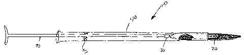
Figure 1 is a pictorial view, partially in cross-section, of a surgical
netting
assembly for deployment from a carrier sheath in the abdomen.
Figure 2 is a pictorial view of the fully deployed netting assembly shown in
Figure 1 with a single netting layer.
Figure 3 illustrates a netting assembly partially deployed, and Figure 4
illustrates the same netting assembly further but yet not fully deployed.
Figure 5 illustrates a threaded connector between the netting assembly 's
deployment rod and frame
Figure 6 illustrates a permeable foam membrane netting layer.
Figure 7 illustrates another embodiment of a permeable netting layer.
Figure 8 is a cross-sectional view of an outer membrane netting layer and
multiple fiber layers for a central netting layer.
Figure 9 illustrates a netting assembly with frame partially deployed via a
basket deployment rod.
Figure 10 illustrates the netting assembly as shown in Figure 9 further
deployed by movement of the outer deployment rod relative to the inner
configuration control rod.
Figure 11 illustrates in cross-sectional view of progressively smaller
passthrough area netting layers for a netting assembly.
Figure 12 illustrates in cross-section an alternative netting assembly with
looped strands.
Figure 13 illustrates a cross-sectional view of another embodiment of the
assembly of the present invention.
Figure 14 is a cross-sectional view to the carrier sheath shown in Figure 13.
7
CA 02529428 2005-12-13
WO 2004/112571 PCT/US2004/018548
Detailed Description of Preferred Embodiments
The inventive device provides a membrane that acts as a trapping
mechanism to immobilize, block or trap stones and stone fragments as they
emerge
from the gallbladder and bile ducts, thus preventing undesirable migration
into the
abdominal cavity. The membrane, which may be a thin layer, or of various three
dimensional geometric configurations, or combination thereof, covers the
subhepatic
space in the abdomen to prevent the stones from migrating after emergence from
either the gallbladder or biliary tract. This invention blocks stone migration
and thus
reduces patient trauma and the common complications that are associated with
stone retrieval from laparoscopic cholecystectomy and laparoscopic common bile
duct exploration procedures.
The netting assembly may be compressed and pre-packaged in a carrier tube
sheath that is deployed by insertion through a carrier sheath within a
laparoscopic
port. The distal end of the device may extracted from the carrier tube sheath
and
expanded manually by the surgeon, or by preformed memory, to form a barrier
which prevents stone migration beneath the liver. As a result of application
of this
device, any stones that are displaced from the gallbladder or common bile duct
remain blocked from migrating behind the liver. The device with stones trapped
therein is removed at the completion of the procedure either by retraction
into the
carrier sheath or by placement into a specimen bag. This stone-immobilization
device thus facilitates the surgeon's ability to remove stones and associated
debris
with an improved rate of operational success and with a reduced risk of post-
operative infection or organ trauma.
The laparoscopic/surgical netting assembly 10 makes use of common
laparoscopic port sizes, typically between 5mm and 12mm. It will be apparent
to
those skilled in the art that the configuration and relative positions of
deployment of
the device is variable and may be tailored to procedural needs and specific
anatomical features. Deployment of the device is typically under the
gallbladder and
to the right side of the common bile duct so as to trap stones and stone
fragments,
i~
CA 02529428 2005-12-13
WO 2004/112571 PCT/US2004/018548
thus avoiding hazardous migration of stones and stone debris during the
surgical
procedure.
An oval, rounded, or rectangular geometry for the frame may be used with a
saddle-shaped configuration, although other geometric configurations, such as
polygonal or trapezoidal configuration, may be chosen. In a preferred
embodiment,
frame 20 when deployed has a generally oval configuration, with a long axis
22,
which is substantially parallel to a central axis of the inward end of the
instrument
guide. The short axis is substantially perpendicular to the long axis. In a
preferred
embodiment, the long axis is from about 3" to about 5" long, and.the short
axis is
from about 1" to about 3" wide. The long axis is preferably about 4" and the
short
axis of about 2" is preferred. The uppermost layer of the netting material is
preferably at least '~2' or more below the short axis at its midpoint. The
substantial
size of the frame 21 when expanded is sufficient, in a preferred embodiment,
such
that the nettings supported on the frame may collect both the gallbladder and
one or
more stones released from the gallbladder or bile duct. This substantial size
also
allows a plurality of stones to be easily collected within the netting, which
may cover
a relatively large area for capturing stones which otherwise may drop into
body
cavities.
The netting layer 30 as shown in Figure 2 may include a thin, flat sponge, a
pierced membrane, a screen with looped elements, or a netting or mesh
material. A
netting material is preferred, with the netting strings defining a passthrough
area to
facilitate passage of fluids, and may include a rectangular, hexagonal,
octagonal or
,1 other selected configuration. In a preferred embodiment, the netting layer
30
comprises at least two layers and preferably at least three layers with each
layer
being spaced from an adjacent layer and having a smaller passthrough area,
such
that stones that effectively become trapped between layers. Figures 6 and 7
illustrate a netting assembly and three layers 30A, 30B and 30C each having a
rectangular area. The passthrough area is intended for catching the large
stones,
which typically are about two centimeters in diameter, while passing through
the
9
CA 02529428 2005-12-13
WO 2004/112571 PCT/US2004/018548
netting small stones which can conventionally be recovered by vacuum, which
are
typically about 3 millimeters in diameter or less. In an exemplary embodiment
as
shown in Figure 11, the netting assembly comprises a frame 20 as discussed
above
and three netting layers 30A, 30B, and 30C. For this exemplary embodiment, the
passthrough areas in the upper layer may have a generally square
configuration, so
that the minimum diameter stone that may pass through the upper layer may have
a
diameter approximating 36A as shown in Figure 1. The second or intermediate
layer may have a smaller passthrough area for a minimum diameter of 36B, and
the
lowest layer 30C may have netting passthrough area of diameter 36C. The
passthrough area for the lowest layer may thus be from about 1 millimeters to
about
5 millimeter, thereby effectively capturing the smallest of the stones which
cannot be
conventionally recovered by vacuum. The intermediate layer may have a
passthrough diameter 36 B of from about 5 millimeters to 10 millimeters, while
the
uppermost layer 38 may have a passthrough area of about 10 millimeters to
about 2
centimeter.
In the alternate embodiment as shown in Figure 12, the netting material may
form loop strands 38 which extend substantially upward in a direction
generally
perpendicular to the plane of the netting layer, as shown in Figure 12.
Alternatively,
3-D shapes with concavity or more complex molded configuration for the netting
layers may be utilized.
The stone barrier or netting layer 30 and the frame 20 may be compressed or
furled in a carrier sheath 40 for insertion through a laparoscopic port. The
netting
assembly 10 may then be extracted by surgeon from the sheath and opened within
the peritoneal cavity. Alternatively, the device may be deployed mechanically
via
pushing a linear rod 90 down the sheath. Upon deployment into the peritoneum,
the
device 10 may assume a predetermined shape related to the predetermined
memory of the frame. A self-sealing valve 41 as shown in Figure 1 may be
provided
to seal between the interior of sheath 40 and the exterior of rod 90, and also
to close
off flow through the sheath 40 when the rod 90 is removed from the sheath, to
CA 02529428 2005-12-13
WO 2004/112571 PCT/US2004/018548
prevent escape of gas. The valve 41 may be employed in all the embodiments,
but
is only shown in Figure 1. The device may be cinched closed thus trapping
stones
by pulling a, purse string 60 (see Figure 2) around the perimeter of the stone
barrier.
Alternative closure methods are envisioned such as twisting, rolling, furling
or
winding of the stone barrier or attached wire or string. The device 10 with
the
trapped stones may then be removed from the peritoneal cavity, and optionally
may
be retrieved through a laparoscopic port.
The permeable membrane 30 may have a high pile, or looped fabric
configuration to entrap stone material. One embodiment of the device provides
a
porous barrier that allows liquid and blood to easily penetrate through, but
screens
out the stone material. The permeable membrane is preferably formed from a
chemical composition that is non-adherent to body tissues.
After insertion of the stone barrier through the port, the surgeon may
position
the instrument in the space below the gallbladder, to the right of the common
bile
duct, inferior to the liver, and superior to the hepatic flecture of the
colon, thus
blocking stone migration to the deep recesses behind the liver. The stone
barrier is
deployed prior to dislodgement of stones from the gallbladder or bile ducts.
The
stone barrier is then gathered and removed after the danger of stone spillage
into
this space has passed.
By minimizing prolonged fishing expeditions for stones spilled into the
peritoneal cavity the efficiency of laparoscopic cholecystectomy and
laparoscopic
common bile duct exploration and the stone removal process may be enhanced and
complications related to retained stones within the peritoneal cavity may be
eliminated, thus benefitting the patient. The surgical netting assembly
collects
stones that are inadvertently spilled from the hepatobiliary tract, including
the
gallbladder and the common bile duct. Alternatively, a suction catheter or
stone
basket may be introduced through the instrument guide to remove stone debris.
Use of the laparoscopic surgical netting assembly for conducting a
laparoscopic gallbladder or bile duct procedure should be apparent to those
skilled
11
CA 02529428 2005-12-13
WO 2004/112571 PCT/US2004/018548
in the art in view of the above disclosure. The procedure may conventionally
be
conducted through a laparoscopic port having an external end above the
abdominal
wall and an internal end within the abdominal cavity, with the laparoscopic
port
including a throughbore extending between the ends to provide a conduit into
the
abdominal cavity, and to facilitate introduction of a pneumoperitoneum to
insuflasuffrate the abdominal cavity. '
The surgical netting assembly may thus be introduced laparoscopically with
the frame collapsed and supporting the fluid permeable membrane along its
perimeter. Insertion through the carrier sheath 40 is accomplished by pushing,
pulling, or rotating the frame control rod 90, which is attached to the
netting
assembly frame 30 by removable connector 80. The frame 20 may be expanded
upon extraction from its carrier sheath 40, whereby its memory assumes a'
substantially greater area than the collapsed frame. The netting 10 supported
on
the expanded frame will thus have a sufficient area so that the surgeon may
place
the netting assembly in its expanded position below the gallbladder and/or
bile duct
for collecting stones which may then be easily collected and removed. The
netting
assembly may remain attached to its frame control rod 90 or may be detached
via
connector 80 to allow the laparoscopic port to be used for other
instrumentation.
Alternatively, the surgeon may elect to use the netting assembly with a
smaller
portion of the netting surface area exposed via partial emergence from the
carrier
sheath, which also allows the device to be used manually by manipulation for
scooping stones or stone debris. The geometric configuration of the netting
surface
may also be controlled and modified by turning and pushing or pulling rod 95.
Rod
95, which may also be considered a deployment rod, acts on the frame when
expanded, as shown in Figure 10. The deployment rod is movable with respect to
control rod 90 within the same through channel that receives the control rod
90, or
the deployment rod 95 may be provided in another one of the through channels
within the carrier sheath. The rod 95 engages the frame at a location spaced
from
12
CA 02529428 2005-12-13
WO 2004/112571 PCT/US2004/018548
the connection of the frame to rod 90, and acts to change the configuration of
the
frame.
The lateral amount of extension of the netting assembly from the carrier
sheath is varied by extension of the frame control rod 90 by the surgeon to
selectively control the frame expansion and netting geometry of the device.
The
netting allows fluids to passthrough the netting during retrieval of the
netting
assembly and trapped stones. Very small stones that passthrough the netting
may
be collected by vacuum line.
Figure 2 shows the generally saddle-shaped oval configuration for a
preferred frame 20, wherein the rearward end of the frame preferably has a
grasping stud 21 secured to the frame. The end of grasping and releasing tool
98
may thus be used to move the netting assembly through the elongated tube 40
and,
as shown Figure 2, may be activated by the surgeon during release of the
netting
assembly from the tube 40. If desired, the langard 60 may be connected with
the
frame, and optionally may by used to assist in retrieval of the netting
assembly with
the stones captured therein. Also, langard 60 may include a retrieval loop as
shown
in Figure 2, which is continuous with a framed loop 62, such that the
combination of
the retrieval and the frame loops form a continuous looped band. The
desirability of
this option is that the surgeon may grasp retrieval loop 60 and pull on the
retrieval
loop, thereby "tightening up" the maximum diameter of the frame loop 62 to
effectively cause the frame to collapse about the netting assembly with the
captured
stones within the netting assembly, in a manner similar to a drawn string on a
bag or
purse. This feature thus further reduces the likelihood of a stone
inadvertently being
released from the netting assembly prior to being retrieved from the patient.
In
another embodiment, the frame may have a generally circular shape, with the
netting layer or layers having a generally funnel shaped configuration.
Figure 5 shows an alternatively threaded connection 80 between the frame
control rod 90 and the frame 20. The netting strands 32 may have generally
rectangular or squared-shaped passthrough openings 34 as shown.
13
CA 02529428 2005-12-13
WO 2004/112571 PCT/US2004/018548
Figure 6 disposes an alternative netting assembly, wherein the netting
assembly is not a conventional net, but is a net in a sense that it provides a
flexible
barrier to capture the stones, but it is sufficiently porous to allow blood
and other
fluids to pass through the netting assembly. Also, the netting assembly 30 as
shown
on Figure 6 does not have a frame. The netting assembly 30 is formed from a
generally plastic sheet 70 which may be rolled into a small diameter to pass
through
the tube 40, then unrolled to occupy the substantially larger area for
desirably
capturing the stones. The plastic layer 70 may have selectively sized
passthrough
holes 72 for fluid flow, and most of these passthrough holes preferably are
generally
circular in cross-section to reduce manufacturing cost and to reduce the
likelihood of
a tear in the sheet 70 during use of the netting assembly.
Figure 7 discloses yet a further alternative, in which the plastic sheet 70
includes a passthrough center hole 72 with a netting assembly 74 secured to
the
edge of the large diameter hole. In this embodiment, the netting assembly 74
may
catch most of the stones, since the stones will move by gravity toward the net
74
may catch most of the stones, since the stones will move by gravity toward the
net
74 due to the contour of the sheet 70.
Figure 8 discloses yet another embodiment, and again depicts in cross-
sections sheet 70 with passthrough holes 72. In this embodiment, the large
hole in
the center of the plastic sheet 70 is filled with a filtering material, which
in one
embodiment may be held in place by netting 74. The top layer 76A of the filter
is
designed to pass the majority of the stones through the layer, so that stones
engage
the second void layer 76B. A third layer 76C has a still smaller passthrough
area,
so that most stones will be captured on top of a layer 76C. The last layer 76D
has
the smallest passthrough area, which is designed to capture the smallest of
the
stones to be retrieved with the netting assembly. Each of the layers 76A, 76C
and
76D may be formed from a fibrous material or a form material, and has the
preferable desired flexibility and low cost to achieve the objectives of the
invention,
while also selectively capturing most stones between different layers, thereby
14
CA 02529428 2005-12-13
WO 2004/112571 PCT/US2004/018548
insuring likelihood that the stones will be removed from the patient at the
completion
of the surgery. Moreover, the various layers 76A, 76C and 76D may be colored
coded, if desired, so that a certain color corresponds to a certain
passthrough area.
If desired, the passthrough area of one or more layers may be selected by the
surgeon based on the specifics of the operation. If the surgeon knows that the
largest stones in the patient will be 10 millimeters in diameter, the surgeon
may
select the netting layers which are most reliable capture all stones down to
the small
diameter stones which are desirably capture by the netting assembly.
In an alternate embodiment, the netting assembly may be prepackaged for
insertion through the sheath 40 in a generally spiraling manner, i.e., both of
the
frame and the netting assembly may be twisted into a small diameter elongate
configuration with the frame and the netting assembly spiraling along a
generally
central axis of the prepackaged assembly. This allows the frame and the
netting
assembly to be controllable released from the sheath 40 in manner that unfolds
in a
reverse spiraling manner as the netting assembly is pushing out the exit of
the
sheath 40. For example, the surgeon may know that insertion of the rod 90 to a
selected point will result in a 50% release of the netting assembly from the
sheath,
and that the further insertion of another inch may result in the simultaneous
rotation
and extension of the netting assembly. The controlled rotation and controlled
axial
position of the netting assembly with respect fo the sheath is to better
control the
configuration of the frame and the position of the netting assembly under the
desired organs to serve its intended purpose. The interior of the sheath 40
may
cooperate with a dog on the rod 90 to slide in within an elongate spiraling
slot in the
sheath to control the release of the netting assembly from the sheath 40.
In another embodiment of the present invention, the carrier sheath is
provided with a plurality of through channels. One of the through channels may
be
sufficient to pass the netting assembly and the frame configuration control
rod. A
surgical tool, such as a scalpel, scissors or other cutting device, may then
be
passed through another of a plurality of through channels so that the surgeon
may
CA 02529428 2005-12-13
WO 2004/112571 PCT/US2004/018548
cut tissue in the area of the bile duct and gallbladder with the netting
assembly
already in place beneath the location of the cut to catch stones released from
the
gallbladder or bile duct.
Figure 13 is a cross-sectional view of an assembly according to the present
invention, illustrating a laparoscopic port 5 which has an external end which
extends
axially above an external surface of the abdomen wall and an abdominal end
which
extends below an internal surface of the abdominal wall and into the abdominal
cavity. A carrier sheath 40 includes a plurality of internal throughbores, and
preferably from two to four internal throughbores, which provide conduits into
the
abdominal cavity. The carrier sheath 40 is provided within the laparoscopic
port
internal throughbore, and has a carrier sheath external end and an instrument
guide
abdominal end. The carrier sheath external end extends above the external
surface
of the abdominal wall and the carrier sheath abdominal end extends below the
laparoscopic port abdominal end and proximal to the gallbladder or bile duct.
As shown in Figure 13, the carrier sheath includes the plurality of through
channels 80 and 82, with one of the channels 80 being somewhat crescent shaped
for passing the netting, and the other channel having a more conventional
circular
cross-section, for conveying surgical tools, such as scalpel 94. Figure 13
illustrates
a control rod 90 for passing the net assembly into and out of the carrier
sheath 40.
At the end of control rod is a pair of wires 70, 72, which provide the frame
for the
netting, with separate frames provided for a lower fine netting 30A and a
course top
netting 308. Each netting assembly has a generally heart-shaped configuration
for
more easily receiving the gallbladder 15 and one or stones. The stones 16 may
thus pass through the netting 30B and be caught in the netting 30A. A fluid
impermeable layer 92 may be provided in the lower layer 30A for capturing
fluid
released from the gallbladder, or for capturing the gallbladder and the fluid
within the
gallbladder. While only a portion of the layer 92 is shown, a fluid
impermeable layer
may be provided above or below the layer 30A, and may have the same area as
netting 30A.
16
CA 02529428 2005-12-13
WO 2004/112571 PCT/US2004/018548
As shown in Figure 13, both the outer frame wire 70 and the inner frame wire
72 pass through one of the through channels in the carrier sheath 40, and exit
the
top of the carrier sheath. Both the outer wire and the inner wire may be
separately
extendable and retractable within the through channel of the carrier sheath
for
changing the configuration of the frame. More particularly, the outer frame
wire 72
may be retracted to be pulled at least partially over the course netting 30B
supported on the inner frame wire, thereby effectively capturing the
gallbladder 15
and/or stones 16 within the netting assembly.
In some applications, the sheath may be eliminated and the tools, including
the netting assembly, installed through the laparoscopic port. In many
applications,
however, the sheath is preferred since its abdominal end may be easily
positioned
proximate to the gallbladder or bile duct.
While preferred embodiments of the present invention have been illustrated
in detail, it is apparent that other modifications and adaptations of the
preferred
embodiments will occur to those skilled in the art. The embodiments shown and
described are thus exemplary, and various other modifications of the preferred
embodiments may be made which are within the spirit of the invention.
Accordingly,
it is to be expressly understood that such modifications and adaptations are
within
the scope of the present invention, which is defined in the following claims.
17