Note : Les descriptions sont présentées dans la langue officielle dans laquelle elles ont été soumises.
CA 02530166 2005-09-07
WO 2004/080284 PCT/US2004/006985
ANTIBODY-TARGETED PHOTODYNAMIC THERAPY
CROSS-REFERENCE TO RELATED APPLICATION
This application claims priority to U.S. Provisional Application No.
60/452,655, filed on March 7, 2003. U.S. Provisional Application No.
60/452,655
is herein incorporated by this reference in its entirety.
BACKGROUND
Photodynamic therapy is a medical procedure used for the treatment of a
large variety of disease states, such as eye disorders, cancer, renal
disorders, and
skin disorders, among others. Photodynamic therapy putatively relies on a
particularly active form of molecular oxygen known as singlet oxygen. Singlet
oxygen, which is generally toxic to cells, can be generated in various ways.
In
photodynamic therapy, singlet oxygen is typically generated photochemically by
irradiating a secondary substance known as a "photosensitizes" in the presence
of
air (oxygen).
As a method to treat cancer, photodynamic therapy attempts to induce
malignant cell death and tumor elimination through a photochemical process
triggered by irradiating diseased tissue with high-intensity light. Prior to
irradiation, the diseased tissue is loaded with a photosensitizes that absorbs
the
light and uses part of its energy to drive a series of photochemical
reactions. These
photochemical reactions are believed to generate reactive toxic products such
as
singlet oxygen, which ultimately damage or destroy the diseased tissue.
A wide variety of photosensitizers have been used for photodynamic
therapy, and the success of the therapy can depend in part on the particular
photosensitizes utilized. For example, photosensitizers that strongly absorb
on the
edge of the visible region and into the near-infrared region are of special
relevance
since the penetration of light into living tissue is substantially better at
wavelengths
beyond 600 nm. The success of photodynamic therapy also rests largely on the
preferential accumulation of photosensitizers by diseased tissue as compared
to the
adjacent healthy tissue, thereby allowing the destruction of such diseased
tissue
CA 02530166 2005-09-07
WO 2004/080284 PCT/US2004/006985
without damaging the surrounding healthy tissue. The limitations of
photodynamic
therapy are most often caused by poor performance of the photosensitizers in
particular environments, or because the uptake and retention of a
photosensitizer
by a distinct diseased tissue is not sufficiently greater than that observed
for the
surrounding normal tissue.
One photosensitizer that has been used in photodynamic therapy is
verteporfin. Verteporfin is sold in an injectable form under the name
MSUDYNE~ (Parkedale Pharmaceuticals; Rochester, MIA. Verteporfin has been
used to treat choroidal neovascular disease (CNV) and has proved effective at
preventing moderate to severe visual loss in eyes with subfoveal predominantly
classic CNV or occult-only CNV caused by age-related macular degeneration
(AMD) and in eyes with subfoveal CNV caused by pathologic myopia. (Lim,
Ophthalmol Clin North Am; 15 (4): 473-478, vii, Dec. 2002). Verteporfin has
also
been used in photodynamic therapy for the treatment of pigment epithelial
detachment (PED).
Photodynamic therapy with photosensitizers such as verteporfin usually
involve a two-step process, beginning with administration of the
photosensitizer,
e.g., by intravenous injection. While circulating in the bloodstream, the
photosensitizer attaches to molecules called lipoproteins. Because cells
undergoing rapid proliferation (cell division and growth) require a greater
amount
of lipoproteins than non-dividing cells, the photosensitizer is delivered more
quickly and in higher concentrations to these types of cells. Once the
concentration of photosensitizer reaches appropriate levels in a cell or
tissue of
interest, it is activated with a pre-calculated dose of light at a particular
wavelength. The light dosage typically used for photodynamic therapy is
usually
much less damaging than thermal or hot laser treatment used in laser
photocoagulation, which can leave permanent blind spots. The activated
photosensitizer subsequently causes the conversion of normal oxygen found in
tissue to singlet oxygen. The singlet oxygen, in turn, causes cell death by
disrupting normal cellular functions.
2
CA 02530166 2005-09-07
WO 2004/080284 PCT/US2004/006985
Although verteporfin accumulates preferentially in choroidal
neovasculature, some non-specific accumulation within the retina occurs. Thus,
retinal structures may be damaged during the application of the light
treatment.
Needed in the art is the ability to increase the selectivity of
photosensitizers like
verteporfin to the specific tissue to be irradiated.
SUMMARY
In accordance with the purposes of the disclosed materials, compositions,
and/or methods, as embodied and broadly described herein, in one aspect, the
disclosed subject matter relates to photosensitizer-antibody conjugates and
methods for making such conjugates. Also disclosed herein are methods for
using
photosensitizer-antibody conjugates in photodynamic therapies for treating
subjects with various diseases.
Additional advantages will be set forth in part in the description which
follows, and in part will be obvious from the description, or may be learned
by
practice of the aspects described below. The advantages described below will
be
realized and attained by means of the elements and combinations particularly
pointed out in the appended claims. It is to be understood that both the
foregoing
general description and the following detailed description are exemplary and
explanatory only and are not restrictive.
BRIEF DESCRIPTION OF DRAWINGS
The accompanying drawings, which are incorporated in and constitute a
part of this specification, illustrate several aspects described below.
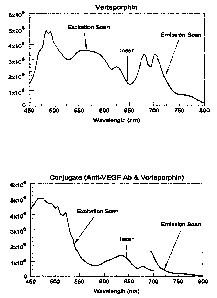
Figure 1 is a pair of fluorescence excitation-emission scans of native
verteporfin (top) and the verteporfin-anti-VEGF-conjugate (bottom). The
spectra
were collected on a Jobin-Yvon SPEX FL-3 spectrofluorimeter.
Figure 2A is a photograph of MS-1 vascular endothelial cells growing in
24-well plastic culture plate. Figure 2B is a close-up photograph of an
individual
well in the culture plate during exposure to the 647 nm emission of a Krypton-
ion
continuous wave laser.
Figure 3 is a graph showing the mean percent cell viability by trypan blue
exclusion over eight experimental runs in 24-well culture plates for VEGF-
3
CA 02530166 2005-09-07
WO 2004/080284 PCT/US2004/006985
expressing endothelial cells in Dulbecco's phosphate buffered saline ("DPBS"),
cells incubated with verteporfin (VISUDYNE) at 40 ~.g/ml ("Verteporfin"), and
cells incubated with verteporfin (VISUDYNE) conjugated to anti-VEGF antibody
("Conjugate"). Error bars indicate 1 standard deviation from the mean. The
cells
received either no laser treatment or were exposed to krypton-ion CW laser
(64T
nm) at a total light dosage of 56.5 J/cm for either 1 hour (left) or 24 hours
(right).
Figure 4A is an image of a confocal bright field view of MS-1 vascular
endothelial cells growing on a glass coverslip. These cells were labeled with
a
VISUDYNE-anti-VEGF antibody conjugate. Figure 4B is an image of the same
field as in "A," taken with fluorescence optics to image the presence of the
conjugate. Images were made with an Olympus IX70 confocal fluorescence
microscope (Olympus America, Inc.; Melville, NY). Fluorescence was excited
with the 633 nm line of a HeNe laser, and the emission was acquired with a 660
nm longpass filter.
Figure SA is a confocal fluorescence microscopic image of MS-1 vascular
endothelial cells incubated with native VISUDYNE for 5 minutes. Figure SB is a
confocal fluorescence microscope image of MS-1 vascular endothelial cells
incubated with the VISUDYNE-anti-VEGF antibody conjugate for 5 minutes.
Figure 6A is a confocal fluorescence microscope image of MS-1 vascular
endothelial cells incubated with native VISUDYNE for 25 minutes. Figure 6B is
a
confocal fluorescence microscope image of MS-1 vascular endothelial cells
incubated with VISUDYNE-anti-VEGF antibody conjugate for 20 minutes.
Figure 7 is a graph of fluorescence intensity of conjugate-labeled and
VISUDYNE-labeled MS-1 vascular endothelial cells as a function of incubation
time. The fluorescence intensity of confocal micrographs was determined with
the
"histogram" analysis tool of the ImagePro image processing software program
(Media Cybernetics; Silver Springs, MD). Intensity is expressed as the
accumulated pixel counts in the image.
Figure 8 is a graph of the photosensitizing properties, i.e., photoinduced
cytotoxicity, of native verteporfin and the verteporfin-anti-VEGF antibody
conjugate as a function of concentration.
4
CA 02530166 2005-09-07
WO 2004/080284 PCT/US2004/006985
DETAILED DESCRIPTION
The disclosed materials, compositions, and methods may be understood
more readily by reference to the following detailed description of specific
aspects
of the materials and methods and the Examples included therein and to the
Figures
and the previous and following description.
Before the present materials, compositions, and/or methods are disclosed
and described, it is to be understood that the aspects described below are not
limited to specific synthetic methods or specific reagents, as such may, of
course,
vary. It is also to be understood that the terminology used herein is for the
purpose
of describing particular aspects only and is not intended to be limiting.
Disclosed are materials, compositions, and components that can be used
for, can be used in conjunction with, can be used in preparation for, or are
products
of the disclosed method and compositions. These and other materials are
disclosed
herein, and it is understood when combinations, subsets, interactions, groups,
etc.
of these materials are disclosed that, while specific reference of each
various
individual and collective combinations and permutation of these compounds may
not be explicitly disclosed, each is specifically contemplated and described
herein.
For example, if a conjugate is disclosed and discussed and a number of
modifications that can be made to a number of photosensitizers andlor
antibodies
in the conjugate are discussed, each and every combination and permutation of
the
conjugate and the modifications to the photosensitizer and/or antibodies that
are
possible are specifically contemplated unless specifically indicated to the
contrary.
Thus, if a class of substituents A, B, and C are disclosed as well as a class
of
substituents D, E, and F and an example of a combination molecule, A-D is
disclosed, then even if each is not individually recited, each is individually
and
collectively contemplated. Thus, in this example, each of the combinations A-
E,
A-F, B-D, B-E, B-F, C-D, C-E, and C-F are specifically contemplated and should
be considered disclosed from disclosure of A, B, and C; D, E, and F; and the
example combination A-D. Likewise, any subset or combination of these is also
specifically contemplated and disclosed. Thus, for example, the sub-group of A-
E,
S
CA 02530166 2005-09-07
WO 2004/080284 PCT/US2004/006985
goat, etc.), laboratory animals (e.g., mouse, rabbit, rat, guinea pig, etc.)
and birds.
"Subject" or "patient" can also include a mammal, such as a primate or a
human.
Reference herein to a "cell" or "tissue" can include a cell or tissue in
vitro.
Alternatively, reference to a "cell" or "tissue" can include a cell or tissue
in vivo,
which can be found in a subject. A "cell" or "tissue" can be a cell or tissue
from
any organism including, but not limited to, a bacterium, a eukaryote, or an
animal.
The terms "higher," "increases," "elevates," or "elevation" refer to
increases above basal levels, e.g., as compared to a control. The terms "low,"
"lower," "reduces," or "reduction" refer to decreases below basal levels,
e.g., as
compared to a control. By "control" is meant either a subject lacking a
disease or a
subject in the absence of a particular variable such as a therapeutic. A
subject in
the absence of a therapeutic can be the same subject before or after treatment
with
a therapeutic or can be a different subject in the absence of the therapeutic.
Comparison to a control can include a comparison to a known control level or
value known in the art. Thus, basal levels are normal in vivo levels prior to,
or in
the absence of, addition of an agent or another small molecule or ligand.
"Disease," as used herein means an impairment of the normal state of a
subject or one of its parts that interrupts or modifies the performance of the
function and is a response to environmental factors (e.g., malnutrition,
industrial
hazards, climate, injury), to infective agents (e.g., bacteria, fungus, or
viruses), to
inherent defects of the organism (e.g., genetic anomalies), or to combinations
of
these factors.
Coniu~ate
Disclosed herein is a method for selectively targeting cells for destruction
or ablation by administering to a subject a photosensitizer conjugated to an
antibody. The antibody allows for the selective targeting of an organ, tissue,
or
protein. That is, the antibody facilitates the localization of the
photosensitizer-
antibody conjugate to cells and tissues for which the antibody is designed to
target.
When light is directed onto the tissue or cell containing the photosensitizer-
antibody conjugate, the photosensitizer becomes activated and the tissue or
cell is
7
CA 02530166 2005-09-07
WO 2004/080284 PCT/US2004/006985
S destroyed where the light has been directed. Such activation can be local,
focused
activation, or can be systemic by general activation.
Photosensitizers
The conjugates disclosed herein contain one or more photosensitizers,
which are compounds that absorb light energy. For example, red light can be
used.
Blue light can also be used. In one example, ambient light can be used. The
photosensitizer can absorb light from about 600 nm to about 800 nm. In one
example, the conjugate contains a photosensitizer that can absorb light from
about
650 nm to about 700 nm. In some aspects, the photosensitizer can absorb light
of
about 600, 601, 602, 603, 604, 605, 606, 607, 608, 609, 610, 611, 612, 613,
614,
615, 616, 617, 618, 619, 620, 621, 622, 623, 624, 625, 626, 627, 628, 629,
630,
631, 632, 633, 634, 635, 636, 637, 638, 639, 640, 641, 642, 643, 644, 645,
646,
647, 648, 649, 650, 651, 652, 653, 654, 655, 656, 657, 658, 659, 660, 661,
662,
663, 664, 665, 666, 667, 668, 669, 670, 671, 672, 673, 674, 675, 676, 677,
678,
679, 680, 681, 682, 683, 684, 685, 686, 687, 688, 689, 690, 691, 692, 693,
694,
695, 696, 697, 698, 699, 700, 701, 702, 703, 704, 705, 706, 707, 708, 709,
710,
711, 712, 713, 714, 715, 716, 717, 718, 719, 720, 721, 722, 723, 724, 725,
726,
727, 728, 729, 730, 731, 732, 733, 734, 735, 736, 737, 738, 739, 740, 741,
742,
743, 744, 745, 746, 747, 748, 749, 750, 751, 752, 753, 754, 755, 756, 757,
758,
759, 760, 761, 762, 763, 764, 765, 766, 767, 768, 769, 770, 771, 772, 773,
774,
775, 776, 777, 778, 779, 780, 781, 782, 783, 784, 785, 786, 787, 788, 789,
790,
791, 792, 793, 794, 795, 796, 797, 798, 799, or 800 nm, where any of the
stated
values can form an upper and/or lower endpoint when appropriate.
The photosensitizer can be any composition that absorbs light and initiates
a photochemical reaction that produces cytotoxic products. For example,
suitable
photosensitizers that can be used in the disclosed conjugates include, but are
not
limited to, haematoporphyrins, photofrins, chlorins such as meta-tetra
hydroxyphenyl chlorin, mono-L-aspartyl chlorin e6, or bacteriochlorins, or
derivatives thereof. The photosensitizer can also include phthalocyanines,
porphyrins, benzoporphyrins, 5-aminolaevulinic acid (ALA), or derivatives
thereof. Other photosensitizers include, but are not limited to, purpurins,
8
CA 02530166 2005-09-07
WO 2004/080284 PCT/US2004/006985
B-F, and C-E are specifically contemplated and should be considered disclosed
from disclosure of A, B, and C; D, E, and F; and the example combination A-D.
This concept applies to all aspects of this disclosure including, but not
limited to,
steps in methods of making and using the disclosed compositions. Thus, if
there
are a variety of additional steps that can be performed it is understood that
each of
these additional steps can be performed with any specific embodiment or
combination of embodiments of the disclosed methods, and that each such
combination is specifically contemplated and should be considered disclosed.
Definitions
In this specification and in the claims which follow, reference will be made
to a number of terms which shall be defined to have the following meanings:
As used in the specification and the appended claims, the singular forms
"a," "an," and "the" include plural referents unless the context clearly
dictates
otherwise. Thus, for example, reference to "a conjugate" includes mixtures of
one
or more conjugates, reference to "an antibody" includes mixtures of one or
more
antibodies, reference to "the photosensitizer" includes one or more such
photosensitizers, and the like.
Ranges may be expressed herein as from "about" one particular value,
and/or to "about" another particular value. When such a range is expressed,
another embodiment includes from the one particular value and/or to the other
particular value. Similarly, when values are expressed as approximations, by
use
of the antecedent "about," it will be understood that the particular value
forms
another embodiment. It will be fiuther understood that the endpoints of each
of the
ranges are significant both in relation to the other endpoint, and
independently of
the other endpoint.
"Optional" or "optionally" means that the subsequently described event or
circumstance may or may not occur, and that the description includes instances
where the event or circumstance occurs and instances where it does not.
As used throughout, the term "subject" or "patient" can include
domesticated animals (e.g., cat, dog, etc.), livestock (e.g., cattle, horse,
pig, sheep,
6
CA 02530166 2005-09-07
WO 2004/080284 PCT/US2004/006985
porphycenes, pheophorbides, and verdins. Purpurins are a class of porphyrin
macrocycle with an absorption band at from about 630 nm to about 715 nm,
typified by tin etiopurpurin (SnET2), which has an extinction coefficient of
40,000
M-lcrri 1 at about 700 nm. Porphycenes, having activation wavelengths of about
635 nm, are also useful. Phorbides are derived from chlorophylls (e.g.
pheophorbide) and are also useful as photosensitizers. Verdins contain a
cyclohexanone ring fused to one of the pyrroles of the porphyrin ring and can
also
be used as a photosensitizer. Psoralens are another example of a
photosensitizer
that can be used in the disclosed conjugates and methods.
Synthetic non-porphyrin compounds can also be used as photosensitizers in
the compositions and methods disclosed herein. Suitable non-porphyrin
compounds include, but are not limited to, phenothiazinium compounds such as
methylene blue, Toluidine blue, cyanines such as Merocyanine-540, acridine
dyes,
derivatives of the tumor marker, Nile blue, and rhodamines such as the
mitochondria-specific Rhodamine 123.
In one aspect, the photosensitizer can be a benzoporphyrin derivative, such
as a benzoporphrin mono acid derivative. In another aspect, the
photosenstitizer
can be verteporfin, which is a benzoporphyrin derivative, or the injectable
form of
verteporfin, VISUDYNE~, from Parkedale Pharmaceuticals, Rochester, MN,
which incorporates preferentially into choroidal neovasculature.
Antibodies
The conjugates disclosed herein also contain one or more antibodies. In
one aspect, the antibodies are monoclonal or polyclonal antibodies to vascular
endothelial growth factor (anti-VEGF).
The term "antibodies" is used herein in a broad sense and includes both
polyclonal and monoclonal antibodies. In addition to intact immunoglobulin
molecules, also included in the term "antibodies" are fragments of
immunoglobulin
molecules and multimers of immunoglobulin molecules (e.g., diabodies,
triabodies,
and bi-specific and tri-specific antibodies, as are known in the art; see,
e.g.,
Hudson and Kortt, J. Immunol. Methods 231:177-189, 1999), fusion proteins
containing an antibody or antibody fragment, which are produced using standard
9
CA 02530166 2005-09-07
WO 2004/080284 PCT/US2004/006985
molecular biology techniques, single chain antibodies, and human or humanized
versions of immunoglobulin molecules or fragments thereof. Any antibody that
specifically binds an antigen in a manner sufficient to deliver a
photosensitizer to
the desired location can be used in the methods disclosed herein.
Whenever possible, antibodies useful in the conjugates and methods
disclosed herein can be purchased from commercial sources, such as Chemicon
International (Temecula, CA). The antibodies of the disclosed conjugates and
methods can also be generated using well-known methods. The skilled artisan
will
understand that either full-length antigens or fragments thereof can be used
to
generate the antibodies of the disclosed conjugates and methods. A polypeptide
to
be used for generating an antibody of the disclosed conjugates and methods can
be
partially or fully purified from a natural source, or can be produced using
recombinant DNA techniques. For example, for antigens that are peptides or
polypeptides, a cDNA encoding an antigen, or a fragment thereof, can be
expressed in prokaryotic cells (e.g., bacteria) or eukaryotic cells (e.g.,
yeast, insect,
or mammalian cells), after which the recombinant protein can be purified and
used
to generate a monoclonal or polyclonal antibody preparation that specifically
binds
the targeted antigen.
One of skill in the art will know how to choose an antigenic peptide for the
generation of monoclonal or polyclonal antibodies that specifically bind the
appropriate antigens. Antigenic peptides for use in generating the antibodies
of the
disclosed conjugates and methods are chosen from non-helical regions of the
protein that are hydrophilic. The PredictProtein Server (http://www.embl-
heidelberg.de/predictprotein/subunitdefhtml) or an analogous program can be
used
to select antigenic peptides to generate the antibodies of the disclosed
conjugates
and methods. In one example, a peptide of about fifteen amino acids can be
chosen and a peptide-antibody package can be obtained from a commercial source
such as AnaSpec, Inc. (San Jose, CA). One of skill in the art will know that
the
generation of two or more different sets of monoclonal or polyclonal
antibodies
maximizes the likelihood of obtaining an antibody with the specificity and
affinity
required for its intended use. The antibodies are tested for their desired
activity by
CA 02530166 2005-09-07
WO 2004/080284 PCT/US2004/006985
known methods (e.g., but not limited to, ELISA and/or immunocytochemistry).
For additional guidance regarding the generation and testing of antibodies,
see,
e.g., Harlow and Lane, Antibodies: A Laboratory Manual, Cold Spring Harbor
Laboratory Press, Cold Spring Harbor, NY, 1988, which is incorporated by
reference herein for methods of making antibodies).
Monoclonal antibodies
The term "monoclonal antibody" as used herein refers to an antibody or
antibody fragment obtained from a substantially homogeneous population of
antibodies or antibody fragments, i.e., the individual antibodies within the
population are identical except for possible naturally occurnng mutations that
can
be present in a small subset of the antibody molecules. The monoclonal
antibodies
herein specifically include "chimeric" antibodies in which a portion of the
heavy
and/or light chain is identical with or homologous to corresponding sequences
in
antibodies derived from a particular species or belonging to a particular
antibody
class or subclass, while the remainder of the chains) is identical with or
homologous to corresponding sequences in antibodies derived from another
species or belonging to another antibody class or subclass, as well as
fragments of
such antibodies, as long as they exhibit the desired antagonistic activity
(See, e.g.,
U.S. Patent No. 4,816,567 and Morrison et al., Proc. Natl. Acad. Sci. USA,
81:6851-6855, 1984).
Monoclonal antibodies useful in the conjugates and methods disclosed
herein can be prepared using hybridoma methods, such as those described by
Kohler and Milstein, Nature, 256:495, 1975. In a hybridoma method, a mouse or
other appropriate host animal is typically immunized with an immunizing agent
to
elicit lymphocytes that produce or are capable of producing antibodies that
will
specifically bind to the immunizing agent. Alternatively, the lymphocytes can
be
immunized in vitro, e.g., using an adapter antigen or an immunogenic fragment
thereof.
The monoclonal antibodies can also be made by recombinant DNA
methods, such as those described in U.S. Patent No. 4,816,567 (Cabilly et
al.).
DNA encoding the monoclonal antibodies of the disclosed conjugates can be
11
CA 02530166 2005-09-07
WO 2004/080284 PCT/US2004/006985
readily isolated and sequenced using conventional procedures (e.g., by using
oligonucleotide probes that are capable of binding specifically to genes
encoding
the heavy and light chains of murine antibodies). Libraries of antibodies or
active
antibody fragments can also be generated and screened using phage display
techniques, e.g., as described in U.S. Patent No. 5,804,440 (Burton et al.)
and U.S.
Patent No. 6,096,441 (Barbas et al.). Recombinant antibodies, antibody
fragments,
and fusions and polymers thereof can be expressed in vitro or in prokaryotic
cells
(e.g., bacteria) or eukaryotic cells (e.g., yeast, insect, or mammalian cells)
and
further purified, as necessary, using well known methods (see, e.g., Sambrook
et
al. Molecular Cloning: a Laboratory Manual, 3d Edition, Cold Spring Harbor
Laboratory Press (2001); and Ausubel et al., Current Protocols in Molecular
Biology, John Wiley & Sons, New York, NY, 2001, which is updated quarterly).
In vitro methods are also suitable for preparing monovalent antibodies.
Digestion of antibodies to produce fragments thereof, particularly, Fab
fragments,
can be accomplished using routine techniques known in the art. For instance,
digestion can be performed using papain. Examples of papain digestion are
described in WO 94/29348 published Dec. 22, 1994 and U.S. Patent No.
4,342,566. Papain digestion of antibodies typically produces two identical
antigen
binding fragments, called Fab fragments, each with a single antigen binding
site,
and a residual Fc fragment. Pepsin treatment yields a fragment that has two
antigen combining sites and is still capable of cross-linking antigen.
Any antibody or antibody fragment useful in the conjugates and methods
disclosed herein, whether attached to other sequences or not, can also include
insertions, deletions, substitutions, or other selected modifications of
particular
regions or specific amino acids residues, provided the activity of the
antibody or
antibody fragment is not significantly altered or impaired compared to the non-
modified antibody or antibody fragment. These modifications can provide for
some additional property, e.g., to remove or add amino acids capable of
disulfide
bonding, to increase its bio-longevity, to alter its secretory
characteristics, etc. In
any case, the antibody or antibody fragment must possess a bioactive property,
such as specific binding to its cognate antigen. Functional or active regions
of the
12
CA 02530166 2005-09-07
WO 2004/080284 PCT/US2004/006985
antibody or antibody fragment can be identified and/or improved by mutagenesis
of a specific region of the protein, followed by expression and testing of the
expressed polypeptide. For example, amino acid sequence variants of antibodies
or antibody fragments can be generated and those that display equivalent or
improved affinity for antigen can be identified using standard techniques
and/or
those described herein. Methods for generating amino acid sequence variants
are
readily apparent to a skilled practitioner in the art and can include site-
specific
mutagenesis or random mutagenesis (e.g., by PCR) of the nucleic acid encoding
the antibody or antibody fragment (Zoller, M.J. Curr. Opin. Biotechnol. 3:348-
354,
1992). Both naturally occurring and non-naturally occurring amino acids (e.g.,
artificially-derivatized amino acids) can be used to generate amino acid
sequence
variants of the antibodies and antibody fragments used in the disclosed
conjugates.
As used herein, the term "antibody" or "antibodies" can also refer to a
human antibody and/or a humanized antibody. Many non-human antibodies (e.g.,
those derived from mice, rats, or rabbits) are naturally antigenic in humans,
and
thus can give rise to undesirable immune responses when administered to
humans.
Therefore, the use of human or humanized antibodies in the methods disclosed
herein serves to lessen the chance that an antibody administered to a human
will
evoke an undesirable immune response.
Human antibodies
The human antibodies useful in the conjugates and methods disclosed
herein can be prepared using any technique. Examples of techniques for human
monoclonal antibody production include those described by Cole et al.
(Monoclonal Antibodies and Cancer Therapy, Alan R. Liss, p. 77, 1985) and by
Boerner et al. (J. Immunol., 147(1):86-95, 1991). Human antibodies (and
fragments thereof) useful in the conjugates and methods disclosed herein can
also
be produced using phage display libraries (Hoogenboom et al., J. Mol. Biol.,
227:381, 1991; Marks et al., J. Mol. Biol., 222:581, 1991; and C.F. Barbas,
D.R.
Burton, J.K. Scott, G.J. Silverman, Phage Display: A Laboratory Manual, Cold
Spring Harbor Laboratory Press, Cold Spring Harbor, NY, 2001 ).
13
CA 02530166 2005-09-07
WO 2004/080284 PCT/US2004/006985
The human antibodies useful in the conjugates and methods disclosed
herein can also be obtained from transgenic animals. For example, transgenic,
mutant mice that are capable of producing a full repertoire of human
antibodies, in
response to immunization, have been described (see, e.g., Jakobovits et al.,
Proc.
Natl. Acad. Sci. USA, 90:2551-255, 1993; Jakobovits et al., Nature, 362:255-
258,
1993; Bruggermann et al., Year in Immunol., 7:33, 1993). Specifically, the
homozygous deletion of the antibody heavy chain joining region (J(H)) gene in
these chimeric and germ-line mutant mice results in complete inhibition of
endogenous antibody production, and the successful transfer of the human germ-
line antibody gene array into such germ-line mutant mice results in the
production
of human antibodies upon antigen challenge.
Humanized antibodies
Antibody humanization techniques generally involve the use of
recombinant DNA technology to manipulate the DNA sequence encoding one or
more polypeptide chains of an antibody molecule. Accordingly, a humanized form
of a non-human antibody (or a fragment thereof) is a chimeric antibody or
antibody
chain (or a fragment thereof, such as an Fv, Fab, Fab', or other antigen-
binding
portion of an antibody), which contains a portion of an antigen binding site
from a
non-human (donor) antibody integrated into the framework of a human
(recipient)
antibody.
To generate a humanized antibody, residues from one or more
complementarity determining regions (CDRs) of a recipient (human) antibody
molecule are replaced by residues from one or more CDRs of a donor (non-human)
antibody molecule that is known to have desired antigen binding
characteristics
(e.g., a certain level of specificity and affinity for the target antigen). In
some
instances, Fv framework (FR) residues of the human antibody are replaced by
corresponding non-human residues. Humanized antibodies can also contain
residues which are found neither in the recipient antibody nor in the imported
CDR
or framework sequences. Generally, a humanized antibody has one or more amino
acid residues introduced into it from a source which is non-human. In
practice,
humanized antibodies are typically human antibodies in which some CDR residues
14
CA 02530166 2005-09-07
WO 2004/080284 PCT/US2004/006985
and possibly some FR residues are substituted by residues from analogous sites
in
rodent antibodies. Humanized antibodies generally contain at least a portion
of an
antibody constant region (Fc), typically that of a human antibody (Jones et
al.,
Nature, 321:522-525, 1986; Reichmann et al., Nature, 332:323-327, 1988; and
Presta, Curr. Opin. Struct. Biol., 2:593-596, 1992).
Methods for humanizing non-human antibodies are well known in the art.
For example, humanized antibodies can be generated according to the methods of
Winter and co-workers (Jones et al., Nature, 321:522-525, 1986; Riechmann et
al.,
Nature, 332:323-327, 1988; and Verhoeyen et al., Science, 239:1534-1536,
1988),
by substituting rodent CDRs or CDR sequences for the corresponding sequences
of
a human antibody. Methods that can be used to produce humanized antibodies are
also described in U.S. Patent No. 4,816,567 (Cabilly et al.), U.S. Patent No.
5,565,332 (Hoogenboom et al.), U.S. Patent No. 5,721,367 (Kay et al.), U.S.
Patent No. 5,837,243 (Deo et al.), U.S. Patent No. 5, 939,598 (Kucherlapati et
al.),
U.S. Patent No. 6,130,364 (Jakobovits et al.), and U.S. Patent No. 6,180,377
(Morgan et al. ).
In addition to antibodies, other delivery systems can be used to facilitate
the
transport of the photosensitizer to the appropriate area of treatment. Heparin
as
well as heparan sulfate proteoglycans can be used. In another aspect, the
peptide
F56 is an effective antagonist of vascular endothelial growth factor (VEGF),
binding to Flt-1 site. In yet another aspect, VEGF binds differentially to
three
receptor tyrosine kinases, namely VEGF-R1, -R2 and -R3, and to the semaphorin
receptors neuropilin l and 2.
Photosensitizer-antibody coupling
Disclosed are compositions containing conjugates made up of an antibody
or an antibody fragment coupled to a photosensitizer. The conjugates can be
readily synthesized using techniques generally known to those of skill in the
art.
The starting materials and reagents used in preparing these conjugates are
either
available from commercial suppliers such as Aldrich Chemical Co., (Milwaukee,
Wis.), Acros Organics (Morris Plains, N.J.), Fisher Scientific (Pittsburgh,
Pa.), or
Sigma (St. Louis, Mo.) or are prepared by methods known to those skilled in
the
CA 02530166 2005-09-07
WO 2004/080284 PCT/US2004/006985
art following procedures set forth in references such as Fieser and Fieser's
Reagents for Organic Synthesis, Volumes 1-17 (John Wiley and Sons, 1991);
Rodd's Chemistry of Carbon Compounds, Volumes 1-5 and Supplementals
(Elsevier Science Publishers, 1989); Organic Reactions, Volumes 1-40 (John
Wiley and Sons, 1991); March's Advanced Organic Chemistry, (John Wiley and
Sons, 4th Edition); and Larock's Comprehensive Organic Transformations (VCH
Publishers Inc., 1989).
In one aspect, the photosensitizer is coupled or linked to the antibody via a
reactive chemical group on the photosensitizer and/or antibody such that the
coupling between the photosensitizer and the antibody results in a covalent
bond
between the two that is resistant to reducing agents. Reactive chemical groups
include, e.g., sulfliydryl groups, amino groups, carboxyl groups, and
imidazole
groups. The reactive chemical group can be in the hinge region of the
antibody.
This location reduces or eliminates interference between the antibody/antigen
interaction and the photosensitizer. In one aspect, the photosensitizer can be
coupled to the antibody, e.g., via a maleimide group.
The coupling of the photosensitizer to the antibody can also involve an
activating agent. Various activating agents that can be used for the coupling
reaction include, but are not limited to, 1-ethyl-3-(3-dimethylaminopropyl)
carbodiimide (EDC), dicyclohexylcarbodiimide (DCC), N,N'-diisopropyl-
carbodiimide (DIP), benzotriazol-1-yl-oxy-tris-(dimethylamino)phosphonium
hexa-fluorophosphate (BOP), hydroxybenzotriazole (HOBt), and N
methylmorpholine (NMM), including mixtures thereof. The coupling reaction can
be carned out in solvents such as N methylpyrrolidone (NMP) or in DMF. In one
aspect, conjugation can involve the use of a conjugation kit, such as the
Imject
Immunogen EDC conjugation kit from Pierce (Rockford, IL). In another aspect,
EDC and NMP can be obtained as separate reagents and formulated into a
reaction
mixture.
The photosensitizer-antibody conjugates in general are specific for an
antigen that allows targeting of the conjugates to an abnormally proliferative
cell.
16
CA 02530166 2005-09-07
WO 2004/080284 PCT/US2004/006985
In one aspect, the photosensitizer-antibody conjugate can be specific for
cells that
express vascular endothelial growth factor (VEGF) or a portion thereof.
The disclosed photosensitizer-antibody conjugates can also include one or
more additional biomolecules. The additional biomolecules can be coupled in
the
same manner as the photosensitizer. The biomolecules in an antibody conjugate
can be the same as the photosensitizer molecule or different. More
specifically, the
biomolecules can have the same structure or different structures. Also, the
disclosed photosensitizer-antibody conjugates can be combined with
pharmaceutically acceptable Garner, as is described in detail herein.
Methods
Disclosed herein is a method of treating a subject with a disease, such as an
ocular disease, by administering to the subject a photosensitizer-antibody
conjugate and irradiating the subject with light. In this way, the subject's
disease
can be efficaciously treated as compared to a control lacking the conjugate or
lacking the irradiation or both. Efficacious treatment is evaluated by
detecting a
reduction in one or more symptoms of the disease. An overall decrease,
slowing,
inhibition, and/or arrest of disease progress (e.g., further
neovascularization), is
indicative of efficacious treatment.
In one aspect, the photosensitizer-antibody conjugate comprises a
benzoporphyrin and an anti-VEGF antibody. In another aspect, the
benzoporphyrin is verteporfin or VISUDYNE.
The photosensitizer-antibody conjugates disclosed herein can be
administered by any suitable means, such as intraperitoneal or intramuscular
injection, intravenous injection, orally, by ocular or intranasal
administration, or
topically. In one aspect, the photosensitizer-antibody conjugate is
administered by
intravenous injection.
The photosensitizer-antibody conjugates disclosed herein can be infused
within a biocompatible fluid, such as a physiological saline solution, or can
be
applied topically to the exterior surface of a tumor, eye, or abnormal tissue.
The light can be in the form of a laser or in the form of a fiber optic source
used to deliver light to the treatment site from a laser. For example, the
laser light
17
CA 02530166 2005-09-07
WO 2004/080284 PCT/US2004/006985
S can be shone through the slit lamp of a microscope into the subject's eye.
In
another example, the subject stands in a whole body light box containing
lights of
an appropriate wavelength. Light irradiation can also be accomplished by
inserting
a light source into the subject's body, e.g., by catheter or endoscope.
The selection of the appropriate light source and wavelength can be
performed by one of skill in the art and will depend on such factors as the
type of
photosensitizes being used, the type of and location of the site to be
treated, and the
like.
Diseases treatable by a photosensitizes-antibody coniu~ate
Inappropriate angiogenesis and neovascularization is a physiological
component of many types of diseases. Vascular endothelial cells both produce
and
respond to vascular endothelial growth factor (VEGF), which stimulates their
proliferation, thereby stimulating angiogenesis and neovascularization. The
photosensitizes-anti-VEGF antibody conjugates and methods disclosed herein can
be used to treat any disease or condition that involves proliferation of
endothelial
cells and/or inappropriate angiogenesis or neovascularization. Either
antibodies
against VEGF or antibodies against the VEGF receptor conjugated to a
photosensitizes such as verteporfin can be used in the methods disclosed
herein to
treat diseases involving abnormal endothelial cell growth and angiogenesis.
Moreover, diseases (such as cancer) that involve any other inappropriately
growing cell (e.g., a tumor cell) that produces and/or responds to VEGF can be
treated used the methods described herein. These methods can be used to treat
diseases or conditions in any human or non-human animal, e.g., but not limited
to,
horses, cows, sheep, goats, birds (e.g., chickens, geese, ducks, parrots,
parakeets),
dogs, cats, ferrets, mice, rats, hamsters, and guinea pigs.
Uses in ophthalmolo~y
Many diseases or abnormal conditions of the eye involve the inappropriate
stimulation of angiogenesis, which can lead to blindness. For example,
proliferative diabetic retinopathy, in which there is abnormal proliferation
of blood
vessels on the surface of the retina, at the optic nerve, or in front of the
eye on the
iris, is a major cause of visual loss in diabetic patients. When vessels grow
in front
18
CA 02530166 2005-09-07
WO 2004/080284 PCT/US2004/006985
of the eye on the iris (iris neovascularization), they can clog the fluid
outflow
channels and cause the pressure to become very high (neovascular glaucoma);
this
condition can also be treated by the conjugates and methods disclosed herein.
Corneal neovascularization is yet another eye disease that can be treated
using the
methods disclosed herein. The normal cornea is avascu1ar; however, corneal
insults, such as irntation secondary to contact lens wear, can induce corneal
neovascularization, which can threaten vision directly or indirectly (e.g.,
through
secondary hemorrhage, scarring, or lipid deposition.)
Choroidal neovascularization (CNV) is yet another ocular disease that
threatens vision if left untreated. CNV can be occult CNV, classic CNV
(minimally or predominately), or combinations thereof. Other diseases
involving
subfoveal neovessels in the eye include, but are not limted to, diabetic
retinopathy,
choroidal hemangioma, polypoidal choroidal vasculopathy, multifocal
choroiditis
and panuveitis, rubella retinopathy, angioid streaks, ocular histoplamosis
syndrome, Vogt-Koyanagi-Harada syndrome, idiopathic subfoveal CNV, CNV in
pathologic myopia, type 2A idiopathic juxtafoveolar retinal telangiectasis,
and
mallatia leventinese. These diseases can be treated using the methods and
conjugates disclosed herein.
In general, any disease or condition that causes retinal hypoxia or
inflammation, thereby inducing abnormal neovascularization, can be treated
using
the methods and conjugates disclosed herein. Such diseases and conditions
include
sickle cell anemia, inflammatory diseases (including inflammatory diseases of
the
retina), hereditary dystrophies of the retina, trauma to the eye, and
retinopathy of
prematurity (i.e., in premature newborns).
The photosensitizer-anti-VEGF antibody conjugates disclosed herein can
readily be administered ophthamically to any patient with a disease or
condition
involving abnormal angiogenesis in the eye. Following administration of the
photosensitizer-antibody conjugate, the treated regions are exposed to the
appropriate wavelength of laser light using routine methods, as will be
understood
by one of ordinary skill in the art, thereby ablating some or all of the
inappropriately proliferating endothelial cells and treating the disease. An
overall
19
CA 02530166 2005-09-07
WO 2004/080284 PCT/US2004/006985
decrease in abnormal neovascularization, and/or a slowing, inhibition, or
arrest of
disease progress (e.g., further neovascularization), is indicative of an
efficacious
treatment.
In one example, VISUDYNE, an injectable form of verteporfin, is used as
the photosensitizer and is coupled to an antibody against VEGF. By increasing
the
affinity of VISUDYNE for endothelial cells, collateral damage is reduced, and
the
specificity of the photodynamic treatment is increased. The amount of
VISUDYNE administered can be reduced to avoid or reduce undesired side-
effects.
Heman iomas
Hemangiomas are birthmarks that consist of small, closely packed blood
vessels, and come in a number of varieties, e.g., strawberry (capillary)
hemangiomas, cavernous hemangiomas (angioma carcernosum), port-wine stains
(nevus flammeus), and salmon patches (stork bites). Although these birthmarks
are usually painless and benign, they are the product of abnormal and
excessive
angiogenesis, and can be disfiguring. Accordingly, by systemic or targeted
administration (as will be understood by one of ordinary skill in the art) of
the
photosensitizer-anti-VEGF antibody conjugates disclosed herein, the described
methods can be used to selectively ablate the endothelial cells within
hemangiomas, thereby lessening the appearance or even fully removing such
birthmarks.
Tumor treatment
Hemangiosarcomas (angiosarcomas) are malignant neoplasms of vascular
origin, which can invade surrounding tissue and spread metastatically
throughout
the body; therefore, such tumors are dangerous if left untreated. The
photosensitizer-anti-VEGF antibody conjugates disclosed herein can be
administered to patients with hemangiosarcoma using routine techniques for
administration of therapeutic compounds and laser light of an appropriate
wavelength. Such lasers are introduced into the body, e.g., using routine
approaches, e.g., via instruments such as endoscopes or catheters, or via
surgical
exposure of the target tissue. Ablation of endothelial cells and a slowing or
arrest
CA 02530166 2005-09-07
WO 2004/080284 PCT/US2004/006985
of tumor growth, or even a reduction or elimination of the tumor burden,
indicates
that the treatment was efficacious.
Neovascularization is known to play an important role in the growth and
spread of solid tumors (e.g., but not limited to, tumors of the liver, colon,
breast,
lung, pancreas, ovary, uterus, prostate, brain, and bone), as such tumors
cannot
spread or grow to a size of more than a few millimeters without developing a
new
blood supply. Many such tumors have been found to stimulate their own
vascularization by producing VEGF. Accordingly, the growth and/or spread of
solid tumors can be slowed, arrested, or even partially or fully reversed by
administration of the photosensitizer-anti-VEGF antibody conjugates disclosed
herein using routine techniques for administration of therapeutic compounds
and
laser light of an appropriate wavelength. Such lasers are introduced into the
body,
e.g., using routine approaches as indicated above.
In addition, certain types of tumor cells contain VEGF receptors on their
cell surfaces and proliferate in response to VEGF. Such tumors include (but
are
not limited to), ovarian carcinoma, prostate carcinoma, pancreatic carcinoma,
melanoma (including iris tumors), neuroblastoma, and Kaposi's sarcoma. Some
tumors even produce their own VEGF, and thereby stimulate their own growth in
an autocrine fashion. Accordingly, the photosensitizer-anti-VEGF antibody
conjugates disclosed herein can be administered to patients as described above
to
ablate any tumor cell that produces and/or binds VEGF. A slowing or arrest of
tumor growth, or even a reduction or elimination of the tumor burden,
indicates
that the treatment was efficacious.
Atherosclerosis
Atherosclerosis is another condition that can be treated with the
photosensitizer-antibody conjugate described herein. Atherosclerosis is the
term
used to describe the buildup of fatty deposits, called plaque, which
accumulate on
arterial walls, causing hardening of the arteries. Trials with rabbits have
shown
that these fatty deposits can be removed photochemically. The photosensitizer-
antibody conjugate concentrates in the target tissue. In a further aspect,
subsequent
21
CA 02530166 2005-09-07
WO 2004/080284 PCT/US2004/006985
treatment with light removes plaque without damage to the surrounding normal
arterial walls.
Dermatolo~y
In another example, using short wavelength blue light is effective in the
treatment of sun-induced precancerous skin lesions such as actinic kerotoses.
With
this procedure, a photosensitizer precursor, S-aminolevulinic acid, is applied
to the
skin. This substance is converted by an enzyme to a naturally occurring
porphyrin
in the epidermal layer. Because the porphyrins absorb blue light many times
more
strongly than red, a much smaller dose of light is needed. Examples of
antibodies
that can be used are Ki-67 or antibodies directed toward KFGF.
Other diseases and conditions
In addition, any appropriate antibody that preferentially targets tumor cells
or any other type of inappropriately growing cells can be conjugated to a
photosensitizer (e.g., verteporfin) and used to treat the disease or
condition.
In one example, epithelial downgrowth into the eye is a common
complication of eye surgery. To treat this condition, a conjugate of a
photosensitizer and an antibody against keratinocyte growth factor (KGF),
which
stimulates epithelial cell growth, or a conjugate of a photosensitizer and an
antibody against the KGF receptor (which is present on epithelial cells) is
administered ophthamically to the eye or eyes of the affected individual,
after
which laser light of the appropriate wavelength is administered to the target
area as
described above, thereby ablating the inappropriately downgrowing epithelial
cells.
In another example, to treat proliferative vitreoretinopathy, an antibody
against RPE-65 (an antigen specific to the retinal pigment epithelium) can be
conjugated to a photodynamic compound and administered ophthalmically as
described above to inhibit inappropriate cell proliferation.
In another example, many types of cancer cells express antigens that are
selective for that particular tumor cell type. For example, an antibody
against a
melanoma antigen (e.g., but not limited to, MAGEA3, MART-1, melan A, or
tyrosinase) can be coupled to a photosensitizer and administered to the
patient to
ablate melanoma cells as described above. A slowing or arrest of tumor growth,
or
22
CA 02530166 2005-09-07
WO 2004/080284 PCT/US2004/006985
even a reduction or elimination of the tumor burden, indicates that the
treatment
was efficacious.
The photosensitizes-antibody conjugate can also be targeted to metastasized
cancer cells, disease causing viruses, disease causing bacteria or other
undesirable
microorganisms that are distributed throughout at least a portion of a
patient's
body. In this instance, the light employed for administering the light therapy
preferably has a relatively long wavelength, e.g., longer than 800 nm, to
enable the
light to pass through several centimeters of tissue. Generally, the longer the
wavelength of the light, the greater its ability to penetrate tissue in the
body of a
patient. The light adsorption waveband of the photosensitizes must be matched
to
the wavelength or waveband of the light that is administered to activate the
photosensitizes. It is contemplated that by passing a long wavelength light
source
over the external surfaces of a subject's body, the majority of the
photosensitizer-
antibody conjugates attached to targeted abnormal tissue can be activated,
thus
destroying the abnormal tissue, even though widely disseminated within the
subj ect's body.
Preventative uses
It is also contemplated that photosensitizes-antibody conjugates can be
employed prophylactically to prevent the development of abnormal tissue
changes
at a prospective treatment site. The disclosed photosensitizes-antibody
conjugates
can provide an alternative prophylaxis by providing for repetitive
administration of
the photosensitizes-antibody conjugate targeted at the type of tumor cells
that
might develop, followed by administration of light therapy using light having
a
waveband corresponding to the light adsorption waveband of the
photosensitizes.
By periodically repeating such prophylactic therapy, development of a tumor
can
be prevented.
Residual tissue
Another application of the disclosed conjugates and methods is for
destroying any residual abnormal tissue that can remain at a tumor resection
site,
following surgical removal of the tumor. A common problem following such
surgery is the regrowth of the tumor. After administering a photosensitizer-
23
CA 02530166 2005-09-07
WO 2004/080284 PCT/US2004/006985
antibody conjugate targeted at antibodies of the tumor, light therapy can be
administered to destroy the residual tumor cells that have linked with the
conjugate, thereby preventing the regrowth of the tumor.
Yet another application of the disclosed conjugates and methods is for
destroying any residual tissue that remains after cataract surgery. For
example, a
photosensitizes conjugated to an antibody against lens epithelial cells can be
used
following cataract surgery to ablate residual lens epithelial cells that are
thought to
be the cause of posterior capsular opacification (PCO) that is a complication
in up
to 15% of cataract cases.
Infectious Agents
A still further application of the disclosed conjugates and methods is for
removing or inactivating infectious agents from blood products. For example, a
photosensitizes conjugated to an antibody against an infectious agent such as
HIV
or other viral particles can be used to inactivate the infectious agent from
transfusion blood.
Bone marrow transplants
Yet another application of the disclosed conjugates and methods is in the
treatment of leukemia or other diseases requiring bone marrow transplant. A
photosensitizes-antibody conjugate targeted at antigens that are
preferentially
expressed by malignant cells in the bone marrow can be administered followed
by
administration of light therapy using light of the appropriate waveband, as
noted
above. This treatment should be effective both pre- and post-bone marrow
transplant to destroy much of the abnormal tissue causing the leukemia, and
can be
employed, in addition to more conventional radiation and chemotherapy
treatments. It is also contemplated that the disclosed conjugates and methods
can
be used for destroying abnormal tissue in bone marrow, thereby avoiding the
need
for a bone marrow transplant. The photosensitizes-antibody conjugate can be
activated with light administered either from an interstitial source or an
external
source, e.g., transcutaneously or from within the subject's body.
24
CA 02530166 2005-09-07
WO 2004/080284 PCT/US2004/006985
S Administration
The disclosed photosensitizer-antibody conjugates can be conveniently
formulated into pharmaceutical compositions composed of one or more of the
conjugates in association with a pharmaceutically acceptable carrier. By
"pharmaceutically acceptable carrier" is meant a material that is not
biologically or
otherwise undesirable. Thus, the carrier can be administered to a subject
without
causing undesirable biological effects or interacting in a deleterious manner
with
any of the other components of the pharmaceutical composition in which it is
contained. The carrier would naturally be selected to minimize any degradation
of
the active ingredient and to minimize any adverse side effects in the subject,
as
would be well known to one of skill in the art. See, e.g., Remington's
Pharmaceutical Sciences, latest edition, by E.W. Martin Mack Pub. Co., Easton,
PA, which discloses typical carriers and conventional methods of preparing
pharmaceutical compositions that can be used in conjunction with the
preparation
of formulations of the conjugates disclosed herein and which is incorporated
by
reference herein.
Pharmaceutical formulations for the disclosed conjugates can include those
suitable for parenteral (e.g., subcutaneous, intradermal, intramuscular,
intravenous,
intraperitoneal, and intraarticular) administration. Alternatively,
pharmaceutical
formulations of the disclosed conjugates can be suitable for administration to
the
mucous membranes of a subject (e.g., intranasal or ocular administration). The
formulations can be conveniently prepared in unit dosage form and can be
prepared by any of the methods well known in the art.
Depending on the intended mode of administration, the pharmaceutical
compositions can be in the form of, for example, solids, semi-solids, liquids,
solutions, suspensions (e.g., incorporated into microparticles, liposomes,
etc.),
emulsions, gels, or the like, preferably in unit dosage form suitable for
single
administration of a precise dosage. The pharmaceutical compositions can
include,
as noted above, an effective amount of the conjugate in combination with a
pharmaceutically acceptable Garner and, in addition, can include other
carriers,
adjuvants, diluents, thickeners, buffers, preservatives, surfactants, etc.
CA 02530166 2005-09-07
WO 2004/080284 PCT/US2004/006985
Pharmaceutical compositions can also include one or more active ingredients
such
as other medicinal agents, pharmaceutical agents, antimicrobial agents, anti-
inflammatory agents, anesthetics, and the like.
Liquid pharmaceutically administrable compositions can, for example, be
prepared by dissolving, dispersing, etc., a conjugate as described herein and
optional pharmaceutical adjuvants in an excipient, such as, for example,
water,
saline aqueous dextrose, glycerol, ethanol, and the like, to thereby form a
solution
or suspension. If desired, the pharmaceutical composition to be administered
can
also contain minor amounts of nontoxic auxiliary substances such as wetting or
emulsifying agents, pH buffering agents and the like, for example, sodium
acetate,
sorbitan monolaurate, triethanolamine sodium acetate, triethanolamine oleate,
etc.
Actual methods of preparing such dosage forms are known, or will be apparent,
to
those skilled in this art; for example see Remington's Pharmaceutical
Sciences,
referenced above.
In one aspect, when liposomes are used to deliver the conjugates disclosed
herein, the liposomes can be lysed thermolitically by using, for example, a
laser of
the appropriate wavelength. Further, the liposome can be formulated to include
agents such as indocyanine green to enhance the infrared laser absorption and
improve the thermolysis of the liposomes. In this aspect, a laser, e.g., a
laser of
810 nm wavelength, can be used to thermolitically lyse the liposomes. Such
liposome thermolysis can be used to achieve local release of the conjugate
near the
target tissue. This can further reduce non-specific binding of the conjugate
to non-
targeted tissue.
Preparations for parenteral administration can include sterile aqueous or
non-aqueous solutions, suspensions, and emulsions which may also contain
buffers, diluents and other suitable additives. Examples of non-aqueous
solvents
are propylene glycol, polyethylene glycol, vegetable oils such as olive oil,
and
injectable organic esters such as ethyl oleate. Aqueous Garners include water,
alcoholic/aqueous solutions, emulsions or suspensions, including saline and
buffered media. Parenteral vehicles include sodium chloride solution, Ringer's
dextrose, dextrose and sodium chloride, lactated Ringer's, or fixed oils.
26
CA 02530166 2005-09-07
WO 2004/080284 PCT/US2004/006985
Intravenous vehicles can include fluid and nutrient replenishers, electrolyte
replenishers (such as those based on Ringer's dextrose), and the like.
Preservatives
and other additives may also be present such as, for example, antimicrobials,
anti-
oxidants, chelating agents, and inert gases and the like.
The dosage of the compositions required will vary from subject to subject,
depending on the species, age, weight, the general condition of the subject,
the
severity of the disorder being treated, the particular active agent used, its
mode of
administration, physician judgment, and the like. Thus, it is not possible to
specify
an exact amount for every composition. However, an appropriate amount can be
determined by one of ordinary skill in the art using only routine
experimentation
given the teachings herein.
In one aspect, the photosensitizer-antibody conjugates described herein are
administered in an effective amount. The term "effective amount" is defined as
any amount necessary to produce a desired physiologic response. Effective
amounts and schedules for administering the compositions can be determined
empirically, and making such determinations is within the skill in the art.
The
dosage ranges for the administration of the compositions are those large
enough to
produce the desired effect in which the symptoms or disorder are affected. The
dosage should not be so large as to cause adverse side effects, such as
unwanted
cross-reactions, anaphylactic reactions, and the like. Generally, the dosage
will
vary with the age, condition, sex and extent of the disease in the patient,
route of
administration, or whether other drugs are included in the regimen, and can be
determined by one of skill in the art. The dosage can be adjusted by the
individual
physician in the event of any contraindications. Dosage can vary, and can be
administered in one or more dose administrations daily, for one or several
days.
Guidance can be found in the literature for appropriate dosages for given
classes of
pharmaceutical products.
In one example, the patient receives an intravenous injection of
photosensitizer-antibody conjugate, and then waits 1 to 48 hours. For example,
the
wait can be from about 1-8 hours, from about 1-12 hours, from about 12-24
hours,
from about 24-36 hours, or from about 36-48 hours. In yet another example, the
27
CA 02530166 2005-09-07
WO 2004/080284 PCT/US2004/006985
wait is from about 24-36 hours. During this waiting period, the
photosensitizer-
antibody conjugate accumulates in the disease tissue to be treated and is
removed
from healthy tissue. After the waiting period, the patient returns to the
clinic and
the diseased tissue is illuminated for a given amount of time. The
illumination
time can be from about 1 minute to 4 hours, from about 5 minutes to 2 hours,
or
from about 10 to 30 minutes. In one aspect, the application of light is aimed
directly at the abnormal tissue. If the tissue is internal, the light can be
directed to
the tissue or organ with devices such as a bronchoscope into the lungs or a
cystoscope into the bladder, or with a catheter (U.S. Patent No. 5,454,794
(Narcisco et al.).
Infection of Antibody/Photosensitizer Coniu~ates
It is generally preferable to introduce the photosensitizes-antibody
conjugates as close as possible to a treatment site, such as by introducing
the
photosensitizes-antibody conjugate directly into a tumor. At times, the
location of
a tumor or other treatment site is such that it is not feasible to localize
the
administration of photosensitizes-antibody conjugate. Furthermore, the cells
that
are targeted for destruction cannot be localized, but instead, can be viruses,
microorganisms or metastasized cancer cells, which are more broadly
distributed
throughout a subject's body. It is therefore contemplated that the
photosensitizer-
antibody conjugate can be injected into the patient's bloodstream to allow the
patient's own circulatory system to deliver the photosensitizes-antibody
conjugates
to the targeted tissue.
For example, a syringe can be used to inject a fluid containing the
photosensitizes-antibody conjugates, which target a particular cell, in
suspension
through the skin and into the bloodstream. A needle passes through the skin
and
through a blood vessel wall; fluid containing the photosensitizes-antibody
conjugates is injected through a needle into the blood. The blood flow in the
vessel carries the photosensitizes-antibody conjugates to one or more
locations
where the targeted cells or microbes are disposed. The antibody will bind
preferentially to the selected site. Therefore, injury to normal tissue is
minimized
during administration of the light, particularly, if the light is administered
from an
28
CA 02530166 2005-09-07
WO 2004/080284 PCT/US2004/006985
external source and must pass through normal tissue to reach the tissue that
has
been targeted.
Methods of Measuring Treatment Effectiveness
Inhibition of cancer or inhibition of cancer formation means partial or total
killing of cancerous cells, reduction in tumor size, disappearance of a tumor,
inhibition of tumor growth, inhibition of vascularization, inhibition of
cellular
proliferation, an induction in dormancy or an apparent induction of dormancy,
or a
decreased metastasis of a tumor or a tumor cell. In one aspect, tumoricidal
activity
is characterized by 10%, 20%, 30%, 40%, 50%, 60%, 70%, 80%, 90%, or 100%
reduction in tumor size or number of tumor cells.
In ocular treatment, verteporfin therapy stems the growth of CNV and
prevents leakage from abnormal blood vessels. In one aspect, improved vision
is
characterized by a 10%, 20%, 30%, 40%, 50%, 60%, 70%, 80%, 90%, or 100%
improvement in sight over a three-month follow-up period or a reduction in the
extent of neurovascularization. In another aspect, an overall decrease,
slowing,
inhibition, and/or arrest of disease progress (e.g., further
neovascularization), is
indicative of efficacious treatment.
Efficacious treatment can also be characterized by from about 30-70%,
from about 40-80%, from about 50-90%, or from about 60-100% reduction in
target cell viability at one hour past exposure, as compared to a control.
Efficacious treatment can further be characterized by from about 40-80%, from
about 50-90%, from about 60-100%, or from about 70-100% reduction in target
cell viability at twenty-four hours past exposure, as compared to a control.
EXAMPLE
The following examples are put forth so as to provide those of ordinary
skill in the art with a complete disclosure and description of how the
compounds,
compositions, and/or methods described and claimed herein are made and
evaluated, and are intended to be purely exemplary and are not intended to
limit
the scope of what the inventors regard as their invention. Efforts have been
made
to ensure accuracy with respect to numbers (e.g., amounts, temperature, etc.)
but
some errors and deviations should be accounted for. Unless indicated
otherwise,
29
CA 02530166 2005-09-07
WO 2004/080284 PCT/US2004/006985
temperature is in °C or is at ambient temperature, and pressure is at
or near
atmospheric. There are numerous variations and combinations of reaction
conditions, e.g., component concentrations, desired solvents, solvent
mixtures,
temperatures, pressures and other reaction ranges and conditions that can be
used
to optimize the product purity and yield obtained from the described process.
Only
reasonable and routine experimentation will be required to optimize such
process
conditions.
Example 1
Rabbit anti-mouse VEGF polyclonal antibody was obtained from
Chemicon International (Temecula, CA). The anti-VEGF antibody was conjugated
to verteporfin (VISUDYNE ~; Parkedale Pharmaceuticals; Rochester, MN) using
the 1-ethyl-3-[3-dimethylaminopropyl]-carbodiimide (EDC) crosslinking reagent
obtained from Pierce (Rockford, IL), following the manufacturer's protocol.
For
each experimental run, 10 pg of anti-VEGF antibody were conjugated to 1 mg of
verteporfin. The reaction product was purified by separation on a size
exclusion
column and eluted with phosphate buffered saline (PBS), thereby eliminating
unconjugated verteporfin. The fluorescence excitation-emission spectrum was
obtained from the conjugate (Fig. 1; bottom) and compared to that of
verteporfin
alone (Fig. 1; top).
The spectra shown in Fig. 1 were collected on a Jobin-Yvon SPEX FL-3
spectrofluorimeter. In the native verteporfin, the excitation peak at about
690 nm
is typically used to produce the photodynamic action. The conjugate retains
the
fluorescence properties of verteporfin, although the relative amplitudes of
the
peaks are changed. The conjugate, however, retains a relative excitation peak
at
690 nm, along with the corresponding emission peak at about 710 nm, similar to
the fluorescence properties of native verteporfin. The arrow at 647 nm
indicates
the laser excitation derived from a Krypton-ion laser that was used in the
example
experiments. As is shown in the Fig. 1, the fluorescence excitation-emission
spectrum of the conjugate (bottom) was found to be similar to that of
verteporfin
alone (top).
CA 02530166 2005-09-07
WO 2004/080284 PCT/US2004/006985
Experiments were conducted in a line of cultured, VEGF-expressing,
marine endothelial cells (MS-1 VEGF, ATCC). Cells were grown in 24-well
culture plates under standard cell culture conditions in Dulbecco's minimal
essential medium (DMEM) with 10% fetal calf serum. Cells were incubated for
one hour with either phosphate buffered saline solution alone, verteporfin at
40
pg/ml, or verteporfin-anti-VEGF antibody conjugate (Fig. 2A). All cells were
washed twice with phosphate buffered saline following incubation to remove
unbound photosensitizes. The photosensitizes was excited by exposing selected
wells to the krypton-ion CW laser (647 nm) at a total light dosage of 56.5
J/cm,
which is the recommended ophthalmic light dosage (Fig. 2A). For each treatment
condition, there were laser-exposed and non-exposed groups. Following laser
exposure, cells were incubated for either 1 or 24 hours, and cell viability
was
assessed using trypan blue exclusion.
The effect of photodynamic therapy was assessed based on changes in cell
viability and the results are shown in Fig. 3. In the absence of laser
exposure, there
was no significant change in cell viability at 1 hour between conjugate and
verteporfin treated cells. In both 1 hour and 24 hour laser-exposed groups,
cells
treated with either conjugate or verteporfin alone exhibited large losses of
viability
(P < 0.0001) compared with Dulbecco's PBS controls. Conjugated treated cells
had uniformly lower viabilities than cells exposed to verteporfin; however,
the
difference did not reach statistical significance. Power calculations suggest
that if
the observed trend were to continue, statistical significance would be reached
with
72 replicates. Cells exposed to verteporfin without laser exposure also showed
reduced viability, indicating possible toxicity or low-level activation of the
agent
by ambient light.
Example 2
To demonstrate that VISUDYNE-anti-VEGF antibody conjugate is
internalized into endothelial cell cytoplasm, a brightfield, confocal image of
MS-1
VEGF-expressing vascular endothelial cells growing on a coverslip was taken.
This image is shown in Figure 4A. A second image was taken of the same
microscopic field of cells incubated with VISUDYNE-anti-VEGF antibody
31
CA 02530166 2005-09-07
WO 2004/080284 PCT/US2004/006985
conjugate for 1 hour and then washed twice. This second image is shown in
Figure
4B. The image shown in Figure 4B was taken with fluorescence optics to image
the presence of the photosensitizes-antibody conjugate. The light areas
represent
the fluorescence due to VISUDYNE. Images were made with an Olympus IX70
confocal fluorescence microscope (Olympus America, Inc.; Melville, NY).
Fluorescence was excited with the 633 nm line of a HeNe laser, and the
emission
was acquired with a 660 nm longpass filter.
Example 3
To demonstrate that VISUDYNE-anti-VEGF antibody conjugate is
internalized more quickly than is native VISUDYNE, a confocal fluorescence
microscopic image of MS-1 VEGF-expressing vascular endothelial cells cultured
on a cover slip and incubated with native VISUDYNE for S minutes was taken.
This image is shown in Figure SA. All light areas represent VISUDYNE
fluorescence. Comparisons were made of Figure SA with the cells in the
micrograph shown in Figure SB, which were incubated for the same time period
with VISUDYNE-anti-VEGF antibody conjugate. Surprisingly, as is evident from
the two images, the VISUDYNE-anti-VEGF antibody conjugate penetrated into
the endothelial cells more quickly than VISUDYNE alone.
The images shown in Figure 6 were from the same conditions as those in
Figure 5 except that the incubation time was 25 minutes in Figure 6A with
native
VISUDYNE and 20 minutes in Figure 6B with VISUDYNE-anti-VEGF antibody
conjugate. Again, it is evident from the two images in Figure 6 that the
VISUDYNE-anti-VEGF antibody conjugate penetrated into the endothelial cells
more quickly than VISUDYNE alone, even though the incubation time with native
VISUDYNE was longer.
Example 4
In order to provide a more quantitative comparison of the binding kinetics
of the conjugate and the native VISUDYNE, the fluorescence intensity of the MS-
1 cells labeled for varying durations was measured in a series of confocal
micrographs using the "histogram" tool of ImagePro image processing program
(Media Cybernetics; Silver Springs, MD). The results of such measurements are
32
CA 02530166 2005-09-07
WO 2004/080284 PCT/US2004/006985
shown in Figure 7. It can be observed that within the first 10 minutes of
labeling,
the conjugate achieves effectively maximum binding to the target cells,
whereas
the native VISUDYNE reaches approximately SO% of maximum binding.
Accordingly, if the laser activation were to be applied within 10 minutes of
application of the conjugate, essentially maximum photodynamic effect would be
realized. Targets not expressing the VEGF factor would not be as strongly
labeled.
This improved selectivity of the conjugate for VEGF-expressing targets is a
distinct advantage over the native VISUDYNE.
Examule 5
The relative efficiencies of native VISUDYNE and the VISUDYNE-anti-
VEGF antibody conjugate were examined with respect to their photosensitizing
properties. For the viability experiments, the MS-1 VEGF-expressing cells were
plated on coverslips and grown in 6-well culture plates. The agents to be
tested
were applied to the cells in the various wells on the plate, incubated for one
hour,
exposed to the 647 nm laser, and incubated for an additional hour following
the
laser. To detect dead and dying cells, the Sytox Orange nuclear stain was
obtained
from Molecular Probes (Eugene, OR) and used at a concentration of 0.25 pM and
an incubation time of 10 minutes. Total cell counts (live and dead) were made
by
counterstaining with Hoechst 33342 (Molecular Probes; Eugene, OR) at 20 p,M
and a 2-minute incubation period. The coverslips containing the cells were
removed from the culture plate, and fluorescent cells were imaged with an
Olympus BX60 epifluoresence microscope (Olympus America, Inc.; Melville,
NY). Images were captured with a frame grabber, and analyzed with ImagePro
software (Media Cybernetics; Silver Springs, MD) to obtain total cell counts
with
each stain. Three fields were measured per experimental condition. A summary
of
these results is shown in Figure 8.
As can be seen in Figure 8, the verteporfin (VISUDYNE)-anti-VEGF
antibody conjugate photosensitizes the MS-1 cells at a lower concentration
than
does the native verteporfin (VISUDYNE). The difference in effect is probably
greater than indicated, because the precise concentration of the conjugate is
not
33
CA 02530166 2005-09-07
WO 2004/080284 PCT/US2004/006985
known. When the conjugate was made, the concentration of the reagents was
adjusted so that the initial concentration of verteporfin (VISUDYNE) was the
same
as in the native verteporfin (VISUDYNE) preparation (40 pg/ml). The efficiency
of the reaction and the recovery efficiency after the purification on the size-
exclusion column, however, have not been determined, but both were likely to
have been less than 100 percent. Therefore, the effective concentration of the
conjugate was probably much less than indicated in Figure 8.
Throughout this application, various publications, patents, and/or patent
applications are referenced in order to more fully describe the state of the
art to
which this invention pertains. The disclosures of these publications, patents,
and/or patent applications are herein incorporated by reference in their
entireties,
and for the subject matter for which they are specifically referenced in the
same or
a prior sentence, to the same extent as if each independent publication,
patent,
and/or patent application was specifically and individually indicated to be
incorporated by reference.
It will be apparent to those skilled in the art that various modifications and
variations can be made in the present invention without departing from the
scope
or spirit of the invention. Other aspects of the invention will be apparent to
those
skilled in the art from consideration of the specification and practice of the
invention disclosed herein. It is intended that the specification and examples
be
considered as exemplary only.
34