Note : Les descriptions sont présentées dans la langue officielle dans laquelle elles ont été soumises.
CA 02532418 2006-01-13
WO 2004/007699 PCT/CA2003/001079
1
METHOD FOR PREPARING ENGINEERED TISSUE
CROSS-REFERENCE TO OTHER APPLICATIONS
This application claims the benefit of United States
provisional Patent Application Serial No. 60/396,004 filed July
16, 2002.
FIELD OF THE INVENTION
The present invention relates to engineered tissue. In
particular, the present invention relates to methods for making
engineered tissue from sheets of living tissue.
BACKGROUND OF THE INVENTION
Tissue engineering may be used to recreate tissues and
organs for grafting onto patients. Engineered tissues and organs
can also serve as in vitro models. A variety of tissue
engineering techniques are known, including tissue in-growth,
seeding of cells on artificial or biodegradable scaffolds, and
collagen gels. Among them, a new method of tissue engineering,
known as the "self-assembly" method, has emerged (L'Heureux
et al.; Michel et al.; Pouliot et al.). In the self-assembly
method, cells are induced to secrete and organise an extracellular
matrix and thereby form a sheet of living tissue. The self-
assembly method takes advantage of the fact that fibroblasts can
produce a suitable extracellular matrix when grown in the presence
of ascorbic acid. To create multi-layer tissue constructs, sheets
of living tissue can be stacked upon each other, folded upon
themselves, or rolled on a tubular support.
The development of tissue engineering methods to
produce reconstructed tissues has focused on the optimization of
morphological and histological properties of the reconstructed
tissues. Most tissue engineering research has focused on
optimizing techniques for growing sheets of tissue. However, in
many cases, it would be desirable to make reconstructed tissue
comprised of several layers of tissue and/or layers of more than
CA 02532418 2006-01-13
W02004/007699
PCT/CA2003/001079
2
one type of tissue. Multi-layer tissue constructs are thicker and
therefore stronger and since multi-layer tissue constructs can
comprise more than one sheet of living tissue, they can be
designed to more closely resemble the tissues that they are
intended to replace. However, in order to create useful multi-
layer tissue constructs, it is essential to be able to fuse
adjacent layers of cell tissue together so that the layers are
bonded together as firmly and reliably as possible and resist
separation. If these layers of tissue are not fused together
well, they may separate or come apart over time, for example,
during handling or in the body of a patient.
Thus, it would be desirable to have a method for
preparing multi-layered engineered tissue constructs with improved
fusion between adjacent layers of tissue.
DESCRIPTION OF BACKGROUND ART
Ye et a/. teach that mechanical stress can enhance the
synthesis and secretion of the principal extracellular matrix
protein, collagen, by fibroblasts, thereby increasing the
mechanical strength of the stiffness of reconstructed tissue. Ye
et al. describe a method for applying mechanical stress wherein
sheets of fibroblast cells are mounted on frames to apply tension.
However, the reconstructed tissues produced by this method have
significantly less collagen than does native tissue and therefore
would not be expected to have mechanical properties that resemble
native tissue.
Kanda et al. teach that mechanical stress induces cell
orientation and phenotypic modulation of cultured smooth muscle
cells.
L'Heureux et a/. describe a method of making a tissue-
engineered blood vessel (TEBV) by wrapping sheets of living tissue
around a tubular support.
Lopez-Valle et al. describe the use of a continuous (as
opposed to punctuated or discontinuous) anchor made of porous
CA 02532418 2006-01-13
W02004/007699
PCT/CA2003/001079
3
glass microfiber material, the pores of which trap collagen fibers
and thereby induce organization of extracellular matrix and
orientation of cells.
SUMMARY OF THE INVENTION
The current invention provides a method for improving
the fusion between adjacent layers of living tissue in a multi-
layered engineered tissue construct. The current invention
further provides a method for making a sheet of living tissue
suitable for use in preparing a multi-layered engineered tissue
construct. Multi-layered reconstructed tissues produced by this
method have improved fusion between layers of tissue and the
layers of tissue are less likely to separate during subsequent
manipulation.
Thus, in one aspect, the invention provides a method
for preparing a human or animal tissue from at least one sheet of
living tissue, the method comprising the steps of: (a) arranging
the at least one sheet of living tissue to form a multi-layer
stack of living tissue; and (b) applying a compressive force in a
direction normal to the surface of the multi-layer stack of living
tissue with a force-applying means at a pressure and for an amount
of time sufficient to compress layers of tissue together for
inducing adjacent layers of tissue to fuse or adhere to each
other.
A particular preferred embodiment provides a method for
producing a tissue by forming a multi-layer stack of living tissue
arranged on a substantially flat support. More preferably, the
multi-layer stack is formed by superimposing two or more sheets of
living tissue and/or by folding a sheet of living tissue upon
itself.
In a further embodiment, the method comprises another
step where the multi-layer stack is then anchored to a
substantially flat support surface with moveable anchors
comprising weights or ingots, wherein the anchors are of a
suitable weight for (1) applying sufficient tension across the
CA 02532418 2006-01-13
WO 2004/007699
PCT/CA2003/001079
4
sheet of living tissue to prevent shrinkage and/or maintain
cellular differentiation and/or induce orientation of cells in at
least one sheet of living tissue and (2) allowing contraction of
at least one sheet of living tissue once a predetermined threshold
of tension is exceeded across the sheet of living tissue. A force
is then applied normal to the surface of the layers of tissue by
way of a weighted device suitable for applying evenly distributed
pressure to the surface of the multi-layer stack of tissue, the
weighted device being at least partially permeable to culture
medium, for inducing adjacent layers of tissue to fuse to each
other.
In a preferred embodiment, the force-applying means in
step (b) of the method comprises a weight device suitable for
applying substantially evenly-distributed pressure to said multi-
layer stack of living tissue, the weight device being at least
partially permeable to tissue-culture medium.
In a further embodiment, the multi-layer stack of step
(a) of the method is formed by rolling a sheet of living tissue on
a tubular support and, more preferably, the force-applying means
of step (b) comprises a tissue-culture medium permeable elastic
sleeve.
There are many different types of sheets of living
tissues that can be used with the method described herein. In an
embodiment, the method utilizes a biopsy as a sheet of living
tissue. In another embodiment, the method utilizes cells cultured
in vitro as a sheet of living tissue. More preferably, the cells
are embryonic stem cells, post-natal stem cells, adult stem cells,
mesenchymal cells, hepatocytes, Islet cells, parenchymal cells,
osteoblasts and other cells forming bone or cartilage, and nerve
cells. The mesenchymal cells can either be fibroblasts,
interstitial cells, endothelial cells, smooth muscle cells,
skeletal muscle cells, myocytes, chrondocytes, adipocytes,
fibromyoblasts, or ectodermal cells. In a further embodiment, the
sheet of living tissue can also be a skin tissue, a corneal
CA 02532418 2006-01-13
W02004/007699
PCT/CA2003/001079
tissue, a cardiac valve tissue, a connective tissue and/or a
mesenchymal tissue.
In another aspect, the invention also provides a multi-
layer tissue made according to the method described herein,
5 wherein the multi-layer tissue comprises at least two different
types of sheets of living tissue. In a preferred embodiment, the
tissue consists essentially of between two and twelve sheets, more
preferably of between three and nine sheets. In another
embodiment, the tissue has a thickness of between about 0.01 mm to
about 0.5 mm, more preferably of between about 0.03 mm to about
0.45 mm.
In a further aspect, the invention provides a method
for preparing a planar human or animal tissue suitable for use in
making a multi-layer tissue construct from at least one sheet of
living tissue, the method comprising the steps of: (a) arranging
said at least one sheet of living tissue on a substantially flat
support surface; and (b) anchoring said at least one sheet of
living tissue to the support surface with an adjustable anchor-
means comprised of a multiplicity of spaced apart anchors, wherein
the anchors are suitable for (1) applying sufficient tension
across the sheet of living tissue to prevent shrinkage and/or
maintain cellular differentiation and/or induce orientation of
cells in said at least one sheet of living tissue and (2) allowing
contraction of said at least one sheet of living tissue once a
predetermined threshold of tension is exceeded across the sheet of
living tissue.
Preferably, the adjustable anchor-means is comprised of
discrete moveable anchors such as weights or ingots.
Alternatively, the adjustable anchor means may also comprise a
moveable frame or a multiplicity of moveable anchors mounted on a
frame.
In a particular preferred embodiment, a planar
construct can be made by forming a multi-layer stack of living
tissue by superimposing two or more sheets of living tissue or by
CA 02532418 2012-12-11
6
folding a sheet of living tissue upon itself. The multi-layer
stack is then anchored to a substantially flat support surface
with moveable anchors comprising weights or ingots, wherein the
anchors are of a suitable weight for (1) applying sufficient
tension across the sheet of living tissue to prevent shrinkage
and/or maintain cellular differentiation and/or induce orientation
of cells in at least one sheet of living tissue and (2) allowing
contraction of at least one sheet of living tissue once a
predetermined threshold of tension is exceeded across the sheet of
living tissue. A force is then applied normal to the surface of
the layers of tissue by way of a weighted device suitable for
applying evenly distributed pressure to the surface of the multi-
layer stack of tissue, the weighted device being at least
partially permeable to culture medium, for inducing adjacent
layers of tissue to fuse to each other.
In a further embodiment, the sheet of living tissue
used to prepare the planar construct is obtained by culturing
cells in vitro.
In yet another aspect, the invention provides a planar
multi-layer tissue consisting essentially of between two to twelve
sheets of living tissue obtained by the method described herein.
In another particular embodiment, a tubular construct
can be made by forming a multi-layer stack of living tissue by
rolling a sheet of living tissue onto itself, for example with the
aid of a tubular support. A culture medium permeable elastic
sleeve can be used to compress the layers of tissue in the multi-
layer stack of living tissue together for inducing the adjacent
layers of tissue to fuse to each other.
CA 02532418 2013-09-26
6a
It is provided a method for preparing a human or animal tissue from at least
one
sheet of living tissue, the method comprising the steps of: (a) arranging the
at least
one sheet of living tissue to form a multi-layer stack of living tissue; and
(b) applying
a compressive force in a direction normal to the surface of the multi-layer
stack of
living tissue with a force-applying means at a pressure and for an amount of
time
sufficient to compress layers of tissue together for inducing adjacent layers
of tissue
to fuse or adhere to each other; wherein: the at least one sheet of living
tissue is
obtained by culturing cells in vitro; and the force-applying means in step (b)
comprises a first weighted device suitable for applying substantially evenly-
distributed pressure to solely the periphery of the multilayer stack of living
tissue, and
a second weighted device cut to fit within the first weighted device, the
second
weighted device being at least partially permeable to tissue-culture medium,
wherein
the second weighted device does not overlap with the first weighted device;
provided
that the human or animal tissue does not comprise any human embryonic cells.
Other embodiments and advantages of the invention will become apparent from
the
details description to follow, together with the accompanying drawings.
CA 02532418 2011-10-13
7
BRIEF DESCRIPTION OF THE DRAWINGS
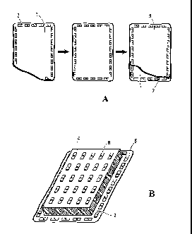
Figure 1 illustrates a preferred method of making a
planar tissue. (IQ A first sheet of living tissue (1) is arranged
on a substantially flat support surface and anchored peripherally
with weights or ingots (2); a second sheet of tissue (3) is
superimposed on the first sheet of tissue, and the weights or
ingots (2) are moved, one-by-one, and placed on the second sheet,
thereby anchoring the superimposed sheets. (B) A sponge (4) that
has been cut to fit Within the ingots is placed on top of the
multi-layer tissue construct (5) so formed; and spaced-apart
weights (6) are placed on the sponge.
Figure 2 illustrates a preferred method of compressing
a tubular tissue construct. An elastic sleeve (7) is placed
around a hollow tube (8) using its tapered end (9). The hollow
tube (8) is larger than a tissue construct (10) which has been
rolled around a mandrel (11). The hollow tube (8) is then placed
around the tissue construct (10). The elastic sleeve (7) is
transferred from the hollow tube (8) to the tissue construct (10)
by gently displacing the tube (8) in one direction and the tissue
construct (10) in the opposite direction.
Figure 3 is a microscopic view of the tissue made
according to the method of the invention, after maturation. The
tissue is assembled from nine sheets of living tissue containing
fibroblasts and extracellular matrix constituents. Magnification
20X, scale bar 50 m.
Figure 4 graphs results of cyclic stress-strain test on
a three-layer tissue construct made as described in Example 1.
DETAILED DESCRIPTION OF THE PREFERRED EMBODIMENTS
In accordance, with the present invention, it has been
found that application of a compressive force normal to the plane
CA 02532418 2006-01-13
W02004/007699
PCT/CA2003/001079
8
of a sheet of tissue enhances fusion between adjacent layers of
sheets of tissue. Compression improves contact between layers of
tissue and encourages fusion of the layers of tissue. By way of
example, a weighted device (for example, a sponge upon which
spaced apart weights are placed) may be applied to a stack of two
or more superimposed planar sheets of tissue, thereby applying a
force normal to the plane of the sheets of tissue (see Figure 13).
In another example, a suitably sized elastic sleeve can be fitted
over a multi-layer stack of tissue made by rolling at least one
sheet of tissue onto a tubular support, whereby the elastic sleeve
applies pressure normal to the two-dimensional plane of the sheet
of tissue (see Figure 2).
Preferably, the compressive force or pressure is
applied evenly on the entire tissue surface. Therefore, it is
preferable that a device adapted to the shape of the tissue be
used to induce the fusion. The amount of pressure applied to the
surface of the tissue stack can be adjusted according to the needs
of the engineered tissue. This pressure is applied for a period
of time sufficient to allow the complete fusion of the tissue
layers, preferably between 24 hours to 7 days.
It is also preferable that the device used to induce
pressure to the surface of the tissue be permeable to culture
media in order to allow the nutrition of the living cells. An
acceptable way to generate this pressure on a flat tissue is to
lay a semi-rigid sponge on the top of the tissue stack.
Additional weight (for example, one or more solid ingots) can be
distributed on top of the sponge to obtain the desired amount of
pressure on the tissue (see Figure 1B). Of course, any other
system using mechanical or hydraulic pressure could be used to
provide this compression.
In the example of a tubular or cylindrical construct,
the compressing device should preferably apply equal pressure on
the external surface of the construct. In this particular case, a
good way to compress the tissue is to apply an elastic and
permeable sleeve around the construct (see Figure 2). The size
CA 02532418 2006-01-13
WO 2004/007699
PCT/CA2003/001079
9
and the elasticity of the sleeve can be adjusted to give the
appropriate pressure for a given tissue.
To wrap the sleeve around the tissue and remove it
without damage, an installation device may be used (see Figure 2).
The elastic sleeve (7) is mounted on a rigid hollow tube (8) using
the tapered end of the tube (9). The tube, having an internal
diameter slightly larger than the construct, is then passed over
the tubular tissue (10) rolled around a mandrel (11). The end of
the sleeve is then anchored to the mandrel and the hollow tube is
carefully removed by pulling from the opposite side, gently
depositing the sleeve on the tissue. To remove the sleeve without
damaging the tissue, it may be for example carefully cut or
unsewn.
It is known that mechanical stress may be used to
induce cellular orientation and phenotypic modulation of cultured
smooth muscle cells (Kanda et a/.; Germain et a/.). Thus,
appropriate forces may be applied to maturating tissue in order to
induce fiber orientation. Such forces may also prevent shrinkage
and maintain the desired cell differentiation.
As an example, it has been shown that a continuous
anchor, such as a frame or a ring of glass microfiber that
circumscribes or encircles a tissue, may be used to induce
cellular orientation (Ye et al.; Kanda et al.). The induction of
cell orientation is thought to occur because the continuous anchor
mechanically restricts the spontaneous contraction of the maturing
cultured tissue, thereby creating a mechanical stress or tension
across the tissue that induces cell orientation.
The underlying mechanism of the orientation response
has not been well elucidated (Kanda et a/.). However, when a
continuous anchor is used, the tension across the tissue continues
to build as the tissue matures and can reach levels that are
detrimental to the health of the cells in the tissue, reducing
viability of cells contained in the tissue and thereby producing
an inferior tissue construct. Therefore, a continuous anchor,
CA 02532418 2006-01-13
W02004/007699
PCT/CA2003/001079
such as a rigid frame, may not be suitable for use with some
tissue types, i.e. those tissues that can create a lot of tension
as they mature.
The current invention provides an improved method of
5 anchoring maturing cultured tissues, the method comprising an
adjustable anchor means, preferably comprising a multiplicity of
spaced apart anchors (such as moveable weights or ingots), wherein
the anchors are suitable for (1) applying sufficient tension
across the sheet of living tissue to prevent shrinkage and/or
10 maintaih cellular differentiation and/or induce orientation of
cells in at least one sheet of living tissue and (2) allowing
contraction of at least one sheet of living tissue once a
predetermined threshold of tension is exceeded across the sheet of
living tissue (for an illustration see Figure 1A).
The anchor means is "adjustable" in that once the
tissue has built up a tension higher than the maximum tension that
can be held by the anchors (i.e. weights or ingots), the tissue
can spontaneously contract and the anchors will be pulled along
with the contracting tissue. Thus, the tension across the tissue
cannot continue to build up when an adjustable anchor means as
described is employed. The maximum tension that can build up
across the tissue can be controlled by choosing suitable anchors
(for example weights or ingots of a certain weight and number, or
an adjustable frame that is designed to move in response to a
certain tension or force). Thus, it is possible to optimize the
amount of tension for any given tissue, for example, to enhance
viability of cells in the tissue.
Anchors according to the current invention may be
"discontinuous" or "punctual". A "discontinuous" or "punctual
anchor" is a device that anchors a tissue substantially at a point
in space. In contrast, in the context of the present invention,
the term "continuous anchor" refers to a device for securing a
tissue around its entire perimeter (such as described by Lopez-
Valle et al.).
CA 02532418 2006-01-13
W02004/007699
PCT/CA2003/001079
11
The anchors of the present invention may be "moveable"
in that they can easily be placed on a sheet of tissue or removed
therefrom.
For making planar sheets of living tissue for use in
making multi-layer tissue constructs, it is preferred that anchors
are arranged to form a closed perimeter near the edge of a sheet
of tissue. This geometry induces cells and extracellular matrix
fibers in the sheet of tissue to orient in the two dimensions of
the plane of the sheet of tissue. This orientation of cells and
extracellular matrix may be beneficial for fusion of adjacent
layers of sheets of tissue and may also improve certain functional
properties of the tissue. For example, in Figure 1A, a first
sheet of living tissue (1) is disposed on a flat surface and
ingots (2) keep the first sheet in place. A second sheet of
living tissue (3) is placed on top of the first sheet. Ingots are
displaced from the first sheet and are arranged on top of the
second sheet to anchor the stack of sheets. The ingots are
arranged to follow the perimeter of the stack of living sheets.
The ingots also provide a discontinuous mechanical force to the
living sheets allowing cellular differentiation and contraction.
The process may be repeated to obtain a multi-layer tissue
construct.
The current invention provides the use of a
multiplicity of spaced apart weights or ingots as anchors for
applying mechanical force to tissue in a punctuated or
discontinuous manner along the edge of the sheet of living tissue.
If the weights or ingots are arranged very close to each other or
so as to contact each other, they may displace each other somewhat
when the tissue contracts. The amount and direction of mechanical
force applied to a sheet of tissue can be controlled by varying
the number, weight and position of the weights or ingots. Hence,
it is possible to optimize or fine-tune the mechanical force
conditions for any particular size or type of tissue.
The current invention is in contrast to the continuous
anchor made of porous glass microfiber material described by
CA 02532418 2006-01-13
W02004/007699
PCT/CA2003/001079
12
Lopez-Valle et al. A porous continuous anchor like that described
in Lopez-Valle et al. is not easily moved, removed or adjusted,
and as a result, does not provide one with the ability to fine-
tune the application of mechanical force to a tissue.
Weights or ingots for use as anchors according to the
current invention may be made from any material that does not
interfere with the development or differentiation of cells in the
sheet of living tissue, such as stainless steel. Magnets or metal
ingots coated with TeflonTm or any polymer material known in the
art to be compatible with tissue culture may also be used.
Suitable weight values for the weights or ingots for use with a
tissue type can be determined empirically. Preferably, weights
are chosen so that cell orientation and/or differentiation are
induced.
The foregoing technique of using adjustable/moveable
anchors and compression to fuse tissues together also may be used
for producing three-dimensional tissue constructs.
Preparation of sheets of living tissue
Sheets of living tissue for use in making multi-layered
reconstructed tissue in accordance with the current invention may
be obtained from biopsy or may be made using any known techniques.
In the case where sheets of living tissue of mesenchymal origin
are prepared using tissue engineering techniques, a preferred
method is the self-assembly approach, which allows normal cell-
cell and cell-extracellular matrix interactions. In addition, the
self-assembly approach allows the secretion of important natural
growth factors and cytokines, and the formation of a mature
connective tissue necessary for functionality of the tissue and
for the cells in the tissue to remain metabolically active and
undergo normal mitosis.
The subsections below describe preparation and use of
human engineered tissue in vitro. However, the invention is not
limited to human engineered tissue and extends to animal tissue
CA 02532418 2006-01-13
W02004/007699
PCT/CA2003/001079
13
and engineered tissue with transformed (human and non-human) cells
as well.
Cell source
A variety of cells can be used in the human engineered
tissue of the present invention. Preferred cell types include
embryonic stem cells, amniotic fluid cells, post-natal stem cells,
adult stem cells, mesenchymal cells, especially fibroblasts,
interstitial cells, endothelial cells, smooth or skeletal muscle
cells, myocytes (muscle stem cells), chrondocytes, adipocytes,
fibromyoblasts, and ectodermal cells, including ductile and skill
cells, hepatocytes, Islet cells, cells present in the intestine
and other parenchymal cells, osteoblasts and other cells forming
bone or cartilage, bone marrow cells and blood cells. In some
cases it may also be desirable to include nerve cells.
Cells can also be genetically engineered to provide
additional or normal function. Methods for genetically
engineering cells with retroviral vectors, polyethylene glycol,
and other methods known to those skilled in the art can be used.
Cells may be autologous, allogeneic or xenogeneic,
however autologous or allogeneic cells are preferred.
Immunologically inert cells, such as embryonic or fetal cells,
stem cells, and cells genetically engineered to avoid the need for
immunosuppression may also be used. Methods and drugs for
immunosuppression are known to those skilled in the art of
transplantation.
In some embodiments, cells are obtained by biopsy and
dissociated using standard techniques, such as digestion with a
collagenase, trypsin or other protease solution. For example, the
dermal layer of a skin biopsy can be digested with collagenase
according to the method of Germain and Auger. After the digestion
of the dermal fragments, mesenchymal cells are harvested following
centrifugation and expanded in cell culture media. All cell
cultures are used between their fourth and eight passages, and
kept incubated at 37 C and 8% CO2. Cells can be easily obtained
CA 02532418 2006-01-13
W02004/007699
PCT/CA2003/001079
14
through a biopsy anywhere in the body, for example, skeletal
muscle biopsies can be obtained easily from the arm, forearm, or
lower extremities, and smooth muscle can be obtained from the area
adjacent the subcutaneous tissue throughout the body. The biopsy
can be readily obtained with the use of a biopsy needle, a rapid
action needle which makes the procedure extremely simple and
almost painless. Cells may also be procured from, for example,
blood vessels, blood, such as umbilical cord blood, valves and
discarded tissues, such as foreskins and tissue obtained during
esthetic or cosmetic surgical procedures.
Fibroblasts, such as dermal fibroblasts or adventitial
fibroblasts, may be used. Fibroblasts are easily available, and
they are the primary collagen secreting cells in connective
tissues. Dermal fibroblasts are typically harvested from normal
adult skin specimens removed during reductive breast surgery, or
from neonatal foreskin. The potential of human fibroblasts for
cardiovascular application is enormous for both allogeneic and
autologous grafts since cells contained in one square-inch of
foreskin can be used to grow many acres of tissue.
Preparation of a sheet of living tissue
The engineered tissue of the present invention is
formed from at least one sheet of living tissue. Each sheet of
living tissue is comprised of cells and an endogenous
extracellular matrix. The extracellular matrix is secreted by
cells, such as mesenchymal cells, embryonic stem cells or adult
stem cells, to name a few. When mesenchymal cells, such as dermal
fibroblasts, are cultured in a planar culture substratum using
L-ascorbic acid or a phosphate derivative of L-ascorbic acid (e.g.
Asc 2-P), serum, and growth factors, they show an abundant
synthesis of extracellular matrix proteins. This creates the
basis of the endogenous extracellular matrix. L-ascorbic acid
plays an important role since it is a cofactor for the
hydroxylation of proline and lysine residues in collagen (Hata and
Senoo), and also it increases both the rate of transcription of
procollagen genes and stability of procollagen mRNA (Tajima and
CA 02532418 2006-01-13
WO 2004/007699
PCT/CA2003/001079
Pinnell). The extracellular material is comprised of different
proteins, such as collagen type I, other collagen types (fibrillar
and non-fibrillar), elastin, fibrillin, glycosaminoglycans (such
as decorin), growth factors, and glycoproteins, to name a few.
5 In the context of the present invention, the resulting
living tissue formed from the cells and the extracellular matrix
as described above is called a "sheet of living tissue".
An exemplary embodiment of methodology for generating
such sheets of living tissue is described in U.S. Patent No.
10 5,618,718 by Auger et al. In summary, Auger et a/. describe that
dermal fibroblasts, at a concentration equivalent to 104 cells/cm2,
are plated into 75 cm2 sterile Petri dishes. Cell medium is
supplemented with a 3:1 DMEM and Ham's F12 modified medium, fetal
bovine serum, penicillin and gentamicin, and with an ascorbic acid
15 solution. For example, a final ascorbic acid solution between
50-100 Ag/m1 can be used every other day. Culture conditions are
kept at 92% air and 8% CO2 at full humidity. Culture time is
approximately three weeks. At the end of the maturation time, the
sheet of living tissue spontaneously detaches from the substratum.
It can be appreciated that a variety of methods can be
used to prepare the sheets of living tissue (e.g. Auger et a/.; Ye
et a/.; L'Heureux et al.; Michel et a/.; Pouliot et al.) and the
present invention is not limited in scope by using one particular
shape (i.e. thickness and size), cell type, origin, age,
maturation time, component concentration, and culture conditions
to generate the sheet of living tissue.
Preparation of Engineered Tissue
The engineered tissue of the present invention is
formed from superimposing a plurality of individual sheets of
living tissue. In an embodiment, the number of sheets varies
between two and twelve. As described above, the sheets of living
tissue are comprised of an extracellular matrix secreted by cells,
such as mesenchymal cells. The extracellular matrix is produced
with many in vivo-like properties including supramolecular
CA 02532418 2006-01-13
WO 2004/007699
PCT/CA2003/001079
16
organization of collagen. Collagen is not only processed, but is
also cross-linked efficiently and the collagen fibrils are
assembled into bundles. When the sheet is layered upon itself,
for example by folding or wrapping, or a plurality of sheets are
stacked or superimposed, a three-dimensional construct having
desired structural characteristics is formed in culture.
In some embodiments, the sheets of living tissue are
stacked in a cell culture dish, either directly superimposed or in
an overlapping fashion. By overlapping, tissues of various shapes
may be formed. For example, rectangular sheets of living tissue
may be arranged in an overlapping fashion to create a circular
layered tissue construct. Or, irregularly shaped cell cultured
sheets may be stacked in a manner to form a regularly shaped
tissue. In addition, the individual sheets may be stacked in the
same orientation or the orientation of the sheets may be varied to
create specific effects in the resulting tissue.
Alternatively or in addition, one or more sheets of
living tissue can be folded to form a multitude of layers. For
example, one sheet may be folded upon itself in an accordion-type
fashion or in repeated halves to superimpose portions of the sheet
upon itself. Or, two or more sheets may be stacked to form a
multi-layer stack of tissue, which multi-layer stack of tissue may
then be folded upon itself to create even more variety of
layering.
Alternatively or in addition, a wrapping technique,
such as wrapping a sheet around itself in the style of a cinnamon
roll, can be used to create a multi-layer stack of tissue. It is
possible to combine different/many techniques, for example by
first creating a multi-layer stack of tissue by (1) layering more
than one sheet of tissue and/or (2) folding a sheet or a multi-
layer stack of sheets of tissue on itself, then wrapping the
multi-layer stack of (1) or (2) around itself.
When layering, the sheets of living tissue are held
together by surface adhesion between the sheets. Any number of
CA 02532418 2006-01-13
W02004/007699
PCT/CA2003/001079
17
sheets of living tissue may be used, preferably five or more, more
preferably seven or more, and more preferably, nine, ten or eleven
or more. The sheets are delicately handled with forceps and
superimposed or otherwise assembled to form the human engineered
tissue construct. By maintaining this construct in culture medium
supplemented with ascorbic acid under conditions similar to those
described in Huynh et al., the sheets of living tissue will fuse
together to form a human engineered tissue resembling the
corresponding mature tissue (Figure 3).
For some applications, it is preferred that the
resulting reconstructed tissue comprises more than one type of
sheet of living tissue. For example, a reconstructed tissue
suitable for use as a skin graft may comprise sheets of a dermal
equivalent and epidermal equivalent. A reconstructed tissue
suitable for use as a corneal graft should comprises the following
layers: an epithelial equivalent; a stromal equivalent; and
endothelial equivalent.
Maturation time of the construct will depend on the
nature of the tissue and the specific mechanical properties
desired. For example, it has been found that mechanical strength
of certain tissue constructs plateau after seven weeks of
maturation (L'Heureux et a/.). For any given tissue construct,
the maturation time necessary to obtain optimal functionality may
be readily determined using routine methods known in the art.
Generally, the engineered tissue is thin enough to
allow oxygen delivery through its surfaces to maintain metabolic
needs yet thick enough to provide desired functionality. The
current embodiments of the engineered tissue of the present
invention are avascular, wherein the tissue does not include a
microvasculature to deliver oxygenated blood to the tissue.
Therefore, the tissue relies on oxygen diffusion from its surfaces
to sustain the tissue. Due to oxygen diffusion limitations, the
tissue thickness is currently an important consideration. (Weind
et a/.). The thickness of the engineered tissue may be controlled
by choosing the number of sheets of living tissue used. The
CA 02532418 2006-01-13
W02004/007699
PCT/CA2003/001079
18
engineered tissue may have a thickness ranging from approximately
0.01 mm to 0.5 mm; more preferably between 0.03 mm to 0.45 mm.
Preferred thickness will vary depending on the tissue type,
intended function of the engineered tissue and the type of cells
used.
If required, mature tissue constructs may be cut into a
desired shape using any suitable method, such as die cutting and
template cutting.
In an embodiment, cells from many different species
and/or transformed cells can be used. Since it is contemplated
that many applications of engineered tissue will concern treatment
of human patients, human engineered tissue is especially
preferred.
Preconditioning
If desired, the tissue-engineered construct may be
preconditioned to reduce shrinkage, for example as described in
the US application by Lafrance et a/.
Although various embodiments of the invention are
disclosed herein, many adaptations and modifications may be made
within the scope of the invention in accordance with the common
general knowledge of those skilled in this art. Such
modifications include the substitution of known equivalents for
any aspect of the invention in order to achieve the same result in
substantially the same way. Numeric ranges are inclusive of the
numbers defining the range. In the claims, the word "comprising"
is used as an open-ended term, substantially equivalent to the
phrase "including, but not limited to". The following examples
are illustrative of various aspects of the invention, and do not
limit the broad aspects of the invention as disclosed herein.
CA 02532418 2011-10-13
19
EXAMPLE 1
Preparation of a reconstructed multi-layered human tissue
construct from sheets of living tissue containing fibroblasts and
extracellular matrix constituents.
The following example describes a method for preparing
a reconstructed multi-layered human tissue construct from sheets
of living tissue containing fibroblasts and extracellular matrix
constituents according to the present invention (see Figure 1 for
illustration of the method). All of the procedures described
below are done under sterile conditions, preferably using a
sterile flow hood. It can be appreciated that a variety of
methods can be used to prepare the multi-layered tissue construct
and this example is not intended to limit the scope of this
invention to the number of sheets of tissue superimposed, to one
particular shape (i.e., thickness and size), cell type, origin,
age, maturation time, component concentration, and culture
conditions to generate the multi-layered human tissue construct.
One skilled in the art can readily appreciate that various
modifications can be made to the method without departing from the
scope of the invention.
In this example, to produce a sheet of living tissue,
750,000 viable sub-cultured human skin fibroblasts are seeded in a
standard 75 cm2 sterile petri dish for a final seeding density of
10 cells/cm?. Cells are fed with culture medium (DME containing
10% fetal calf serum (FCS), 100 IU/ml penicillin and 25 g/ml
gentamicin), and cultivated for 4 weeks to form sheets that can be
manipulated. The culture medium is changed three times per week.
A freshly prepared solution of ascorbic acid is added each time
the medium is changed to obtain a final concentration of 50 g/ml
of ascorbic acid. During culture, cells are kept in a humidified
atmosphere (92% air and 8% CO2).
After the sheets of tissue are formed, they are peeled
from the dishes, and three separate sheets of living tissue are
superimposed using the following technique. A first sheet of
CA 02532418 2006-01-13
WO 2004/007699
PCT/CA2003/001079
living tissue is put into a petri dish and culture media is added
over the sheet to keep it wet and to help to spread it. Stainless
steel ingots (approximately 1 mm X 2 mm X 8 mm) are placed around
the tissue sheet perimeter to keep the tissue sheet anchored and
5 stretched to its maximal area on the surface of the petri dish.
Another sheet of tissue is then placed on top of the first sheet
of tissue. One by one, the ingots are carefully pushed aside from
the first sheet and other ingots were placed around the tissue
sheet perimeter of the second layer, spreading it over the first
10 sheet of tissue. These steps were repeated to obtain a three-
layered tissue construct.
A semi-rigid sponge permeable to the culture media is
then cut to fit the size of the tissue construct between the
ingots and applied to the surface of the construct (see Figure
15 1B). The sponge should closely fit the perimeter delimited by the
ingots, but not overlap or exceed it. Ingots are then evenly
distributed on the sponge surface to put some weight on it (in
this case, 11 g/40 cm2 [0.275 g/cm2]). The sponge as well as the
ingots are removed 24 hours to 7 days following the stacking.
20
Seven days after the stacking of the sheets of tissue,
three three-layered tissue constructs were superimposed to form
the final nine-layered tissue construct using the same technique
as described above. The constructs were further incubated for up
to 8 weeks and culture medium refreshed 3 times a week. The
tissue constructs are then ready for shipment processing.
EXAMPLE 2
Microscopic analysis of the tissue construct
The tissue construct is prepared according to the
procedure described in Example 1. In this example, the tissue
construct is assembled from nine sheets of living tissue.
CA 02532418 2006-01-13
W02004/007699
PCT/CA2003/001079
21
Biopsies of the living tissue construct are first fixed
in BouinhsTM solution. Cross-sections of the fixed tissue are
embedded in paraffin. The cross-sections are stained with
Masson's trichrome. Microscopic observations are done on a Nikon
TS100Tm microscope at 20X magnification.
Figure 3 shows a microscopic cross-section of the
tissue construct obtained after the stacking and maturation of 9
sheets of living tissue containing fibroblasts and extracellular
matrix constituents. This light microscopy demonstrates a tissue
construct resembling that of a native tissue with dense
extracellular matrix. In addition, the 9 superimposed sheets of
living tissue have fused together to form one single tissue
construct.
EXAMPLE 3
Biomechanical properties of the tissue construct
Mechanical properties of the tissue are determined by
simple tensile tests and cyclic tensile tests. These tests are
performed using a Tytron' 250 MicroForce Testing System, (MTS
Systems Corporation). This machine allows the loading and
unloading of the tissue at different speed rates, and makes data
acquisition of the stress and the deformation applied to the
tissue. Both tests are made on 7.9 mm width tissue slices, for a
total of three slices per tissue. Traction speed is set to 1 mm/s
for both tensile and cyclic tests.
A simple tensile test consists in stretching the tissue
until the load becomes high enough to break it. It allows the
measure of the modulus of elasticity and the ultimate tensile
strength of the tissue. These two values give the relative
stiffness and resistance of the tissue.
Cyclic tensile tests allow determination of the
percentage of plastic deformation of the tissue following a
stretch. The percentage of plastic deformation evaluates the
capacity of a tissue to recover its original shape after a load is
CA 02532418 2011-10-13
22
applied to it. The cyclic tensile test is performed by stretching
the tissue until 10% of the ultimate tensile strength of the
tissue is reached. Then the traction is stopped and the load
removed from the tissue at the same rate it was applied
previously. This result gives the amount of irreversible
deformation the tissue had to endure while it was stretched.
Figure 4 graphs cyclic stress-strain test on a mature
three-layer tissue constructs made as described in Example 1. The
tissue construct is resistant to tensile stress. It also has the
capacity to recover its original shape after a 10% strain.
CA 02532418 2006-01-13
WO 2004/007699 PCT/CA2003/001079
23
REFERENCES
Auger et al. U.S. Patent No. 5,618,718 issued April 8, 1997.
Germain and Auger. "Tissue engineered biomaterials: biological
and mechanical characteristics", In: Wise, Trantolo, et al.
editors: "Encyclopedic handbook of biomaterials and
bioengineering", NY, NY: Marcel Dekker Inc., 1995, pp. 699-734.
Germain et al. Patent application WO 03/045458, published June 5,
2003.
Huynh et a/. U.S. Patent No. 5,928,281 issued July 27, 1999.
Hata and Senoo. J Cell Physiol. (1989) 138,8-16.
Kanda et a/. ASAIO Journal (1993) 39, M686-90.
Lafrance et al. US application Serial No. 20030027332 published
February 6, 2003.
L'Heureux et a/. The FASEB Journal (1998) 12, 47-56.
Lopez-Valle et al. British Journal of Dermatology (1992) 127,
365-371.
Michel et al. In Vitro Cell Dev Biol Anim (1999) 35, 318-26.
Pouliot et al. Transplantation (2002) 73, 1751-7.
Tajima and Pinnell. Biochem Biophys Res Commun. (1982) 106,
632-7.
Weind et a/. J Thorac Cardiovasc Surg (2002) 123, 333-40.
Ye et al. European Journal of Cardio-thoracic Surgery (2000) 17,
449-454.