Note : Les descriptions sont présentées dans la langue officielle dans laquelle elles ont été soumises.
CA 02538199 2013-07-24
SYSTEMS AND METHODS FOR INTRA-OPERATIVE STIMULATION
Related Application
This application claims the benefit of United States Provisional Patent
Application
Serial No. 60/657,277, filed March 1, 2005, and entitled "Systems and Methods
for
Intra-Operative Stimulation".
Field of the Invention
The invention relates generally to nerve and muscle identification and
integrity
testing, and more particularly to systems and methods for safeguarding against
nerve
and muscle injury during surgical procedures, identification and assessment of
nerve
and muscle integrity following traumatic injuries, and verification of range
of motion and
attributes of muscle contraction during reconstructive surgery.
Background of the invention
Even with today's sophisticated medical devices, surgical procedures are not
risk-free. Each patient's anatomy differs, requiring the surgeon to be ever
vigilant to
these differences so that the intended result is accomplished. The positioning
of nerves
and other tissues within a human or animal's body is one example of how
internal
anatomy differs from patient to patient. While these differences may be
slight, if the
surgeon fails to properly identify one or several nerves, the nerves may be
bruised,
stretched, or even severed during an operation. The negative effects of nerve
damage
can range from lack of feeling on that part of the body to loss of muscle
control.
Traumatic injuries often require surgical repair. Determining the extent of
muscle
and nerve injury is not always possible using visual inspection. Use of an
intra-operative
stimulator enables accurate evaluation of the neuromuscular system in that
area. This
evaluation provides valuable knowledge to guide repair/reconstructive surgery
following
traumatic injury, and when performing a wide range of surgeries.
1
CA 02538199 2013-07-24
Summary of the Invention
The invention provides devices, systems, and methods for intra-operative
stimulation. The intra-operative stimulation enables accurate evaluation of
the
neuromuscular system to guide repair or reconstructive surgery.
One aspect of the invention provides devices, systems, and methods comprising
a tissue stimulation system comprising a housing, such as a tubular shaped
housing,
having a proximal end and a distal end, an operative element having an
electrically
conductive surface sized and configured for electrical stimulation of a
targeted tissue
region, the operative element extending from the distal end of the housing,
and wherein
the electrical stimulation is in the form of a signal having an amplitude and
a duration for
providing a first indication to the user of close proximity of the operative
element to the
targeted tissue region, and a stimulation control device electrically coupled
to the
operative element, the stimulation control device comprising stimulation
signal
generating circuitry. The housing may include a first control device for
turning the
stimulation signal to the operative element on and off and for providing
adjustment of
the stimulation signal amplitude, the first control device being electrically
coupled to the
stimulation control device. The housing may also include a second control
device for
providing adjustment of the stimulation signal duration, the second control
device being
electrically coupled to the stimulation control device.
Additional aspects of the invention provide a tissue stimulation system that
may
be sterilized and prepackaged for single use. The stimulation signal of the
tissue
stimulation system includes an amplitude that may range between about zero
milliamps
and about 20 milliamps, allowing for accurate selective stimulation of both
muscles and
nerves, and also identification of nerves and muscles, muscle attachments, or
to
contract muscles to assess the quality of surgical interventions. The tissue
stimulation
signal duration may include a range between about zero microseconds and about
200
microseconds, for example. The first indication provided by the tissue
stimulation
system may include, for example, audio and visual indications. The tissue
stimulation
system may further include a second indication means to provide confirmation
of power
on to the device and delivery of a stimulation signal to the electrically
conductive
surface. The operative element of the tissue stimulation system may comprise a
probe,
2
CA 02538199 2013-07-24
= for example, where the electrically conductive surface of the probe
comprises between
about 1 millimeter and about 10 millimeters of the distal end of the probe,
and the probe
comprises a diameter between about 0.5 millimeters and about 1 millimeter. The
tissue
stimulation system may also further include a return electrode electrically
coupled to the
stimulation control device.
Additional aspects of the invention provide a tissue stimulation system, such
as a
medical device comprising a housing having a proximal end and a distal end,
the
housing sized and configured to be held by a user, a probe having an
electrically
conductive surface sized and configured for electrical stimulation of a
targeted tissue
region, the probe extending from the distal end of the housing, and wherein
the
electrical stimulation is in the form of a signal having an amplitude and a
duration for
providing a physical motor response, a stimulation control device electrically
coupled to
the probe and sized and configured to be positioned within the housing, the
stimulation
control device comprising stimulation signal generating circuitry. The housing
may
include a first control device for turning the stimulation signal to the probe
on and off
and for providing adjustment of the stimulation signal amplitude, the first
control device
being electrically coupled to the stimulation control device. The housing may
also
include a second control device for providing adjustment of the stimulation
signal
duration, the second control device being electrically coupled to the
stimulation control
device.
According to another aspect of the invention, a stimulation control device
electrically coupled to at least one surgical tool, which can comprise, e.g.,
a cutting,
grasping, drilling, screwing, and/or viewing tool. The application of
stimulation voltage or
current to the device allows the clinician to observe muscle contraction or
changes in
the nervous system response when the surgical tool is in close proximity to
viable nerve
or muscle tissue. The surgical tool thus becomes a neural/muscular stimulating
electrode. In use, different surgical tools, individually deployed in
association with
different medical procedures, can make use of a singe, stimulation control
device, to
which a selected surgical tool can be temporarily coupled for use.
3
CA 02538199 2013-07-24
According to yet another aspect of the invention, the stimulation control
device
may be embedded within the surgical tool to provide a medical device capable
of
providing stimulation, as described above.
Another aspect of the invention provides devices, systems, and methods
comprising a stimulation monitor or probe and at least one electrode. In one
embodiment, a hand held stimulation probe or monitor includes the stimulation
control
device and at least one stimulation electrode within a unified housing to
provide an
ergonomic stimulation device. The hand held stimulation probe can be a
sterile, single
use instrument intended for use during surgical procedures to identify nerves
and
muscles, muscle attachments, or to contract muscles to assess the quality of
surgical
interventions or the need for surgical interventions, or to evaluate the
function of nerves
already identified through visual or audible means, or by other nervous system
monitoring instruments.
Additional aspects of the invention provide a stimulation control device
electrically coupled to a tissue cutting instrument, or a stimulation control
device
electrically coupled to a drilling instrument, or a stimulation control device
electrically
coupled to a pilot auger for hard surface rotary probing prior to pilot hole
drilling, or a
stimulation control device electrically coupled to a fixation device, which is
commonly
used in spinal stabilization procedures and internal bone fixation procedures.
Features and advantages of the inventions are set forth in the following
Description and Drawings, as well as the appended description of technical
features.
Brief Description of the Drawings
Fig. I is a diagrammatic view of a system usable in association with a family
of
different monitoring and treatment devices for use in different medical
procedures.
Fig. 2 is a perspective view showing an exemplary embodiment of the system
shown in Fig. 1, the stimulation control device being removably coupled to a
stimulation
probe, and showing the stimulation signal path through the system.
Fig. 3A is a perspective view showing the stimulation control device in use
with a
stimulation probe.
4
CA 02538199 2013-07-24
Fig. 3B is a perspective view with a portion broken away and in section
showing
the stimulation probe having the stimulation control device embedded within
the
stimulation probe, and showing an optional needle-like return electrode.
Fig. 4 is a block diagram of a circuit that the stimulation control device
shown
throughout the Figs. can incorporate.
Figs. 5A and 5B are perspective views showing the stimulation control device
in
use with a cutting device.
Figs. GA & GB are perspective views showing the stimulation control device in
use with a drilling or screwing device.
Figs. 7A & 7B are perspective views showing the stimulation control device in
use with a pilot auger device.
Figs. 8A and 8B are perspective views showing the stimulation control device
in
use with a fixation device.
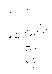
Fig. 9 is a view showing how the geometry of the stimulation control device
shown in Fig. 2 aids in its positioning during a surgical procedure.
The invention may be embodied in several forms without departing from its
spirit
or essential characteristics. The scope of the invention is defined in the
appended
claims, rather than in the specific description preceding them.
Detailed Description
This Specification discloses various systems and methods for safeguarding
against nerve, muscle, and tendon injury during surgical procedures or
confirming the
identity of nerves, muscles, and tendons and evaluating their function or the
function of
muscles enervated by those nerves. The systems and methods are particularly
well
suited for assisting surgeons in identification of nerves and muscles in order
to assure
nerve and muscle integrity during medical procedures using medical devices
such as
stimulation monitors, cutting, drilling, and screwing devices, pilot augers,
and fixation
devices. For this reason, the systems and methods will be described in the
context of
these medical devices.
The systems and methods desirably allow the application of a stimulation
signal
at sufficiently high levels for the purpose of stimulating and evaluating
nerve or muscle,
5
CA 02538199 2013-07-24
or both nerve and muscle integrity in numerous medical procedures, including,
but not
limited to, evaluating proximity to a targeted tissue region, evaluating
proximity to a
nerve or to identify nerve tissue, evaluating if a nerve is intact (i.e.,
following a traumatic
injury) to determine if a repair may be needed, evaluating muscle contraction
to
determine whether or not the muscle is innervated and/or whether the muscle is
intact
and/or whether the muscle is severed, and evaluating muscle and tendon length
and
function following a repair or tendon transfer prior to completing a surgical
procedure.
Still, it should be appreciated that the disclosed systems and methods are
applicable for use in a wide variety of medical procedures with a wide variety
of medical
devices. By way of non-limiting example, the various aspects of the invention
have
application in procedures requiring grasping medical devices and internal
viewing
devices as well.
I. Overview of the System
Fig. 1 shows an illustrative system 20 for safeguarding against nerve injury
during surgical procedures. In the illustrated embodiment, the system 20 is
configured
for monitoring and stimulating nerves and other structures throughout the
body. The
system 20 includes a stimulation control device 22 operating in conjunction
with one or
more of a family of stimulating medical devices including, for example, a
stimulation
monitor or probe 100, a cutting device 200, a drilling or screwing device 300,
a pilot
auger 400, and a fixation device 500.
In an exemplary embodiment, and as can be seen in Fig. 2, the stimulation
control
device 22 functions in the system 20 to generate an electrical stimulation
signal 29. The
stimulation signal 29 flows from the stimulation control device 22 through a
lead 24 to a
medical device (e.g., stimulation probe 100). The stimulation signal 29 then
flows
through a predefined insulated path 124 within the stimulation probe 100 and
to an
operative element, such as an electrically conductive surface, i.e., a coupled
electrode
110. The electrode is to be positioned on or near a region of a patient to be
stimulated.
In monopolar operation, a return electrode (or indifferent electrode) 38
provides an
electrical path from the body back to the control device 22. The stimulation
control
6
CA 02538199 2013-07-24
device 22 may operate in a monopolar or bipolar configuration, as will be
described
in gr eater detail later.
The stimulation signal 29 is adapted to the stimulation signal 29 is adapted
to
provide an indication. The indication may include a physical motor response
(e. g.,
twitching), and/or a visual or audio signal from the stimulation control
device 22, which
indicate to the surgeon close proximity of the electrode 110 to a nerve, or a
muscle, or a
nerve and a muscle. The stimulation control device may also indicate to the
surgeon
that the stimulation control device is operating properly and delivering a
stimulus
current.
II. Medical Devices
The configuration of the stimulating medical devices that form a part of the
system can vary in form and function. Various representative embodiments of
illustrative
medical devices will be described.
A. Stimulation Probe
Figs. 3A and 3B show various embodiments of a hand held stimulation monitor or
probe 100 for identification and testing of nerves and/or muscles during
surgical
procedures. The stimulation probe 100 is preferably a sterile, single use
instrument
intended for use during surgical procedures to identify nerves and muscles,
muscle
attachments, or to contract muscles to assess the quality of surgical
interventions or the
need for surgical interventions, or to evaluate the function of nerves already
identified
through visual means.
The stimulation probe is preferably sized small enough to be held and used by
one hand during surgical procedures. The angle of the stimulating tip
facilitates access
to deep as well as superficial structures without the need for a large
incision. A visual or
audio indicator 126 incorporated in the housing provides reliable feedback to
the
surgeon as to the request and delivery of stimulus current.
In one embodiment, the stimulation probe 100 includes a housing 112 that
carries an insulated lead 124. The insulated lead 124 connects to an electrode
110
positioned at the housing's proximal end 114. The lead 124 within the housing
112 is
7
CA 02538199 2013-07-24
insulated from the housing 112 using common insulating means (e.g., wire
insulation,
washers, gaskets, spacers, bushings, and the like). The electrode 110 is
positioned in
electrical conductive contact with at least one muscle, or at least one nerve,
or at least
one muscle and nerve.
In an additional embodiment, the stimulation probe 100 is mono-polar and is
equipped with a single electrode 110 at the housing proximal end 114.
Electrode 38
may be any of a variety of electrode types (e. g. , paddle, wire, or surface),
depending
on the surgical procedure being performed. In an alternative embodiment, the
stimulation device 100 itself may be bipolar, which precludes the use of the
return
electrode 38.
As shown in Figs. 3A and 3B, the stimulation probe 100 may accommodate
within the housing 112 the electrical circuitry of a stimulation control
device 22. In this
arrangement, the stimulation probe 100 may have two operational slide
controls, 155
and 160. Power switch 155 serves a dual purpose of turning the stimulation
signal to the
probe 100 on and off, and also can be stepped to control the stimulation
signal
amplitude selection within a predefined range (e.g., 0.5, 2.0, and 20 mA). The
pulse
control switch 160 allows for adjustment of the stimulation signal pulse width
from a
predefined range (e.g., 0 through 200 microseconds).
An operative element, such as a stimulus probe 110, exits the housing at the
proximal
end 114 to deliver stimulus current to the excitable tissue. The probe or
electrode 110
comprises a length and a diameter, and is desirably fully insulated with the
exception of
the most distal end, e. g. about 1.0 millimeters to about 10 millimeters, and
desirably
about 4 millimeters to about 6 millimeters, which is non-insulated and serves
as the
stimulating surface to allow the surgeon to deliver the stimulus current only
to the
intended tissue. The small area of the probe (the active electrode) ensures a
high
current density that will stimulate nearby excitable tissue. The probe
diameter may
range between about 0.5 millimeters to about 1.0 millimeters, and may be
desirably
about 0.75 millimeters.
In monopolar operation, a return electrode (or indifferent electrode) 130
provides
an electrical path from the body back to the control device 22. The return
electrode 130
may be placed on the surface of intact skin (e. g., surface electrodes as used
for ECG
8
CA 02538199 2013-07-24
monitoring during surgical procedures) or it might be needle-like 131 (see
Fig. 36), and
be placed in the surgical field or penetrate through intact skin. The
housing's distal end
118 can incorporate a connector or jack 120 which provides options for return
current
pathways, such as through a surface electrode 130 a or a needle electrode 131,
having
Additionally, the device 100 may desirably incorporate a visual or audio
indicator 126 for
the surgeon. This visual or audio indicator 126 allows the surgeon to confirm
that the
stimulator 100 is delivering stimulus current to the tissue it is contacting.
Through the
use of different tones, colors, different flash rates, etc., the indicator 126
(which can take
Audio feedback also makes possible the feature of assisting the surgeon with
monitoring nerve integrity during surgery. The insulated lead 124 connects to
an
electrode 110 that, in use, is positioned within the surgical field on a nerve
distal to the
surgical site. Stimulation of the nerve causes muscle contraction distally.
The
Alternatively, as Fig. 2 shows, the stimulation control device 22 may be
housed in
9
CA 02538199 2013-07-24
The present invention includes a method of locating a nerve in a patient that
comprises
the steps of providing a hand-held stimulation probe 100 as set forth above,
engaging a
patient with the first electrode 110 and the second electrode 130, moving the
power
switch 155 to an activation position causing a stimulation signal 29 to be
generated by
the stimulation control device 22 and transmitted to the first electrode 110,
through the
patient's body to the second electrode 130, and back to the stimulation
control device
22,
B. The Stimulation Control Device
As Fig. 4 shows, the stimulation control device 22 includes a circuit 32 that
generates electrical stimulation waveforms. A battery 34 internal to the
stimulator 100
desirably provides the power. The pulse generator 28 also desirably includes
an on-
board, programmable microprocessor 36, which carries embedded code. The code
expresses pre-programmed rules or algorithms for generating the desired
electrical
stimulation waveforms using the stimulus output circuit 46 and for operating
the visible
or audible indicator 126 based on the controls actuated by the surgeon.
In one form, the size and configuration of the stimulation control device 22
makes
for an inexpensive device, which is without manual internal circuit
adjustments. It is
likely that the stimulation control device 22 of this type will be fabricated
using
automated circuit board assembly equipment and methods.
C. Incorporation with Surgical Devices
A stimulation control device 22 as just described may be electrically coupled
through a lead, or embedded within various devices commonly used in surgical
procedures.
1. Cutting Device
In Figs. 5A and 5B, a device 200 is shown that incorporates all the features
disclosed in
the description of the stimulation probe 100, except the device 200 comprises
the
additional feature of providing an "energized" surgical device or tool. Fig.
5A shows the
CA 02538199 2013-07-24
tool to be a cutting device 200 (e.g., scalpel) removably coupled to a
stimulation control
device 22.
In the embodiment shown, the cutting device 200 includes a body 212 that
carries an insulated lead 224. The insulated lead 224 connects to an operative
element,
such as electrode 210, positioned at the body proximal end 214 and a plug-in
receptacle 219 at the body distal end 118. The lead 224 within the body 212 is
insulated
from the body 212 using common insulating means (e.g., wire insulation,
washers,
gaskets, spacers, bushings, and the like).
In this embodiment, the electrode 210 performs the cutting feature (e.g.,
knife or
razor). The electrode 210 performs the cutting feature in electrical
conductive contact
with at least one muscle, or at least one nerve, or at least one muscle and
nerve. The
cutting device 200 preferably includes a plug-in receptacle 216 for the
electrode 210,
allowing for use of a variety of cutting electrode shapes and types (e.g.,
knife, razor,
pointed, blunt, curved), depending on the specific surgical procedure being
performed.
In this configuration, the lead 224 electrically connects the electrode 210 to
the
stimulation control device 22 through plug-in receptacle 219 and lead 24.
In one embodiment, the cutting device 200 is mono-polar and is equipped with a
single electrode 210 at the body proximal end 214. In the mono-polar mode, the
stimulation control device 22 includes a return electrode 38 which functions
as a return
path for the stimulation signal. Electrode 38 may be any of a variety of
electrode types
(e.g., paddle, wire, or surface), depending on the surgical procedure being
performed.
The return electrode 38 may be attached to the stimulation device 22 by way of
a
connector or plug-in receptacle 39. In an alternative embodiment, the cutting
device 200
may be bipolar, which precludes the use of the return electrode 38.
In the embodiment shown in Fig. 53, the cutting device 200 accommodates
within the body 212 the electrical circuitry of the stimulation control device
22. In this
arrangement, the cutting device 200 may have at least two operational slide
controls,
255 and 260. Power switch 255 serves a dual purpose of turning the stimulation
signal
to the cutting device 200 on and off, and also is stepped to control the
stimulation signal
amplitude selection from a predefined range (e.g., 0.5,2.0, and 20 mA). The
pulse
11
CA 02538199 2013-07-24
control switch 260 allows for adjustment of the stimulation signal pulse width
from a
predefined range (e.g., 0 through 200 microseconds).
At the body distal end 218, a second plug-in receptacle 220 may be positioned
for receipt of a second lead 222. Lead 222 connects to electrode 230 which
functions as
Additionally, the device 200 may incorporate a 20 visual or audio indicator
for the
surgeon, as previously described.
The present invention includes a method of locating a nerve in a patient that
15 22.
2. Drilling Device
In Figs. 6A and 6B, a device 300 is shown that incorporates all the features
disclosed in
the 35 description of the stimulation probe 100, except the device 300
comprises the
In the embodiment shown, the drilling device 300 includes a body 312 that
carries an insulated lead 324. The insulated lead 324 connects to an operative
element,
In this embodiment, the electrode 310 performs the drilling feature. The
electrode
12
CA 02538199 2013-07-24
The drilling device 300 preferably includes a plug-in receptacle or chuck 316
for
the electrode 310, allowing for use of a variety of drilling and screwing
electrode shapes
and sizes (e.g., 1/4 and 3/8 inch drill bits, Phillips and flat slot screw
drivers), depending
on the specific surgical procedure being performed. In this configuration, the
lead 324
electrically connects the electrode 310 to the stimulation control device 22
through plug-
in receptacle 319 and lead 324.
In one embodiment, the drilling device 300 is mono-polar and is equipped with
a single
electrode 310 at the body proximal end 314. In the mono-polar mode, the
stimulation
control device 22 includes a return electrode 38 which functions as a return
path for the
stimulation signal. Electrode 38 may be any of a variety of electrode types
(e.g., paddle,
wire, or surface), depending on the surgical procedure being performed. The
return
electrode may be attached to the stimulation device 22 by way of a connector
or plug-in
receptacle 39. In an alternative embodiment, the drilling device 300 may be
bipolar,
which precludes the use of the return electrode 38.
In Fig. 6B, the drilling device 300 is shown to accommodate within the body
312
the electrical circuitry of the stimulation control device 22. The drilling
device 300 may
have at least two operational slide controls, 355 and 360. Power switch 355
serves a
dual purpose of turning the stimulation signal to the drilling device 300 on
and off, and
also is also stepped to control the stimulation signal amplitude selection
from a
predefined range (e.g., 0.5, 2.0, and 20 mA). The pulse control switch 360
allows for
adjustment of the stimulation signal pulse width from a predefined range
(e.g., 0 through
200 microseconds). At the body distal end 318, a second plug-in receptacle 320
may be
positioned for receipt of a second lead 322. Lead 322 connects to electrode
330 which
functions as a return path for the stimulation signal when the drilling device
300 is
operated in a mono-polar mode.
Additionally, the device 300 may incorporate a visual or audio indicator for
the
surgeon, as previously described.
The present invention includes a method of locating a nerve in a patient that
comprises the steps of providing a drilling device 300 as set forth above,
engaging a
patient with the first electrode 310 and the second electrode 330, moving the
power
switch 355 to an activation position causing a stimulation signal 29 to be
generated by
13
CA 02538199 2013-07-24
the stimulation control device 22 and transmitted to the first electrode 310,
through the
patient's body to the second electrode 330, and back to the stimulation
control device
22.
3. Pilot Auger
An additional aspect of the invention provides systems and methods for
controlling operation of a family of stimulating devices comprising a
stimulation control
device electrically coupled to a pilot auger for hard surface rotary probing.
This embodiment incorporates all the features disclosed in the description of
the
stimulation probe 100, except this embodiment comprises the additional feature
of
providing an "energized" surgical device or tool. Fig. 7A shows a pilot auger
device 400
removably coupled to a stimulation control device 22. In the embodiment shown,
the
pilot auger device 400 includes a body 412 that carries an insulated lead 424.
The
insulated lead 424 connects to an operative element, such as an electrode 410,
positioned at the body proximal end 414 and a plug-in receptacle 419 at the
body distal
end 418. The lead 424 within the body 412 is insulated from the body 412 using
common insulating means (e.g., wire insulation, washers, gaskets, spacers,
bushings,
and the like). In this embodiment, the electrode 410 performs the pilot
augering feature,
The electrode 410 performs the pilot augering feature in electrical conductive
contact
with a hard structure (e.g., bone). The pilot auger device 400 preferably
includes a plug-
in receptacle or chuck 416 for the electrode 410, allowing for use of a
variety of pilot
augering electrode shapes and sizes (0. g., 1/32, 1/16, and 1/8 inch),
depending on the
specific surgical procedure being performed. In this configuration, the lead
24
electrically connects the electrode 410 to the stimulation control device 22
through plug-
in receptacle 419 and lead 24.
In one embodiment, the pilot auger device 400 is mono-polar and is equipped
with a
single electrode 410 at the body proximal end 414. In the mono-polar mode, the
stimulation control device 22 includes a return electrode 38 which functions
as a return
path for the stimulation signal. Electrode 38 may be any of a variety of
electrode types
(e.g., paddle, wire, or surface), depending on the surgical procedure being
performed.
The return electrode 38 may be attached to the stimulation device 22 by way of
a
14
CA 02538199 2013-07-24
connector or plug-in receptacle 39. In an alternative embodiment, the pilot
auger device
400 may be bipolar, which precludes the use of the return electrode 38.
As Fig. 7B shows the pilot auger device 400 may accommodate within the body
412 the electrical circuitry of the stimulation control device 22. At the body
distal end
418, a second plug-in receptacle 420 may be positioned for receipt of a second
lead
422. Lead 422 connects to electrode 430 which functions as a return path for
the
stimulation signal when the pilot auger device 400 is operated in a mono-polar
mode.
The pilot auger device 400 includes a power switch 455. When moved to an
activation
position, a stimulation signal is generated by the stimulation control device
22.
Additionally, the device 400 may incorporate a visual or audio indicator for
the surgeon,
as previously described.
The present invention includes a method of locating a nerve in a patient that
comprises the steps of 25 providing a pilot auger device 400 as set forth
above,
engaging a patient with the first electrode 410 and the second electrode 430,
moving
the power switch 455 to an activation position causing a stimulation signal to
be
generated by the stimulation control device 22 and transmitted to the first
electrode 410,
through the patient's body to the second electrode 430, and back to the
stimulation
control device 22.
D. Incorporation with Fixation Devices
An additional aspect of the invention provides systems and methods for
controlling
operation of a family of stimulating devices comprising a stimulation control
device
electrically coupled to a fixation device or a wrench or screwdriver for
placing the
fixation device. A fixation device (e.g., orthopedic hardware, pedicle screws)
is
commonly used during spinal stabilization procedures (fusion), and internal
bone
fixation procedures.
This embodiment incorporates all the features disclosed in the description of
the
stimulation probe 100, except this embodiment comprises the additional feature
of
providing an "energized" fixation device or tool. Fig. 8A shows a fixation
device 500
removably coupled to a stimulation control device 22. In the embodiment shown,
the
fixation device 500 includes a rectangularly shaped body 512 that also serves
as an
CA 02538199 2013-07-24
operative element, such as electrode 510. The fixation device 500 may take on
an
unlimited number of shapes as necessary for the particular procedure taking
place.
Pedicle screws 535 may be used to secure the fixation device to the bony
structure. The
electrode 510 performs the fixation feature in electrical conductive contact
with a hard
In yet an additional alternative embodiment (see Fig. 8B), the fixation device
may
be a pedicle screw 535. The pedicle screw 535 is removably coupled to a
stimulation
In the mono-polar mode, the stimulation control device 22 includes a return
electrode 38 which functions as a return path for the stimulation signal.
Electrode 38
16
CA 02538199 2013-07-24
_
on the surgical procedure being performed. In an alternative embodiment, the
fixation
device 500 may be bipolar, which precludes the use of the return electrode 38.
The present invention includes a method of locating a nerve in a patient that
comprises
the steps of providing a fixation device 500 as set forth above, engaging a
patient with
the first electrode 510 and the second electrode 38, turning power on to the
stimulation
control device 22 through the I/O controls 26, causing a stimulation signal 29
to be
generated by the stimulation control device 22 and transmitted to the first
electrode 510,
through the patient's body to the second electrode 38, and back to the
stimulation
control device 22.
IV. Technical Features
The stimulation control device 22 can incorporate various technical features
to
enhance its universality.
A. Small Size
According to one desirable technical feature, the stimulation control device
can
be sized small enough to be held and used by one hand during surgical
procedures, or
to be installed within a stimulation probe or surgical device. The angle of
the stimulating
tip facilitates access to deep as well as superficial structures without the
need for a
large incision. Visual and/or audible indication incorporated in the housing
provides
reliable feedback to the surgeon as to the request and delivery of stimulus
current.
According to an alternative desirable technical feature, the stimulation
control
device 22 may also be sized small enough to be easily removably fastened to a
surgeon's arm or wrist during the surgical procedure, or positioned in close
proximity to
the surgical location (as shown in Fig. 9), to provide sufficient audio and
visual feedback
to the surgeon.
B. Power Source
According to one desirable technical feature, power is provided by a primary
battery for single use mounted inside the housing on or near the circuit board
22.
17
CA 02538199 2013-07-24
=
= =
C. The Microprocessor/Microcontroiler
According to one desirable technical feature, the stimulation control device
22
desirably uses a standard, commercially available micro-power, flash
programmable
microcontroller 36. The microcontroller 36 reads the controls operated by the
surgeon,
controls the timing of the stimulus pulses, and controls the feedback to the
user about
the status of the instrument (e.g., an LED or 1, 2, or more colors that can be
on, off, or
flashing).
The microcontroller operates at a low voltage and low power. The
microcontroller
send low voltage pulses to the stimulus output stage that converts these low
voltage
signals into the higher voltage, controlled voltage, or controlled current,
stimulus pulses
that are applied to the electrode circuit. This stimulus output stage usually
involves the
use of a series capacitor to prevent the presence of DC current flow in the
electrode
circuit in normal operation or in the event of an electronic component
failure.
The foregoing is considered as illustrative only of the principles of the
invention.
Furthermore, since numerous modifications and changes will readily occur to
those
skilled in the art, it is not desired to limit the invention to the exact
construction and
operation shown and described. While the preferred embodiment has been
described,
the details may be changed without departing from the invention, which is
defined by
the claims.
18