Note : Les descriptions sont présentées dans la langue officielle dans laquelle elles ont été soumises.
CA 02539850 2006-03-21
WO 2005/044074 PCT/US2004/029853
SURGICAL GEL SEAL
s Background of the Invention
Cross Reference to Related Applications
This application claims the benefit of non-provisional application Serial No.
101695,295 filed on October 28, 2003 and entitled "Surgical Gel Seal", which
is fully
to incorporated herein by reference.
Field of the Invention
This invention relates generally to medical and surgical devices and more
is specifically to access seals adapted for use in urological procedures.
Discussion of the Relevant Art
There are many procedures which involve the exploration, visualization and
2o manipulation of body conduits such as the vascular system, digestive tract,
and urinary
tract. Notwithstanding the wide application of the present invention, a more
detailed
description will be undertaken only with respect to a single field of use.
In the urinary tract it is well known that stones, which commonly form in the
kidneys and the bladder, often need to be surgically removed. This procedure
is
as typically undertaken using a ureteroscopec having a working channel that is
accessible
-1-
CA 02539850 2006-03-21
WO 2005/044074 PCT/US2004/029853
through an exit port. Initially, a guidewire is threaded through the urinary
tract, perhaps
with the assistance of a cystoscope. Once the guidewire is in place, the
ureteroscope
can be advanced over the guidewire which is back-loaded into the working
channel of
the ureteroscope. Once the ureterscope is in place, it is used to visualize
the interior of
s the kidney, for example.
This visualization is enhanced by irrigating the kidney through the
ureterscope.
This irrigation fluid which is typically introduced to a separate channel in
the
ureteroscope nevertheless tends to fill the working channel. Within the
working
channel, the irrigation fluid would flow retrograde through the exit port
contaminating the
to surgical site were it not for a urology seal placed over the exit port. In
the past, these
urology valves have typically been provided with an elastomeric septum or
duckbill
having characteristics for forming both a zero seal in the absence of an
instrument,
such as the guidewire and to form an instrument seal in the presence of the
instrument.
Both of these valves rely upon force more than compliance for their sealing
is characteristics. Accordingly, there is significant resistance associated
with the
introduction and removal of instruments through these urology valves. These
valves
also accommodate a very limited range of instrument sizes and tend to degrade
over a
short period of time.
CA 02539850 2006-03-21
WO 2005/044074 PCT/US2004/029853
SUMMARY OF THE INVENTION
In accordance with the present invention, an access valve is provided for use
in
establishing a zero seal or instrument seal across any body conduit. In the
urology
procedure previously discussed, the valve is of primary interest.
It is of particular advantage that the access valve of the present invention
is
provided with a gel material which provides the sealing characteristics for
the valve.
Within the valve, the gel functions with properties that are partly liquid and
partly solid.
The gel has solid properties to the extent that it can be provided with an
initial form, and
cohesion properties sufficient to maintain the gel in a single contiguous
piece. The gel
io has liquid properties to the extent that it can be pushed to flow in the
direction of least
resistance and is generally non-compressible. These and other features of the
gel are
disclosed in applicant's co-pending PCT application, serial number
PCTlUS01/29682
filed on September 21, 2001 and entitled Surgical Access Apparatus and Method
which
is incorporated herein in its entirety by reference.
is In order to accommodate the gel within the valve and otherwise provide
additional features and advantages for the valve, other structural
modifications can be
made. For example, the housing for the valve can be formed with both a distal
portion
and a proximal portion which define a gel cavity. (It should be noted that
throughout this
specification, the words "proximal" and "distal" are measured relative to the
surgeon not
2o the patient.) Lead-in tubes can be provided in both portions to facilitate
both forward
and retrograde loading of the valve. The lead-in tube on the proximal portion
of the
housing can extend through the housing wall into the gel cavity to contact and
-3-
CA 02539850 2006-03-21
WO 2005/044074 PCT/US2004/029853
compress the gel material during assembly. This will facilitate formation of a
circumferential seal between the gel and the housing and will also tend to
close an
instrument channel through the gel to facilitate formation of a zero seal.
Both zero
seals and instrument seals can be formed while leaving expansion space within
the gel
s cavity to accommodate displacement of the gel by an instrument.
Alternatively, expansion space can be controlled within the gel cavity in
order to pressurize the incompressible gel material and thereby produce a
variably
locking force on an instrument. Finger tabs can be provided on the distal and
proximal
portions to facilitate control of the variable pressure through various de-
tented positions
io of the tabs.
In one aspect, the invention includes a surgical valve having an axis that
extends between a proximal end and a distal end. The valve includes a housing
having
a proximal housing portion and a distal housing portion which cooperate to
define a gel
cavity. A seal material is disposed in the gel cavity and includes a gel
having non-
is compressible characteristics. A proximal guide tube which extends axially
proximally
from the proximal housing portion, facilitates insertion of a surgical
instrument into the
seal material. A distal guide tube which extends axially distally from the
distal housing
portion, facilitates retrograde insertion of the surgical instrument into the
seal material.
The proximal guide tube includes interior portions which extend distally of
the proximal
2o housing portions to contact the gel around an axial channel extending
through the gel.
In another aspect, a surgical valve includes a first housing portion defining
a gel cavity, and a seal material including a gel and having a node and an
axial
-4-
CA 02539850 2006-03-21
WO 2005/044074 PCT/US2004/029853
channel. A subassembly includes the seal material disposed in the gel cavity,
the seal
material being formed with a channel in an open state. A second housing
portion,
disposed in juxtaposition to the first housing portion, applies a force to the
seal material
in the subassembly, the force being of a magnitude sufficient to place the
channel in a
s closed state. This force has a magnitude which is also sufficient to create
a
circumferential seal between the seal material in the first housing portion.
The force is
created by contact between an axial guide tube of the second housing portion
which
extends into the gel cavity contacting the node and applying the force to the
seal
material.
io In a further aspect of the invention, a surgical valve is adapted to form a
seal around a surgical instrument extending through the valve. First and
second
housing portions define a gel cavity having a volume and being adapted to
receive a gel
having properties including flowability and incompressibility. The gel also
has
characteristics for creating a pressure on the instrument to form a seal with
the
is instrument. Means is provided to move the second housing portion relative
to the first
housing portion to increase the pressure of the incompressible gel on the
instrument
and to create a locking force tending to inhibit movement of the instrument
relative to
the valve. The moving means can include complimentary screw threads, disposed
on
the first and second housing portions, which facilitate axial movement to
reduce the
2o volume of the gel cavity and increase the pressure of the incompressible
gel on the
instrument.
-5-
CA 02539850 2006-03-21
WO 2005/044074 PCT/US2004/029853
In another aspect, the invention includes a method for manufacturing a
surgical valve including the step of providing a seal material in the form of
a gel having
non-compressible characteristics. A housing is provided, including a first
housing
portion and a second housing portion which define a gel cavity. The seal
material is
s mounted in the first housing portion in a loose-fit relationship, and the
second housing
portion is moved into a proximal relationship with the first housing portion.
During this
moving step a force is applied to the gel which causes the gel to flow into a
sealing
relationship with at least the first housing portion. This force is applied
through a guide
tube of the second housing portion which extends into the gel cavity. During
the
to mounting step, the first and second housing portions, as well as the seal
material, can
be mounted on a mandrel in order to facilitate axial alignment during the
moving step.
In still a further aspect, the invention includes a method for accessing a
kidney of a patient in a urological procedure. A guidewire is placed in the
patient, the
guidewire having a proximal end, and a distal end extending through a urethra,
a
is bladder, and into the kidney of the patient. The proximal end of the
guidewire is
inserted retrograde into a channel of an endoscope. The endoscope is then
moved
over the guidewire to access the kidney, leaving the proximal end of the
guidewire
extending from the channel of the endoscope. A urological valve is provided,
having a
proximal end and a distal end, and a seal material in the form of an
incompressible gel
zo disposed therebetween. The valve is loaded retrograde onto the proximal end
of the
guidewire to form a seal between the incompressible gel and the guidewire.
Mounting
-6-
CA 02539850 2006-03-21
WO 2005/044074 PCT/US2004/029853
the distal end of the valve to the endoscope seals the channel of the
endoscope around
the guidewire.
These and other features and advantages of the invention will become
more apparent with a discussion of preferred embodiments and reference to the
s associated drawings.
DESCRIPTION OF THE DRAWINGS
FIG. 1 is a schematic view of the left side of a urinary tract showing a
guidewire
extending from the urethra into a kidney;
io FIG. 2 is a schematic view of the urinary tract showing a ureteroscope
threaded
over the guidewire and a surgical valve of the present invention positioned to
extend
over the guidewire to close an exit port of a working channel;'
FIG. 3 is a schematic view showing the handle of the ureteroscope with the
guidewire removed and the surgical valve ready to receive various instruments;
is FIG. 4 is a side elevation cross sectional exploded view of one embodiment
of
the present invention;
FIG. 5 is a cross sectional assembled view of the embodiment illustrated in
FIG.
4;
FIG. 6 is a side elevation cross sectional exploded view of a further
embodiment
20 of the invention;
FIG. 7 is a cross sectional assembled view of the valve embodiment illustrated
in
Figure 6;
-7-
CA 02539850 2006-03-21
WO 2005/044074 PCT/US2004/029853
FIG. 8 is an axial cross-section view of an embodiment which provides control
of
the expansion cavity to produce a locking force on the instrument;
FIG. 9 is an axial cross-section view illustrating substantial elimination of
the
expansion cavity to increase the area and pressure of the instrument seal;
s FIG. 10 is an axial cross-section illustrating two tab pairs operable to
alternatively
increase and decrease the instrument locking pressure;
FIG. 11 ,is a front planned view illustrating compression of one of the tab
pairs to
increase the locking pressure;
FIG. 12 is a top plan view illustrating compression of the other tab pair to
reduce
io the locking pressure;
FIG. 13 is a top plan view taking along lines 13-13 of FIG. 8 and illustrating
one
embodiment of a detent mechanism;
FIG. 14 is an axial cross section view illustrating another embodiment of a
detent
mechanism; and
is FIG. 15 is a top plan view taken along lines 15-15 of FIG. 14.
DESCRIPTION OF PREFERRED EMBODIMENT
AND BEST MODE OF THE INVENTION
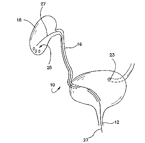
2o A urinary tract is illustrated in Figure 1 and designated by the reference
numeral 10. Only the right side of the urinary tract 10 is illustrated showing
the urethera
_g_
CA 02539850 2006-03-21
WO 2005/044074 PCT/US2004/029853
12, bladder 14, right ureter 16 and right kidney 18. The junction of the
ureter 18 and
bladder 16 is commonly referred to as a urethral orifice 21. A similar orifice
23 is
illustrated for the left urinary tract but will not be further discussed in
order to simplify
the disclosure.
s As with most body conduits, many procedures can be undertaken with
respect to the urinary tract 10, most of which require some degree of
exploration,
visualization and manipulation of the tract 10.
By way of example, a plurality of stones 25 are illustrated in the kidney 18
to facilitate discussion of a common stone removal' procedure. As illustrated
in Figure
io 1, a guidewire 27, having a floppy distal end 30 and a proximal end 32, is
initially
passed through the urinary tract 10 beginning at the urethera 12 and ending at
the
kidney 18. In order to facilitate placement of the floppy guidewire 27, a
cystoscope (not
shown) may be used primarily to facilitate introduction of the guidewire 27
into the
urethral orifice 21.
is Once the guidewire is in place, a ureterscope 34 can be introduced into
the urinary tract 10. The ureterscope 34 has an elongate shaft 36 extending
from a
handle 38, and typically includes fiberoptics
(not shown) to facilitate visualization, and a working channel 41 which
terminates
proximally at an exit port 43 on the handle 38.
ao With the ureterscope 34 thus positioned over the guidewire 32,
visualization of the interior regions of the kidney 18 can be undertaken. This
visualization is greatly facilitated by irrigating and aspirating the kidney
18 with saline
_g_
CA 02539850 2006-03-21
WO 2005/044074 PCT/US2004/029853
which is typically introduced through a separate channel in the ureteroscope
34. During
this procedure, the irrigation fluid will have a tendency to flow retrograde
through the
working channel and out the exit port where it can severely contaminate the
surgical
site. In order to prevent this contamination, a urology valve 50 at the
present invention
can be placed over the exit port to provide a zero seal in the absence of an
instrument
and an instrument seal in the presence of an instrument, such as the guidewire
27.
With the proximal end 32 of the guidewire 27 extending from the exit port
43, the urology valve 50 can nevertheless be positioned by introducing the
guidewire 27
retrograde into the valve 50, and attaching the valve 50 to the exit port 43
by means of
io a Luer fitting 52.
This retrograde insertion of the guidewire 27 into the valve 50 has
presented a particular problem in the past where elastomeric sealing materials
have
been used to form. a duckbill or septum valve. These valves are commonly
configured
to facilitate introduction of instruments in a forward direction and do not
easily
is accommodate retrograde insertion.
Once the urology valve 50 is in place over the exit port 43 as illustrated in
Figure 3, the guidewire 27 can be removed to vacate the working channel 41. A
zero
seal is immediately formed by the valve 50 in the absence of an instrument.
The introduction of various instruments to facilitate the engagement capture
and
zo withdrawal of the stones 25 (Figure 1 ) can now be introduced through the
urology valve
50 , through the exit port 43, and into the working channel 41. By way of
example, a
grasper 54 and a stone basket 56 are illustrated in Figure 3. These
instruments will
-10-
CA 02539850 2006-03-21
WO 2005/044074 PCT/US2004/029853
commonly have an operative device, such as grasper arms 58 at the distal end
of an
elongate flexible shaft 61. In an operative state, the operative device will
have a high
profile, but in an insertion state the operative device is retracted into the
shaft 61 to
provide a low-profile state. In the low-profile state, the shaft 61 of such
devices will
s typically have a diameter less than .070 inches, a common size for the
through-channel
of the valve 50.
In order to fulfill all of the functions desired for the urology valve 50, it
must accommodate retrograde insertion of a guidewire as well as forward
insertion of
the instruments, such as the grasper 54. A zero seal must be maintained across
the
to exit port 43 in the absence of an instrument while an instrument seal must
be
maintained across the exit port 43 in the presence of an instrument. These
functions
are accomplished in preferred embodiments of the invention which are
illustrated, for
example, in the exploded views of Figures 4 and Figures 6. In both of these
embodiments, a seal material 70 in the form of a gel 72 is provided to
facilitate
is formation of both a zero seal and an instrument seal. The gel 72 has
excellent
elongation and cohesive properties which facilitate both the manifestation and
operation
of the valve 50. It also has fluid properties which easily accommodate
insertion of
instruments in both a forward and retrograde direction.
The embodiment of Figure 4 includes a proximal housing portion 74 and a
2o distal housing portion 76 which in this case functions as a cap. A
preferred
embodiment of the urology valve 50 is illustrated in the cross sectional
exploded view of
Figure 4. This embodiment includes a distal housing portion 74, and a proximal
-11 -
CA 02539850 2006-03-21
WO 2005/044074 PCT/US2004/029853
housing portion 76 that combine to define a gel cavity 78. This cavity is
sized and
configured to~receive a plug 81 of seal material which in this case
advantageously
includes the gel 72. A male luer fitting 85 and associated screw cap 87 extend
distally
of the distal housing portion 76.
s The walls of the distal housing portion 76 include a pair of cylindrical
walls
90 and 92 with respective diameters which increase in the proximal direction.
The
proximal housing portion 74 in this embodiment includes a planar walls which
extends
radially, and together with a cylindrical wall 96, forms a cap for the distal
housing
portion 76. A hole 97 is formed centrally in the wall 94.
to The plug 81 including the gel 72 will typically be formed with a
cylindrical
configuration and an axial channel 101 which is open in an uncompressed state.
The
diameter of the plug 81 is preferably only slightly more than the inside
diameter of the
wall 90 to facilitate loading the plug into the gel cavity 78 of the distal
housing portion
76.
is In its assembled state, illustrated in the cross sectional view of Figure
5,
the plug 81 is disposed in the cavity 78 and the proximal housing portion 74
is brought
into a capping relationship with the cylindrical wall 92 of the distal housing
portion 76.
In this assembled state, the plug 81 is compressed within the seal cavity 78
in order to
form a circumferential seal with the cylindrical wall 90 of the distal housing
portion 76,
2o and a face seal with the wall 94 of the proximal housing portion 74. Since
the plug 81 is
composed of the gel 72, it is non-compressible so the applied force is
distributed
throughout the gel to produce the seals. The pressure within the gel 72 will
also tend to
-12-
CA 02539850 2006-03-21
WO 2005/044074 PCT/US2004/029853
create an annular bulge 103, which extends into an expansion portion 105 of
the cavity
78 defined by the cylindrical wall 92. This portion 105 of the cavity remains
to provide
free space into which the gel 72 can further expand as an instrument is
inserted
through the hole 97 and the channel 101. The pressure within the gel 72 also
is
s sufficient to close the channel 101 so that the plug 81 can function as a
zero seal in the
absence of an instrument.
In a further embodiment of the invention, illustrated in Figure 6, structure
elements similar to those previously described will be provided with the same
reference
numeral followed by the lower case letter "a". For example, in this embodiment
the
io valve 50a is illustrated in a cross sectional exploded view to include a
proximal housing
portion 74a, a plug 81 a of gel 72a, and a distal housing portion 76a with a
luer fitting
85a and an associated screw cap 87a. In this case, the gel cavity 78a is
defined
progressively proximally by a radially wall 107 having a shoulder 110, and a
coaxial
cylindrical wall 112.
is The plug 81 a is similar to the plug 81 in Figure 4 in that it includes a
cylindrical portion 114 and the axial channel 101a. In this case, however, a
spherical
node 116 is formed integral with and proximal of the cylindrical portion 114.
The
channel 101 a also extends through the node 116 and is provided with a lead-in
funnel
118 on the proximal end of the plug 81 a and a lead-in funnel 121 on the
distal end of
2o the plug 81 a.
The proximal housing portion 74a is similar to the housing portion 74 of
Figure 4 in that it includes a wall 94a that extends radially outwardly to a
coaxial
-13-
CA 02539850 2006-03-21
WO 2005/044074 PCT/US2004/029853
cylindrical wall 96a. In this case however, the proximal housing portion 74a
includes a
lead-in tube 123 which defines the hole 97a and extends through the wall 94a
with a
proximal portion 125 and a distal portion 127.
In order to facilitate assembly of the valve 58, the diameter of the
s cylindrical portion 114 of the plug 81 a is preferably provided with a
diameter only slightly
more than that of the wall 112 which defines the seal cavity 78a. With this
lesser
dimension, the plug 81 a is easily inserted axially into the seal cavity 78a
of the distal
housing portion 76a. At this point, the plug 81 a is only slightly compressed
so the
channel 101 a remains in an open state.
io As the proximal housing portion 74a is moved axially to cap the distal
housing portion 76a, the cylindrical wall 96a initially engages the
cylindrical wall 112
and may ultimately abuts the shoulder 110 of the distal housing portion 76a.
As this
axial movement progresses, the distal portion 127 of the lead-in tube 123
contacts the
node 116 around the funnel 118 of the plug 81a. Further axial movement of the
is proximal housing portion 74a applies a force to the node 116 which is
converted into a
pressure throughout the incompressible gel 52a. This pressure forces the
channel
101 a to a closed state and also moves the cylindrical portion 114 into a
sealing
relationship with the walls 107 and 112 of the distal housing portion 67a.
In the assembled state illustrated in Figure 7, it can be seen that
2o compression of the plug 81 a by the distal portion 127 of the lead-in tube
123 also forms
an annular bulge 103a which extends slightly into the open cavity portion
105a. This
-14-
CA 02539850 2006-03-21
WO 2005/044074 PCT/US2004/029853
cavity portion 105a accommodates further expansion of the plug 81 a when an
instrument is inserted through the channel 101 a.
In a further embodiment illustrated in Figure 8, elements of structure
similar to those previously discussed are designated with the same reference
numeral
s followed by the lower case letter "b". Thus, the embodiment of Figure 8
includes the
proximal housing portion 75b, with the radial wall 94b and associated
cylindrical wall
96b. This embodiment also includes the distal housing portion 76b with a
generally
radial wall 107b and shoulder 110b integral with the axial wall 112b, the plug
81 b of gel
72b is disposed in the gel cavity 78b.
io The guidewire 27b is also illustrated in Figure 8 after it has been loaded
into the valve 50b in the manner previously discussed. During this loading
step, the
guidewire 27b takes advantage of the low friction forces but the high ceiling
characteristics provided by the gel 72b. While these advantages can be
particularly
appreciated when the guidewire 27b is loaded into the valve 50b, it may be
desirable
is after the loading step, to increase the resistance between the gel 72b and
the guidewire
27b or otherwise lock the guidewire 27b in place. In the embodiment of Figure
8, this
locking features is facilitated by screw threads 130 which are disposed on the
cylindrical
wall 96b of the proximal housing portion 74b, and the axial wall 112b of the
distal
housing portion 76b. As the proximal housing portion 74b is rotated on its
axis relative
2o to the distal housing portion 76b, the radial wall 94b approaches the
radial wall 107b.
This has two effects. Initially, it tends to eliminate or at least reduce the
size of the
expansion cavity portion 105b. It also creates a force on the plug 81 b. Since
the plug
- 15-
CA 02539850 2006-03-21
WO 2005/044074 PCT/US2004/029853
18b is formed of the gel 72b with non-compressible characteristics, any force
applied to
the gel 72b results in a pressure within the plug 81 b. This pressure is
represented in
Figure 9 by arrows 129. With the gel cavity 78b defined by rigid walls 94b,
107b and
112b, the pressure within the gel 72b forces the gel 72b to move into any open
space.
s With the elimination of the expansion cavity portion 105b, as illustrated in
Figure 9, the
gel tends to expand into any openings which may exist between the guidewire
27b and
the valve 50b. In Figure 9, this movement is shown by a pair of bulges 132
which
notably increase the area of contact between the gel 72b and the guidewire
27b. With
an increase in the pressure within the gel 72b, a force shown by arrow 134 is
directed
to against the guidewire 27b tending to lock it in place relative to the valve
50b.
The screw threads 130 are of particular advantage in producing the
locking force shown by arrow 134, because they can provide a significant
mechanical
advantage. However, it will be appreciated by those skilled in the art that
there are
many other mechanisms which can be used to compress the cavity 78b between the
is walls 94b and 107b.
As illustrated in Figure 10, the treads 130 offer a further advantage in
facilitating one-handed control of the size of the cavity 78b. As illustrated
in the side
view of Figure 10, a pair of tabs 136 and 138 can be attached to the housing
portions
74b and 76b, respectively, in an upper region of the valve 50b. Similarly, a
pair of tabs
ao 141 and 143 can be attached to the housing portions 74b and 76b,
respectively, in a
lower region of the valve 50b. These tab pairs are further illustrated in the
front
elevation view of Figure 11 where the tabs 136 and 141 can be separated by an
-16-
CA 02539850 2006-03-21
WO 2005/044074 PCT/US2004/029853
angular distance such as 150 degrees on the proximal housing portion 74b.
Similarly,
the tabs 138 and 143 can be separated by an angle such as 150 degrees.
When the upper tab pair including the tabs 136 and 138 are pinched into
juxtaposition as illustrated in Figure 11, the proximal housing portion 74b
moves axially
s toward the distal housing portion 76b at a rate dependent on the pitch of
the threads
130. As this movement occurs by the simple pinching action on the two tabs 136
and
138, the size of the gel cavity 72b is decreased and the locking force
represented by
the arrow 134 is applied to the guidewire 27b. When it is desired to remove
the
guidewire 27b, this locking force can be removed by merely pinching the lower
tab pair
to including the tabs 141 and 143. This will place the tabs 141 and 143 in
juxtaposition as
illustrated in Figure 12. The proximal housing portion 74b rotates
counterclockwise
relative to the distal housing portion 76b thereby increasing the size of the
gel cavity
78b and appropriately decreasing the locking force represented by arrow 134.
With a structure similar to that in Figures 8-12, the magnitude of the
is locking force represented by the arrow 134 is dependent upon the degree of
separation
between the tabs 136 and 38. The maximum locking force is provided when the
tabs
136 and 138 are positioned as illustrated in Figure 11. The minimum force is
applied
when the tabs 136 and 138 are maximally separated as illustrated in Figure 12.
Between these two extremes, a detent mechanism 145 can be provided so that the
ao desired degree of locking force 134 can be achieved by movement of the tabs
136 and
138. This mechanism can include interfering projections 152 and 154 are formed
on
the cylindrical walls 96b and shoulder 110b, respectively, as illustrated in
Figure 13. As
-17-
CA 02539850 2006-03-21
WO 2005/044074 PCT/US2004/029853
the tabs 136 and 138 are closed, to move the proximal housing portion 74b
axially
toward the distal housing portion 76b, these interfering projections 152 and
154 will
detent to define a variable locking force represented by the arrow 134. The
detent
projections 152 and 154 in this case are disposed axially relative to each
other.
s In a further embodiment illustrated in Figure 14, a detent mechanism 145
is formed between projections 156 and 158 that are oriented in a radial
direction. The
projection 156 is disposed to extend radially on the underside of the
cylindrical wall 96b
while the projections 158 extend upwardly from the axial wall 112b. As in the
previous
embodiment, the detent mechanism 145 of Figures 14 and 15 will function to
define a
io variable locking force represented by the arrow 134.
In another aspect, a preferred method for manufacturing the valve 15 will
be discussed. Initially it will be noted that the various parts of the valve
50, including
the proximal housing portion 74, the distal housing portion 76, the plug 81,
and the
screw cap 87, each have an axial hole or channel in the assembly of the valve.
This
is enables the various parts to be threaded onto a mandrel 160 which can be
used to
guide the various structural elements axially to their assembled relationship.
In a preferred method of manufacture, the distal housing portion 76a is
initially placed on the mandrel 160. Then the plug 81 a can be threaded onto
the
mandrel 160 through the channel 101 a. The mandrel 160 holds these two
elements in
zo exact axial alignment so that the plug 81 a can merely be pushed along the
mandrel 160
and into the gel cavity 78a. At this point, the lead-in tube 125 of the
proximal housing
portion 74a can be threaded onto the mandrel 160. Again, the mandrel holds
this part
-18-
CA 02539850 2006-03-21
WO 2005/044074 PCT/US2004/029853
in axial alignment with the prior subassembly. With this alignment ensured,
the
proximal housing portion 74a can merely be pushed generally onto the distal
housing
portion 76a to compress the plug 81 a. The wall 96a of the proximal housing
portion
74a and the wall 112 of the distal housing portion 76a can then be joined by a
snap fit,
s glue or preferably a sonic weld. Finally, the luer cap 87a can be threaded
onto the
mandrel 160 and moved into a snap fit, rotatable relationship with the luer
fitting 85.
It will be understood that many other modifications can be made to the
various disclosed embodiments without departing from the spirit and scope of
the
concept. For example, various sizes of the surgical device are contemplated as
well as
io various types of constructions and materials. It will also be apparent that
many
modifications can be made to the configuration of parts as well as their
interaction. For
these reasons, the above description should not be construed as limiting the
invention,
but should be interpreted as merely exemplary of preferred embodiments. Those
skilled in the art will envision other modifications within the scope and
spirit of the
is present invention as defined by the following claims.
-19-