Note : Les descriptions sont présentées dans la langue officielle dans laquelle elles ont été soumises.
CA 02544775 2006-05-04
WO 2005/046795 PCT/US2004/037205
-1-
BRACHYTHERAPY APPARATUS AND METHOD FOR TREATING
A TARGET TISSUE THROUGH AN EXTERNAL SURFACE OF THE TISSUE
BACKGROUND OF THE INVENTION
The present invention relates generally to apparatus and methods for use in
treating proliferative tissue disorders, and more particularly to apparatus
and methods
for the treatment of such disorders in the body by the application of
radiation to a tissue
surface.
Malignant tumors are often treated by surgical resection of the tumor to
remove
as much of the tumor as possible. Infiltration of the tumor cells into normal
tissue
surrounding the tumor, however, can limit the therapeutic value of surgical
resection
because the infiltration can be difficult or impossible to treat surgically.
Radiation
therapy can be used to supplement surgical resection by targeting the residual
tumor
margin after resection, with the goal of reducing its size or stabilizing it.
Radiation
therapy can be administered through one of several methods, or a combination
of
methods, including external-beam radiation, stereotactic radiosurgery, and
permanent or
temporary interstitial brachytherapy. The term "brachytherapy," as used
herein, refers to
radiation therapy delivered by a spatially confined radiation source inserted
into the
body at or near a tumor or other proliferative tissue disease site. Owing to
the proximity
of the radiation source, brachytherapy offers the advantage of delivering a
more
localized dose to the target tissue region.
For example, brachytherapy is performed by implanting radiation sources
directly into the tissue to be treated. Brachytherapy is most appropriate
where 1)
malignant tumor regrowth occurs locally, within 2 or 3 cm of the original
boundary of
the primary tumor site; 2) radiation therapy is a proven treatment for
controlling the
growth of the malignant tumor; and 3) there is a radiation dose-response
relationship for
the malignant tumor, but the dose that can be given safely with conventional
external
beam radiotherapy is limited by the tolerance of normal tissue. In
brachytherapy,
radiation doses are highest in close proximity to the radiotherapeutic source,
providing a
high tumor dose while sparing surrounding normal tissue.
CA 02544775 2006-05-04
WO 2005/046795 - PCT/US2004/037205
-2-
One example of a brachytherapy device is disclosed in U.S. Patent No.
5,030,195
of Nardi, entitled "Radioactive Seed Patch for Prophylactic Therapy." Nardi
describes a
method and apparatus for treating tissue surrounding a surgically excised
tumor with
radioactive emissions to kill any cancer cells that may be present in the
tissue
surrounding the excised tumor. In order to implement the radioactive
emissions, Nardi
provides a low-energy, nonabsorbable radioactive seed patch made from a
plastic mesh
having Iodine-125 seeds threaded therein. The patch is put in place during the
time of
surgery after the resection of the tumor, and remains therein indefinitely.
While the apparatus described in Nardi provides some advantages, the patch is
limited to use with permanently implanted radioactive seeds, which in some
applications
can be less effective than other radiation sources. Moreover, Nardi does not
disclose
methods for tailoring the radiation dosage to avoid fully dosing sensitive
tissue or to
reduce the amount of radiation that escapes into the body.
Accordingly, there is still a need for a device that can be used to
effectively
deliver radiation from a solid and/or liquid radioactive source to target
tissue within the
human body.
SUMMARY OF THE INVENTION
The present invention generally provides a brachytherapy device for treating
target tissue surrounding a surgical extraction site. In one embodiment the
device
includes an insertion member having a proximal portion, a distal portion, and
at least
one lumen extending therethrough. A fluid retaining member is mated to the
distal
portion of the insertion member and has a first surface shaped to conform to a
predetermined external surface area of a tissue to be treated, and at least
one cavity
formed therein in fluid communication with the at least one lumen in the
insertion
member. A plurality of anchor members can be distributed about a periphery of
the first
surface to anchor the first surface to an external surface area of a tissue to
be treated.
In use, the brachytherapy device is adapted to receive a radiation source
through
the at least one lumen into a cavity in the fluid retaining member for
delivering radiation
to the tissue to be treated. Preferably, the fluid retaining member is shaped
to provide a
uniform radiation dosage throughout the first surface when the fluid retaining
member is
filled with a radioactive fluid. The fluid retaining member can also include a
second
CA 02544775 2006-05-04
WO 2005/046795 PCT/US2004/037205
-3-
surface opposed to the first surface, and a peripheral wall extending between
the first
and second surfaces to define the cavity therein. The peripheral wall
preferably has a
substantially uniform depth. In an exemplary embodiment, the fluid retaining
member
can be substantially disk-shaped or oval-shaped.
In another embodiment, the fluid retaining member can be movable between a
closed position in which the fluid retaining member is disposed adjacent the
insertion
member, and an open position in which the fluid retaining member extends
outward
from the insertion member. Preferably, the fluid retaining member is an
expandable
balloon member that is inflated in the open position and deflated in the
closed position.
The expandable balloon member can have a predetermined shape in the open
position
such that, when inflated, the expandable balloon member is effective to cover
a
predetermined area of tissue. While the predetermined shape can vary, in an
exemplary
embodiment the predetermined shape of the expandable balloon member is
substantially
disk-shaped or oval-shaped. In yet another embodiment, the fluid retaining
member can
be formed of a shape memory material, and can have a three-dimensional shape
in the
open position, and a substantially folded shape in the closed position. Again,
the fluid
retaining member is preferably substantially disk-shaped or oval-shaped in the
open
position.
In other aspects of the present invention, a brachytherapy device is provided
having an elongate catheter member with a proximal portion, a distal portion,
and at
least one lumen extending therethrough. A balloon member is disposed around
the
distal portion of the elongate catheter member and has a cavity formed therein
and in
fluid communication with at least one lumen in the elongate catheter. The
balloon
member includes a first tissue contacting surface shaped to conform to a
predetermined
external surface area of a tissue to be treated. The device further includes a
radiation
source in the form of a liquid disposed within the cavity of the balloon.
The catheter member can be mated with the balloon member at any position on
the balloon. Exemplary sites for connecting the catheter member and the
balloon
member include the balloon treatment surface, a surface opposite the treatment
surface
and the periphery of the balloon member.
CA 02544775 2006-05-04
WO 2005/046795 PCT/US2004/037205
-4-
In yet another embodiment of the present invention, a method for treating
tissue
surrounding a surgical extraction site is provided. The method includes the
step of
providing at least one brachytherapy apparatus for delivering radioactive
emissions. The
apparatus preferably includes a catheter member having proximal and distal
ends and at
least one lumen extending therethrough, and at least one fluid retaining
member
disposed proximate to the distal end of the catheter member. The fluid
retaining
member includes a cavity formed therein in communication with the at least one
lumen
in the catheter member, and a first surface shaped to conform to a
predetermined
external surface area of a tissue to be treated. The method further includes
the steps of
intraoperatively placing the at least one brachytherapy apparatus on an
external tissue
surface of a tissue to be treated, and introducing a controlled dose of a
radiation source
through the at least one lumen in the catheter to the fluid retaining member
to treat
tissue. Preferably, the radiation source is placed into the brachytherapy
apparatus after
placement of the apparatus on a tissue surface, and is removed from the
apparatus before
removal of the apparatus. The method can also include the step of attaching
the fluid
retaining member to the predetermined external surface area of a tissue to be
treated.
BRIEF DESCRIPTION OF THE DRAWINGS
The foregoing features, objects and advantages of the invention will become
apparent to those skilled in the art from the following detailed description
of a preferred
embodiment, especially when considered in conjunction with the accompanying
drawings in which:
FIG. 1 is perspective view illustration of one embodiment of a brachytherapy
device according to the present invention;
FIG. 2 is a side view illustration of the distal portion of the device shown
in FIG.
1;
FIG. 3 is an illustration of another embodiment of the brachytherapy device of
the present invention shown in a perspective view;
CA 02544775 2006-05-04
WO 2005/046795 PCT/US2004/037205
-5-
FIG. 4A is an illustration of another embodiment of the brachytherapy device
containing multiple cavities;
FIG. 4B is an illustration of another embodiment of the brachytherapy device
containging multiple cavities;
FIG. 5A is an illustration of a patient's lung having a lesion surgical
resected
therefrom;
FIG. 5B is an illustration of the lung shown in FIG. 5A having the resected
lesion
that is closed by sutures; and
FIG. SC is an illustration of the lung shown in FIG. 5B having a brachytherapy
device according to the present invention attached thereto.
DETAILED DESCRIPTION OF THE INVENTION
The present invention generally provides a radiotherapy device, and preferably
a
brachytherapy device, for delivering radiation to tissue andlor bone. While
the system
can be used for a variety of purposes, the system is preferably used to treat
tissue
proximate to a resected tumor site, and more particularly, to treat an
external surface of
tissue surrounding a closed tumor resection site, for example, in a patient's
lungs. FIGS.
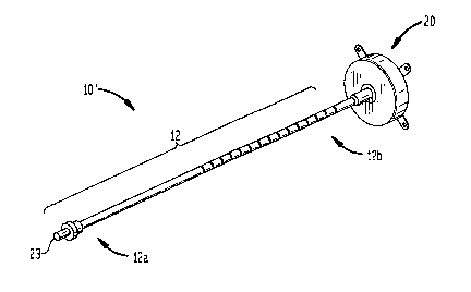
1 and 2 illustrate one embodiment of a brachytherapy device 10 which generally
includes an insertion member, e.g:, a catheter member 12, having a proximal
portion
12a, a distal portion 12b, and at least one lumen 12c extending therethrough.
An
expandable surface element, illustrated as fluid retaining member 20, can be
mated to
the distal portion 12b of the catheter member 12 and includes a cavity 21
formed therein
in fluid communication with at least one lumen 12c in the catheter member 12.
In use,
the fluid retaining member 20 is disposed on an external surface area of a
tissue to be
treated, and the cavity 21 is effective to receive a radiation source,
typically either in
liquid or solid form, for delivering radiation to the tissue to be treated.
CA 02544775 2006-05-04
WO 2005/046795 PCT/US2004/037205
-6-
The catheter member 12 can have a variety of configurations, but is preferably
a
semi-flexible or flexible elongate member having a proximal portion 12a, a
distal
portion 12b, and at least one lumen 12c formed therein that extends through
the
proximal and distal portions 12a, 12b. The lumen 12c can terminate at or near
a distal
port 14 formed in the distal portion 12b of the catheter 12. As shown in FIG.
1, the
proximal end 12a of the catheter 12 preferably includes a percutaneous port 23
for
providing access to the fluid retaining member 20 once the device 10 is
implanted in a
patient. While only one inner lumen 12c is illustrated in FIG. 2, a person
skilled in the
art will readily appreciate that the catheter member 12 can have one or more
inner
lumens, or that other means known in the art can be used to deliver fluid
and/or air to the
fluid retaining member 20.
The fluid retaining member 20 can have a variety of configurations, shapes,
and
sizes. However, the fluid retaining member preferable includes a cavity 21,
formed
therein in fluid communication with at least one lumen 12c formed in the
catheter
member 12. In one embodiment, the fluid retaining member 20 is configured and
adapted for receiving a fluid radiation source. Moreover, at least one outer
surface of
the fluid retaining member 20 is preferably a tissue-contacting surface that
is adapted to
be positioned on, and optionally conform to, a predetermined external surface
area of a
tissue to be treated. In addition, a person skilled in the art will appreciate
that the fluid
retaining member can include any number of cavities formed therein, and one or
more
surfaces can be adapted to be positioned on an external tissue surface to be
treated.
As shown in FIG. 2, the fluid retaining member 20 includes a first, tissue
contacting surface 24, a second, opposed surface 22, and a peripheral sidewall
26
extending therebetween. The first and second surfaces 24, 22 can each have
virtually
any size, but preferably the first surface 24 has a size that is sufficient to
cover a
predetermined external surface area of a tissue to be treated. The shape of
the first and
second surfaces 24, 22 can also vary, but the first surface 24 should be
adapted to be
positioned on an external tissue surface. In an exemplary embodiment, the
first surface
24 is substantially planar, but is preferably flexible or semi-flexible to
allow the surface
24 to conform to the tissue surface. The second surface 22 can also vary in
shape and
size, but preferably has a shape and size substantially the same as the first
surface 22 to
provide for uniform radiation dosage where a liquid radioisotope within fluid
retaining
CA 02544775 2006-05-04
WO 2005/046795 PCT/US2004/037205
member 20 provides the radiation dose. In one embodiment, uniform radiation
dosage
can be achieved by providing a peripheral sidewall 26 having a substantially
constant
width w extending between the first and second surfaces 24, 22. The uniform
width w of
the sidewall 26 facilitates the even distribution of radioactive fluid within
the fluid
retaining member 20, thereby providing a uniform radiation dosage, with the
exception
of edge effects, throughout the first surface 24 when the fluid retaining
member is filled
with a radioactive fluid.
While the embodiment of FIGS. 1 and 2 illustrates a substantially disk-shaped
fluid retaining member 20, which is advantageous because it can provide a
substantially
uniform radiation dosage, a person skilled in the art will appreciate that the
fluid
retaining member 20 can have a variety of configurations. By way of non-
limiting
example, the first and second opposed surfaces of the fluid retaining member
can be
square, oval, rectangular, etc. In one preferred embodiment, the first surface
is oval in
shape. The inventors have discovered that an oval shape can provide complete
coverage
of a target tissue region while being easier to manipulate, in particular
during surgery for
treatment of lung tumors, than other shapes. Moreover, while the size of the
fluid
retaining member 20I can be predetermined, the size can be selectable during
treatment
by inflating the fluid retaining member to a desired level. In certain
embodiments of the
invention, the surface area of the first surface can be between about 4 cm2 to
100 cm2.
The fluid retaining member 20 also preferably includes at least one anchor
member 28a-d formed thereon or mated thereto for attaching the fluid retaining
member
20 to the tissue surface. The anchor members 28a-d can be formed on or mated
to any
portion of the fluid retaining member 20, and can have a variety of
configurations.
Preferably, each anchor member 28a-d is disposed around'a periphery of the
first surface
22. In the illustrated embodiment, such a configuration results in anchor
members 28a-d
being placed around the peripheral wall 26 of, or adjacent to, the first
surface 24. A
variety of anchor members 28a-d can be used including, for example, eyelets,
hooks,
adhesives, and combinations thereof. FIG. 2 illustrates anchor members 28a-d
in the
form of eyelets. In use, each anchor member 28a-d can be sutured or otherwise
attached
to the tissue surface to securely implant the fluid retaining member 20 within
the patient.
CA 02544775 2006-05-04
WO 2005/046795 PCT/US2004/037205
-g-
In the embodiment illustrated in FIGS. 1 and 2, catheter 12 is attached to
fluid
retaining member 20 at second surface 22, and possibly also at first surface
24. In a
further embodiment, illustrated in FIG. 3, catheter 12 is attached to fluid
retaining
member 20 at one portion, and preferably at two opposed portions, of side wall
26. By
attaching catheter 12 along sidewall 26, a device 10 geometry is obtained that
is
preferred for insertion of the device to a desired treatment area in certain
treatment
procedures. For example, the geometry of the embodiment illustrated in FIG. 3
can be
preferred for use in treating lung tumors where device 10 must be moved
laterally
beneath a patient's ribs. While in the embodiment illustrated in FIG. 3 the
catheter 12 is
centrally connected to fluid retaining member 20, a person skilled in the art
will
recognize that other configurations are possible.
The fluid retaining member 20 can also include a variety of other features not
shown or described herein. In another embodiment, the fluid retaining member
20 can
be adapted to shield radiation-sensitive tissue. By way of non-limiting
example, all or a
portion of the second surface 22 and/or the peripheral wall 26 can be formed
from, or
coated with, a radio-opaque material that is effective to shield tissue
surrounding the
treatment site. In an exemplary embodiment, the entire fluid-retaining member
20,
except the tissue contacting surface 24, is radio-opaque. The coating (not
shown) can be
strategically positioned to shield radiation sensitive tissue, and/or to
provide an
asymmetric isodose curve as described in U.S. Patent No. 6,482,142, issued on
November 19, 2002, and entitled "Asymmetric Radiation Dosing Apparatus and
Method," which is incorporated herein by reference.
Radio-opaque materials suitable for coating include, for example, barium,
tungsten, bismuth, tantalum, and tin. As an alternative to coating portions of
the fluid
retaining member 20, a radiation-blocking or absorbing shield (not shown) can
be
positioned between within or around particular areas of the fluid retaining
member 20 to
produce a desired isodose curve. A person skilled in the art will appreciate
that other
configurations may be employed to achieve the desired isodose curves and/or
shielding
of radiation sensitive tissue.
In yet another embodiment, the fluid retaining member can be adapted to
provide
spacing between the radiation source and the tissue. By way of non-limiting
example, as
shown in FIGS. 4A and 4B, the fluid retaining member 20' can include a divider
CA 02544775 2006-05-04
WO 2005/046795 PCT/US2004/037205
-9-
disposed therein to separate the inner cavity into first and second cavities
21a', 21b'.
Each cavity 21a', 21b' is preferably in communication with a port 14a', 14b'
to allow a
radiation source to be delivered to the first cavity 21a', and fluid or air to
be delivered to
the second cavity 21b'. In use, the second cavity 21b' is effective to space
the radiation
source a distance apart from the tissue surface. By providing a uniform
spacing between
the radiation source (in the illustrated embodiment, a fluid radiation source
would be
preferred, such as Iotrex, available from Proxima Therapeutics, Inc. of
Alpharetta,
Georgia), a uniform prescribed radiation dose can penetrate into the target
tissue while
minimizing the chance of necrosis of healthy tissue in contact with or
proximate to fluid
retaining member 20' as described in U.S. Patent No. 6,413,204 to Winkler et
al, which
is incorporated herein by reference in its entirety. A person skilled in the
art will
appreciate that a variety of techniques can be used to provide spacing between
the
radiation source and.the tissue. By way of non limiting example, the fluid
retaining
member 20' can include a second balloon member disposed around the fluid
retaining
member for providing spacing, as described in U.S. Patent No. 6,413,204.
In addition to providing spacing, a second inner cavity can be used to deliver
a
therapeutic agent to the target tissue. For example, at least a portion of
retaining
member 20 can be defined by a porous material, and can be used to deliver a
therapeutic
agent from cavity 21b' to adjacent tissue. In one embodiment, tissue
contacting surface
24 is defined by a porous membrane through which a therapeutic agent can be
delivered.
U.S. Patent No. 6,083,148 to Williams discloses exemplary brachytherapy
methods and
apparatus using porous balloon walls, and is incorporated herein by reference
in its
entirety. The therapeutic agent is preferably a medically useful agent, for
example a
chemotherapy agent, an anti-neoplastic agent, an anti-angiogenesis agent, an
immunomodulator, a hormonal agent (including agonists and antagonists), an
immunotherapeutic agent, an antibiotic, or combinations thereof. Other
therapeutic
agents and useful porous materials are disclosed in U.S. Patent No. 6,200,257
to Winkler
which is incorporated herein by reference in its entirety.
In use, the fluid retaining member 20 is preferably movable between a closed,
unexpanded form, and an open, expanded form in which the fluid retaining
member 20
has a predetermined shape, as shown in FIGS. 1, 2 and 3. When positioned in
the
expanded form, the predetermined shape is preferably adapted to cover an
external
CA 02544775 2006-05-04
WO 2005/046795 PCT/US2004/037205
-10-
surface area of a target tissue to be dosed with radiation. Movement of the
fluid
retaining member 20 between the open and closed positions can be accomplished
by a
variety of techniques. While the fluid retaining member 20 is preferably
inflated using
liquid, air, or a radiation source, movement can optionally be accomplished
using an
actuating member (not shown), such as a wire, pulley assembly, lever, or
similar device,
effective to move the fluid retaining member to one of the open or closed
positions. A
person skilled in the art will readily appreciate that a variety of different
actuating
members can be used to position the fluid retaining member 20 within the
patient
adjacent the extraction site, and to move the fluid retaining member 20
between the open
and closed positions.
In an exemplary embodiment, the fluid retaining member 20 is an expandable
balloon member having a predetermined shape in the expanded position. It will
be
understood that the term "balloon" is intended to include distensible devices
which can
be, but need not be, constructed of an elastic material. In an alternative
embodiment, the
fluid retaining member 20 can be formed from a shape memory material, wherein
the
fluid retaining member 20 has a three-dimensional shape in the open position,
and a
substantially folded shape in the closed position.
With no limitation intended, the fluid retaining member 20 can be formed from
a
polymeric film wall, which may comprise a biocompatible, radiation resistant
polymer.
Suitable polymers include, for example, silastic rubbers, polyurethanes,
polyethylene,
polypropylene, polyester, and PVC. Still further, the fluid retaining member
20 can be
formed according to the balloon and/or expandable surface elements described
in U.S.
Patent No. 6,413,204, which is incorporated herein by reference in its
entirety.
The present invention also provides a method for treating a target tissue
through
an external surface area of the target tissue. FIGS. SA-SC illustrate one
embodiment of
a method for using a brachytherapy device to treat a resected lung tumor by
application
of a brachytherapy device to an external surface of the lung. In FIG. 5A, the
cancerous
tissue has been resected from the lung creating a resected cavity or "wedge"
50. After
wedge resection, the cavity 50 is sutured or stapled closed, as shown in FIG.
5B, which
illustrates sutures 52. A brachytherapy device according to the present
invention can
then be intra-operatively placed into the patient's' body and the fluid
retaining member
20 can be positioned over an external surface of the lung proximate to the
sutured
CA 02544775 2006-05-04
WO 2005/046795 PCT/US2004/037205
-11-
resection site 52 (a predetermined external surface area). If the fluid
retaining member
20 is introduced in a closed configuration, the fluid retaining member 20 can
be inflated
or otherwise moved to the open position, at which point it is preferably
attached to the
tissue using one or more of the anchor members 28a-d. Inflation can be
achieved with
air or other fluids, such as saline or a radiation absorbing fluid such as a
contrast media
used in angiography, or alternatively the radioactive fluid can be pre-loaded
into the
fluid retaining member prior to anchoring the fluid retaining member to the
tissue.
Preferably, however, the radioactive fluid is introduced into the fluid
retaining member
20 after it is anchored to the tissue surface. The radioactive source dwells
in the fluid
retaining member 20 until the prescribed dose of radiotherapy is delivered, or
the
radioactive source can be inserted for prescribed amounts of time on a daily
or other
scheduled basis until the prescribed dosage has been achieved. The radioactive
source is
then retrieved and the catheter 12 is removed.
The application of radiotherapy using a radioactive source can also be
performed
according to the many descriptions and examples provided in LT.S. Patent No.
6,413,204,
issued July 2, 2002, and entitled "Interstitial Brachytherapy Apparatus and
Method for
Treatment of Proliferative Tissue Diseases," which has been incorporated
herein by
reference above. The radiation treatment may end upon removal of the
brachytherapy
apparatus, or the brachytherapy may be supplemented by further doses of
radiation
supplied externally. By way of non-limiting example, the radioactive material
can be a
fluid made from any solution of radionuclide(s), e.g., a solution of I-125 or
I-131, or a
radioactive fluid can be produced using a slurry of a suitable fluid
containing small
particles of solid radionuclides, such as Au-198, Y-90. Moreover, the
radionuclide(s)
can be embodied in a gel. One radioactive material useful in the invention is
IotrexTM, a
sterile single use, non-pyrogenic solution containing sodium
3-(izsl)iodo-4-hydroxybenzenesulfonate (lasl-HBS), available from Proxima
Therapeutics, Inc. of Alpharetta, Georgia.
In addition, the radiation source employed in the brachytherapy device and
method of the invention can be solid or another non-liquid radiation source
such as an x-
ray emitter. Again, by way of non-limiting example, a solid radiation source
for use
with the invention could include radioactive micro spheres of the type
available from the
3M Company of St. Paul, Minnesota. This radioactive source can either be
preloaded
CA 02544775 2006-05-04
WO 2005/046795 PCT/US2004/037205
-12-
into the catheter at the time of manufacture or loaded into the device after
it has been
implanted. The solid radiation emitting material can be inserted through
catheter 12 on a
wire, for example, using an afterloader (not shown). Such a solid radioactive
core
configuration offers an advantage in that it allows a wider range of
radionuclides than if
one is limited to liquids. Such radionuclides that could be used with the
delivery device
of the present invention are currently generally available as brachytherapy
radiation
sources. In this embodiment, a solid spherical radiation source is surrounded
by fluid
retaining member 20, defining a spatial volume between the radiation source
and the
fluid retaining member that can be occupied by a radioactive ray absorbing
material,
such as air, water, or a contrast material.
In a further embodiment, the radiation source, instead of comprising a single
solid sphere, may comprise a plurality of radiation emitting particles 44
strategically
placed within the fluid retaining member 20 so as to radiate all directions,
or more
particularly toward the target tissue through the tissue contacting surface
24, with a
substantially equal intensity. This plurality of radiation emitting particles
can be
mounted on the distal end of a plurality of wires that are routed through the
catheter
body 12 and exit a plurality of ports formed through the wall of the catheter
body. This
arrangement allows the exact positioning of the individual radiation sources
so as to
generate a desired resultant profile.
A person having skilled in the art will appreciate that the foregoing is only
illustrative of the principles of the invention, and that various
modifications can be made
by those skilled in the art without departing from the scope and spirit of the
invention.
All references cited herein are expressly incorporated by reference in their
entirety.
What is claimed is: