Note : Les descriptions sont présentées dans la langue officielle dans laquelle elles ont été soumises.
CA 02545677 2006-05-11
WO 2005/046643 PCT/IL2004/001041
METIHOD FOR DRUG L0~4DING 1N LIPOSOMES
Field of the Invention
(ooo~~ The present invention relates to a method and the product obtained
thereby of loading therapeutic agents into preformed liposomes, in particular,
loading of protonatable compounds by an ammonium ion gradient having
glucuronate as the balancing anion.
Background of the Invention
(0002] Delivery of therapeutic agents via liposomal compositions has
drastically changed the drug pharmacokinetics and biodistribution of some
agents
(Martin, F.M., in MEDICAL APPLICATIONS OF LIPOSOMES, Lasic, D.D. and D.
Papahadjopaulos, eds., p. 635-88, Elsevier, Amsterdam (1998)). For example,
doxorubicin, which is known for its dose limiting cardiac-toxicity, shows no
apparent (clinical and functional) cardiac-toxicity in patients with solid
tumors
when administered entrapped in liposomes (Doxil~, ALZA Corporation, Mountain
View, CA; Uziely, B. et al., J. Clin. Onco., 13:1777-1785 (1995); Working,
P.K. et
al., J. Pharmaco. Exp. Ther., 289:1128-1133 (1999)). A cardiac biopsy study of
acquired immune deficiency syndrome (AIDS)-related Kaposi sarcoma (KS)
patients receiving large cumulative dosages of Doxil~ showed no tissue damage,
which suggests that the liposomal formulation may have a cardioprotective
effect
on doxorubicin (Berry, G. et al., Ann. Oncol., 9:71-76 (1998)). The lack of
cardiac-
toxicity is attributed, in part, to the long circulation half-life of
liposomes
(polyethylene glycol coated liposomes known as Stealth~, ALZA Corporation,
Mountain View, CA) and the stable drug retention, such that most of the
administered dose reaches tissues in liposome-encapsulated form with only
minimal amounts of drug (< 5%) leaking from liposomes during circulation and
distributed to tissue as free drug (Martin, F.M., supra, (1998); Gabizon, A.
et al.,
Cancer Res., 54:987-92 (1994)).
[OOO3~ It is known that long-circulating liposomes accumulate preferentially
(10
fold) in tissues with increased microvascular permeability, which includes
most
1
CA 02545677 2006-05-11
WO 2005/046643 PCT/IL2004/001041
tumors with active neoangiogenesis (Wu, N.Z., et al., Cancer Res., 53:3765-
3770
(1993); Yuan, F., etal., CancerRes., 54:3352-3356 (1994)). Long circulating
liposomes also accumulate in various healthy and susceptible tissues such as
the
skin (Gabizon, A. et al., Adv. Drug Deliv. Rev., 24:337-344 (1997)) and
probably
the mucosas. On prolonged exposure, accumulation of liposome-entrapped
doxorubicin in the skin may cause palmer-plantar erythrodysestheris (PPE, also
known as hand foot syndrome; Lyass et al., Cancer 89:1037-1047 (2000)). The
onset of PPE may be prevented by prolongation of dosing intervals, however,
dose andlor schedule modifications may reduce efficacy against certain tumors,
e.g., breast carcinoma (Lyass et al., supra, (2000); Ranson, M.R. et al., J.
Clin.
Oncol., 15:3185-3191 (1997)).
[0004] Current preclinical and clinical data on the long circulating, liposome-
entrapped doxorubicin (Doxil~) indicate that there is negligible release of
drug
from circulating liposomes (<5% of the injected dose). Once the liposomes have
extravasated into extracellular tissue fluids, little is known of the
processes
determining drug release. It is believed that gradual loss of the proton
gradient
retaining the drug, enzymatic breakdown of liposomal phospholipids by
phospholipases, andlor endocytosis by scavenger macrophages likely contribute
to drug release. Doxorubicin when entrapped in the commercially -available
liposomal Doxil~forms a salt with the divalent sulfate anion. The salt
precipitates
or gels due to its low solubility in the aqueous internal liposomal
compartment.
This gel formation stabilizes the entrapped doxorubicin in the lipid vesicle
and
decreases its rate of efflux.
[0005] Altering the holding capability of the anion on doxorubicin could have
a
major impact on the rate of drug release. For example, accelerating the rate
of
drug release from Doxil~ liposomes, without intertering with its long-
circulating,
tumor-homing properties, may be of significance for the following reasons: (1
) the
tumor-inhibitory activity may increase because of more time-intense exposure
of
tumors to the drug, and (2) the skin toxicity may decrease because this class
of
toxicity is mainly a function of prolonged exposure of skin tissues to the
drug.
[0006] Accordingly, a liposome composition that varied the release of an
entrapped compound, and in particular, doxorubicin, from liposomes is
desirable.
2
CA 02545677 2006-05-11
WO 2005/046643 PCT/IL2004/001041
A method for entrapping therapeutic compounds in preformed liposomes which
retains the advantages of the ammonium sulfate gradient, e.g., efficiency and
stability, yet enables the entrapped compound to be release at a higher rate
would
be desirable.
Summary of the Invention
[0007j In one aspect, the invention provides a liposomal composition
liposomes comprised of vesicle forming lipids and having an entrapped
ionizable
therapeutic agent~in association with a glucuronate anion. The therapeutic
agent
so loaded has a higher release rate than that loaded by an ammonium gradient
having sulfate as the balancing, or counter, anion.
[000s] In one embodiment, the vesicle forming lipids forming the liposomes are
phospholipids. In another embodiment, the liposomes further comprise between
about 1-20 mole percent of a vesicle-forming lipid derivatized with a
hydrophilic
polymer, such as polyethylene glycol.
[0009] In another embodiment, the vesicle-forming lipid is hydrogenated soy
phosphatidylcholine (HSPC) and said vesicle-forming lipid derivatized with a
hydrophilic polymer is distearoyl phosphatidylethanolamine (DSPE) derivatized
with polyethylene glycol. In yet another embodiment, the liposomes further
comprise cholesterol. An exemplary composition is HSPC, cholesterol, and
DSPE-PEG in a molar ratio of is 92.5:70:7.5.
[0010] In another embodiment, the therapeutic agent is an anthracycline
antibiotic. Exemplary anthracycline antibiotic include doxorubicin,
daunorubicin,
and epirubicin.
[0011] The composition described above is used, in another aspect, for
treating a patient. The composition is used, in another aspect, for treating a
neoplasm in a patient.
[0012] In another aspect, the invention includes an improved method of
preparing liposomes that have an entrapped ionizable therapeutic agent, where
the therapeutic agent is loaded into pre-formed liposomes against an ammonium
ion gradient with sulfate as a counterion. The improvement comprises loading
the
3
CA 02545677 2006-05-11
WO 2005/046643 PCT/IL2004/001041
ionizable therapeutic agent into liposomes by an ammonium ion gradient having
glucuronate as a counterion.
[0013] In this improved method, loading includes preparing a suspension of
liposomes, each fiposome having at least one internal aqueous compartment that
contains ammonium glucuronate at a first concentration, in one embodiment.
[0014] In another embodiment, the iMproved method includes preparing
liposomes suspended in an external bulk medium having a second concentration
of ammonium glucuronate, wherein the first concentration is higher than the
second concentration thereby establishing an ammonium ion concentration
gradient across lipid bilayers of the liposomes.
[0015] In another embodiment, the improved method includes adding an
amount of the therapeutic agent to the suspension of liposomes.
[0016] In another aspect, the invention includes a method of preparing
liposomes, comprising forming liposomes having an internal compartment and a
bifayer lipid membrane. The liposomes have a concentration gradient of
ammonium glucuronate across their bilayer lipid membranes. The, the liposomes
are contacted with an ionizable therapeutic agent to achieve transport of the
agent into the internal compartment.
[0017] In one embodiment, the method includes (i) preparing a suspension of
liposomes, each liposome in the suspension having at least one internal
aqueous
compartment that contains ammonium glucuronate at a first concentration, the
liposomes suspended in an external bulk medium comprising ammonium
glucuronate at the first concentration; (ii) reducing the first concentration
of
ammonium glucuronate in the external bulk medium to a lower, second
concentration of ammonium glucuronate, thereby establishing an ammonium ion .
concentration gradient across lipid bilayers of the liposomes.
[0018] In various embodiments, the step of reducing is achieved by dilution,
dialysis, diafiltration, or ion exchange.
[0019] In still another aspect, the invention includes a method for loading a
protonatable compound into pre-formed liposomes, comprising preparing a
suspension of liposomes having a greater concentration of ammonium
glucuronate inside the liposomes Than outside the liposomes thereby
establishing
4
CA 02545677 2006-05-11
WO 2005/046643 PCT/IL2004/001041
an ammonium ion concentration gradient from the inside to outside of the
liposomes. The gradient is capable of active transport of said protonatable
compound towards the inside of the liposomes. The method also includes adding
an amount of protonatable compound to the suspension, and allowing the
protonatable compound to transport into the liposomes to achieve a content of
said protonatable compound inside the liposomes fio be greater than that
outside
of the liposomes. '
[0020) In one embodiment, the method includes forming the liposomes in the
presence of an ammonium glucuronate solution having a first concentration; and
entrapping said ammonium glucuronate solution of said first concentration
inside
said liposomes; and reducing said first concentration of said ammonium
glucuronate solution outside of the liposomes to a second concentration which
is
less than that of said first concentration.
j0021~ The method of the invention has a high loading efficiency. In one
embodiment greater than 50% of the amount of protonatable compound added to
the suspension is transported to the inside of the liposomes. In another
embodiment approximately 90% of the amount of protonatable compound added
to the suspension is transported to the inside of the liposomes. In specific
embodiments, the loading efficiency for doxorubicin is greater than 90% and
the
doxorubicin to phospholipid ratio is in the range of about 100-150 pg/pmol.
[0022] These and other objects and features of the invention will be more
fully
appreciated when the following detailed description of the invention is read
in
conjunction with the accompanying~drawings.
Brief Description of the Drawincrs
[0023] Figs. 1A-1 E are growth inhibition curves plotting the growth rate, as
a
percent of untreated control cells, of mouse cell fines M109ST (Fig. 1A),
M109R
(Fig. 1 B) and of human cell lines C-26 (Fig. 1 C), KB (Fig. 1 D), and KB-V
(Fig. 1 E),
against doxorubicin concentration (in nM}, after treatment with free
doxorubicin
(circles), liposome-entrapped doxorubicin, where the doxorubicin was remotely
loaded into the liposomes against an ammonium sulfate gradient (triangles,
"lipo-
dox-AS") or against an ammonium glucuronate gradient (squares, "lipo-dox-AGn);
CA 02545677 2006-05-11
PFii''tetl ~~t~9~~OQ5t~~~~PAI1~D
~...::~_;~ ~ _.... _... .~_ __...~ ~ ;.,_~. ..
wo Zoasra:~~sa3 gcTnLZOOaramo-tz
[ot~2~4] ~ Fig. ~ ShClwS the in vitro leakage rate of doxorut~icin from
lipr~somes,
where the doxarubicin was remotely loaded into the iiposomes against an
ammonium sulfate gradient {triangles, niipa-dox-ASp) or against an ammonium
glucuranate gradient (squares, "tipo-dox-AG"j;
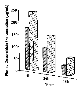
joD25] Fig. 3 is a bar graph showing doxorubicin concentration (Nglml_) in
mouse plasma at various times after fhe injection of liposomes containing
doxorubicin, where the doxarubicin was rert~otely Loaded into the liposomes
against an ammonium sulfate gradient {dotted bars) or against an ammonium
glucuranate gradient (hatched barsj;
~Da2&I Fig_ 4 is a plot of mean footpad thickness, in mm, in mice inoculated
v~rith M~to9-S cells as a function c~f days after treatment with saline (dosed
squares), free doxarubicin (circles), ar doxorubicin entrapped in llposomes,
where
the doxorvbicin was remotely loaded into the Iigasornes against an ammonium
sulfate gradient (triangles, °lipo-dox-AS°) or against an
ammoniurr~ glueuronate
gradient {open squares, °lipo-dox AG");
jD02?j Fig. 5 is a plat of mean fiootpad thickness, in mrn, in mice inoculated
with M9 D8R cells ~doxacublcin-resistant tumor rails) as a function of days
after
treatment with saline {closed squares), free daxarubicin {circles), or
doxorubicin
entrapped in liposames, where the doxorubicin was remotely Loaded into the
lipasomes against an ammonium sulfate gradient {friangfes, "lipa-dox-
AS°) or
against an ammonium glucuronate gradient {open squares, ~tipa-dox AG°y;
and
joia28~ Fg. 6 is a plot of number of surviving mice as a function of days
altar
lnoculatiorr with C-~26 tumor cells and treatment with free doxorubicin
{circles) or
with doxorubicin entrapped in lipasomes, where the doxonJbicin was remotely
loaded iota the liposomirs against an ammonium sulfate gradient ttriangles,
"lipo-
dax-AS'j yr against an ammaniurn glucuronate gradient (squares, alipo-dox-
AGa).
Detailed Description of the invention
~0029~ Tl~e ir~ventlon provides a liposomat compositar~ where an ionizable
therapeutic agent is entrapped in the internal tiposori~ai compartment{s) in
the
form of an ionic salt with monovalent glucuranate anions. As will be shown
below,
the entrapped therapeutic agent has a faster release rate from the liposomes
CA 02545677 2006-05-11
WO 2005/046643 PCT/IL2004/001041
compared to the release rate of the agent entrapped in the liposomes in the
form
of an ionic salt with divalent sulfate anions. The invention also provides a
remote
loading procedure for loading therapeutic agents into pre-formed liposomes
against an ammonium glucuronate gradient. The faster rate of release of the
therapeutic agent from the liposomes affords flexibility to adjust dosing
schedules
without compromising the biological efficacy of the therapeutic agents. The
method of the invention therefore provides a beneficial alternative to loading
by
ammonium sulfate.
[0030] Similar to the conventional ammonium sulfate gradient method, the
ammonium glucuronate remote loading method does not require the liposomes to
be prepared in acidic pH, nor to alkalinize the extraliposomal aqueous medium.
The approach also permits the loading of therapeutic agents in a broad
spectrum
of liposomes of various types, sizes, and compositions, including sterically-
stabilized liposomes, immunoliposomes, and sterically-stabilized
immunoliposomes. "Entrapped" as used herein refers to an agent entrapped
within the aqueous spaces of the liposomes or within the lipid bilayers.
[0039) The higher release rate is a result of using glucuronate as the
balancing
anion. While not wishing to be bound by theory, it is hypothesized that the
glucuronate ion, being monovalent and containing several hydroxyl functional
groups on its six-membered ring, is less effective compared to a sulfate ion
at
inducing aggregation and precipitation of the therapeutic agent after being
transported inside the liposomes. The inventors have observed that the
solubility
of doxorubicin is approximately 100-fold greater in a 250 mM ammonium
glucuronate (AG) solution than in a 250 mM ammonium sulfate (AS) solution. In
addition, doxorubicin precipitates at less than 2 mM concentration in the
presence
of sulfate ions, while a much higher concentration of doxorubicin is required
for
precipitation to occur in the presence of glucuronate ions. Accordingly, when
glucuronate is the balancing anion, more of the therapeutic agent is in a
soluble
form and therefore it is more available for release from the liposomes.
Further,
the permeability of glucuronate through the liposomal membranes is very low,
possibly due to its low pKa, its bulkiness and/or polarity, making if very
efficient for
maintaining the ammonium ion gradient for loading of the therapeutic agents.
7
CA 02545677 2006-05-11
WO 2005/046643 PCT/IL2004/001041
[0032 The method of the invention can be used to remotely load essentially
any therapeutic agent which is protonatable (can exist in a positively charged
state) when dissolved in an appropriate aqueous medium. Preferably, the agent
should be relatively lipophilic so that it will partition into the lipid
vesicle
membranes. Also, preferably, the therapeutic compound for loading is a weak
amphipathic compound, that is a compound having either weak basic or acidic
moieties. Examples of therapeutic agents which can be loaded into liposomes by
the method of the invention include, but are not limited to, doxorubicin,
mitomycin,
bleomycin, daunorubicin, streptozocin, vinblastine, vincristine,
mechlorethamine
hydrochloride, melphalan, cyclophosphamide, triethylenethiophosphoramide,
carmustine, lomustine, semustine, fluoruracil, hydroxyurea, thioguanine,
cytarabine, floxuridine, decarbazine, cisplatin, procarbazine, ciprofloxacin,
epirubicin, carcinomycin, N-acetyladriamycin, rubidazone, 5-ienidodaunomycin,
N-
acetyldaunomycine, all anthracyline drugs, daunoryline, propranolol,
pentamindine, dibucaine, tetracaine, procaine, chlorpromazine, pilocarpine,
physostigmine, neostigmine, chloroquine, amodiaquine, chloroguanide,
primaquine, mefloquine, quinine, pridinol, prodipine, benztropine mesylate,
trihexyphenidyl hydrochloride, propranolof, timolol, pindolol, quinacrine,
benadryl,
promethazine, dopamine, serotonin, epinephrine, codeine, meperidine,
methadone, morphine, atropine, decyclomine, methixene, propantheline,
imipramine, amitriptyline, doxepin, desipramine, quinidine, propranolol,
lidocaine,
chlorpromazine, promethazine, perphenazine, acridine orange, prostaglandins,
fluorescein, carboxyfluorescein, and other molecules similar to these above.
[0033] In addition to loading a single therapeutic agent, the method can be
used to load multiple therapeutic agents, either simultaneously or
sequentially.
Also, the iiposomes into which the protonatable therapeutic agents are loaded
can
themselves be pre-loaded with other pharmaceutical agents or drugs using
conventional encapsulation techniques (e.g., by incorporating the drug in the
buffer from which the liposomes are prepared). The method of the invention
therefore provides great flexibility in preparing liposome encapsulated "drug
cocktails" for use in therapies. Of course, if desired, one or more of the
protonatable drugs listed above can be pre-loaded and then the same or a
8
:I'i'l~'t~s~ g~2Df?b
CA 02545677 2006-05-11
WO 2QOS/Oa66:~3 PCTiIi.,200~i00i0:~1
dififerent drug can be added to the lipvsomes using the ammonium gtucuronate
gradient of the present invention.
[n~34~ The method is particularly suitable for loading weakly amphipathic
drugs such as doxorubicin. Doxorubicln loaded in Iiposomes having an externs!
surface coa~ng of hydmphitic polymer chains by an ammonium glucuronate
gradient treferred to herein as "fipo-dox-P'G's exhibits a faster release rate
than
doxorubicin loaded in tiposomes having an external surface coating of
hydrophilic
polymer chains by an ammonium sulfate gradient preferred to herein as "Iipo-
dox-
AS"; commercially known as Doxii~}, and has similar biological efficacy. ft is
contemplated that the faster release of drug when loaded info iiposornes
against
an ammonium gluouronate gradient lessens the duration of the drug in the blood
and lowers the opportunity for doxorubicin to accumulate in the skin to cause
palrnar-plantar etythrodysesthesia (PPS, also known as hand-foot syndrome, a
side effect observed with liposomahentrapped doxotubicin is administered.
[DD35~ In studies performed in support of the invention, tiposomes containing
entrapped doxvrubicin were prepared, where the doxorubicin was remotely loaded
into prefonned Iipasomes against an ammonium sulfate gradient or against an
ammonium glucuronate gradient. in Section I below, ifie Iiposome composition
and the remote loading procedure will be described. 'These tiposomes were
characterized in vitro to determine their cylotaxicity, cellular drag uptake,
and
plasma leakage rate, also described in Section I. In Sections tt and Itt, the
in viuo
plasma clearance rate and the therapeutic activity of the lipasome-entrapped
doxon3bicin are discussed.
l, ~;posome Components and Preoaratian
A Lic~osome Comnanent
[oo36a hiposomes suitable for.use in the compositions of fhe present invention
include those composed primarily of vesicle forming lipids. Vesicle forming
lipids,
exemplified by the phaspholipids, farm spontaneously info bilayer vesicles in
water at physiological pN and temperatures. The tipasomes can also include
other lipids, incorporated into the lipid bilayers, with the hydrophobic
moiety in
contact with the interior, hydrophobic region of the bitayer membrane, and the
s
CA 02545677 2006-05-11
WO 2005/046643 PCT/IL2004/001041
head group moiety oriented toward the exterior, polar surtace of the bilayer
membrane.
(0037 The vesicle-forming lipids are preferably ones having two hydrocarbon
chains, typically acyl chains, and a head group, either polar or nonpolar.
There
are a variety of diacyl synthetic vesicle forming lipids and naturally-
occurring
vesicle-forming lipids, such as phospholipids, diglycerides, dialiphatic
glycolipids,
single lipids such as sphingomyelin and glycosphingolipid, cholesterol and
derivatives thereof, alone or in combinations andlor with or without liposome
membrane rigidifying agents. As defined herein, "phospholipids" include
phosphatidylcholine (PC), phosphatidylethanolamine (PE), phosphatidic acid
(PA), phosphatidylinositol (PI), phosphatidylserine (PS), sphingomyelin,
plasmalogens, and phosphatidylcholine lipid derivatives where the two
hydrocarbon chains are typically between about 14-22 carbon atoms in length,
and have varying degrees of unsaturation. The above-described lipids and
phospholipids whose acyl chains have varying degrees of saturation can be
obtained commercially or prepared according to published methods.
[0038) Cationic lipids are also suitable for use in the liposomes of the
invention, where the cationic lipid can be included as a minor component of
the
lipid composition or as a major or sole component. Such cationic lipids
typically .
have a lipophilic moiety, such as a sterol, an acyl or diacyl chain, and where
the
lipid has an overall net positive charge. Preferably, the head group of the
lipid
carries the positive charge. Exemplary cationic lipids include 1,2-dioleyloxy-
3-
(trimethylarnino) propane (DOTAP); N-[I-(2,3,-ditetradecyloxy)propyl]-NN-
dimethyl-N-hydroxyethylanimonium bromide (DMRIE); N-[l-(2,3,-
dioleyloxy)propyl]-NN-dimethyl-N-hydroxy ethylammonium bromide (DORIE); N-
[1-(2,3-dioleyloxy) propyl]-N,N,N-trimethylammoniurn chloride (DOTMA); 30[N-
(N',N'-dirnethylaminoethane) carbarnoly] cholesterol (DC-Chol); and
dimethyldioctadecylammonium (DDAB).
[0039] The cationic vesicle forming lipid may also be a neutral lipid, such as
dioleoylphosphafidyl ethanolamine (DOPE) or an amphipathic lipid, such as a
phospholipid, derivatized with a cationic lipid, such as polylysine or other
CA 02545677 2006-05-11
WO 2005/046643 PCT/IL2004/001041
polyarnine lipids. For example, the neutral lipid (DOPE) can be derivatized
with
polylysine to form a cationic lipid.
[0040 The vesicle-forming lipid can be selected to achieve a specified degree
of fluidity or rigidify, to control the stability of the liposome in serum and
to control
the rate of release of the entrapped agent in the liposome. Liposomes having a
more rigid lipid bilayer, or a liquid crystalline bilayer, are achieved by
incorporation of a relatively rigid lipid, e.g., a lipid having a relatively
high phase
transition temperature, e.g., above room temperature, more preferably above
body
temperature and up to 80°C. Rigid, i.e., saturated, lipids contribute
to greater
membrane rigidity in the lipid bilayer. Other lipid components, such as
cholesterol, are also known to contribute to membrane rigidity in lipid
bilayer
structures.
[0041] Lipid fluidity is achieved by incorporation of a relatively fluid
lipid,
typically one having a lipid phase with a relatively low liquid to liquid-
crystalline
phase transition temperature, e.g., at or below room temperature, mare
preferably,
at or below body temperature.
j0042] The liposomes may optionally include a vesicle-forming lipid
derivatized
with a hydrophilic polymer, as has been described, for example in U.S. Patent
No.
5,013,556 and in WO 98107409, which are hereby incorporated by reference.
Incorporation of a hydrophilic polymer-lipid conjugate into the liposomal
bilayer
polymer provides a surface coating of hydrophilic polymer chains on both the
inner and outer surfaces of the liposome lipid bilayer membranes. The
outermost
surtace coating of hydrophilic polymer chains is effective to extend the blood
circulation lifetime in vivo relative to liposomes lacking the polymer chain
coating.
The inner coating of hydrophilic polymer chains extends into the aqueous
compartments in the liposomes, f.e., between the lipid bilayers and into the
central
core compartment, and is in contact with any entrapped agents. Vesicle-forming
lipids suitable for derivatization with a hydrophilic polymer include any of
those
lipids listed above, and, in particular phospholipids, such as distearoyl
phosphatidylethanolamine (DSPE).
[0043] Hydrophilic polymers suitable for derivatization with a vesicle-forming
lipid include polyvinylpyrrolidone, polyvinylmethylether, polymethyloxazoline,
11
CA 02545677 2006-05-11
WO 2005/046643 PCT/IL2004/001041
polyethyloxazoline, polyhydroxypropyloxazoline,
polyhydroxypropylmethacrylamide, polymethacrylainide, polydirnethylacrylamide,
polyhydroxypropyhnethacrylate, polyhydroxyethylacrylate,
hydroxymethylcellulose, hydroxyethylcellulose, polyethyleneglycol, and
polyaspartamide. The polymers may be employed as homopolymers or as block
or random copolymers.
[0044, A preferred hydrophilic polymer chain is polyethylenegiycol (PEG),
preferably as a PEG chain having a molecular weight between about 500 and
about 10,000 Daltons, more preferably between about 500 and about 5,000
Daltons, most preferably between about 1,000 to about 2,000 Daltons. Methoxy
or ethoxy-capped analogues of PEG are also preferred hydrophilic polymers,
commercially available in a variety of polymer sizes, e.g., 120-20,000
Daltons.
[0045] Preparation of vesicle forming lipids derivatized with hydrophilic
polymers has been described, for example in U.S. Patent No. 5,395,619.
Preparation of liposomes including such derivatized lipids has also been
described, where typically, between 1-20 mole percent of such a derivatized
lipid
is included in the liposome formulation. It will be appreciated that the
hydrophilic
polymer may be stably coupled to the lipid, or coupled through an unstable
linkage which allows the coated liposomes to shed the coating of polymer
chains
as they circulate in the bloodstream or in response to a stimulus, as has been
described, for example, in U.S. Patent No. 6,043,094, which is incorporated by
reference herein.
B. Liposome Preparation
[0046 Liposomal suspensions comprised of liposomes having an ion gradient
across the liposome bilayer (also referred to as a'transmembrane gradient')
for
use in remote loading can be prepared by a variety of techniques, such as
those
detailed in Szoka, F., Jr., et al., Ann Rev Biophys Bioeng 9:467, (1980).
Multilarnellar vesicles (MLVs) can be formed by simple lipid-film hydration
techniques. In this procedure, a mixture of liposome-forming lipids of the
type
described above is dissolved in a suitable organic solvent and the solvent is
later
evaporated off leaving behind a thin film. The film is then covered by an
aqueous
12
CA 02545677 2006-05-11
WO 200510~6ba3 PC'T/IL2QO~/OQ10.t1
medium, containing the solute species, e.g., ammonium glucuronate, which farms
fhe aqueous phase in the liposome.interior spaces and also the extraliposomal
suspending solution. The lipid film hydrates to form f~tLVs, typically with
sizes
between about 0.1 to 1 D microns.
~o0d.'T~ The 11I7td5 used in forming the lipvsomes of the present invention
are
preferably present in a molar ratio of about 70-100 mole percent vesicle-
forming
lipids, optionally 1 20 mole percent of a Lipid derivatized with a hydrophilic
polymer chain. One exemplary formulation includes 80-90 mole percent
phosphatidylethanolamine, '1 20 male percent of PEG-DSPE. Cholesterol may be
included in the formulation at between about 'I-50 mole percent. fn a
preferred
embodiment, the lipid components are hydrogenated soy phosphatidylcholine
(HSPG), cholesterol (Chol) and methoxy-capped polyethylene glycol derivatized
distearyl phosphatidylethanofamine {mPEG(~t~at3}~l?SPl=) in a molar ratio of
82.5:70:7.5_
~0048~ For preparation liposomes having an ammonium gfucuronate gradient,
the hydration medium contains ammonium glucurvnate. The concentration of
ammonium glucuronate would depend on fhe amount of therapeutic agent to be
loaded. Typically, the concentration is between '1110 to 30EJ mNf of ammonium
glucuranate. In ono preferred embadimertt, the hydration medium contains 250
mlitl ammonium glucuronate.
[OO4s~ The vesicles formed by the thin film method may be sized to achieve a
size distribution within a selected range, according to known methods.
Preferably,
the iiposames are uniformly sized to a size range between 0.04 to 0.2~ prn.
Small
unilamellar vesicles (SUVs), fypicaify in the 0.04 to O.OB pm range, can be
prepared by~post formation sonicatian or homogenization. I-tomogeneously sized
iiposames having sizes in a selected range between about 0.08 to i7.4 pm can
be
produced, e.8., by exirusivn through polycarbonate membranes or other defined
pore size membranes having selected uniform pore sizes ranging from 0.03 to
0.5
urn, typically, 0.05, 0.08, 0.'f, or 0.2 ~rrr~. The pure size of the membrane
corresponds roughly to the largest size of fiposomes produced by extrusion
through that membrane, particularly where the preparation is extruded two or
more times.through the same membrane. The sizing is preferably carried out in
'13
CA 02545677 2006-05-11
WO 2005/046643 PCT/IL2004/001041
the original lipid-hydrating buffer, so that the liposome interior spaces
retain this
medium throughout the initial liposome processing steps. Preparation of an
exemplary liposomal formulation is described in Example 1.
[0050 Generally, a therapeutic agent is loaded into the liposomes after
sizing.
A "remote" or "active" loading process results from exchange of the
therapeutic
agent in the external or bulk medium in which the liposomes are suspended with
an ammonium ion in internal liposomal compartment. The efficiency of loading
depends, at least in part, on an ammonium ion gradient, where the
concentration
of the ammonium ion inside the liposomes is higher than the concentratration
of
ammonium ion in the external, bulk suspension medium. The magnitude of this
gradient determines to a large extent the level of encapsulation; the larger
the
gradient, generally the higher the encapsulation.
[0061 An ammounium glucuronate gradient across the liposomal lipid bilayer,
where the ammonium ion concentration is higher on the inside of the liposomes
than in the external suspension medium (i.e., a higher inside/lawer outside
ammonium ion gradient) may be formed in a variety of ways, e.g., by (f)
controlled
dilution of the external medium, (ii) dialysis against the desired final
medium, (iii)
molecular-sieve chromatography, e.g., using Sephadex G-50, against the desired
medium, or (iv) high-speed centrifugation and resuspension of pelleted
liposomes
in the desired final medium. The final external medium selected will depend on
the mechanism of gradient formation and the external ion concentration
desired.
The gradient is measured as the ratio of ammonium glucuronate inside to that
outside of the liposomes. Generally, the gradient is in the range of 1000-10
inside/outside. Preferably, the gradient is in the range of 500-50.
[o052~ The concentration of ammonium glucuronate in an external medium that
also contains electrolytes may be measured as ammonia concentration at pH 13-
14 (Bolotin, E.M., et al., Journal of Liposome Research 4(i):455-479 (1994))
by an
ion analyzer, e.g., a Coming 250 pHlion analyzer (Corning Science Products,
Corning, NY) equipped with a Corning 476130 ammonia electrode and an
automatic temperature compensation (ATC) stainless steel probe. if the final
external medium lacks electrolytes the ammonium glucuronate gradient may be
confirmed by conductivity measurements using a conductivity meter, e.g., a
type
14
CA 02545677 2006-05-11
WO 2005/046643 PCT/IL2004/001041
CDM3 conductivity meter equipped with a CDC 304 immersion electrode with
manual temperature compensator type CDA 100 (Radiometer, Copenhagen,
Denmark).
[0053 In one approach, the ammonium ion gradient is created by controlled
dilution. This method gives a diluted liposome preparation. After suing, the
liposomal suspension has a selected first concentration of ammonium
glucuronate
inside the liposome and in the external bulk medium. The external bulk medium
is
diluted with a second medium containing no ammonium glucuronate. Exemplary
second medium include aqueous solutions containing electrolytes (sodium
chloride or potassium chloride) or aqueous solutions containing non
electrolytes
(glucose or sucrose). The internal and external: media are preferably selected
to
captain about the same osmolarity, e.g., by suitable adjustment of the
concentration of buffer, salt, or low molecular weight solute, such as
sucrose. A
preferred second medium is 15 mM HEPES buffer containing 5% dextrose at
approximately pH 7.
[0054 In another approach, a proton gradient across the lipid bilayer is
produced by dialysis in which the external bulk medium is exchanged for one
lacking ammonium ions, e.g., the same buffer but one in which ammonium
glucuronate is replaced by a salt such as NaCI or KCI, or by a sugar that
gives the
same osmolarity inside and outside of the liposomes. For small-scale
preparation, the gradient can be created by four consecutive dialysis
exchanges
against 25 volumes of the dialysis buffer. For large-scale preparation, the
gradient may be prepared by a three-step tangential flow dialysis, e.g., using
a
Minitan ultrafiltration system (Millipore Corp., Bedford, MA) equipped with
"300 K"
polysulfone membranes. The dialysis buffer contain electrolytes (e.g., sodium
chloride or potassium chloride) or non electrolytes (glucose or sucrose). In
one
preferred embodiment, the dialysis buffer is 15 mM HEPES containing 5%
dextrose at approximately pH 7. Using either of the dialysis approaches (large
or
small-scale) and under conditions in which the hydration medium was 60-250 mM
ammonium glucuronate, a gradient of 1,000 or higher can be obtained without
dilution of the liposomal dispersion.
CA 02545677 2006-05-11
WO 2005/046643 PCT/IL2004/001041
[0055] The ionization events that occur when loading an ionizable drug into
liposomes against an ammonium ion gradient are described in the art (see U.S.
Patent No. 5, 192,549). Briefly, after formation of the liposomes and
establishment of a gradient across the liposomal bilayers, ammonium ions
inside
the liposomes dissociate and are in equilibrium with ammonia and protons.
Ammonia gas is permeable in the lipid bilayer, with a permeability coefficient
of
around 1.3 x 10-' cm/second, and is able to permeate the liposomal bilayer.
The
efflux of ammonia shifts the equilibrium within the liposome towad production
of
protons which results in a [H+] gradient, with the intraliposomal
concentration
higher than that in the extraliposomal medium. Unprotonated drug crosses the
liposomal bilayer, becomes protonated inside the liposome, and is stabilized
by
the anions present in the internal aqueous compartment of the liposome.
Formation of a drug-glucuronate salt elevates the intraliposomal pH and
induces
formation of NH3 inside the liposmes. This cycle repeats repeated until
essentially
all the ammonium ions are effluxed from the liposomal internal compartment as
NH3. A therapeutic agent, e.g., doxorubicin, may be loaded into the liposomes
by
adding a solution of the agent to a suspension of liposomes having an ammonium
ion gradient across the liposomal membranes. The suspension is treated under
conditions effective to allow passage of the compound from the external medium
into the liposomes. Incubation conditions suitable for drug loading are those
which (i) allow diffusion of the compound, which is in an uncharged form, into
the
liposomes, and (ii) preferably lead to high drug loading concentration, e.g.,
5-500
mM drug encapsulated, more preferably between 20-300 mM, most preferably
between 50-200 mM.
(0o56~ The loading is preferably carried out at a temperature above the phase
transition temperature of the liposome lipids. Thus, for liposomes formed
predominantly of saturated phospholipids, the loading temperature may be as
high as 60°C or mare. The loading period is typically between 15-120
minutes,
depending on permeability of the drug to the liposome bilayer membrane,
temperature, and the relative concentrations of liposome lipid and drug. In
one
preferred embodiment, the loading is performed at 60°C and for 60
minutes.
16
CA 02545677 2006-05-11
WO 2005/046643 PCT/IL2004/001041
[0057] Thus, with proper selection of liposome concentration, external
concentration of added compound, and the ion gradient, essentially all of the
added compound may be loaded into the liposomes. For example, with an
ammonium ion gradient of approximately 1000, encapsulation of doxorubicin can
be greater than 90%. Knowing the calculated internal liposome volume, and the
maximum concentration of loaded drug, one can then select an amounf of drug in
the external medium which leads to substantially complete loading into the
liposomes.
[x058] If drug loading is not effective to substantially deplete the external
medium of free drug, the liposome suspension may be treated, following drug
loading, to remove non-encapsulated drug. Free drug can be removed, for
example, by ion exchange chromatography, molecular sieve chromatography,
dialysis, or centrifugation. In one embodiment, the non-entrapped drug is
removed using Dowex 50WX-4 (Dow Chemical, MI). For example, free
doxorubicin (but not liposomal doxorubicin) binds to a cation exchange resin
(Storm, G. et al., Biochim Biophys Acta, 818:343 (1985)).
(l. In vitro Characterization
A. In vitro Cytotoxicily
[0059] The in vitro cytotoxicity of free doxorubicin (free-DOX), of liposome-
entrapped doxorubicin loaded against an ammonium sulfate gradient (lipo-dox-
AS) or against an ammonium glucuronate gradient (lipo-dox-AG) were tested
against two mouse cell lines (M109-S and M109-R) and three human tumor cell
lines (C-26, KB, and KB-V). M109-R and KB-V cell lines are doxorubicin-
resistant
sublines of M109-S and KB, respectively. The cells were exposed continuously
to
the drug formulation for 72 hours following the experimental details described
by
Horowitz, et al., Biochimica ef Biophysics Acta, 1109:203-209 (1992) and also
in
Example 2.
[0060] Table 1 shows the doxorubicin concentration needed to inhibit 50% of
cell grow (1C50 values) for free doxorubicin (F-DOX), lipo-dox-AG, and lipo-
dox-
AS. Doxorubicin in free from is mare cytotoxic than either of the two
liposomal
doxorubicin formulations. Doxorubicin loaded into liposomes against an
17
CA 02545677 2006-05-11
WO 2005/046643 PCT/IL2004/001041
ammonium glucuronate gradient is more cytotoxic than when loaded into
liposomes against an ammonium sulfate gradient, suggesting that the drug is
more bioavailable from a glucuronate salt than from a sulfate salt.
Table 1: Inhibitory Concentration (1C50) Values
IC50 (pM)
Cell Line F-DOX Lipo-DOX-AS Lipo-DOX--AG
M 109-S 0.56 9.8 1.4
M109-R 2.00 >300 28.0
KB 0.04 7.6 1.4
KB-V 0.69 >300 21.0
C26 0.96 >200 64.0
..
[0061 That lipo-dox-AG is more cytotoxic than lipo-dox-AS is further
demonstrated by the inhibition curves shown in Figs. 1A-1 E, which show the
growth rate of the cells, as a percent of cells not treated with drug
(control),
against the amount of doxorubicin added to the growth medium. Figs. 1 A-1 E
are
inhibition curves for fhe mouse cell lines, M109-S (Fig. 1A), M109-R (Fig. 1
B) and
the human cell lines C-26 (Fig. 9 C), KB (Fig. 1 D), and KB-V (Fig. 1 E). The
doxorubicin concentration, in nM, of the difFerent formulations are
represented as
tree doxorubicin (circles), lipo-dox-AG (squares), and lipo-dox-AS
(triangles). All
the drug formulations at doxorubicin concentrations between 102 to 106 were
cytotoxic to each of the tumor cell lines tested. In all cases, with
variations in the
growth rate inhibition, lipo-dox-AG was more cytotoxic than lipo-dox-AS,
showing
that drug from the liposomal-ammonium glucuronate platform was more readily
bioavailable than drug from the liposomal-ammonium sulfate platform.
B. In vitro Drug Uatake by Tumor Cells
[0062] In vitro accumulation of doxorubicin in mouse tumor cells was studied
by
exposing KB, KB-V, and M109-R cells to free doxorubicin, lipo-dox-AS, or lipo-
18
CA 02545677 2006-05-11
WO 2005/046643 PCT/IL2004/001041
dox-AG for 1, 5, and 24 hours, as described in Example 2. Table 2 shows the
results of the study. There is a greater drug accumulation in cells treated
with
iipo-dox-AG than in those treated with lipo-dox-AS. This is consistent with
the in
vitro cytotoxicity results described above (Figs. 1A-1E), which showed that
lipo-
dox-AG was more cytotoxic than lipo-dox-AS.
Table 2: In vitro Uptake of Doxorubicin into Tumor Cells
I
Doxorubicin
Uptake
(ng DOX/106
cells)
Cell Line, ExposureFree dox Lipo-dox-AS Lipo-dox-AG
Time
KB, 1 hr 426 (33) 5.0 (0.4) 7.8 (0.6)
KB, 5 hr 937 (46) 8.8 (0.4) 15.0 (0.4)
KB, 24 hr 840 (15) 24.0 (1 ) 154.0 (9)
KB-V, 1 hr 311 (22) 5.4 (0.8) 9.6 (1.5)
KB-V, 5 hr 931 (21) 12.3 (1.2) 18.0 (0.8)
M 109-R, 24 80 (3) 7.0 (2) 25.0 (5)
C. In vitro Leakage in Plasma
[0063 To determine the in vitro leakage of liposome-encapsulated drug in
plasma, lipo-dox-AS and lipo-dox-AG were incubated in 90% human plasma at
37°C with continuous shaking in incubation flask containing Dowex
cation-
exchange resin beads. The resin beads bind released drug, whether free or
protein bound. At pre-scheduled intervals, samples were taken for acidified
alcohol extraction and fluorometric determination of the fraction of drug
remaining
associated with liposomes (i.e., not trapped by the resin beads). The results
are
shown in Fig. 2 and indicate that doxorubicin from lipo-dox-AG from the
liposome
faster than drug from lipo-dox-AS. The difference between the two preparations
begins to manifest after 24 hr of incubation. At end of incubation (96 hr),
lipo-dox-
AG has released about twice as much doxorubicin as lipo-dox-AS (~80% vs.
40%).
19
CA 02545677 2006-05-11
WO 2005/046643 PCT/IL2004/001041
Ili. in Vivo Characterization
A. Plasma Clearance
[0064] The pharmacokinetics of doxorubicin entrapped in liposomes by loading
against an ammonium glucoronate gradient were evaluated in 3-month-old
BALB/c female mice. As described in Example 3A, liposomes with entrapped
doxorubicin loaded against ammonium sulfate or ammonium glucuronate were
injected intravenously into the mice. Blood samples were taken at selected
intervals and analyzed for doxorubicin concentration. Fig. 3 shows the plasma
doxorubicin concentration for mice treated with lipo-dox-AG (cross hatched
bars)
or with lipo-dox-AS (dotted bars). The half life of doxorubicin when
administered
from a lipo-dox-AG platform is approximately 16 hours, while that of
doxorubicin
when administered from a lipo-dox-AS platform is approximately 24 hours. It is
also apparent that lipo-dox-AG is cleared faster than lipo-dox-AS. The lipo-
dox-
AG blooc concentrations were 25% lower at 4 hours post intravenous
administration, S3% lower at 24 hours, and almost 50% lower at 48 hours post
intravenous administration. Since the composition and size of the liposomes
were
identical, the rate of uptake by the reticuloendothelial system (RES.) should
be
similar. Accordingly, the faster clearance is probably the result of a faster
release
rate in vivo of doxorubicin from the lipo-dox-AG formulation, consistent with
the
the in vitro experiments.
B. In Vivo Theraipeutic Activity
[00S5] To determine whether the faster clearance of doxorubicin when
admininstered from liposomes containing a doxorubicin-glucuronate salt has an
impact on therapeutic efficacy, the liposomal formulations were administered
to
tumor-bearing mice.
[0066] As described in Example 3B, mice were inoculated with M109S tumor
cells (106 cells) and treated with a single dose of doxorubicin at 10 mg/kg of
either
free doxorubicin, lipo-dox-AS, or lipo-dox-AG post tumor inoculation. Fig. 4
shows
the mean (n=10) footpad thickness, in mm, against days post-doxorubicin .
treatment. Both liposomal preparations were more effective in suppressing
tumor
growth than the free drug (circles). There was a slight, but insignificant,
CA 02545677 2006-05-11
WO 2005/046643 PCT/IL2004/001041
improvement in efficacy when mice were treated with lipo-dox-AG (squares)
compared to lipo-dox-AS (triangles).
[0067] In another study, also described in Example 3B, mice were inoculated
with M109R cells (106 cells). Ten days after inoculation, the mice were
treated
with either free doxorubicin, lipo-dox-AS, or lipo-dox-AG at a dose of 8
mg/kg.
The same dose was administered again one week and three weeks later. Fig. 5
shows the mean (n=10) footpad thickness, in mm, as a function of days post
tumor
inoculation. Both liposomal preparations (triangles, open squares) were more
effective in inhibiting tumor growth than free drug (circles), despite the
progressive
tumor growth in all test groups, probably due to the resistant nature of this
tumor.
[0068] In another study, also described in Example 3B, mice were inoculated
with C-26 cells (106 cells) to induce a tumor and treated, five days after
tumor
inoculation, with either free doxorubicin, lipo-dox-AS, or lipo-dox-AG at a
doxorubicin dose of 10 mg/kg. Fig. 6 shows the number of surviving mice as a
function of time post tumor inoculation. Untreated (control) mice died quickly
with
a median survival of 13 days (not shown). Mice treated with free doxorubicin
(circles) showed a neglible increase in mean survival time (4 days more than
control, i.e., 17 days). Both liposomal preparations (squares, triangles) were
more
effective in extending survival time in tumor-bearing mice than was free
doxorubicin.
[0069] All the above models are carcinoma-type tumors. An additional model
tested (results not shown) was the J6456 lymphoma of BALB/c mice with an
experimental design similar to the C-26 model (intraperitoneal 1 O6 tumor
cells,
intravenous therapy with a dose of 10 mg/kg on day 5 post-tumor inoculation).
The liposomal formulations were more effective than free drug, with no
significant
differences between mouse survival time after treatment with lipo-dox-AS or
lipo-
dox-AG.
[0070] From the foregoing, it can be seen how various objects and features of
the invention are met. Liposomes having a drug entrapped in the form of a
glucuronate salt provide a higher release rate of drug than does a similar
liposome where the drug is entrapped in the form of a sulfate salt, without
significant effect on drug efficacy. Clinical data with liposome-entrapped
21
CA 02545677 2006-05-11
WO 2005/046643 PCT/IL2004/001041
doxorubicin (Doxil~) indicate that the incidence and severity of PPE decrease
with
a shortening of the circulation half-life of Doxil~, the faster release, and
shorter
circulation of doxorubicin in the form of lipo-dox-AG provides a good
alternative
for doxorubicin delivery. It will be appreciated that the findings specific to
doxorubicin extend to other drugs capable of remote loading against an
ammonium ion gradient, such as those recited herein.
IV. Examples
j0071~ The following examples further illustrate the invention described
herein
and are in no way intended to limit the scope of the invention.
E?~4MPLE 'I
Liposorne Preparation anti Loadin4
A. Liposome Preparation
[0072 Liposomes containing ammonium glucuronate in the aqueous
compartments were prepared as follows. The lipid component, hydrogenated soy
phosphatidylcholine (HSPC), cholesterol and methoxy-capped polyethylene glycol
derivatized distearyl phosphatidylethanolamine (mPEG(200)-DSPE) in a molar
ratio of 92.5:70:7.5, were dissolved in chloroform. The solvent was evaporated
using a rotary evaporator under reduced pressure leaving behind a dried lipid
thin
film. The dried lipid thin film was hydrated with a 250 mM aqueous ammonium
glucuronate buffer solution (pH 5.5), forming liposomes containing ammonium
glucuronate in the internal aqueous compartments and suspended in an
ammonium glucuronate external bulk medium . The liposomes were then sized by
extrusion through 0.5 pm pore size membranes.
[0073] Following extrusion, the external ammonium glucuronate buffer was
exchanged by dialysis against a dialysis buffer containing 5% dextrose and 15
mM Hepes at pH 7.
[0074] A comparative liposome formulation containing 250 mM ammonium
sulfate in the interior aqueous compartments was similarly prepared by using
250
arm ammonium sulfate as the hydration buffer. The batches obtained were
similar
22
CA 02545677 2006-05-11
WO 2005/046643 PCT/IL2004/001041
to the ammonium glucuronate preparations in vesicle size, drug-loading
efficiency,
and drug-to-phospholipid ratio.
B. Remote Loading
[0075 Doxorubicin was loaded into the liposomes containing ammonium
glucuronate (lipo-dox-AG) and into the liposomes containing ammonium sulfate
(lipo-dox-AS) by incubating the liposomes prepared as described in A. above
with
a solution of doxorubicin for 1 hour at 60°C. Encapsulation of
doxorubicin
proceded to >90% efficency. The final drug to phospholipid ratio was 100-150
pglpmol.
[0076 Free doxorubicin (i.e., doxorubicin not entrapped in a liposome) in the
external bulk medium was removed by chromatography on a Sephadex G-50
column eluted with degassed dextrose-Hepes buffer.
E~MPLE 2
In vitro Characterization
A. In vitro C tour toxicity
[0077 Free doxorubicin and liposomal formulations of doxorubicin, prepared
as described above in Example 1, were tested against five mouse and human
tumor cell lines (M109-S, M109-R, C 26, KB, KB-V).
[0078] Cells for each line were exposed continuously to drug for 72 hours.
Other experimental details were as described by Horowitz et al., Biochem
Biophys
Acta, 1109(2):203 (1992). Briefly, 5x10³ cells from exponentially growing
cultures
in 200 NL aliquots were plated onto 96-well flat-bottom microtiter plates.
Following 20 hours in culture, during which cells attached and resumed growth,
20
pL of the tested drug formulation (free doxorubicin, lipo-dox-AS, iipo-dox-AG)
were added to each well. For each 10-fold increase in drug concentration, six
drug concentration points were tested. Each test was performed in triplicate
wells
and in two parallel plates. The cells were treated continously for 72 hours.
The
cultures were fixed by the addition of 50 pL 2.5% glutaraldehyde to each well
for
minutes. The plates were washed three times with de-ionized wafer, once with
0.1 M borate buffer (pH 8.5) and then stained for 60 minutes with 100 NL
23
CA 02545677 2006-05-11
WO 2005/046643 PCT/IL2004/001041
methylene blue (1 % in 0.1 M buffer borate, pH 8.5) at room temperature. The
plates were rinsed in five baths of de-ionized water to remove non-cell bound
dye
and were then dried. The dye was extracted with 200 NL 0.1 M HCI for 60 min at
37°C and the optical density was determined using a microplate
spectrophotometer.
[0079 The growth rate was calculated by dividing the doubling times of drug-
treated cells with those of the control cells. The drug concentration which
caused
a 50% inhibition of the control growth rate (1C50) was calculated yb
interpolation
of the two closest values of the growth inhibition curve.
[0080] Table 1 shows the IC50 values for free doxorubicin, lipo-dox-AS, and
lipo-dox-AG for each of the cell lines, and the corresponding growth
inhibition
curves are shown in Figs. 1 A-1 E.
B. In vitro Drua Updake
[0081 Cellular accumulation of doxorubicin was assayed by a method similar
to that described in Chambers, S. K. et al., Cancer Res., 49:6275-6279 (1989).
Monolayers of KB, KB-V, and M109-R cells (exponentially growing cultures of
about 106 cells in 35-mm plates) were incubated with free doxorubicin, lipo-
dox-
AS, and lipo-dox-AG for 1, 5, and 24 hours. At the end of the incubation, the
cells
were rinsed three times with PBS and the drug was extracted from the cells
with 1
mL acidified isopropanol (0.075 M HC1 in 90% isopropanol), for 20 hours at
4°C.
Doxorubicin concentration was determined spectrofluorometrically using an
excitation wavelength of 470 nm and an emission wavelength of 590 nm. The
fluorescence intensity emitfied was translated into doxorubicin-equivalents
based
on a doxorubicin standard curve, after readings of untreated background cells
were subtracted.
[0082 The result of drug uptake by KB, KB-V, and M109-R cells after exposure
to free doxorubicin, lipo-dox-AS, and lipo-dox-AG for 1, 5, and 24 hr are
shown in
Table 2.
24
CA 02545677 2006-05-11
WO 2005/046643 PCT/IL2004/001041
C. In vitro Plasma Leakage.
Materials
[0083] Lipo-dox-AS and lipo-dox-AG were prepared as described in Example 1
at a concentration of >500 pg doxorubicinimL.
[0084] 2 mL of 50% Dowex~ cation exchage resin beads (Sigma, 50W-
hydrogen, 50% pre-cleaned in saline) were added to 15 mL plastic tissue
culture
round bottom tubes. The tubes were centrifuged for 10 min at 2,000 rpm (850g),
and the liquid was decanted. The liposomal preparations were diluted with
human
plasma to approximately 5 pg doxorubicinlmL in 90% human plasma. Duplicate
tubes for each liposomal preparation were prepared, and tubes containing the
liposomal preparations absent Dowex resin beads were prepared.
[0085] An acidic isopropanol solution was prepared from 10% 0.75N HCI in
90% isopropanol, volume/volume. The reagents were reagent grade chemicals
obtainable from Sigma.
[0086] All the materials used in the study were sterile, and all the
experiments
were performed in sterile conditions.
A- ssay
[0087 Dowex~ cation exchange resin beads bind doxorubicin in human plasma
whether the drug is free or protein bound. In this assay lipo-dox-AG and lipo-
dox-
AS were incubated in the tubes containing the resid beads and human plasma (as
described above) at 37°C with continuous shaking using a rotary shaker
to
prevent sedimentation of the resin beads. At preschedufed intervals, samples
were taken for acidified alcohol extraction and fluorometric defiermination of
the
fraction of drug remaining associated with liposomes (i.e., not trapped by the
resin
beads). The following stepwise protocol for the analysis was followed.
1. Add 30 mL of human plasma into a 50 mL-tube.
2. Add 2 mL of 50% sterile Dowex~ resin beads in saline to the
centrifugation tubes (15 mL, plastic round bottom) and centrifuge for
min 2,000 rpm (850g). Decant the supernatant fluid from the
centrifuge tubes.
CA 02545677 2006-05-11
WO 2005/046643 PCT/IL2004/001041
3. Using liposomal preparations prepared as described in Example 1,
add an amount of the liposome suspension to the 50 mL tubes
containing 30 mL human plasma to obtain a stock solution having a
final concentration of 5 pg/mL doxorubicin.
4. Add 9 mL of the 5 pglmL doxorubicin liposomal suspension stock
solution to each of the centrifuge tubes containing resin beads (Tube
Nos. A, B), and add 10 mL of the liposomal stock solution to an tube
absent any resin beads (Tube no. C). Mix.
5. Remove 1 mL aliquots from each tube (A, B, C) and centrifuge for 3
minutes at 14,000 rpm. Remove 200 pL from the supernatants for a
time zero reading, and freeze the samples at -20°C until analysis.
6. Incubate the tubes at 37°C with continuous shaking on a rotary
shaker that grips and rotates the tubes 360°C at slow motion, with
sufficient speed to prevent sedimentation of the resin beads.
7. Remove a 1 mL aliquot from each of the tubes at 1, 4, 24, 48, 72,
and 96 hours. Centrifuge each aliquot at 14,000 rpm for 3 minutes,
remove a 200 pL aliquot of the clear supernatant. Freeze the aliquot
at about -20°C until analysis.
8. For analysis of the samples, 1.8 mL of acidified isopropanol was
added to to the 200 pL samples to extract doxorubicin from the
liposomes. The samples were incubated overnight at 4°C, and then
centrifuged to remove the precipitate (2,000 rpm for 10 minutes).
The clear supernatants were examined in a spectrofluorimeter
equipped with high wavelength photomultiplier, excitation at 470 nm
and emission at 590 nrn. Doxorubicin concentration was determined
based on a standard calibration curve, where the concentration
obtained represented the amount of doxorubicin retained in the
liposomes.
[0088 The results are shown in Fig. 2.
26
CA 02545677 2006-05-11
WO 2005/046643 PCT/IL2004/001041
EXAMPLE 3
In vivo Characterization
A. In vivo Plasma Clearance Rate
[0089] Three month-old BALB/c female mice were injected intravenously with
mglkg of either lipo-dox-AS or with lipo-dox-AG, prepared as described in
Example 1. Blood samples were taken 4, 24 and 48 hours after injection for
analysis of plasma doxorubicin levels. The results are shown in Fig. 3.
B. In Vivo Therapeutic Activity.
[0090] Thirty mice were inoculated in the footpad with M109-S cells (106
cells).
Seven days later, when the footpad thickness increased from a normal value of
approximately 1.5 mm to an average of 2.0-2.5 mm, the mice were divided into
three groups of 10 each and the mice groups were injected intravenously with
either free doxorubicin, lipo-dox-AS, or lipo-dox-AG at a doxorubicin dose of
10
mg/kg. Thereafter, the footpad thickness was measured twice a week with
alipers
to follow tumor growth and effect of therapy. The results are shown in Fig. 4.
[0091] In a separate study, thirty mice were inoculated in the footpad with
the
doxorubicin-resistant tumor cell line M109R cells (106 cells). Ten days later,
when
the footpad thickness increased from a normal value of approximately 1.5 mm to
an average of 2.0-2.5 mm, the mice were divided into three groups for
intravenous
treatment with free doxorubicin, lipo-dox-AS, or fipo-dox-AG at a doxorubicin
dose
of 8 mglkg. Two additional injections were given at the same dose 1 week and 3
weeks later. The footpad thickness was measured twice a week with calipers and
the results are shown in Fig. 5.
[0092] In another study, mice were inoculated i.p. with C-26 cells (106
cells).
Five days later, the mice were separated into three groups of 10 mice each,
and
each group of mice was injected intravenously with either free doxorubicin,
lipo-
dox-AS, or lipo-dox-AG at a dose of 10 mg/kg. The survival of these mice was
followed and survival curves are shown in Fig. 6.
[0093] Although the invention has been described with respect to particular
embodiments, it will be apparent to those skilled in the art that various
changes
and modifications can be made without departing from the invention.
27