Note : Les descriptions sont présentées dans la langue officielle dans laquelle elles ont été soumises.
CA 02549567 2006-06-14
1
DESCRIPTION
ENDOSCOPE SYSTEM AND ENDOSCOPE
TECHNICAL FIELD
[0001] The present invention relates to an endoscope
system including an endoscope having an objective optical
system with a different viewing angle in an observation
window at a distal end of an insertion unit, in which an
installation position of a treatment instrument channel
opening varies according to the viewing angle of the
objective optical system.
BACKGROUND ART
[0002] In recent years in a medical field, the endoscope
device has been utilized to observe and operate organs
within a body cavity by employing the endoscope which has
at least a long insertion unit having the observation
window, an illumination window, and the treatment
instrument channel opening at its end part and ail operating
unit positioned at an proximal end of the insertion unit.
Also, this endoscope device has been utilized in observing
the inside of pipes in industrial fields.
[00031 The insertion unit of this endoscope device is
comprised of a distal end portion in which the observation
window, the illumination window, and the treatment
instrument channel opening are provided, a bendable part
adjacent to the proximal end of the distal end portion, and
a flexible soft tube unit connected to the proximal end of
the bendable part and a distal end of the operating unit.
Also, an image guide, a light guide, and a treatment
instrument channel are provided in the insertion unit. An
end of the image guide is arranged in the objective optical
CA 02549567 2006-06-14
2
system provided in the observation window. An end of the
light guide is arranged in the illumination optical system
provided in the illumination window. An end of the
treatment instrument channel is communicated to the
treatment instrument channel opening.
[0004] The operating unit of the endoscope device, has a
bending operation knob, an eye piece, a treatment
instrument insertion hole, and a universal cable. The
bending operation knob enables its bendable part to be
controlled as pulling a bending wire extended between the
bending operation knob and the bendable part of the
insertion unit. The eye piece enables an operator to view
the observed region image as an ocular optical system is
arranged at the proximal end of the image guide. The
treatment instrument insertion hole communicates with the
proximal end of the treatment instrument channel for a
treatment instrument to be inserted therein. The universal
cable has the built-in light guide to be connected to a
light source.
[0005] Also, there is another endoscope in which a solid
image element is provided at an image formation position of
the objective optical system of an observation window and a
signal cable sending and receiving a generated image signal
as driving the solid image element is provided instead of
the image guide.
[0006] Regarding the endoscope device of this type, the
bendable part of the insertion unit is operated to be bent
according to an internal shape of a tube to be inserted
therein, the reflected light is introduced from the
observed region illuminated with illumination light emitted
from the illumination window of the distal end portion, and
the operator observes the observed region image displayed
on the eye piece of the operating unit as being transmitted
CA 02549567 2006-06-14
3
by the image guide.
[0007] The viewing angle of the objective optical system
of the observation window provided at the distal end
portion of the insertion unit depends on observed region,
and for example the objective optical system with a wide
viewing angle is used to facilitate the observation of a
lesioned part such as a back side of large intestine bag
which is difficult to be observed. Also, a wide angle
endoscope device, enabling to achieve a wide range observed
region image, is proposed for example in Patent Document 1,
wherein when the bending angle of the bendable part is
limited according to the shape and size of the observed
region, for example, the viewing angle of the objective
optical system of the endoscope used to observe the
observed region, which has a wide space and may have a
large bending angle of the bendable part thereof, does not
need to be widened as much, while the viewing angle of the
objective optical system of the endoscope used to observe
the observed region, which has a relatively narrow space
and may have a small bending angle of the bendable part, is
set wide.
[0008] Patent Document 1: Japanese Patent Application
Laid-open No. H04-102432 Publication
DISCLOSURE OF INVENTION
PROBLEM TO BE SOLVED BY THE INVENTION
[0009] Conventionally, the wide angle endoscope device
with the wide angle objective optical system, as proposed
in Patent Document 1, arranged in the observation window
has been used so as to facilitate the observation of a
lesioned part which is difficult to be observed, for
example, the back side of the complex shape in the tube
hole to be observed, such as a back side of large intestine
CA 02549567 2006-06-14
4
bag.
[0010] On the other hand, while observing the position
subject to be observed (observed region) by the endoscope
device, the treatment instrument is projected from the
treatment instrument channel opening toward the observed
region to perform various operations such as organism
system incision sampling. In the process of performing
various organism system operations by this treatment
instrument, the operator recognizes a physical relationship
between the observed region and the treatment instrument
from the position of the treatment instrument displayed by
the observation figure obtained from the observation window
of the distal end portion of the insertion unit to be
observed at the eye piece or the observation image
displayed on the monitor (hereinafter both are referred to
as an observation image) and operates the treatment
instrument.
[0011] In the case that the physical relationship
between the actual observed region and the treatment
instrument are the same, the physical relationship between
the observed region and the treatment instrument in the
observing image for the endoscope device, which has the
objective optical system of a predetermined angle (e.g.,
120 to 150 degrees) at the observation window of the distal
end portion of the insertion unit and has a predetermined
viewing angle, is mutually different from the physical
relationship between the observed region and the treatment
instrument in the observing image for the endoscope, which
has the objective optical system of wider viewing angle
(e.g., 151 degrees or more) at the operation window of the
distal end part of the insertion unit than the viewing
angle of the endoscope having the predetermined viewing
angle (hereinafter referred to as a wide angle endoscope
CA 02549567 2006-06-14
device)
[0012] That is, for example, if distances, i.e., the
physical distances between the observation window and the
treatment instrument at the distal end part of the
5 insertion unit with respect to the endoscope device having
the predetermined viewing angle and the wide angle
endoscope device, are the same, the extent of projection of
the treatment instrument projecting from the respective
treatment instrument channel opening to reach the viewing
angle of the objective optical system of the observation
window with respect to the wide angle endoscope device is
smaller than the extent of the projection with respect to
the endoscope device having the predetermined viewing angle.
That is, the wide angle endoscope device, because of the
wide angle objective optical system of the observation
window, reaches the viewing angle of the wide angle
objective optical system while the amount of projection of
the treatment instrument projecting from the treatment
instrument channel is being small.
[0013] Therefore, when the operator operates the
treatment instrument with the observation image of the wide
angle endoscope, the treatment instrument appears in the
observation image at the position where the treatment
instrument slightly projects from the treatment instrument
channel opening. As such, the operator may misunderstand
that the treatment instrument projects to the same position
for the endoscope with the predetermined viewing angle.
Accordingly, there is a problem that the operator feels
uncomfortable because of the difference in the physical
relationship of the treatment instrument between the
endoscope device with the predetermined viewing angle and
the wide angle endoscope device.
[0014] Furthermore, generally, a fixed focus optical
CA 02549567 2006-06-14
28964-126
system is used in the objective optical system provided in
the observation window of the endoscope. Therefore,
especially, regarding the wide angle objective optical
system, surrounding of the viewing angle comparing to the
central part of the viewing angle slightly becomes out of
focus. Accordingly, the treatment instrument immediately
after being projected in the viewing angle of the wide angle
objective optical system of the wide angle endoscope device
temporary becomes unclear which possibly gives uncomfortable
feeling to the operator.
[0015] This invention is made in consideration of the
above-points, and it is an object of this invention to
provide an endoscope system capable of being operated with
the same feeling for the operator when using the endoscope
with the predetermined viewing angle and the wide angle
endoscope device.
MEANS FOR SOLVING PROBLEM
[0016] An endoscope system according to one aspect of the
present invention includes a first endoscope that includes a
first distal end portion, a first observation window
provided at the first distal end portion, a first objective
optical system optically coupled to the first observation
window and having a first viewing angle, and a first
treatment instrument channel opening positioned at a first
distance away from the first observation window; and a
second endoscope that includes a second distal end portion,
a second observation window provided at the second distal
end portion, a second objective optical system optically
coupled to the second observation window and having a second
viewing angle wider than the first viewing angle, and a
second treatment instrument channel opening positioned at a
second distance longer than the first distance.
6
CA 02549567 2006-06-14
28964-126
[0017] An endoscope system according to another aspect of
the present invention includes a first endoscope that
includes a first distal end portion, a first observation
window provided at the first distal end portion, a first
objective optical system optically coupled to the first
observation window and having a first viewing angle, and a
first treatment instrument channel opening into which a
first treatment instrument is inserted; and a second
endoscope that includes a second distal end portion, a
second observation window provided at the second distal end
portion, a second objective optical system optically coupled
to the second observation window and having a second viewing
angle different from the first viewing angle, and a second
treatment instrument channel opening into which a second
treatment instrument is inserted. Under a condition where
an amount of projection of the first treatment instrument
from the first treatment instrument channel opening is
substantially the same as an amount of projection of the
second treatment instrument from the second treatment
instrument channel opening, a first distance between the
first treatment instrument channel opening and the first
observation window and a second distance between the second
treatment instrument channel opening and the second
observation window are set so that the first treatment
instrument is in the first viewing angle and the second
treatment instrument is in the second viewing angle.
[0018] In the endoscope system, the second viewing angle
may be wider than the first viewing angle, and the second
distance may be longer than the first distance.
[0019] An endoscope used in a system according to still
another aspect of the present invention includes a first
distal end portion; a first objective optical system which
has at least a first viewing angle and is provided in the
7
CA 02549567 2006-06-14
28964-126
first distal end portion; and a first treatment instrument
channel opening into which a first treatment instrument is
inserted and which is provided in the first distal end
portion. A first distance between the first treatment
instrument channel opening and the first objective optical
system at the first distal end portion is longer than a
second distance between a second treatment instrument
channel opening provided in a second distal end portion and
a second objective optical system having a second viewing
angle narrower than the first viewing angle, the second
distal end portion, the second objective optical system, and
the second treatment instrument channel opening constituting
another endoscope used in the system.
[0020] An endoscope according to still another aspect of
the present invention includes a distal end portion; an
objective optical system having at least a predetermined
viewing angle and provided in the distal end portion; and a
treatment instrument channel opening into which a treatment
instrument is inserted and is disposed at a distance,
determined based on the viewing angle, away from the
objective optical system.
[0021] In the endoscope, the distance may be determined
through the objective optical system when an amount of
projection of the treatment instrument from the treatment
instrument channel opening reaches a reference value, and be
set so that an image of an end of the treatment instrument
appears in a reference area in an observation image of an
area determined by the viewing angle.
[0022] In the endoscope, the reference area may be a
peripheral region of the observation image.
[0023] In the endoscope, the objective optical system may
8
CA 02549567 2006-06-14
28964-126
have an viewing angle of 150 degrees or more, and the
distance may be set so that a part in a space area of a
field of view of the objective optical system determined by
the viewing angle is set to be constantly farther than a
near point of the objective optical system, with the
treatment instrument inserted into the treatment instrument
channel opening.
[00241 According to the endoscope system of the present
invention, in the endoscope with the predetermined viewing
angle and the endoscope with wider viewing angle than the
endoscope with the predetermined viewing angle, if the
actual physical relationship between the observed region and
the treatment instrument are mutually identical, the
physical relationship between the observed region and the
treatment instrument in the observation image respectively
can become substantially identical by the endoscope with the
predetermined viewing angle.
EFFECT OF THE INVENTION
[00251 According to the endoscope system of the present
invention, utilizing the endoscopes with a predetermined and
wide viewing angle, from the treatment instrument in the
operation image observed respectively, the operator can
recognize the actual physical relationship between the
observed region and the treatment instrument with
9
CA 02549567 2010-07-30
28964-126
substantially identical feeling, thereby preventing the
operator from feeling uncomfortable.
According to one aspect of the present invention,
there is provided an endoscope system, comprising: a first
endoscope that includes a first distal end portion, a first
observation window provided at the first distal end portion,
a first objective optical system optically coupled to the
first observation window and having a first viewing angle,
and a first treatment instrument channel opening positioned
at a first distance away from the first observation window;
and a second endoscope that includes a second distal end
portion, a second observation window provided at the second
distal end portion, a second objective optical system
optically coupled to the second observation window and
having a second viewing angle wider than the first viewing
angle, and a second treatment instrument channel opening
positioned at a second distance longer than the first
distance, wherein a first treatment instrument inserted into
the first treatment instrument channel opening projects in a
range of the first viewing angle to be observed from the
first observation window and a second treatment instrument
inserted into the second treatment instrument channel
opening projects in a range of the second viewing angle to
be observed from the second observation window, and an
amount of projection of the first treatment instrument from
the first treatment instrument channel opening is the same
as an amount of projection of the second treatment
instrument from the second treatment instrument channel
opening.
CA 02549567 2009-07-03
28964-126
inserted, wherein under a condition where an amount of
projection of the first treatment instrument from the first
treatment instrument channel opening is the same as an
amount of projection of the second treatment instrument from
the second treatment instrument channel opening, a first
distance between the first treatment instrument channel
opening and the first observation window and a second
distance between the second treatment instrument channel
opening and the second observation window are set so that
the first treatment instrument is in the first viewing angle
and the second treatment instrument is in the second viewing
angle.
10a
CA 02549567 2006-06-14
11
32 Second electronic endoscope
33 First objective optical system
37 Second objective optical system
BEST MODE(S) FOR CARRYING OUT THE INVENTION
[0028] Exemplary embodiments of an endoscope system
according to the present invention will be explained with
reference to FIGS. 1 to 5. FIG. 1 is an explanatory view
illustrating the relationship between the observation
window and the treatment instrument channel opening
provided in the insertion unit of the endoscope employed in
the endoscope system according to the present invention;
FIG. 2 is a block diagram showing general structure of the
endoscope device employed in the endoscope system according
to the present invention; FIG. 3 is a front view
illustrating the structure of the distal end portion
provided in the insertion unit of the endoscope employed in
the endoscope system according to the present invention;
FIG. 4 is a cross-sectional view illustrating the structure
of the distal end portion provided in the insertion unit of
the endoscope employed in the endoscope system according to
the present invention; and FIG. 5 is a block diagram
showing the structure of the endoscope device employed in
the endoscope system according to the present invention.
[0029] To begin with, general structure of the endoscope
device employed in the endoscope system according to the
present invention will be explained with reference to FIG.
2. The endoscope device is comprised of an endoscope 1, a
light source 9, a video processor 10, and a monitor 11.
The endoscope 1 includes an insertion unit 2 composed of a
distal end portion 4, a bendable part 5, and a flexible
part 6, an operating unit 3 jointed to a proximal end of
the insertion unit 2, a universal cable 7 extending out
CA 02549567 2006-06-14
12
from the operating unit 3, and an endoscope connector 8
provided at the end of the universal cable 7.
[0030] The distal end portion 4 of the endoscope 1
provided with such as an illumination window, an
observation window, a treatment instrument channel opening,
and a water and air feeding opening, which are not shown in
the figures. At the observation window of the distal end
portion 4, the objective optical system to guide the
reflecting light from the observed region therein is
provided, and a solid imaging element is provided at an
image formation position of the objective optical system.
The bendable part 5 jointed to the distal end portion 4 has
plural curved coma and is vertically and horizontally bent
by a bending wire extended out from the bending operation
knob as an example of a bending operation input unit
provided at the operating unit 3. The flexible part 6
jointed to this bendable part 5 is made from a long
flexible member.
[0031] A light guide, a signal cable, a treatment
instrument channel, and a water and air feeding channel are
provided at the distal end portion 4, the bendable part 5,
and the flexible part 6. An end of the light guide is
arranged at the illumination window of the distal end
portion 4. The end of the signal cable is connected to the
solid imaging element provided at the observation window.
The end of the treatment instrument channel is arranged at
the treatment instrument channel opening of the distal end
portion 4. The end of the water and air feeding channel is
arranged at the water and air feeding opening of the distal
end portion 4.
[0032] The proximal end of the light guide is connected
to the light source 9 from the operating unit 3 via the
universal cable 7 and the endoscope connector 8. The
CA 02549567 2006-06-14
13
proximal end of the signal cable is connected to the video
processor 10 from the operating unit 3 via the universal
cable 7 and the endoscope connector 8. The proximal end of
the treatment instrument channel is connected to the
treatment instrument insertion hole provided at the
operating unit 3. The proximal end of the water and air
feeding channel is connected to the sleeve of the water and
air feeding channel provided at the operating unit 3 and
feed water and air by a water and air feeding switch
provided at the operating unit 3.
[0033] The light source 9 has an illumination lump and a
light control circuit for the illumination lump and
projects emitting light to the proximal end of the light
guide of the endoscope connector 8. The video processor 10
drives a solid imaging element provided at the distal end
portion 4 and receives an image signal of the observed
region obtained by the solid imaging element therein to
process a predetermined signal processing relative to the
image signal, thereby generating the image signal. The
monitor 11 displays the image of the observed region imaged
by the solid imaging element (hereinafter referred to as
the observation image) on the endoscope image display area
lla based on the image signal generated at the video
processor 10. Furthermore, the monitor 11 displays, in
addition to the observation image, information of, for
example, name, age, and gender of the patient, and date of
endoscope observation on a patient information display area
llb at the same time.
[0034] The structure of the distal end portion 4 of the
insertion unit 2 in the endoscope device will be explained
with reference to FIGS. 3 and 4. Here, FIG. 3 is a front
view of the end surface of the distal end portion 4, viewed
form the front thereof, and FIG. 4. is a cross-sectional
CA 02549567 2009-07-03
28964-126
view taken along cutting line X-X of FIG. 3 as cutting the
distal end portion 4 in an axial direction.
[0035) At the end surface 4a of the distal end portion 4,
as shown in FIG. 3, there are provided an observation
window 15, plural illumination windows 16a, 16b, and 16c
arranged at substantially equal intervals around the
observation window 15, a treatment instrument channel
opening 17, a water and air feeding nozzle 18 feeding water
and air to the observation window 15, and a forward water
feeding opening 19.
[0036) The distal end portion 4 includes an end cap 4b
and a cylindrical cover 4c, and an internal structure, as
shown in FIG. 4, is designed such that an objective optical
system 21 including plural optical lenses with an viewing
angle a is arranged at the observation window 15 provided
on an end surface 4a of the end cap 4b. A solid imaging
element 22 is arranged at the image formation position of
the objective optical system 21. A circuit board 23, which
has circuit functions to perform the drive control of the
solid imaging element 22 and to receive the image signal
generated by photoelectric conversion, is connected at a
rear side of the solid imaging element 22. The signal
cable 24 is connected to the circuit board 23, and the
proximal end of the signal cable 24 is connected to the
video processor 10.
[0037] The treatment instrument channel opening- 17
provided at the end surface 4a of the eiid cap 4b
communicates the treatment instrument channel 26 via a
substantially cylindrical treatment instrument insertion
cylinder 25. Also, the illumination lens, not shown in the
figures, is provided at the illumination windows 16a to 16c
provided at the end surface 4a of the distal end portion 4,
and the end of the light guide is arranged at the
14
CA 02549567 2006-06-14
illumination lens. Furthermore, the water and air feeding
channel and the forward air feeding channel, not shown in
the figures, communicate the water and air feeding nozzle
18 and the forward water feeding opening 19, respectively.
5 [0038] The endoscope system employing the endoscope 1
having the distal end portion 4 with the above-structure
employed in the endoscope system 1 relating to the present
invention will be explained with reference to FIG. S. This
endoscope system includes a first electronic endoscope 31
10 and a second electronic endoscope 32 equivalent to the
endoscope 1, the video processor (hereinafter referred to
as VPU) 10, and the monitor 11. Furthermore, the light
source for generating illumination light to be projected to
the observed region from the electronic endoscopes 31, 32
15 is not shown in the figures.
[0039] The first electronic endoscope 31 includes a
first objective optical system 33 composed of plural lenses
with general viewing angle (120 to 150 degrees) al, a
first solid imaging element (hereinafter referred to as a
first CCD) 34 arranged at the image formation position of
the first objective optical system 33 to image the observed
region, a CDS circuit 35 performing a correlation double
sampling processing of the image signal generated by the
first CCD 34, and an analog-digital conversion circuit
(hereinafter referred to as an A/D circuit) 36 converting
analog image signal processed at the CDS circuit 35 to
digital image signal.
[0040] The second electronic endoscope 32 includes a
second objective optical system 37 composed of plural
lenses with greater viewing angle (151 degrees or more) a2
(al < (x2) than that of the first objective optical system
33 of the first electronic endoscope 31, a second solid
CA 02549567 2006-06-14
28964-126
imaging element (hereinafter referred to as a second CCD) 38
arranged at the image formation position of the second
objective optical system 37 to image the observed region, a
CDS circuit 39 performing a correlation double sampling
processing of the image signal generated by the second CCD
38, and an analog-digital conversion circuit (hereinafter
referred to as an A/D circuit) 40 converting analog image
signal processed at the CDS circuit 39 to digital image
signal.
[0041] The VPU 10 includes a separating process circuit
(hereinafter referred to as S/P circuit) 41, a digital
signal process circuit (hereinafter referred to as DSP
circuit) 42, a text information superimposing circuit 43, an
alphabetic information input circuit 44, a digital-analog
signal conversion circuit (hereinafter referred to as D/A
circuit) 45, an image display signal circuit 46, a reference
signal generating circuit (hereinafter referred to as SSG)
47, a timing signal generating circuit (hereinafter referred
to as T/G circuit) 48, and a display image switch input
circuit 49.
[0042] The S/P circuit 41 separates, for example, a
luminance signal and a color signal for the digital image
signal from the A/D circuit 36 of the first electronic
endoscope 31 or the digital image signal from the A/D
circuit 40 of second electronic endoscope 32. The DSP 42
performs a predetermined digital signal process with respect
to the separated luminance signal and color signal at the
S/P circuit 41 and at the same time performs correction such
as white balance and y correction, thereby generating the
digital endoscope image signal.
[0043] The text information superimposing circuit 43
superimposes the text information indicating the information
16
CA 02549567 2006-06-14
28964-126
about the patient such as patient's name, age, gender, and
date of endoscope observation on the digital endoscope image
signal signal-processed in the DSP circuit 42. In the text
information superimposing circuit 43, the superimposed text
information signal, in the text information input circuit
44, is generated by the patient information input by the
operator through a keyboard, not shown in the figures. In
this text information superimposing circuit 43, the digital
endoscope image signal, on which the text information is
superimposed, is converted into the analog endoscope image
signal in the D/A circuit 45 to be output to the image
signal circuit 46. Furthermore, in the text information
superimposing circuit 43, the digital endoscope image
signal, on which the generated text information signal is
superposed, is recorded in a memory 30 detachably provided
on the VPU 10.
[0044] The image display signal circuit 46 converts and
generates the image signal for displaying the observation
image and the patient information on the monitor 11 based on
the analog endoscope image signal supplied from the D/A
circuit 45. This image display signal circuit 46 changes
and sets, for example, the display position of the
observation image and the patient information and size of
the display image to be displayed on the monitor 11 by the
control signal from the display image switch input circuit
49. For the display image switch input circuit 49, although
it is not shown in the figures, it is possible to send
commands for, e.g., the observation image, the display
position of the patient information, and size of the display
image that the operator displays on the monitor 11.
17
CA 02549567 2006-06-14
28964-126
[00451 The SSG circuit 47 generates and outputs a
reference signal which controls driving of the S/P circuit
41, the DSP circuit 42, the text information superimposing
circuit 43, the D/A circuit 45, and the image display
17a
CA 02549567 2006-06-14
18
signal circuit 46. The T/G circuit 48 generates the timing
signal of the drive control of the first and second CCDs 34,
38 for the first and second electronic endoscopes 31, 32,
respectively from the reference signal from the SSG circuit
47.
[0046] In addition, the first electronic endoscope 31
and the second electronic endoscope 32 are connected to the
VPU 10 using a connector as necessary or constantly
connected, thereby enabling to switch the connection by a
switch which is not shown in the figures.
[0047] Next, the structure of the distal end portion 4
of the insertion unit 2, in which the first and the second
objective optical systems 33, 37 of the first and the
second electronic endoscopes 31, 32 are arranged
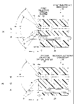
respectively, will be explained with reference to FIG. 1.
Here, this FIG. 1 shows a model relationship between the
first and the second objective optical system provided in
the observation window 15 at the distal end portion 4 and
the treatment instrument channel opening 17.
[0048] FIG. 1(a) shows the distal end portion 4 of the
first electronic endoscope 31. The observation window 15,
the treatment instrument channel opening 17, and the
illumination windows 16a to 16c, the water and air feeding
nozzle 18, and the forward water feeding opening 19, which
are not shown in the figures, are provided at the distal
end portion 4 of the first electronic endoscope 31. The
first objective optical system 33 having the viewing angle
al is arranged in the observation window 15 of the first
electronic endoscope 31. A center axle of the observation
window 15 of this distal end portion 4 and a center axle of
the treatment instrument channel opening 17 on the end
surface are arranged to have the physical relationship with
an interval as appeared as tl in the figure. Because of
CA 02549567 2006-06-14
19
the physical relationship between the observation window 15
provided at the first objective optical system 33 of the
viewing angle al and the treatment instrument channel
opening 17 positioned with a distance tl from the
observation window 15, the treatment instrument 51 inserted
and projected in the direction toward observed region from
the treatment instrument channel opening 17 needs to be
projected beyond an amount of projection 1 in the figure so
as to project in a range of the viewing angle a of the
first objective optical system 33. The treatment
instrument 51 projecting in the viewing angle al can be
observed from the observation window 15, and the physical
relationship of the observed region and the treatment
instrument 51 can be recognized.
[0049] Next, the observation windows 15 has the physical
relationship with the treatment instrument channel opening
17 away therefrom to the distance of tl, and when the
second objective optical system 37 with the viewing angle
a2, which is wider than the viewing angle al of the first
objective optical system 33 is installed in the observation
window 15 of the distal end portion 4 to constitute the
second electronic endoscope 32, the treatment instrument 51
projecting from the treatment instrument channel opening 17
proceeds in the range of viewing angle a2 with an amount
of projection 1'(l > 1') in the figure much smaller than
the amount of projection 1.
[0050] As such, the treatment instrument 51 proceeds in
the range of the wide viewing angle a2 of the second
objective optical system 37 with slight amount of
projection 1'. The amount of projection l' of the
treatment instrument 51 in this viewing angle a2 is not
sufficient in view of the physical relationship with the
CA 02549567 2006-06-14
observed region.
[0051] Then, as shown in FIG. 1(b), at a distal end
portion 4' of the second electronic endoscope 32, the
second objective optical system 37 with the viewing angle
5 a2 is arranged in an observation window 15', and the
center axle of a treatment instrument channel opening 17'
is arranged at a distance t2 (tl < t2) shown in the figure
away from the center axle of the observation window 15' of
the distal end portion 4'. From the physical relationship
10 between the observation window 15' provided at the second
objective optical system 37 of the viewing angle a2 and
the treatment instrument channel opening 17' positioned at
the distance t2 from the observation window 15', the
treatment instrument 51 penetrating and projecting from the
15 treatment instrument channel opening 17' toward the
observed region needs to exceed the amount of projection 1
in the figures to project in order to proceed in the range
of the viewing angle a2 of the second objective optical
system 37. That is, by projecting the treatment instrument
20 51 of the second electronic endoscope 32 more than the
amount of projection 1, the treatment instrument 51 can
proceed in the range in the wide viewing angle a2 of the
second objective optical system 37.
[0052] That is, as the treatment instrument 51 of the
second electronic endoscope 32 is operated to project the
same amount of projection 1 as the treatment instrument 51
of the first electronic endoscope 31 as shown in FIG. 1(a),
the treatment instrument can be recognized in the
respective observation image. Accordingly, the operator
feels substantially the same when operating to project the
respective treatment instrument 51 for the first and the
second electronic endoscopes 31, 32, and at the same time
CA 02549567 2006-06-14
21
the amount of projection of the treatment instrument 51
from the end surface of the distal end portions 4, 4'
becomes substantially the same, and thus the physical
relationship between the observed region and the treatment
instrument 51 is substantially the same.
[0053] As described above, the physical relationship
between the observation window 15 provided at the distal
end portion 4 of the insertion unit 2 and the treatment
instrument channel opening 17, while the amount of
projection of the treatment instrument 51 projecting from
the treatment instrument channel opening 17 being
substantially the same, is set according to the viewing
angle of the objective optical systems 33, 37 provided in
the observation window 15, thereby preventing the operator
from feeling uncomfortable as to the operational feeling of
the treatment instrument 51 and the physical relationship
between the observed region and the treatment instrument.
[0054] In addition, as applying the above concept,
regarding a single electronic endoscope, it is beneficial
that the distance between the objective optical system at
the insertion unit end and the treatment instrument channel
opening is determined based on the viewing angle of the
objective optical system. Concretely, for example, the
distance between the objective optical system and the
treatment instrument channel opening should be determined
in order for the image of the end of the treatment
instrument to be displayed in the predetermined reference
area on the observation image obtained by the objective
optical system when the amount of projection of the
treatment instrument projecting from the treatment
instrument channel opening reaches the predetermined value.
[0055] As described above, the conventional electronic
endoscope, which sets the distance between the objective
CA 02549567 2006-06-14
22
optical system and the treatment instrument channel opening
based on such as the diameter of the insertion unit and the
size of the objective optical system, when the field of
view of the objective optical system is different, can
adversely affect the operation of the operator because of
the different amount of projection of the treatment
instrument even if the treatment instrument is displayed at
the identical position in the observation image. For the
purpose of resolving the above problem, it is beneficial to
realize the electronic endoscope in which the distance
between the objective optical system and the treatment
instrument channel opening is set so as to display the
image of the end of the treatment instrument in the
predetermined reference area in the observation image when
the treatment instrument projects the predetermined
reference value for the predetermined amount of projection
of the treatment instrument. The distance to satisfy this
condition is determined according to the viewing angle of
the objective optical system as described above, and for
example in the case of FIG. 1(a), the distance between the
objective optical system and the treatment instrument
channel opening is tl based on the viewing angle al of the
objective optical system, and in the case of FIG. 1(b), the
distance between the objective optical system and the
treatment instrument channel opening is t2 based on the
viewing angle a2. As such, by employing the electronic
endoscope with the determined distance between the
objective optical system and the treatment instrument
channel opening based on the viewing angle, even if the
electronic endoscope of different viewing angle is used,
the operator can refer to the observation image to
understood how much the treatment instrument is projected
easily, thereby significantly improving the operability of
CA 02549567 2006-06-14
23
the electronic endoscope.
[0056] The reference area on the observation image can
be arbitrary area; however, it is preferable that the
peripheral region of the observation image is set as the
reference area as shown in the example of FIG. 1. As such,
by setting the reference area, when the treatment
instrument is gradually projected, it is an advantage that
the operator can easily recognize that the amount of
projection of the treatment instrument reaches the
predetermined reference value immediately upon displaying
the image of the end of the treatment instrument in the
observation image.
[0057] As an example of the electronic endoscope, which
has the determined distance between the objective optical
system and the treatment instrument channel opening based
on the viewing angle of the objective optical system, it is
preferable that the distance between the objective optical
system and the treatment instrument channel opening is set
so that a part of the treatment instrument projecting from
the treatment instrument channel opening, which exists
within the field of view of the objective optical system
determined based on the viewing angle, is arranged at a
position farther from the objective optical system than a
near point. A near point means a point at the shortest
distance for the operative system to be able to produce the
image. By setting the distance between the objective
optical system and the treatment instrument channel opening
so that the treatment instrument is arranged at a point
farther from the objective optical system than the near
point within the field of view of the objective optical
system, the image of the treatment instrument displayed in
the observation image always becomes clear, and the
operator can easily recognize that the treatment instrument
CA 02549567 2006-06-14
24
is displayed in the observation image.
[0058] FIG. 1 is referred for the explanation of an
example of determining the distance. Furthermore, in order
to simplify the explanation below, the near points for the
first objective optical system 33 and the second objective
optical system 37 exist in the area where the distance from
the objective optical system is d0 and establishes the
relationship of dl > d0 > d2 relative to the later
described dl, d2.
[0059] In FIG. 1(a), when the viewing angle of the first
objective optical system 33 is al, the minimum value of
the distance d between the part of the treatment instrument
51 existing in the field of view of the first objective
optical system 33 and the first objective optical system 33
(precisely, the observation window 15 forming the first
objective optical system 33 in FIG. 1(a)) becomes dl (>d0),
and the treatment instrument 51, within the viewing angle,
is positioned farther from the first objective optical
system 33 than the near point. Therefore, at the first
electronic endoscope 31 equipped with the first objective
optical system 33 of the viewing angle a1, the image of
the treatment instrument 51 displayed on the observation
image is very clear, which eliminates problems such as
giving a possibility of the operator to feel uncomfortable.
[0060] On the other hand, in FIG. 1(a), the viewing
angle a2 has the different situation. When the first
objective optical system 33 has the viewing angle a2, as
shown in FIG. 1(a), the minimum value of the distance d
between the part of the treatment instrument 51 existing in
the field of view of the first objective optical system 33
determined by the viewing angle a2 and the first objective
operative system 33 becomes d2, which creates a situation
CA 02549567 2006-06-14
where the distance from the first objective optical system
33 in the field of view becomes smaller than the distance
dO to the near point. Therefore, in the case of the wide
viewing angle a2, the image of the treatment instrument 51
5 in the observation image may become unclear, which gives
the problems such as giving the operator uncomfortable
feeling.
[0061] In consideration of the above two cases of
viewing angle al and a2, it is necessary for the minimum
10 value of the distance d between the first objective optical
system 33 and the treatment instrument 51 to become dO or
above in order to avoid the problems. Furthermore, as
shown in FIG. 1(a), the minimum value of the distance d can
be determined by the distance between the area where the
15 limit of field of view of the first objective optical
system 33 and the treatment instrument 51 cross and the
first objective optical system 33. Accordingly, when the
viewing angle is enlarged, it is preferable to form the
treatment instrument channel 26 so that the distance
20 between the cross area and the first objective optical
system 33 becomes dO or above, and more concretely,
preferably, the distance between the treatment instrument
channel opening 17 and the first objective optical system
33 is determined to be dO or above. By setting the
25 position of the treatment instrument channel opening 17
satisfying the conditions above, for example as shown in
FIG. 1(b), similarly for the viewing angle a2, the minimum
value of the distance d can be dl (>d2), thereby displaying
the image of the treatment instrument 51 on the observation
image clear. In addition, such a structure is preferably
employed in the electronic endoscope equipped with the wide
angle objective optical system with 150 degrees or more of
CA 02549567 2006-06-14
26
viewing angle. This is because especially when the field
of view is wide angle, the image of the treatment
instrument on the observation image tends to become unclear.
[0062] Here, if the number of pixels of the first CCD 34
and the second CCD 38 of the first and second electronic
endoscopes 31, 32 and the aspect ratio thereof are the same,
the range of the observed region image formed in the first
and second CCDs 34, 38 varies for each of the first
objective optical system 33 of the viewing angle al and
the second objective optical system 37 having the viewing
angle a2 with wider angle than the viewing angle al of the
first objective optical system 33.
[0063] That is, comparing to the area of the observed
region displayed in the endoscope image display area lla on
the monitor 11 as being imaged by the first electronic
endoscope 31, the area of the observed region displayed in
the same endoscope image display area on the monitor 11 as
being imaged by the second electronic endoscope 32 is wider.
However, individual observed region among observed regions
with wide area being displayed on the monitor 11 as being
imaged by the second electronic endoscope 32 is smaller
comparing to individual observed region among observed
regions being displayed on the monitor 11 as being imaged
by the first electronic endoscope 31. Furthermore, because
the number of pixels of individual observed region among
the wide observed regions is small, there is a possibility
of displaying unclear image.
[0064] Here, high pixel CCD which provides higher number
of pixels than the first CCD 34 of the first electronic
endoscope 31 is used for the second CCD 38 of the second
electronic endoscope 32 having the second objective optical
system 37 having the wide viewing angle a2.
CA 02549567 2006-06-14
27
[0065] By increasing the pixel of the second CCD 38 of
the second electronic endoscope 32, based on the second
objective optical system 37 having the viewing angle a2,
the pixel of the image (observation image 9 of the observed
region displayed on the monitor 11 from the image signal is
higher than that of the first electronic endoscope 31,
thereby improving the resolution of the entire image of the
observed region of the second electronic endoscope 32 and
the individual observed region among the images of the
observed regions. Furthermore, a zoom process is
electronically performed on the high pixel image of the
observed region generated by the high pixel second CCD 38
of the second electronic endoscope 32, thereby restricting
the degradation of the resolution because of its high pixel
image even if a zoomed observed region is displayed.
[0066] As explained above, the electronic endoscope
having the objective optical system with wide viewing angle
restricts the degradation of the image quality for the
individual observed region within the wide image area and
electronically zoomed observed region by using the zoomed
high pixel CCD.
[0067] NOTES
According to the above-described embodiments, the
following structures can be formed.
[0068] Note 1: An endoscope system using an endoscope to
observe and treat an observed region, including:
a first endoscope that includes a first objective
optical system provided in an observation window provided
at a distal end portion of an insertion unit, and a
treatment instrument channel opening positioned at a
predetermined distance away from the observation window,
the first objective optical system having a predetermined
viewing angle; and
CA 02549567 2006-06-14
28
a second endoscope that includes a second objective
optical system provided in an observation window provided
at a distal end portion of an insertion unit, and a
treatment instrument channel opening positioned at a
distance longer than the distance between the observation
window and the treatment instrument channel opening of the
first endoscope, the second objective optical system having
an viewing angle wider than the viewing angle of the first
objective optical system.
[0069] Note 2: An endoscope system, including:
a first endoscope that includes a first objective
optical system having a predetermined viewing angle
provided in an observation window at a distal end portion
of an insertion unit to be inserted into a subject; and
a second endoscope that includes a second objective
optical system having an viewing angle different from the
viewing angle of the first objective optical system in an
observation window at a distal end portion of an insertion
unit to be inserted into the subject, wherein
under a condition where an amount of projection of a
treatment instrument projecting from a treatment instrument
channel opening of the first endoscope is substantially the
same as an amount of projection of a treatment instrument
projecting from a treatment instrument channel opening of
the second endoscope, a distance between the treatment
instrument channel opening and the observation window of
the first endoscope and a distance between the treatment
instrument channel opening and the observation window of
the second endoscope are set so that the respective
treatment instruments are in respective viewing angles of
the first objective optical system and the second objective
optical system.
[0070] Note 3: The endoscope system according to Note 2,
CA 02549567 2006-06-14
29
wherein
the viewing angle of the second objective optical
system of the second endoscope is wider than the viewing
angle of the first objective optical system of the first
endoscope, and
a distance between the observation window of the
second endoscope and the treatment instrument channel
opening is longer than a distance between the observation
window and the treatment instrument channel opening of the
first endoscope.
[0071] Note 4: The endoscope system according to any one
of Notes 1 to 3, wherein in the first and second endoscopes
having the solid imaging element at the respective image
formation position of the first and second objective
optical system, the solid imaging element with higher pixel
than the solid imaging element provided at the image
formation position of the first objective optical system is
used at the image formation position of the second
objective optical system.
[0072] Note 5: An endoscope that includes an objective
optical system having at least a predetermined viewing
angle, and a treatment instrument channel opening into
which a treatment instrument is inserted, the objective
optical system and the treatment instrument channel opening
being provided at a distal end portion of an insertion unit,
wherein
the opening is formed at a predetermined distance away
from the objective optical system, the distance being
determined based on the viewing angle.
[0073] Note 6: The endoscope according to Note 5 capable
of being connected to the signal processing device
identical to the endoscope having the objective optical
system with narrower viewing angle than the endoscope
CA 02549567 2006-06-14
[0074] Note 7: An endoscope that includes an objective
optical system having at least a predetermined viewing
angle and provided at a distal end portion of an insertion
unit; and a solid imaging element having a greater number
5 of pixels than that of a solid imaging element used in an
objective optical system with narrower viewing angle than
the endoscope.
INDUSTRIAL APPLICABILITY
10 [0075] As described above, the endoscope of the present
invention is effective when being employed in the endoscope
system equipped with the endoscope with wide viewing angle
or plural endoscopes having different viewing angle.