Note : Les descriptions sont présentées dans la langue officielle dans laquelle elles ont été soumises.
CA 02556183 2010-01-28
63189-677
BONE GRAFT SUBSTITUTE
FIELD OF THE INVENTION
[0001] This invention relates to biocompatible bone graft materials for
repairing bone defects and the application of the bone graft materials
disclosed herein.
The present invention incorporates the benefits of inorganic shaped bodies
having
macro, meso, and microporosity and polymers such as collagen. The bone
restoratives may also be useful as delivery vehicles for therapeutic materials
such as
bone marrow aspirate, blood, plasma, cells, cell signaling materials, growth
factors,
proteins, or medicaments.
BACKGROUND OF TILE INVENTION
[0002] There has been a continuing need for improved bone graft materials.
Although autograft, the current gold standard, has the ideal properties and
radiopacity,
the use of autogenous bone exposes the patient to risk of a second surgery,
pain, and
morbidity at the donor site. Allograft devices, which are processed from donor
bone,
also have ideal radiopacity, but carry the risk of disease transmission. The
devices are
1
CA 02556183 2010-01-28
63189-677
restricted in terms of variations on shape and size and have sub-optimal
strength
properties that decrease after implantation. The quality of the allograft
devices varies
because they are natural. Also, since companies that provide allograft
implants obtain
their supply from donor tissue banks, there tend to be limitations on supply.
In recent
years, synthetic materials have become a viable alternative to autograft and
allograft
devices. One such synthetic material is Vitoss Scaffold Synthetic Cancellous
Bone
Void Filler (Orthovita, Inc., Malvern, PA, assignee of the present
application).
Synthetic graft materials, like autograft and allograft, serve as
osteoconductive
scaffolds that promote the ingrowth of bone. As bone growth is promoted and
increases, the graft material resorbs and is eventually replaced with new
bone.
[0003] Many synthetic bone grafts include materials that closely mimic
mammalian bone, such as compositions containing calcium phosphates. Exemplary
calcium phosphate compositions contain type-B carbonated hydroxyapatite
[Cas(PO4)3,(CO3).(OH)], which is the principal mineral phase found in the
mammalian body. The ultimate composition, crystal size, morphology, and
structure
of the body portions formed from the hydroxyapatite are determined by
variations in
the protein and organic content. Calcium phosphate ceramics have been
fabricated
and implanted in mammals in various forms including, but not limited to,
shaped
bodies and cements. Different stoichiometric compositions such as
hydroxyapatite
(HAp), tricalcium phosphate (TCP), tetracalcium phosphate (TTCP), and other
calcium phosphate salts and minerals, have all been employed to match the
adaptability, biocompatibility, structure, and strength of natural bone. The
role of
pore size and porosity in promoting revascularization, healing, and remodeling
of
bone has been recognized as a critical property for bone grafting materials.
The
preparation of exemplary porous calcium phosphate materials that closely
resemble
2
CA 02556183 2010-01-28
63189-677
bone have been disclosed, for instance, in U.S. Patent Nos. 6,383,519
(hereinafter the
`519 patent) and 6,521,246, (hereinafter the `246 patent).
[0004] There has been a continued need for improved bone graft systems.
Although calcium phosphate bone graft materials are widely accepted, they lack
the
strength, handling and flexibility necessary to be used in a wide array of
clinical
applications. Heretofore, calcium phosphate bone graft substitutes have been
used in
predominantly non-load bearing applications as simple bone void fillers and
the like.
For more clinically challenging applications that require the graft material
to take on
load, bone reconstruction systems that pair a bone graft material to
traditional rigid
fixation systems are used. The prior art discloses such bone reconstruction
systems.
For instance, MacroPore OSTM Reconstruction System is intended to reinforce
and
maintain the relative position of weak bony tissue such as bone graft
substitutes or
bone fragments from comminuted fractures. The system is a resorbable graft
containment system composed of various sized porous sheets and sleeves, non-
porous
sheets and sleeves, and associated fixation screws and tacks made from
polylactic
acid (PLA). However, the sheets are limited in that they can only be shaped
for the
body when heated.
[0005] The Synthes SynMeshTM consists of flat, round, and oval shaped
cylinders customized to fit the geometry of a patient's anatomical defect. The
intended use is for reinforcement of weak bony tissue and is made of
commercially
pure titanium. Although this mesh may be load bearing, it is not made entirely
of
resorbable materials that are flexible and also lacks an absorbant component
for the
delivery of materials of the types described herein.
3
CA 02556183 2010-01-28
63189-677
[0006] Many bone graft materials have limited interconnectedness that
substantially limits their ability to retain and deliver therapeutic materials
and fluids at
a bony site. As such, these graft materials would not be suitable as carriers
for
therapeutic materials and fluids such as cells, cell signaling materials,
proteins, bone
marrow aspirate, and blood. It is also known that most bone graft materials
lack the
structural integrity necessary to provide support.
[0007] Conversely, metals, which are capable of providing structural support
typically are not readily absorbent and cannot retain fluid. This is also due
in part to
their low porosity or macro-hole structures.
[0008] It would be of great benefit in the art to use graft materials for the
retention and delivery of therapeutic materials or fluids. Currently, bone
grafts often
are incapable of adequately retaining fluids once a surgeon attempts to
implant the
graft into a bony space. The majority of the fluids are flushed out of the
graft when
manipulated by the surgeon. Thus, there is a need in the art for a bone graft
capable
of retaining and delivering therapeutic materials that are at least partially
load bearing.
[0009] There is a need for resorbable bone grafts with improved handling,
which are flexible and not brittle, and are compression resistant. It has been
discovered that admixing highly porous resorbable inorganic bodies with
resorbable
polymeric materials greatly improves upon handling, yet still provides an
osteoconductive implant with good resorption and bone formation properties. It
will
be appreciated that such an implant would offer an easy-to-use dose of
composite
material and would be an advancement over current bone reconstruction systems
for
certain clinical applications in that it eliminates the need to have both a
graft material
and rigid fixation system.
4
CA 02556183 2010-01-28
63189-677
[0010] There is a need in the art to provide biocompatible graft materials
with
exceptional osteoconductive properties; provide pre-sized graft materials in a
variety
of forms, including strips and cylinders for restoring defects in bone; to
provide bone
graft materials that can be shaped; and to provide bone graft materials with
improved
handling properties, so that the graft material can be cut dry or after being
wetted and
does not crumble.
[0011] Currently, bone grafts often are incapable of retaining fluids once a
surgeon attempts to implant the graft into a bony space. The fluids are
flushed out of
the graft when manipulated by the surgeon. There is also a need to provide
bone graft
materials with some compression resistance, such that the brittleness often
associated
with inorganic or ceramic bone graft materials is eliminated; to provide bone
graft
materials with integrity that are at least partially load bearing; to provide
bone graft
materials with improved pliability that still retain high degrees of porosity
over a
broad pore size distribution to maintain superior resorption and bone ingrowth
properties; to provide bone graft materials with fluid wicking and retention
properties
even under compressive loads; and to provide bone grafts that provide easy
implantation into a bony space and with decreased tendency to wash away when
imbibed with fluid. Additional objects, advantages, and novel features of this
invention will become apparent to those skilled in the art upon examination of
the
following descriptions, figures and claims thereof, which are not intended to
be
limiting.
SUMMARY OF THE INVENTION
[0012] The present invention is directed to biocompatible bone graft materials
that comprise a biocompatible, resorbable polymer and the oxidation-reduction
reaction product of at least one metal cation, at least one oxidizing agent,
and at least
CA 02556183 2011-03-18
63189-677
one oxidizable precursor anion. The reaction product may be selected to suit
the
needs of one skilled in the art but may be inorganic compositions comprising
calcium
phosphate, biphasic calcium phosphate, or beta tri-calcium phosphate (13-TCP).
The
present invention is also directed to pliable bone restorative comprising an
osteoconductive foam comprising biocompatible, resorbable polymer and calcium
phosphate that at least partially surrounds a biocompatible mesh.
According to one aspect of the present invention, there is provided a
biocompatible bone graft material comprising a homogenous composite of
biocompatible, resorbable collagen and calcium phosphate, the biocompatible
bone
graft having macro-, meso-, and microporosity.
According to another aspect of the present invention, there is provided
use of biocompatible, resorbable collagen and the oxidation-reduction reaction
product of at least one metal cation, at least one oxidizing agent, and at
least one
oxidizable precursor anion in preparation of a bone graft material for
restoring or
repairing bone in a mammal.
According to still another aspect of the present invention, there is
provided a bone graft for long bone reinforcement in the form of a sleeve, the
graft
comprising a homogenous composite of biocompatible, resorbable collagen and
calcium phosphate, the graft having interconnected macro-, meso-, and
microporosity.
According to yet another aspect of the present invention, there is
provided a graft for the restoration of bone in the form of a shaped body, the
shaped
body comprising a homogenous composite of collagen and beta-tricalcium
phosphate, the graft having interconnected macro-, meso-, and microporosity;
the
body shape being selected to conform to a mammalian, anatomical tissue
structure;
and further comprising a mesh affixed to a side of the composite.
6
CA 02556183 2011-03-18
63189-677
According to a further aspect of the present invention, there is provided
a pliable bone restorative comprising a biocompatible mesh and a pliable bone
graft
material comprising a homogenous composite of biocompatible, resorbable
collagen,
and the oxidation-reduction reaction product of at least one metal cation, at
least one
oxidizing agent, and at least one oxidizable precursor anion, wherein said
bone graft
material has macro-, meso-, and microporosity and at least partially surrounds
said
mesh, and wherein said bone restorative is wetted with a fluid.
According to yet a further aspect of the present invention, there is
provided a pliable bone restorative comprising a biocompatible mesh and a
pliable
bone graft material comprising a homogenous composite of biocompatible,
resorbable collagen and biocompatible, resorbable calcium phosphate, wherein
said
bone graft material has macro-, meso-, and microporosity, wherein said bone
restorative is wetted with a fluid and wherein at least a portion of the
biocompatible
mesh is in contact with the bone graft material.
According to another aspect of the present invention, there is provided
a pliable bone restorative comprising a biocompatible mesh; and a pliable,
biocompatible, resorbable homogenous blend of collagen and a biocompatible,
resorbable material comprising the oxidation-reduction product of at least one
metal
cation, at least one oxidizing agent, and at least one oxidizable precursor
anion and
having interconnected macro-, meso-, and microporosity, wherein said blend at
least
partially surrounds said biocompatible mesh, and wherein said bone restorative
is
wetted with a fluid.
According to yet another aspect of the present invention, there is
provided a pliable bone restorative for the restoration of bone in the form of
a shaped
body, the shaped body selected to conform to a mammalian, anatomical tissue
structure, said body comprising a biocompatible mesh; and a pliable bone graft
material comprising a homogenous composite of collagen and biocompatible,
resorbable beta tricalcium phosphate, having interconnected macro-, meso-, and
6a
CA 02556183 2011-12-19
67044-100
microporosity; wherein said bone graft material at least partially surrounds
said mesh,
and wherein said bone restorative is wetted with a fluid.
According to another aspect of the present invention, there is provided
use of biocompatible, resorbable collagen and the oxidation-reduction reaction
product of at least one metal cation, at least one oxidizing agent, and at
least one
oxidizable precursor anion in preparation of a bone restorative for delivering
therapeutic material wherein said bone restorative is imbibed with the
therapeutic
material.
According to still another aspect of the present invention, there is
provided use of biocompatible, resorbable collagen and calcium phosphate in
preparation of a pliable bone restorative for delivering therapeutic material
wherein
the bone restorative is imbibed with said therapeutic material.
According to yet another aspect of the present invention, there is
provided use of a biocompatible, resorbable collagen and calcium phosphate
having
macro-, meso-, and microporosity in preparation of a pliable bone restorative
for
delivering therapeutic material wherein the bone restorative is imbibed with a
therapeutic fluid.
According to a further aspect of the present invention, there is provided
use of a biocompatible mesh and a bone graft material comprising
biocompatible,
resorbable collagen and calcium phosphate in preparation of a ductile bone
restorative for delivering a therapeutic material wherein the bone restorative
is
imbibed with the therapeutic material contained in wells in the bone graft
material.
[0013] Other embodiments of the present invention include pliable bone
restoratives comprising a biocompatible mesh and at least partially
surrounding the
mesh, a biocompatible, resorbable polymer, and the oxidation-reduction
reaction
product of at least one metal cation, at least one oxidizing agent, and at
least one
oxidizable precursor anion. Disclosed herein are also pliable bone
restoratives
comprising a biocompatible mesh and a bone graft material comprising
6b
CA 02556183 2011-03-18
63189-677
biocompatible, resorbable collagen and calcium phosphate. A further embodiment
of
the present invention is a pliable bone restorative comprising a biocompatible
mesh
and a biocompatible bone graft material comprising biocompatible, resorbable
collagen and calcium phosphate having macro, meso, and microporosity. Also
disclosed within are pliable bone restoratives comprising a biocompatible,
resorbable
substantially homogenous blend of a first polymeric material and a second
material
having interconnected macro, meso- and microporosity, with said blend at least
partially surrounding a biocompatible mesh. A further embodiment that may be
preferred is in the form of a shaped body selected to conform generally to a
mammalian, anatomical tissue structure. The shaped body comprises a polymer
and
beta tricalcium phosphate partially surrounding a biocompatible mesh. The
graft may
have interconnected macro-, meso-, and microporosity. Suitable polymers may
include structural proteins such as collagen.
6c
CA 02556183 2010-01-28
63189-677
[0014] Still other embodiments of the present invention are directed to
methods for delivering therapeutic materials comprising: providing a bone
restorative
comprising biocompatible, resorbable polymer, and the oxidation-reduction
reaction
product of at least one metal cation, at least one oxidizing agent, and at
least one
oxidizable precursor anion; imbibing said bone restorative with a therapeutic
material;
and placing said bone restorative into a bony space. In some embodiments that
may
be preferred the bone restorative may be pliable for optimum shaping
capability. The
present invention is also directed to methods where the bone restorative also
has
macro-, meso-, and microporosity; and to methods where the bone restorative is
capable of wicking and delivering materials via its interconnected structure.
The
therapeutic materials may comprise cells, cell signaling materials, proteins,
bone
marrow aspirate, plasma, blood, growth factors, or medicaments. The cells may
comprises stem cells. In some embodiments, the selected polymer may be
collagen.
In many embodiments that may be preferred the reaction product may be calcium
phosphate or (3- tricalcium phosphate in other embodiments. The bone
restorative
may comprise cell wells for containing therapeutic materials or an admixture
of
autogenous bone chips, synthetic bone graft, or medicaments. The therapeutic
materials imbibed into the bone restorative or those contained within the cell
wells
may release them over time.
[0015] The present invention is an improvement upon the shaped bodies
disclosed in U.S. Patent Nos. 6,383,519 ("'519 patent") and 6,521,246 ("'246
patent"), and the RPR process disclosed in U.S. Patent Nos. 5,939,039 ("039
patent")
and 6,325,987 ("'987 patent"), all assigned to the present assignee. The
oxidation-reduction reaction product of the present invention shares the
same unique porosity of those shaped bodies of the `519
7
CA 02556183 2010-01-28
63189-677
and `246 patents. The reaction product grants the present invention graft
material
macro, meso, and microporosity, which allow the graft material to have
extraordinary
imbibation and absorption properties. Further, the inclusion of a polymer in
the
present invention material lends improved handling and flexibility. The graft
materials can have a finite shape for some applications and are compression
resistant
or at least partially load bearing. When imbibed with fluids, the bone graft
materials
are flexible, bendable/deformable, and scalpable, without crumbling or falling
apart.
Some embodiments have a mesh or plate affixed to the bone graft material for
added
support. The bone graft materials may be imbibed with fluids such as bone
marrow
aspirate, blood, or saline. The graft materials may be provided in any basic
shape,
including cylinders, blocks, strips, sheets, and wedges. In one embodiment,
the graft
materials are provided in basic cylinder or strip form. In other embodiments,
the graft
materials may have a finite shape or custom shape for specific applications
(e.g.,
semi-spherical for graft acetabular containment, half-tubular long bone wrap
or
sleeve), or may be "shredded" and housed within a delivery vessel. Yet, in
other
embodiments, the graft materials may serve as a coating on any orthopaedic
appliance
such as an intermedullary rod, pedicle screw, plate, hip stem, acetabular cup
component and the like. The bone graft materials of the present invention also
have
the ability to attach to Bone Morphogenic Proteins (BMP).
[0016] This invention gives rise to biocompatible, resorbable composites that
may have up to about 30% by weight of the biocompatible polymer and 70% by
weight of the reaction product. The amount of biocompatible polymer within the
bone graft materials may also be up to about 20% by weight or up to about 10%
by
weight, or alternatively up to about 50% by weight.
BRIEF DESCRIPTION OF THE DRAWINGS
8
CA 02556183 2010-01-28
63189-677
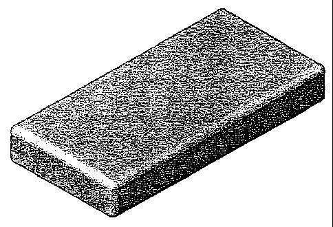
[0017) Figure IA illustrates one basic form of the biocompatible graft
material
in cylinder form. Figure 1B depicts the graft material in cylindrical form 80
inserted
into a bone void 83 below the femur 81 in the tibial plateau 82 within a human
knee.
[0018] Figure 2 illustrates another basic form of the present invention in
strip
form.
[0019) Figure 3A illustrates one embodiment of the biocompatible graft
material of the present invention in semi-spherical form used as a graft
containment
device. Figure 3B depicts a semi-spherical form of the graft material 102 used
to
accommodate an artificial implant 103. The graft material 102 contains an
acetabular
cup 106, which holds a polyethylene cup 105, in this embodiment, and 101
represents
cancellous bone.
[0020] Figure 4A illustrates the bone restorative of the present invention in
disc form. Figure 4B illustrates another embodiment of the biocompatible graft
material of the present invention used as a cranio-maxillofacial, zygomatic
reconstruction and mandibular implant.
[0021] Figure 5 illustrates one embodiment of a bone graft material described
shaped into a block/wedge form and used as a tibial plateau reconstruction
that is
screwed, bonded, cemented, pinned, anchored, or otherwise attached in place.
[0022] Figures 6A and 6B illustrate synthetic resorbable defect filling bone
graft materials 272 for bone restoration having mesh 270 attached to one side.
Figure
6C depicts a synthetic resorbable defect filling bone graft material block in
which the
mesh 270 is placed between the graft material 272.
[0023] Figure 7A, 7B, and 7C illustrate the shapes of some embodiments in
semi-tubular form used as a long bone reinforcement sleeve. As shown in the
figures,
the semi-tube may have a moon cross-section with a uniform thickness (Figure
7A);
9
CA 02556183 2010-01-28
63189-677
or a crescent moon cross-section with a tapered radius that comes to a point
(Figure
7B) or a tapered radius that is rounded on the edges (Figure 7C).
[0024] Figure 8 depicts the semi-tubular shaped embodiment 200 placed on a
metacarpal bone.
[0025] Figure 9 depicts a tubular shaped embodiment 200 fitted around the
femur.
[0026] Figures I OA and I OB depicts a tubular shaped embodiments 200 showing
different configurations for placing the biocompatible mesh 270 and graft
material
272.
[0027] Figure 11 is a representative XRD spectra of a bone graft material of
the present invention (top) vs. [3-TCP (bottom).
[0028] Figure 12 is a representative FTIR spectrum of bone graft material of
the present invention vs. l-TCP (beta-TCP) and Predicate.
[0029] Figure 13 is an SEM of the bone graft material, 20x.
[0030] Figure 14 is an SEM of the bone graft material, 50x.
[0031] Figure 15 is an SEM of the bone graft material, 250x.
[0032] Figure 16 depicts the Ultimate Indentation Strength for one
embodiment of the bone graft material vs. control normalized by adjacent bone
at 12
weeks.
[0033] Figure 17 is an SEM of air-dried gelatin treated inorganic material,
23x.
[0034] Figure 18 is an SEM of sheep trabecular bone, 25x.
[0035] Figure 19 is an SEM of the material shown in Figure 14, 2000x
CA 02556183 2010-01-28
63189-677
[0036] Figure 20A depicts an embodiment of the bone restorative having
channels
255 in the graft material 272 so that a surgeon can see the fixation holes 260
in the
mesh 270 to accommodate mechanical fixation with screws. Channels 255 may also
used to soak and hold therapeutic materials. Figure 20B depicts a side view of
the
restorative.
[0037] Figure 21 depicts an embodiment having wells 265 to soak and hold
therapeutic materi al and also may be used for assisting in fixation.
[0038] Figures 22 and 23 depict the restorative with crimp zones 275 for
localized bending.
[0039] Figure 24 depicts a discoid shaped embodiment placed at appropriate
sites on the femur; note the cut line 160 for guiding a surgeon to shape the
restorative
200 for optimal fitting at appropriate sites on the femur.
[0040] Figure 25 depicts the restorative used on the iliac crest.
[0041] Figures 26A, 26B, and 26C depict an embodiment having crimp
zones 275 that guide a surgeon to forming a bowl shaped restorative. Figure
26A
shows the restorative with mesh 270 side up and 26B shows the restorative with
foam 272 side up. Figure 26C depicts the embodiment after being guided into a
bowl shape.
[0042] Figure 27 depicts an embodiment of the present invention having a
gradient of interconnectedness.
DETAILED DESCRIPTION OF PREFERRED EMBODIMENTS
(0043] The terms "bone graft material" and "foam" may be used
interchangeably in this description. Disclosed in that application were, mater
alia,
11
CA 02556183 2010-01-28
63189-677
biocompatible bone graft material may comprise resorbable polymer, such as
collagen, and certain inorganic materials, especially calcium phosphate. The
present
invention provides improvements to bone graft materials, by integrating mesh
or other
flexible support that result in the present bone restoratives. One class of
these
advancements that are of particular utility are pliable bone restoratives
comprising an
osteoconductive foam comprising biocompatible, resorbable polymer and calcium
phosphate that at least partially surrounds a biocompatible mesh. The present
invention also provides improvements to bone graft materials having
exceptional
carrier properties and are suited for use in methods for delivering
therapeutic
materials to a bony site.
12
CA 02556183 2006-08-01
WO 2005/074614 PCT/US2005/003251
[0045] The present invention finds utility in a wide variety of applications
and
may provide an alternative to autografts and other implantation materials
comprised
of cadaver bone, bovine bone, or the like. The porous bone restoratives formed
herein
can be used in medicine, such as, but not limited to, the restoration of bony
defects.
The bone restoratives can also be used for the delivery of medicaments that
are
internal to the defect, or can be used to promote cellular, bone, or tissue
growth. In
this way, the can be partially filled with materials that either comprise or
carry a
medicament or therapeutic such as proteins, growth hormones, antibiotics, or
cell
signaling materials. Indeed, the larger porous spaces within some of the bone
restoratives of the present invention can be used for culturing cells within
the human
body. In this regard, the larger spaces are amenable to the growth of cells
and can be
permeated readily by bodily fluids such as certain blood components. In this
way,
growing cells can be implanted in an animal through the aegis of implants in
accordance with the present invention. These bone restoratives are implants
that give
rise to important biochemical or therapeutic uses.
[0046] The present bone restoratives are exceptional fluid carrier support
systems. The bone restoratives can retain and deliver fluids to a bone defect
site due
to the porous and interconnected structure of the carrier, the material
composition of
the carrier, and the design of the carrier. Additionally, the bone
restoratives may have
structural integrity that is at least partially load-bearing with a mesh
component.
[0047] Preferably, the graft materials can be shaped or formed and are
pliable.
It will be appreciated that one particularly beneficial aspect of some
embodiments of
this invention is that it provides unprecedented utility in the surgical
operation where
a reconstructive surgeon, relying upon the pliability of the bone restorative,
may
manipulate the restorative into shapes which are particularly amenable to the
bony
13
CA 02556183 2006-08-01
WO 2005/074614 PCT/US2005/003251
areas to be reconstructed. The restoratives have the capability of being
fashioned into
a new form and have varying degrees of pliability. It may be preferred that
the
restorative is deformable by human finger pressure and, when in the shape of a
strip,
can be rolled upon itself when wetted. Alternatively, simple hand tools such
as
forceps, and other common tools used in the operatory, may be employed to
shape
restoratives of the invention. As will readily be perceived, this enables the
surgeon to
tailor the precise shape of the restorative to that which is required in a
particular
circumstance very conveniently and at the point of use. The pliability of the
restoratives of the present invention makse this possible. Relatively hard
restoratives,
which cannot be molded conveniently by hand or with the use of common hand
tools,
require extraordinary processing techniques including machinery, heat, or
highly
leveraged manipulative devices that are much less useful than the pliable
restoratives
of the present invention. Other relatively brittle bone graft materials in the
art are not
shapeable without crumbling.
[00481 In accordance with some embodiments of the present application, there
are pliable bone restoratives comprising a biocompatible mesh at least
partially
surrounded by a biocompatible, resorbable polymer and the oxidation-reduction
reaction product of at least one metal cation, at least one oxidizing agent,
and at least
one oxidizable precursor anion.
[00491 It will be appreciated that a number of alterations may be made to
customize the restoratives for specific needs. There may be radiopaque
embodiments.
Other embodiments may be coated with titanium plasma spray to significantly
increases implant surface area and mechanical retention in the bone at the
time of
placement. The mesh may also be acid etched titanium or sodium treated
titanium to
aid in mechanical interlock of the foam.
14
CA 02556183 2006-08-01
WO 2005/074614 PCT/US2005/003251
[0050] In accordance with the present invention, graft materials are provided
comprising a biocompatible polymer such as collagen, the oxidation-reduction
reaction product of at least one metal cation, at least one oxidizing agent,
and at least
one oxidizable precursor anion. Graft materials are also provided that
comprise a
collagen and macro-, meso-, and microporous calcium phosphate. Some
embodiments may comprise up to 100% Type I collagen. In other embodiments, the
collagens used may be predominantly, or up to about 90%, of Type I collagen
with up
to about 5% of Type III collagen or up to about 5% of other types of collagen.
The
Type I bovine collagen may be native fibrous insoluble collagen, soluble
collagen,
reconstituted collagen, or combinations thereof. The biocompatible polymer may
be
combined with the reaction product in slurry form, or combined by blending or
kneading, to form a substantially homogenous mixture. As used in this context,
substantially homogenous means that the ratio of components within the mixture
is
the same throughout. This, upon treatment using various preferred freeze-
drying and
crosslinking techniques, produces a form of the present invention graft
material that
may be preferred.
[0051] Collagen has been found to be particularly suitable in the present
invention for service as the biocompatible polymer. The admixture of the
collagen
with the highly porous reaction product results in a graft that is highly
porous with a
broad pore size distribution, increased handling properties, and pliability
beyond that
which is achievable with some forms of the reaction product alone, for
instance
calcium phosphate. The resorption profile of some of the embodiments of the
present
invention may vary depending upon the amount, nature, and source of the
collagen or
other polymer used. Typically, by twelve weeks in vivo about 80%-90% of the
present invention is resorbed. One reason that may explain the superior
resorption
CA 02556183 2006-08-01
WO 2005/074614 PCT/US2005/003251
properties of the Y gent invention is the high degree of porosity retained
even upon
admixing the collagen with the reaction product. The collagen may be in a
polymerized fibrous form that has a long three-dimensional architecture with
multiple
cross-links.
[0052] Preferable collagens have beneficial biochemical attributes such as
10% to 20% nitrogen, 10% to 15% of hydroxyproline, or up to 2.5% of ash
content.
In some embodiments, the collagens may be 10.5% to 17% nitrogen, 10.5% to 14%
of
hydroxyproline, or up to 2.5% of ash content. The percent nitrogen of a
collagen is a
measurement of nitrogen in a sample. In the presence of sulfuric acid, the
amino
nitrogen of organic material is converted to ammonium sulfate. The ammonium
sulfate is distilled from an alkaline medium, and further decomposes from
which the
ammonia is absorbed into a boric acid solution containing a pH indicator. The
ammonia (nitrogen) concentration determined colorimetrically by back titrating
the
boric acid solution with a standard acid.
[0053] The percent hydroxyproline of a collagen is a measure of
hydroxyproline in a sample. Collagen is hydrolyzed with dilute Hydrochloric
Acid,
filtered and diluted. The solution is reacted with several reagents and then
measured
using ultraviolet (UV)/Vis analysis along with a standard hydroxyproline
solution.
Using the sample and standard absorbances, the percentage of hydroxyproline
can be
calculated [(Sample Abs)(Std)(Weight)(dilution factor)]/[(Sample weight)(Std.
Abs)(dilution factor)].
[0054] The ash content of collagen is a measure of the amount of residual
elements in collagen materials. When collagen is heated to extremely high
temperatures, it is converted to mainly carbon dioxide and water. Elements
other than
collagen and hydrogen are converted to oxides and salts. A small sample of
material
16
CA 02556183 2006-08-01
WO 2005/074614 PCT/US2005/003251
is heated until there is only ash left. The weight of this ash is considered
the gross
amount of inorganic/organic material of the original sample.
[0055] Bone graft materials of this invention that may be preferred are held
together in surgically relevant shapes and sizes by foaming the inorganic
reaction
product with the collagen. The resulting osteoconductive foam articles retain
substantially all of the biological and chemical properties of the shaped
bodies taught
in the `519 and `246 patents, while forming a shapeable, flexible unit dose.
The
osteoconductive foam or bone graft materials may be manufactured (with or
without
mesh) into strips and cylinders of prescribed dimensions and volumes. Other
shapes
include but are not limited to block, hemisphere, half pipe, rod, funnel, cup,
sleeve, or
discoid. As seen in Figure 8, the half pipe shaped embodiment 200 has a mesh
on top
of the foam portion of the restorative. The graft material portion is in
contact with the
metacarpal bone and the mesh is outward facing. A full pipe embodiment 200 may
be
seen in Figure 9 that completely surrounds the femur. This shape may be called
a
bone cuff. Alternatively, the foam 272 could completely surround the mesh. The
foam aids in assisting bony incorporation of the mesh and eliminates the
surgical step
of having to add graft material to the structural mesh portion of the
restorative device.
The graft material will resorb following delivery in the surgical site and
exhibit the
same beneficial biological responses (e.g., bone formation) as the
aforementioned
shaped bodies.
[0056] The foam may be further manufactured to have a number of physical
features that may assist in placing the restorative in the bony site adding
support to
surrounding bone. The foam may have channels 255 or wells 265 as seen in
Figures
20A and 21. In embodiments where the mesh is embedded within the foam, these
wells 265 allow a surgeon to see the mesh and the location on the mesh where a
screw
17
CA 02556183 2006-08-01
WO 2005/074614 PCT/US2005/003251
or suture will be fixated. These channels 255 and wells 265 may also soak and
hold
therapeutic materials, as seen in Figure 20, and may aid in the delivery of
therapeutic
materials to the bony site. The wells may also vary in size and diameter such
that
they are suitable for helping in the fixation of surgical screws, sutures, or
wires.
These channels 255 and wells 265 serve not only as a micro-repository for
cells, but
also as macro-encasements for admixtures of autogenous bone chips, synthetic
bone
grafts, or other medicaments. The admixture of the latter can be considered a
bone
graft pate. These chambers may also serve as time-release depositories in
which
medicaments or therapeutic materials are released over time.
[0057] Another useful aspect of the wells 265 and channels 255 will be
appreciated in those embodiments where the mesh is embedded within the foam
material. The channels 255, for instance, expose the mesh so that an operator
can
easily affix a screw, suture, or the like to the mesh. The wells 265 may allow
for easy
fixation of a screw through the foam portion directly to the mesh. The
channels 255
will allow for easy fixation of wires and sutures through the foam.
[0058] Certain presently preferred embodiments of the present invention may
be described as a pliable bone restorative comprising a biocompatible mesh and
a
bone graft material comprising biocompatible, resorbable collagen and calcium
phosphate. Other embodiments may be a pliable bone restorative comprising a
biocompatible, resorbable substantially homogenous blend of a first polymeric
material and a second material having interconnected macro, meso-, and
microporosity, said blend at least partially surrounding a biocompatible mesh
with
said bone restorative. The first polymeric material may be collagen. The
second
material may comprise calcium phosphate.
18
CA 02556183 2006-08-01
WO 2005/074614 PCT/US2005/003251
[0059] In some embodiments, the bone graft materials may have up to about
30% by weight of biocompatible polymer. The biocompatible polymer may also be
up to about 25% by weight in other embodiments. It will be appreciated that
embodiments exist wherein the bone graft materials have up to about 20% or 10%
by
weight of a biocompatible polymer. In other embodiments where the polymer
chosen
is a collagen, the present invention exhibits a unique mineral (13-TCP) to
collagen ratio
that is unlike the ratios shared by other bone grafts. One skilled in the art
may obtain
bone graft materials of variable ratios depending on their particular needs.
In one
effective embodiment, the mass ratio of the reaction product and the collagen
is
80:20. In others, it may be 90:10 or 70:30. The mass ratio may be altered
without
unreasonable testing using methods readily available in the art. It will be
appreciated
that this ratio is contrary to the mineral R-TCP to collagen ratios one
skilled in the art
would find in previous bone grafts while still maintaining all the properties
(e.g.,
porosity, pore size distribution) that attribute to an effective bone graft
(e.g.,
simultaneous bone formation, strength and graft resorption).
[0060] Due to the high porosity and broad pore size distribution (1 m -
1000 m) of the present invention graft, the implant is not only able to
wick/soak/imbibe materials very quickly, but is also capable of retaining
them. A
variety of fluids could be used with the present invention including blood,
bone
marrow aspirate, cell concentrate, liquid hemostat, fibrin, sealant, saline,
antibiotics
and proteins such as bone morphogenetic proteins (BMPs).
[0061] Materials of the present invention can also be imbibed with blood,
cells
(e.g. fibroblasts, mesenchymal, stromal, marrow and stem cells), protein rich
plasma
other biological fluids and any combination of the above. This capability has
utility
in cell-seeding, drug delivery, and delivery of biologic molecules as well as
in the
19
CA 02556183 2006-08-01
WO 2005/074614 PCT/US2005/003251
application of bone tissue engineering, orthopaedics, and carriers of
pharmaceuticals.
As used herein materials or fluids are materials such as bone marrow aspirate
(BMPs),
blood, plasma or protein rich plasma, cells, cell signaling materials, growth
factors or
hormones, proteins, antibiotics, or medicaments. Cells useful in this
invention
comprise fibroblasts, mesenchymal, stromal, marrow, adipose, myoblasts,
lysosomes,
and stem cells. Suitable stem cells may be stem cells of embryonic, fetal, or
adult
tissue lineage, such as embryonic stem cells, fetal stem cells or mesenchymal
stem
cells. Also suitable would be cells derived from these lineages such as
osteoprogenitors, osteoblasts, osteocytes, adipocytes, myoblasts,
chondrocytes,
lysosomes, and the like. As used herein, stem cells may be considered those
undifferentiated cells capable of self-renewal and differentiation into
multiple
lineages of mature cells.
[0062] Cell signaling materials may be described as those materials capable of
provoking a cell to react. Signaling materials, growth factors, and proteins
may
include signaling molecules under the Transforming Growth Factor (TGF)
Superfamily of proteins, specifically proteins under the TGF-beta (TGF-(3),
Osteogenic Protein (OP)/Bone Morphogenic Protein (BMP), VEGF (VEGF-1 and
VEGF-2 proteins) and Inhibin/activtin (Inhibin-beta A, Inhibin-beta B, Inhibin-
alpha,
and MIS proteins) subfamilies. In may be preferred in many embodiments that
the
exemplary therapeutic materials are proteins under the TGF-(3 and OP/BMP
subfamilies. The TGF-(3 subfamily includes the proteins Beta-2, Beta-3, Beta-4
(chicken), Beta-1, Beta-5 (xenopus) and HIF-1 alpha. The OPBMP subfamily
includes the proteins BMP-2, BMP-4, DPP, BMP-5, Vgr-1, OP-1BMP-7, Drosophila
60A, GDF-1, Xenopus Vg-1 and BMP-3. Representative proteins of these types
include: OP-1/rhBMP-7 (Stryker Corporation, Kalamazoo, MI), rhBMP-2 (Genetics
CA 02556183 2010-01-28
63189-677
Institute/American Home Products, Madison, NJ), rhIGF-1 (Insulin-like Growth
Factor-1) (Cephalon, West Chester, PA), TGF beta (Genentech, S.an Francisco,
CA),
MP52 (Biopharm GmbH, Heidelberg, Germany/DePuy Acromed, Raynham, MA).
Other proteins, genes and cells outside the TGF Superfamily may also be
included in
the exemplary types of therapeutic materials to be used in conjunction with
the
TM
present invention. These other proteins and genes include PepGen P-15
(Ceramed,
Lakewood, CO); LMP-1 (LIM Mineralized Protein-1 gene) (Emory University,
TM
Atlanta, GA/Medtronic Sofamor Danek, Minneapolis, MN); Chrysalin TP 508
Synthetic Peptide (Chrysalis Biotechnology, Galveston, TX); GAM (parathyroid
hormone) (Selective Genetics, San Diego, CA); rhGDF-5 (Orquest, Mountain View,
CA/DePuy Acromed, Raynham, MA); cells lines and FGF (Fibroblast Growth
Factor), such as BFGF (Basic Fibroblast Growth Factor), FGF-A (Fibroblast
Growth
Factor Acidic), and FGFR (Fibroblast Growth Factor Receptor); and certain cell
lines
such as osteosarcoma cell lines. The therapeutic materials to be used with the
present
invention material may be combinations of those listed above. Such mixtures
include
TM
products like Ne-Osteo GFm (growth factor mixture) (Sulzer Orthopaedics,
Austin,
TX/Zimmer, Warsaw, IN) or mixtures of growth factors, proteins, genes, and
cells
produced by devices such as AGF (Autologous Growth Factor) (Interpore Cross
International, Irvine, CA/EBI, Parsippany, NJ), Symphony Platelet Concentrate
TM
System (Harvest Technologies, Belton, TX/DePuy, Warsaw, IN), GPS
(Gravitational
TM
Platelet System) (Biomet, Warsaw, IN), Magellan platelet separator
(Medtronic), and
the like. The materials to be used with the present invention material may
also be
combinations of those listed above. Such mixtures include products like Ne-
Osteo
GFm (growth factor mixture) (Sulzer/Ziminer), or mixtures of growth factors,
proteins, and genes produced by devices such as AGF (Interpore Cross
21
CA 02556183 2010-01-28
63189-677
TM
InternationalTEBl), Symphony BM Concentrator (D.ePuy), and the like. Further,
materials such as ascorbic acid, anti-bone resorption drugs, chemotherapeutic
agents,
chemicals, genes, fibrin sealants, liquid hemostats, vectors, vitamin D, and
sodium
fluoride may also be used.
[00631 Bone graft materials of the present invention that may be preferred
exhibit high degrees of porosity. It is also preferred that the porosity occur
in a broad
range of effective pore sizes. In this regard, persons skilled in the art will
appreciate
that preferred embodiments of the invention may have, at once, macroporosity,
mesoporosity, and microporosity. Macroporosity is characterized by pore
diameters
greater than about 100 m and, in some embodiments, up to about 1000 m to
2000 m. Mesoporosity is characterized by pore diameters between about 100 m
and
101im, while microporosity occurs when pores have diameters below about 10 m.
It
is preferred that macro-, meso-, and microporosity occur simultaneously and
are
interconnected in products of the invention. It is not necessary to quantify
each type
of porosity to a high degree. Rather, persons skilled in the art can easily
determine
whether a material has each type of porosity through examination, such as
through the
preferred methods of mercury intrusion porosimetry, helium pycnometry and
scanning electron microscopy. While it is certainly true that more than one or
a few
pores within the requisite size range are needed in order to characterize a
sample as
having a substantial degree of that particular form of porosity, no specific
number or
percentage is called for. Rather, a qualitative evaluation by persons skilled
in the art
shall be used to determine macro-, meso-, and microporosity. Therefore, some
embodiments of the present invention include a pliable bone restorative
comprising a
biocompatible mesh and a biocompatible bone graft material comprising
22
CA 02556183 2006-08-01
WO 2005/074614 PCT/US2005/003251
biocompatible, resorbable collagen and calcium phosphate having macro, meso,
and
microporosity.
[0064] It will be appreciated that in some embodiments of the overall porosity
of materials prepared in accordance with this invention be high. This
characteristic is
measured by pore volume, expressed as a percentage. Zero percent pore volume
refers to a fully dense material, which, perforce, has no pores at all. One
hundred
percent pore volume cannot meaningfully exist since the same would refer to
"all
pores" or air. Persons skilled in the art understand the concept of pore
volume,
however and can easily calculate and apply it. For example, pore volume may be
determined in accordance with W. D. Kingery, Introduction to Ceramics, 1960 p.
416
(Wiley, 1060), who provides a formula for determination of porosity.
Expressing
porosity as a percentage yields pore volume. The formula is: Pore Volume=(1-
ff)
100%, where fp is fraction of theoretical density achieved.
[0065] Porosity is measured by Helium Pycnometry. This procedure
determines the density and true volume of a sample by measuring the pressure
change
of helium in a calibrated volume. A sample of known weight and dimensions is
placed in the pycnometer, which determines density and volume. From the
samples
mass, the pycnometer determines true density and volume. From measured
dimensions, apparent density and volume can be determined. Porosity of the
sample
is then calculated using (apparent volume - measured volume)/apparent volume.
Porosity and pore size distribution may also be measured by mercury intrusion
porosimetry.
[0066] Pore volumes in excess of about 30% may be achieved in accordance
with this invention while materials having pore volumes in excess of 50% or
60%
may also be routinely attainable. Some embodiments of the invention may have
pore
23
CA 02556183 2006-08-01
WO 2005/074614 PCT/US2005/003251
volumes of at least about 70%. Some embodiments that may be preferred have
pore
volumes in excess of about 75%, with 80% being still more preferred. Pore
volumes
greater than about 90% are possible as are volumes greater than about 92%. In
some
preferred cases, such high pore volumes are attained while also attaining the
presence
of macro- meso-, and microporosity as well as physical stability of the
materials
produced. It is believed to be a great advantage to prepare graft materials
having
macro-, meso-, and microporosity simultaneously with high pore volumes that
also
retain some compression resistance and flexibility when wetted. It is also an
advantage to prepare graft materials with interconnected porosity, which
increases the
capillary action and wicking capabilities of the material. One embodiment of
the
present invention is capable of rapidly wicking and retaining materials, and
then
allowing for sustained release over time
[0067] In accordance with certain preferred embodiments of the present
invention, a reactive blend in accordance with the invention may be imbibed
into a
material that is capable of absorbing it. It may be preferred that the
material have
significant porosity, be capable of absorbing significant amounts of the
reactive blend
via capillary action, and that the same be substantially inert to reaction
with the blend
prior to its autologous oxidation-reduction reaction. Due to this porosity,
the bone
graft materials disclosed herein may soak and hold fluids. Fluids would not be
squeezed out as seen in other bone grafts found in the art. The restorative
soaks and
retains an approximate 1:1 volume of fluids. There are embodiments that retain
over
95% soaked fluid with an applied 500g mass. Some embodiments exhibit a
wettability wherein bone graft material becomes fully saturated within 120
seconds
with at least a 100% mass increase. In some embodiments, the graft material
experiences a 150% mass increase and yet, in others, an approximate 200%-300%
24
CA 02556183 2006-08-01
WO 2005/074614 PCT/US2005/003251
mass increase. Fluids that may be used in the present invention may be bone
marrow
aspirate, blood, saline, antibiotics and proteins such as bone morphogenetic
proteins
(BMPs) and the like.
[0068] Wettability determines the amount of fluid taken up by sample material
and if the material absorbs an appropriate amount of fluid within a specified
time.
Pieces of the material are randomly selected, weighed, and placed in a
container of
fluid for 120 seconds. If the samples adequately take up fluid, they are then
weighed
again to determine the percentage of mass increase from fluid absorption.
[0069] Some embodiments may be described as pliable bone restoratives
comprising a biocompatible mesh and, at least partially surrounding said mesh,
a
biocompatible, resorbable polymer and the oxidation-reduction reaction product
of at
least one metal cation, at least one oxidizing agent, and at least one
oxidizable
precursor anion. Still further, other embodiments may be described as bone
graft
materials partially comprised of materials, or morsels, resulting from an
oxidation-
reduction reaction. These materials may be produced by methods comprising
preparing an aqueous solution of a metal cation and at least one oxidizing
agent. The
solution is augmented with at least one soluble precursor anion oxidizable by
said
oxidizing agent to give rise to the precipitant oxoanion. The oxidation-
reduction
reaction thus contemplated is conveniently initiated by heating the solution
under
conditions of temperature and pressure effective to give rise to said
reaction. In
accordance with preferred embodiments of the invention, the oxidation-
reduction
reaction causes at least one gaseous product to evolve and the desired
intermediate
precursor mineral to precipitate from the solution.
[0070] The intermediate precursor mineral thus prepared can either be used
"as is" or can be treated in a number of ways. Thus, it may be heat-treated
greater
CA 02556183 2006-08-01
WO 2005/074614 PCT/US2005/003251
than about 800 C or, preferably, greater than about 1100 C in accordance with
one or
more paradigms to give rise to a preselected crystal structure or other
preselected
morphological structures therein. In accordance with preferred embodiments,
the
oxidizing agent is nitrate ion and the gaseous product is a nitrogen oxide,
generically
depicted as NOx(g). It is preferred that the precursor mineral provided by the
present
methods be substantially homogenous. As used in this context, substantially
homogenous means that the porosity and pore size distribution throughout the
precursor mineral is the same throughout.
[0071] In accordance with other preferred embodiments, the intermediate
precursor mineral provided by the present invention may be any calcium salt.
Subsequent modest heat treatments convert the intermediate material to e.g.
novel
monophasic calcium phosphate minerals or novel biphasic (3-tricalcium
phosphate ((3 -
TCP)+type-B, carbonated apatite (c-HAp) [(3 -Ca3 (P04)2 +Cas(P04)3_.
(C03),t(0H)]
particulates. More preferably, the heat treatment converts the intermediate
material to
a predominantly (3 -TCP material.
[0072] It will be appreciated that the porosity is similar to that of
inorganic
shaped bodies disclosed in the `519 and `246 patents. The bone graft materials
of the
present invention are indeed improvements on the shaped bodies disclosed in
the `519
and `246 patents. For some embodiments of the present invention, the shaped
bodies
of the `519 and `246 patents are modified using various natural and synthetic
polymers, film forming materials, resins, slurries, aqueous mixtures, pre-
polymers,
organic materials, metals, and other adjuvants. Materials such as collagen,
wax,
glycerin, gelatin, polycaprolactone, pre-polymeric materials such as
precursors to
various nylons, acrylics, epoxies, polyalkylenes, and the like, were caused to
permeate
all or part of the shaped bodies formed in accordance with the `519 and `246
patents.
26
CA 02556183 2006-08-01
WO 2005/074614 PCT/US2005/003251
The soak and hold properties of some graft materials disclosed herein exhibit
at least a
greater than 100% mass increase of blood. Many of the bone graft materials
have a
tough structural integrity with improved clinical handling when compared to
the
bodies of the `519 and `246 patents.
[0073] An embodiment of the present invention includes pliable restoratives
for the restoration of bone in the form of a shaped body, the shaped body
selected to
conform generally to a mammalian, anatomical tissue structure, said body
comprising
a polymer and beta tricalcium phosphate, and partially surrounding a
biocompatible
mesh; the graft having interconnected macro-, meso-, and microporosity.
[0074] The bone restoratives have improved handling that can provide a unit
dose delivery. The addition of a polymer in the graft material greatly
enhances the
ability of the product to be shaped or cut without crumbling. The bone
restorative
(graft materials) may be shaped or cut using various instruments such as a
scapel or
scissors. This feature finds utility in a variety of surgical applications,
particularly
since the bone graft can be formed "in situ" in an operating room to suit the
needs of
the patient in cases where the bone void to be filled is an irregular shape.
Some graft
materials disclosed may also be delivered into the bony site directly, shaped,
and
allowed to wick bodily fluids by an operator while during an operation. The
present
invention is also osteoconductive with a structure capable of supporting
revascularization unlike metals and low porosity materials that lack an
interconnected
structure.
[0075] It will be appreciated that the handling ability of the restoratives
will
fall under a number of descriptions. The restoratives may be described as
being very
pliable, malleable, or even formable. As used herein, pliable means supple
enough to
bend freely without breaking. In the context of the present application,
malleable
27
CA 02556183 2006-08-01
WO 2005/074614 PCT/US2005/003251
means capable of being altered or controlled by outside forces or influence.
As used
herein, formable means to become formed or shaped. It can also be said that
the
restoratives have high ductility, which means easily molded or shaped in the
present
context. A surgeon using one of the embodiments of the present invention
should be
able to shape and form the restorative using the force of his hands or
fingers.
[0076] The bone graft materials may be sterilized and may be preferably
gamma irradiated at a range of about 25kGy to 40kGy.
[0077] Many of the embodiments disclosed herein are to fill bony voids and
defects and may not be intrinsic to the stability of the surgical site. It
will be
appreciated that applications for the embodiments of the present invention
include,
but are not limited to, filling interbody fusion devices/cages (ring cages,
cylindrical
cages), placement adjacent to cages (i.e., in front cages), placement in the
posterolateral gutters in posteriolateral fusion (PLF) procedures, backfilling
the iliac
crest, acetabular reconstruction and revision hips and knees, large tumor
voids, use in
high tibial osteotomy, burr hole filling, and use in other cranial defects.
The bone
graft material strips may be suited for use in PLF by placement in the
posterolateral
gutters, and in onlay fusion grafting. Additional uses may include
craniofacial and
trauma procedures that require covering or wrapping of the injured/void site.
The
bone graft material cylinders may be suited to fill spinal cages and large
bone voids,
and for placement along the posterolateral gutters in the spine.
[0078] Due to the wide range of applications for the embodiments of the
present invention, it should be understood that the present invention graft
material
could be made in a wide variety of shapes and sizes via standard molding
techniques.
For instance, blocks and cylinders of the present invention may find utility
in bone
void filling and filling of interbody fusion devices; wedge shaped devices of
the
28
CA 02556183 2006-08-01
WO 2005/074614 PCT/US2005/003251
present invention may find utility in high tibial osteotomies; and strips may
find
utility in cranial defect repairs. In general, the bone restorative may take a
variety of
forms including cylindrical, block, or discoid shapes. Of particular interest,
may be
the use of some of the graft materials as semi-spherical (Figure 3A), semi-
tubular
(Figures 7A-7C) or disc-shaped (Figure 4A) strips for graft containment
devices. An
embodiment of the semi-spherical form 102 in use is depicted in Figure 3B.
Some
embodiments are ring shaped with the mesh being partially surrounded by the
graft
material. The graft material may also be surrounded by the mesh in this
embodiment.
[0079] It will be appreciated that these shapes are not intended to limit the
scope of the invention as modifications to these shapes may occur to fulfill
the needs
of one skilled in the art. The benefits of the graft containment materials
that, for
instance, may be used in acetabular reconstruction made from the present
invention
are several-fold. The graft materials may act as both a barrier to prevent
migration of
other implants or graft materials and serves as an osteoconductive resorbable
bone
graft capable of promoting bone formation. The graft containment device may be
relatively non-load bearing, or partially load bearing, or may be reinforced
to be fully
load bearing as described below. Depending on the form, the graft materials
have
barrier properties because it maintains its structural integrity.
[0080] In applications requiring graft materials with load bearing
capabilities,
the graft materials of the present invention may have meshes or plates
affixed. The
meshes or plates may be of metal, such as titanium or stainless steel, or of a
polymer
or composite polymer such as polyetheretherketone (PEEK), or nitinol. As
depicted
in Figures 6A and 6B, a metallic mesh 270 may be placed to one side of the
bone
graft material 272 to add strength and load bearing properties to the implant.
In
Figure 6A, the mesh plate 270 sits affixed to one surface of the graft
material 272. In
29
CA 02556183 2006-08-01
WO 2005/074614 PCT/US2005/003251
Figure 6B, the mesh plate 270 penetrates one surface of the graft material 272
with
one side of mesh exposed on top. In Figure 6C, the mesh plate 270 is immersed
more
deeply than in Figure 6B within the graft material 272. Figures 7A-7C depict
another
embodiment of the graft material 272 in semi-tubular form. A mesh may be
affixed to
a surface for further support in long bone reinforcement. Due to the unique
properties
of the present invention graft material, the mesh may be affixed in the body
using
sutures, staples, screws, cerclage wire or the like.
[0081] One skilled in the art may place the mesh in any-location necessary for
a selected procedure in a selected bodily void. For instance, a composite of
mesh and
graft material could be used in a craniomaxillofacial skull defect with the
more pliable
graft surface being placed in closer proximity to the brain and the more
resilient mesh
surface mating with the resilient cortical bone of the skull. In this manner,
the mesh
or plate may be affixed to one side of the graft material. Alternatively, the
mesh or
plate may be affixed to both sides of the graft material in sandwich fashion.
Likewise, graft material could be affixed to both sides of the mesh or plate.
In some
embodiments, the mesh may be immersed within the graft material. The meshes
may
be flat or may be shaped to outline the graft material such as in a semi-
spherical,
semi-tubular, or custom form. These embodiments may be unique due to their
integral relation between the graft material and the mesh.
[0082] The mesh may also comprise crimped areas for localized bending or
shaping as shown in Figure 22. This crimp line may also guide a surgeon in
cutting
the restorative before placing it on bone. These zones assist an operator in
manipulating the restorative into predetermined shapes. For instance, as shown
in
Figure 22, the disc is crimped or scored in concentric circles so that an
operator will
be guided to bend the disc to make a cup. In some embodiments of the present
CA 02556183 2006-08-01
WO 2005/074614 PCT/US2005/003251
invention as shown in Figure 27, the bone restorative may exhibit a gradient
of
interconnectedness with tuneable properties. This embodiment is one in which
the
restorative exhibits a designated porosity in one area of the bone restorative
and the
porosity gradually changes towards another area of the restorative. For
instance, the
gradient may represent an integration of materials and properties such that
the left-
most portion of the restorative is comprised of a first relatively dense
material with a
first porosity (p1), the left middle portion of the restorative is the same
first relatively
dense material but with a second porosity (p2), the right middle portion of
the
restorative is a second relatively porous material with a third porosity (p3),
and the
right-most portion of the restorative is the same second relatively porous
material but
with a fourth porosity (p4), wherein p4>p3>p2>pl, thus creating a gradient. In
other
embodiments, the gradient is one of stiffness or of load bearing capabilities
that
gradually increases or decreases from one portion of the restorative to the
other
portion of the restorative. In order to have such a porosity, stiffness or
load-bearing
gradient, the materials and their properties, such as porosity, to be
integrated may
vary. That is, the first material of the bone restorative may be comprised of
a metal,
polylactic acid, carbon-fiber reinforced composite, collagen, or mesh that is
integrated
as described above with the second material comprising calcium phosphate, bone
graft materials, bone graft substitutes, or porous resorbable structures. In
other
embodiments, the gradient could be one of both porosity and stiffness. In this
manner, the type of material, the thickness of the material, and the porosity
all play a
role. Such an embodiment should be useful in applications requiring controlled
release of therapeutics, drug delivery applications, and even bone
reconstruction in
which the properties of the local tissues vary and, therefore, require a
restorative with
a gradient of properties .
31
CA 02556183 2006-08-01
WO 2005/074614 PCT/US2005/003251
[0083] The entire mesh material in some embodiments will be uniform
throughout. In some embodiments, the porosity of the device will be from about
30%
to about 95%. However, it will be appreciated that some embodiments may have
meshes having multiple zones of porosity and thickness. A lower degree of
porosity
may be needed in an area of the restorative where that area will be used for
load
bearing applications. In non-load bearing zones, the restorative may have
increased
mesh porosity. The mesh, on some embodiments with have a thickness between
about 0.Imm to about 2.5mm. In other embodiments that may be preferred, the
thickness can be about 0.5mm. The thickness of the mesh may be equal
throughout or
may vary as with porosity such that it is thicker in areas requiring load-
bearing
capabilities and thinner in non-load bearing zones. Total device thickness may
be
from about Imm to about 4cm. In some embodiments that may be preferred, the
total
thickness maybe 4mm.
[00841 This is contrary to other products in the field in which the graft
material is placed adjacent to the structural implant or, in the case of a
cage, within
the implant, with distinct boundaries between the graft material and the
structural
implant.
[0085] In accordance with the present invention, another embodiment
provides a bone graft for long bone reinforcement comprising a biocompatible,
resorbable semi-tubular shape, or sleeve, of a polymer and beta-tricalcium
phosphate,
the graft having interconnected macro-, meso-, and microporosity. A mesh may
be
affixed to the surface of the sleeve or may be immersed in the sleeve. The
mesh may
be made of titanium, stainless steel, nitinol, a composite polymer, or
polyetheretherketone. In some embodiments that may be preferred, the polymer
may
be collagen. The beta-tricalcium phosphate and polymer may be in a mass ratio
of
32
CA 02556183 2006-08-01
WO 2005/074614 PCT/US2005/003251
about 90:10 to about 70:10, or about 85:15 to about 75:25. The cross-section
of the
sleeve may be in the shape of a crescent shape moon (Figure 7B).
[0086] The mesh may also comprise crimped areas for localized bending or
shaping as shown in Figure 22. This crimp line may also guide a surgeon in
cutting
the restorative before placing it on bone. These zones assist an operator in
manipulating the restorative into predetermined shapes. For instance, as shown
in
Figure 22, the disc is crimped or scored in concentric circles so that an
operator will
be guided to bend the disc to make a cup. As shown in Figure 27, the foam
portion of
the bone restorative may exhibit a gradient of interconnectedness with
tuneable
properties. This embodiment is one in which restorative exhibits a designated
porosity in one area of the bone restorative and the porosity gradually
changes
towards another area of the restorative. For instance, the gradient may
represent an
integration of materials and properties such that the left-most portion of the
restorative
is comprised of a first relatively dense material with a first porosity (p1),
the left
middle portion of the restorative is the same first relatively dense material
but with a
second porosity (p2), the right middle portion of the restorative is a second
relatively
porous material with a third porosity (p3), and the right-most portion of the
restorative
is the same second relatively porous material but with a fourth porosity (p4),
wherein
p4>p3>p2>pI, which creates the gradient. In other embodiments, there is a
stiffness
gradient that is a measure of load bearing capabilities that gradually
increases or
decreases from one portion of the restorative to the other portion of the
restorative. In
order to have such a porosity, stiffness, or load-bearing gradient, the
materials and
their properties, such as porosity, to be integrated may vary. That is, the
first material
of the bone restorative may be comprised of a metal, polylactic acid, carbon-
fiber
reinforced composite, collagen, or mesh that is integrated as described above
with the
33
CA 02556183 2006-08-01
WO 2005/074614 PCT/US2005/003251
second material calcium phosphate, bone graft materials, bone graft
substitutes or
porous resorbable structures. In other embodiments, the gradient could be one
of both
porosity and stiffness. In this manner, the type of material, the thickness of
the
material, and the porosity all play a role.
[0087] The mesh material may also exhibit variable porosity. The entire mesh
material in some embodiments will be uniform throughout. In some embodiments,
the porosity of the device will be from about 30% to about 95%. However, it
will be
appreciated that some embodiments may have meshes having multiple zones of
porosity and thickness. A lower degree of porosity may be needed in an area of
the
restorative where that area will be used for load bearing applications. In non-
load
bearing zones, the restorative may have increased mesh porosity. The mesh, on
some
embodiments with have a thickness between about 0.1mm to about 2.5mm. In other
embodiments that may be preferred, the thickness can be about 0.5mm. The
thickness
of the mesh may be equal throughout or may vary as with porosity such that it
is
thicker in areas requiring load-bearing capabilities and thinner in non-load
bearing
zones. Total device thickness may be from about 1mm to about 4cm. In some
embodiments that may be preferred, the total thickness maybe 4mm.
[0088] The surface texture of the mesh may also vary depending on the need
and depending upon the degree of adhesion that is required between the mesh
and the
foam to be integrated. The surface texture may be measured by a roughness
measurement. It will be appreciated that in some embodiments, the roughness
measurement will be high like that of sandpaper. In other embodiments, the
mesh
may have a low roughness measurement like that of a sheet of ice. It is
foreseeable
that one skilled in the art will vary the surface texture to fit their
particular need.
34
CA 02556183 2006-08-01
WO 2005/074614 PCT/US2005/003251
[0089] In other embodiments, there is a graft for the restoration of bone in
the
form of a shaped body, the shaped body comprising a polymer and beta-
tricalcium
phosphate, the material of the graft having interconnected macro-, meso-, and
microporosity; the body shape being selected to conform generally to a
mamalian,
anatomical bone structure. The shapes will vary depending on the area of the
body
being repaired. Some basic shapes may be a disk, semi-sphere, semi-tubular, or
torus.
In some embodiments, the shape will conform generally to the acetabulum.
[0090] Other graft materials of the present invention having load-bearing
capabilities may be open framed, such that the bone graft material is embedded
in the
central opening of the frame. The frame may be made of a metal such as
titanium or
of a load-bearing resorbable composite such as PEEK or a composite of some
form of
poly-lactic acid (PLA). In the case of the latter, the acid from the PLA co-
acts, or
interacts with the calcium phosphate of the embedded bone graft material to
provide
an implant with superior resorption features.
[0091] The graft materials can also be imbibed with any bioabsorbable
polymer or film-forming agent such as polycaprolactones (PCL), polyglycolic
acid
(PGA), poly-L-Lactic acid (PL-LA), polysulfones, polyolefins, polyvinyl
alcohol
(PVA), polyalkenoics, polyacrylic acids (PAA), polyesters and the like. The
resultant
graft material is strong, carveable, and compressible. The grafts of the
present
invention coated with agents such as the aforementioned may still absorb
blood.
[0092] In another embodiment of the present invention, the graft materials
may be used as an attachment or coating to any orthopaedic implant such as a
metal
hip stem, acetabular component, humeral or metatarsal implant, vertebral body
replacement device, pedicle screw, general fixation screw, plate or the like.
The
coating may be formed by dipping or suspending the implant for a period of
time in a
CA 02556183 2006-08-01
WO 2005/074614 PCT/US2005/003251
substantially homogenous slurry of polymer and mineral and then processing via
freeze-drying/lypholization and crosslinking techniques. As used in this
context,
substantially homogenous means that the ratio of elements within the slurry is
the
same throughout. Alternatively, a female mold may be made of the implant and
the
slurry may be poured into the mold and processed, as described above, to form
the
coating.
[0093] In yet another embodiment of the present invention, the graft material
may be shredded or cut into small pieces. These smaller shredded pieces could
then
be used as filler or could be placed in a syringe body. In this fashion,
fluids could be
directly aspirated into or injected into the syringe body thereby forming a
cohesive,
shapeable bone graft mass "in situ" depending upon the application
requirements.
The shredded pieces find particular use as filler for irregular bone void
defects.
Further, unlike traditional bone graft substitutes they are highly
compressible and
therefore can be packed/impacted to insure maximum contact with adjacent bone
for
beneficial healing.
[0094] It will be appreciated that methods of treating bony defects are
foreseen by the embodiments of the present invention. A method for restoring
or
repairing bone in an animal comprising accessing a site to be restored; and
implanting
into a bony space a bone graft material comprising biocompatible, resorbable
collagen, the oxidation-reduction reaction product of at least one metal
cation, at least
one oxidizing agent, and at least one oxidizable precursor anion. The graft
material
used in this method may be chosen by one skilled among those disclosed in the
present application.
EXAMPLES
EXAMPLE 1
36
CA 02556183 2006-08-01
WO 2005/074614 PCT/US2005/003251
[0095] One embodiment was comprised of (3-TCP, with a cation to anion ratio
of Ca3(P04)2; and medical grade Type I bovine collagen, manufactured in the
following manner. Inorganic scaffolds were made using the RPR process
disclosed in
U.S. Patent Nos. 5,939,039 and 6,325,987. The resultant inorganic scaffolds
were
crushed and sieved to obtain morsels in the size range of .25mm-4mm. The
morsels
were added to a fibrous collagen slurry in a wet processing room and the
resultant
slurry was further mixed and casted/molded into various shapes in a cleanroom.
The
shapes were freeze-dried and crosslinked using dehydrothermal (DHT) treatment
to
produce resultant bone graft material shaped products.
EXAMPLE 2
Mineral Component of Bone graft material
[0096] Approximately 78%-82% by weight of some bone graft materials of
the present invention is (3-TCP, with the cation to anion ratio of Ca3(PO4)2.
Each lot
of the mineral component of these bone graft materials was tested using X-ray
diffraction (XRD) to confirm phase pure (3-TCP in accordance with ASTM F1088-
87,
Standard Specification for Beta-Tricalcium Phosphate for Surgical
Implantation. In
addition to XRD, Inductively Coupled Plasma Chromatography (ICP) was used to
demonstrate that the levels of heavy metals in the predicate bone graft
material are
below those established in ASTM F-1088-87. Fourier Transform Infrared
Spectroscopy (FTIR) analyses of the bone graft material were also performed.
[0097] The quantitative XRD results show that the mineral component of the
bone graft material is 98.25% pure (3-TCP, which matches well with the ICDS
standard plot for (3-TCP pictured with the representative XRD pattern of the
bone
graft material (Figure 11). The ICP results for the bone graft material show
that the
levels of heavy metal contaminants- arsenic (As), cadmium (Cd), mercury (Hg),
and
37
CA 02556183 2006-08-01
WO 2005/074614 PCT/US2005/003251
lead (Pb), are below the method detection limits of 2.25ppm, 1.80ppm, 2.25ppm
and
4.5ppm, respectively, thus below the limits set forth in ASTM F-1088-87.
Qualitative
FTIR results show a 95% match of the bone graft material to greater than 99%
pure 13-
TCP. A representative FTIR spectrum is shown in Figure 12.
EXAMPLE 3
Bulk Density
[0098] Bulk density of bone graft material was calculated from three
representative samples. Each sample was measured in triplicate to provide an
average
calculated density of 0.46 g/cc +/- 0.03 g/cc.
EXAMPLE 4
Porosity and Pore Size Distribution
[0099] In one embodiment of the present invention, as determined by mercury
intrusion porosimetry, pore diameters in the graft range from 1 m to 1000 m.
Approximately 5% to 15 % of the pores are greater than 100 m, approximately
50%-
70% of the pores are between 10 m -100 m, and approximately 20%-35% of the
pores are less than 10 m. The larger macro pores (greater than 100 m) allow
bone to
grow in apposition to the calcium phosphate surfaces of the implant. The
smaller
meso (10 m-100 m) and micro (less than 10 m) interconnected pores allow for
fluid
communication and nutrient transport. Total porosity is approximately 70%-80%.
EXAMPLE 5
Scanning Electron Microscopy Evaluation
[00100] Scanning electron micrographs (SEM) of one embodiment of the
present invention graft material are provided in Figures 13, 14, and 15.
EXAMPLE 6
In-Vivo
[00101] A GLP animal study was performed at North American Science
Associates, Inc. (NAMSA), Northwood, OH, to evaluate the biological effects of
the
38
CA 02556183 2006-08-01
WO 2005/074614 PCT/US2005/003251
bone graft material and a control in metaphyseal defects of adult dogs.
Sixteen dogs
were implanted both with one embodiment of the present invention and the
control.
Animals were sacrificed at each of the time periods of 3, 6, 12, and 24 weeks.
Gross
evaluation, radiographic assessment, histological evaluation,
histomorphometry, and
mechanical evaluations were performed.
[00102] In this animal study, the control was placed in the proximal humerus,
and the present invention was placed in the femoral condyle.
Quantitative Histology
[00103] Qualitatively, by 12 weeks approximately 80%-90% of the bone graft
material implant was resorbed and the amount of new bone in the implant was
approximately 20%-25%. For the predicate (control) at 12 weeks, approximately
80%-90% of the implant was resorbed and the amount of new bone in the implant
was
approximately 30%-35%. By 24 weeks, the estimated amount of new bone in the
implant was approximately 25-35% for both, with equivalent resorption of each
material.
Mechanical Evaluation
[00104] In addition to histology, half of each specimen from the animal study
was utilized for biomechanical indentation testing. In brief, a flat-head
indentor with
a diameter equal to half the diameter of the defect (e.g., 5mm diameter
indentor for
10mm humeral defects and 4mm diameter indentor for 8m-in femoral condyle
defects)
was lowered (compression) into the center of the defect in order to evaluate
the
structural properties of the repaired defect at 3, 6, 12, and 24-week time
points. For
comparison purposes, the indentor was also lowered in an area adjacent to the
defect
to evaluate the structural properties of the adjacent bone. Ultimate
indentation load,
yield load, stiffness, and ultimate indentation strength were quantified.
39
CA 02556183 2006-08-01
WO 2005/074614 PCT/US2005/003251
[00105] By twelve weeks, strength between the bone graft material and control
was similar, and not significantly different. In addition, the strength and
stiffness of
each material at,this time point were statistically similar to the respective
adjacent
bone.
[00106] The similarities in strength and stiffness between the bone graft
material repaired defect site and the control repaired defect site are readily
apparent
after normalization with the adjacent bone.
EXAMPLE 7
Gelatin Modification
[00107] A piece of the inorganic material was immersed in a solution prepared
by dissolving 7.1g food-grade gelatin (CAS #9000-70-0) (Knox Unflavored
Gelatin,
Nabisco Inc., East Hanover, N.J. 07936) in 100.Og deionized water at
approximately
90 C. The inorganic material readily imbibed the warm gelatin solution and,
after
several minutes, the largely intact piece of inorganic material was carefully
removed
from the solution and allowed to cool and dry overnight at room temperature.
The
gelatin solution gelled on cooling and imparted additional strength and
improved
handling properties to the inorganic material. Although no pH or
electrolyte/nonelectrolyte concentration adjustments were made to the system
described in this example, it is anticipated that such adjustments away from
the
isoelectric point of the gelatin would impart additional rigidity to the
gelatin gel and,
thereby, to the gelatin-treated inorganic material. Significant additional
strength and
improved handling properties were noted in the gelatin-treated inorganic
material
after the gelatin was allowed to thoroughly dry for several days at room
temperature.
Some shrinkage of the gelatin-treated inorganic materials was noted on drying.
The
shrinkage was nonuniform with the greatest contraction noted near the center
of the
CA 02556183 2006-08-01
WO 2005/074614 PCT/US2005/003251
body. This central region was, of course, the last area to dry and, as such,
was
surrounded by hardened inorganic material which could not readily conform to
the
contraction of the core as it dehydrated. The material exhibited considerable
improvement in compression strength and a dramatically reduced tendency to
shed
particulate debris when cut with a knife or fine-toothed saw. It is presumed
that the
film-forming tendency of the gelatin on drying induced compressive forces on
the
internal cellular elements of the inorganic sponge material, thereby
strengthening the
overall structure.
[00108] Cylindrical plugs could be cored from pieces of the air dried gelatin-
treated inorganic material using hollow punch tools ranging from 1/2 inch down
to
1/8 inch in diameter.
[00109] Figure 17 is a SEM of the air-dried gelatin treated inorganic
material.
Figure 18 is a SEM of sheep trabecular bone. The highly porous macrostructure
of
sheep trabecular bone is representative of the anatomical structure of
cancellous bone
of higher mammals, including humans. The sample of sheep trabecular bone was
prepared for SEM analysis by sputter coating a cross-sectional cut from a
desiccated
sheep humerus. Figure 19 is a higher magnification SEM of the air-dried
gelatin
treated inorganic material depicted in Figure 17. From this SEM micrograph,
the
presence of meso- and microporosity in the calcium phosphate matrix is readily
apparent.
EXAMPLE 8
Sterilization
[00110] Samples of gelatin-treated inorganic material were prepared as
described in Example 7 and allowed to thoroughly dry at room temperature for
longer
than one week. Pieces of this dry gelatin-treated material were subjected to
prolonged
41
CA 02556183 2006-08-01
WO 2005/074614 PCT/US2005/003251
oven treatments in an air atmosphere within a Vulcan model 3-550 oven to
simulate
conditions typically encountered in "dry heat" sterilization procedures. The
following
table summarizes these experiments
Temperature ( C) Time (h) Observations
130 3 No color change
130 6 Very slight yellowing
130 15 Very slight yellowing
150 4 Very slight yellowing
170 1 Very slight yellowing
170 3.5 Pale yellow at surface,
white interior
[00111] It was assumed that temperature equilibration between the samples and
the oven was rapidly attained due to the significant porosity and low thermal
mass of
the materials. Clearly, there was no significant degradation of the gelatin
under these
heat treatment regimens. Furthermore, a subjective assessment of the strength
of these
dry heat treated specimens showed no apparent changes.
EXAMPLE 9
Template Residues
[00112] A reactant solution was prepared as described in the `162 patent. A
variety of shapes, including disks, squares, and triangles, were cut from a
sheet of
3/32 inch thick sponge material (Spontex, Inc., P.O. Box 561, Santa Fe Pike,
Columbia, Tenn. 38402) using either scissors or hollow punches. The cut pieces
of
compressed sponge were fully imbibed with the reactant solution after which
they
swelled to form cylinders, cubes, and wedges. These solution saturated sponge
articles were placed into an oven preheated to 500 C and held at that
temperature for
1 hour. After cooling, the inorganic sponge pieces were carefully removed from
the
considerable amount of crusty white solid resulting from the exudate material.
All
samples had been converted to an inorganic replica of the original organic
sponge
structures. The vestigial structures represented positive versions of the
original
42
CA 02556183 2006-08-01
WO 2005/074614 PCT/US2005/003251
sponge structures with faithful replication of the cellular elements and
porosity. The
vestigial masses were fragile with very low apparent density, but they were
robust
enough to be handled as coherent blocks of highly porous solid once they were
removed from the exudate material. After refiring the samples between 800 C to
1100 C (Vulcan furnace) for 15 minutes, the final inorganic sponge samples
were
completely white. The integrity of the various samples made from the
controlled
porosity cellulose sponge was improved over corresponding samples prepared
from
the commercial cellulose sponge materials. The samples were then crushed and
sieved to obtain morsels in the size range of 0.25mm-4mm. The morsels were
added
to a collagen slurry in a wet processing room and the resultant slurry was
further
mixed and casted/molded into various shapes in a cleanroom. The shapes were
freeze-dried and crosslinked to produce resultant bone graft material shaped
products.
EXAMPLE 10
Modified Templates
[00113] Pieces of an inorganic sponge material were immersed in a gelatin
solution prepared as described in Example 7 except that 7.1g of Knox gelatin
was
dissolved in 200g deionized water rather than 100g of deionized water. The
inorganic
sponge material readily imbibed the warm gelatin solution and, after several
minutes,
the largely intact pieces of inorganic sponge material were carefully removed
from the
solution and allowed to cool and dry at room temperature. Significant
additional
strength and improved handling properties were noted in the gelatin-treated
inorganic
sponge material after the gelatin was allowed to thoroughly dry for several
days. The
material exhibited considerable improvement in compression strength and a
dramatically reduced tendency to shed particulate debris when cut with a knife
or
fine-toothed saw.
43
CA 02556183 2010-01-28
63189-677
[00114] Several pieces of gelatin treated sponge which had been drying in air
TM
for over 1 week were subjected to a burnout of the organic material at 800'C
(Vulcan
furnace) for 30 minutes. The snow white inorganic sponge samples were weighed
after firing and it was determined that the level of gelatin in the treated
samples was
13.8+/-1.0 wt % (with respect to the inorganic sponge material).
EXAMPLE 11
Rewetting
[00115] Several pieces of air-dried gelatin-treated inorganic sponge material
from Example 7 were placed in deionized water to assess the
rewetting/rehydration
behavior. Initially, the pieces floated at the water surface but, after
approximately 2
hours, the sponge pieces began to float lower in the water indicating liquid
uptake.
After 24 hours, the samples were still floating, but greater than 50% of the
sponge
volume was below the liquid surface. After 48 hours, the inorganic sponge
samples
were completely submerged suggesting complete rehydration of the gelatin and
complete water ingress into the structure via interconnected porosity.
EXAMPLE 12
Shaped Calcium Phosphates
[00116] Several pieces of the inorganic sponge material made from U.S. Patent
Nos. 5,939,039 and 6,325,987 were immersed in a 50 wt % solution of disodium
glycerophosphate hydrate in 10.08 deionized water. The inorganic sponge
material
readily imbibed the disodium glycerophosphate solution and, after several
minutes,
the largely intact pieces of saturated inorganic sponge material were
carefully
removed from the solution. The wetted pieces were placed in a Vulcan model 3-
550
oven preheated to 150 C. Immediately, temperature was ramped to 850 C followed
by a 60 minute hold. After cooling to room temperature, the surface of the
treated
inorganic sponge material had a glassy appearance, and significant additional
strength
44
CA 02556183 2006-08-01
WO 2005/074614 PCT/US2005/003251
and improved handling properties were noted. Upon examination of the pieces
with a
LeicaTM zoom stereo microscope, the presence of a glassy surface was confirmed
and
rounding of the features was evident indicating that some level of sintering
had
occurred. Considerable shrinkage of the pieces was also noted.
EXAMPLE 13
Discoid Bodies
[00117] A reactant solution was prepared as described in the `519 patent.
Disks were cut from a sheet of 3/32 inch thick compressed sponge using a 3/8
inch
diameter hollow punch and a model No. 3393 Carver hydraulic press (Carver
Inc.,
1569 Morris St., P.O. Box 544, Wabash, Ind. 46992) to ensure uniform sizing.
The
disks were distended by immersion in deionized water and the resulting sponge
cylinders, each approximately 3/8 inch diameter by 1 inch length, were then
blotted
on paper towel to remove as much excess water as possible. The damp sponge
cylinders were then imbibed with approximately seven times their weight of the
reactant liquid. Nine of the solution imbibed pieces were placed horizontally
and
spaced uniformly in a 100mm x 20mm Pyrex petri dish. Two petri dishes,
containing
a total of 18 imbibed sponge cylinders, were irradiated for a total of two
minutes.
After 30 seconds of exposure, the reactant liquid, which had exuded from the
sponge
cylinders, had reacted/dehydrated to form a crusty white deposit in the petri
dishes.
After several additional cycles of exposure, the fully dried sponge cylinders
were
removed. The dried, solid-filled cylindrical sponge pieces were arrayed in a
rectangular alumina crucible (21/2" W x 6" L x 1/2" D) and placed in a furnace
preheated to 500 C. The furnace temperature was ramped at 40 C/minute to 800 C
and held at 800 C for 45 minutes. The resultant cylindrical white porous
inorganic
sponge samples were robust and exhibited strengths qualitatively similar to
those
CA 02556183 2006-08-01
WO 2005/074614 PCT/US2005/003251
attained from the fully dried gelatin-treated samples prepared as described in
Example
10.
EXAMPLE 14
Bone Wrap and Universal Plate
[0104] A 100mm x 100mm square mesh was formed. The foam portion had a
thickness of 3mm to 3.5mm and the mesh portion had a thickness of 0.5mm to
1mm.
Ovoid shaped holes were made in the thin, malleable mesh. The bone
restorative,
mesh plus foam, had a total thickness of 4mm. The mesh was then embedded in
the
foam portion.
[0105] A 50mm x 100mm mesh was formed. The approximate thickness of
the foam portion was about 3mm thick. A mesh shaped foam portion was formed
having the characteristic variety of pore sizes. A slightly malleable mesh
having
about 1mm thickness was formed and placed on top portion of the foam. Screw
holes
to accommodate standard titanium trauma screws were formed extending through
the
bone restorative.
46