Note : Les descriptions sont présentées dans la langue officielle dans laquelle elles ont été soumises.
CA 02558306 2006-06-02
WO 2005/041942 PCT/EP2004/012384
DESCRIPTION
Preparation of three-dimensional mammalian ovarian
follicular cell and ovarian follicle culture systems in a
biocompatible matrix.
Field of the invention
The present invention relates to semi-permeable membrane
capsules containing cells or follicles of various types
for the preparation of organs, tissues or biological
substances both in vitro and in vivo.
In recent years there has been great interest in
the study of novel technologies suitable for the
encapsulation within semi-permeable and biocompatible
living cell membranes, with the aim of transplanting
cells, tissues or tissue parts into living organisms
without resorting to the use of immunosuppressant drugs
(Uludag et al., 2000). Currently, the culture of
isolated living cells is performed predominantly in
liquid media or on mono-layers on suitable culture
dishes whilst maintaining appropriate conditions of
temperature and humidity. Both the above methods may
only simulate, in a very limited manner, the complexity
of an entire organism since the cells are deprived of
their tissue specific extracellular matrix. During
culture, in the absence of extracellular matrix, the
CA 02558306 2006-06-02
WO 2005/041942 PCT/EP2004/012384
cells frequently undergo alterations to their morphology
and their biochemical and functional properties, above
all due to the adhesion of the cells to an unsuitable
substrate, an inadequate supply of nutrients and two
dimensional growth conditions. (Sittinger et al.,
1996). In their natural environment, cells are found in
a complex three-dimensional system constituted by an
intricate network of proteins and polysaccharides which
plays a dynamic role in the regulation of cellular
functionality (Li, 1998). Hence, in order to achieve the
development of cells or tissues in vitro, the formation
of an extracellular matrix, as close to that found
physiologically, allowing the three-dimensional
organisation of the cells, is indispensable. Such an
arrangement, potentially similar to that found in living
tissues is able to obviate the aggregation of the cells
into dense clumps with the consequent loss of efficiency
and functionality.
Many authors have used different types of polymer-
based matrices (scaffolds), in order to allow the
development of isolated cells .zn vitro. Such matrices
have high porosity and are able to provide attachment
sites suitable for the orientation and growth of a
sufficient number of cells, so as to guarantee survival
and functionality, similar to that found in vivo
2
CA 02558306 2006-06-02
WO 2005/041942 PCT/EP2004/012384
(Shapiro and Cohen, 1997). In order to achieve adequate
growth of the cells, the structural uniformity of the
polymeric scaffold, which must be constituted by
biocompatible materials with appropriate mechanical
characteristics (Kuo and Ma, 2001) is necessary.
A different approach for the attainment of three
dimensional culture systems is the encapsulation of
cells, by entrapping a population of living cells inside
an artificial extracellular matrix bounded by semi-
permeable membranes, thus physically isolating them from
the external environment. The extracellular matrix
within the capsule is essential so that the cells auto-
organise themselves into structures functionally similar
to tissues in vivo.
It was Chang who succeeded in obtaining "artificial
cells": systems constituted by polymeric materials,
suitable for encapsulating proteins, enzymes or cells
(Chang, 1964). One of the first applications has been
the vehicularisation of pancreatic cells in alginate
capsules for the treatment of diabetes (Lim and Sun,
1980). Cells or tissues were suspended in sodium
alginate, and such suspension was extruded into a
solution containing bivalent rations, such as calcium
ions: the ions bring about the polymerisation of the
polymer and the transformation of the suspension into a
3
CA 02558306 2006-06-02
WO 2005/041942 PCT/EP2004/012384
rigid matrix (bead). Through subsequent treatment with
a solution of poly-L-lysine, a permanent semi-permeable
membrane forms on the surface of the capsules, the
porosity of which could be adjusted depending on the
molecular weight and concentration of the poly-L-lysine,
and depending on the concentration and type of alginate
used (De Vos et al., 1993).
Recently, Mauchamp et a1. (1998) have found that
isolated porcine thyroid follicular cells organise
themselves into pseudofollicles if they are allowed to
adhere onto a type-I collagen matrix. Such structures
are not obtained with cell cultures in monolayers.
Adequate permeability of the polymeric membrane
(cut off) is indispensable for the survival and auto-
organisation of encapsulated living cells. The ideal
membrane should allow the entry of molecules essential
for the survival of the cells and the elimination of
secreted substances and the waste substances from
cellular metabolism (Cotton, 1996); further, it should
result in a state of immuno-isolation, inhibiting the
entry of effectors of the organism's immune response
into the cellular environment.
Semi-permeable membranes, with precise molecular
cut-offs, allow the diffusion of cellular secretions,
catabolites and metabolites. The permeability and
4
CA 02558306 2006-06-02
WO 2005/041942 PCT/EP2004/012384
selectivity of the membranes thus represent a first
criti cal aspect in the development of such types of
systems. Adequate mechanical properties for the
capsules, both in terms of resistance to breakage, and
in terms of elasticity, size distribution and surface
properties are indispensable.
Primordial ovarian follicles are structures
characterised by a single layer of flat cells, similar
to epithelial cells: such cells, during the maturation
of the follicles, become cuboidal in shape and begin to
divide, differentiating into outer theca cells, inner
theca and the granulosa. During the entire in vivo
maturation period giving rise to the Graaf follicle, the
granulosa cells are able to produce predominantly
oestrogens through aromatase enzyme system, which uses
androgens and progesterone as substrates. Following the
ovulation process, the granulosa cells differentiate
morphologically and functionally moving towards
progesterone biosynthesis.
Recently, a novel living cell encapsulation
technology, particularly for porcine spermatozoa
(EP0922451), has been developed. Divalent ions are
added. to the seminal material and such suspension is
extruded into an aqueous solution of sodium alginate.
Upon contact with the alginate solution the divalent
CA 02558306 2006-06-02
WO 2005/041942 PCT/EP2004/012384
ions diffuse towards the outer surface thus inducing the
gelification of the alginate around the cellular
suspension. Such capsules may have their outer surfaces
cross-linked using polyamines, such as for example
protamine, thus altering the mechanical properties and
the permeability of the membrane.
The advantage of this technology with respect to
other encapsulation and micro-encapsulation technologies
is that the process steps are reduced and the cells thus
contained do not undergo any chemical or physical
stresses which would compromise their functionality and
structure.
Detailed description of the iri,vention.
To date, no attempts have been reported in the
literature of the encapsulation of mammalian ovarian
follicular cells or ovarian follicles. The use and
culture of ovarian follicles and granulosa cells of
bovines, equines, caprines, porcines, canines, felines,
lagomorphs, mouse and rat and laboratory species in
general, as well as humans, but preferably porcines and
bovines, is particularly interesting in that such cells,
when suitably cultivated, produce hormones or proteins
and/or biologically active substances analogously to
those which said cells are able to produce in vivo.
6
CA 02558306 2006-06-02
WO 2005/041942 PCT/EP2004/012384
These physiologically produced substances contribute
towards the maturation of the oocyte.
The present invention relates to an encapsulation
technology for ovarian follicular cells, mature and
immature gametes, embryos and ovarian follicles at
various stages of mammalian development in a
biocompatible matrix, enclosed within a membrane of a
divalent or trivalent metal salt of alginic acid,
optionally cross-linked on the inner and/or outer
surface and/or on both surfaces. Besides the
aforementioned cellular species, stem cells of various
origins may be vehicularised within the capsules;
indeed, the latter show morphological and functional
characteristics similar to the granulosa cells which
constitute the primordial follicles. Further,
genetically modified male and female somatic cells may
be vehicularised within the capsules, for example
pancreatic and thyroidal cells. Cells, tissue or organ
parts, tissues or organs, gametes or embryos may be
preserved whilst awaiting encapsulation at laboratory
temperature, or by refrigeration, freezing,
cryopreservation or lyophilisation.
The cells vehicularised within the capsules auto-
arrange themselves in vitro into three-dimensional
follic~u.lar, parenchymatose or alveolar structures, which
7
CA 02558306 2006-06-02
WO 2005/041942 PCT/EP2004/012384
permit the in vitro growth of tissues and multicellular
structures functionally similar to the organs within the
whole organism.
Said cellular structures express biological
functions which may not be currently reproduced in vitro
with other cell culture technologies. The capsule
structure allows the attainment of a microenvironment
similar to that found physiologically, characterised by
the presence of an extracellular matrix and a semi-
permeable membrane which acts as a basal membrane.
The cell cultures obtained using this methodology
are useful for the production of peptides, proteins,
hormones, for the biological assay of drugs, hormones
and hormone precursors, for the evaluation of the
efficacy of drugs and the toxicity and teratogenicity of
chemical and pharmacological substances, for improving
the in vi tra yields of oocell, follicle and embryo
cultures and co-cultures in experimental practices and
reproductive biotechnology applications. Further, such
cell cultures may be implanted into individuals as
hormonal-type replacement therapies, indeed the
polymeric film (i.e. the membrane coating the capsule)
which surrounds the artificial tissue, vehicularised
within the capsule, constitutes an immuno-protective
barrier which allows the obviation of the use of
8
CA 02558306 2006-06-02
WO 2005/041942 PCT/EP2004/012384
immunosuppressive drugs .
Part icularly, the encapsulated mammalian ovarian
follicular cells and ovarian follicles are capable of
producing progesterone (P4) and 17j3-oestradiol (E2)
analogously to that which occurs in vivo.
The capsules are essentially constituted by:
- a nucleus containing mammalian stem cells,
ovarian follicular cells, gametes and embryos or ovarian
follicles and/or a biocompatible and/or biodegradable
polymer;
by a semi-permeable membrane constituted by a
divalent or trivalent metal salt of a biocompatible and
biodegradable polymer such as for example alginic acid,
optionally cross-linked on its inner and/or outer
surface and/or on both surfaces, optionally
vehicularising a second cellular type.
Within said nucleus the cells are suspended in a
gelatinous medium.
The organs or tissues are removed from various
mammalian species, such as bovines, equines, caprines,
lagomorphs, porcines, canines, felines, rodents and
possibly humans, but preferably from porcines and
bovines. Such removal may be carried out at the time of
slaughter, during the removal of biopsy material or
whilst performing surgical operations, but for livestock
9
CA 02558306 2006-06-02
WO 2005/041942 PCT/EP2004/012384
preferably at the time of normal slaughter. The tissues
or organs of interest are removed, preferably the female
gonads.
In the case whe re the organs of interest are the
ovaries, these are appropriately removed and washed in a
physiological solution, as known to experts of the art.
The somatic cells within the follicle and the
gametes are isolated from the tissues by aspiration,
centrifugation of the follicular liquids, or digestion
of the intracellular matrix as known to those skilled in
the art. Following centrifugation, the cellular sediment
is washed by repeated passages in culture medium and
recovered by removal of the supernatant. The cellular
concentration of the sediment is determined by direct
counting using a Makler chamber, or Biirker chamber, or
by cytofluorimetry, or by using semi-automated and
automated cell counters.
The isolated ce1 is may be suspended in culture or
maintenance media until their encapsulation, preserving
them in an environment at a temperature between room
temperature and - 2 O 0 °C and humidity between 40o and
1000, as known to those skilled in the art.
As culture or maintenance media, the followings may
be used: physiological solution (isotonic saline),
glucosate solution, Basal Medium Eagle (BME) and
CA 02558306 2006-06-02
WO 2005/041942 PCT/EP2004/012384
derivatives thereof, Hanks salts solution and
derivatives thereof, tissue culture medium 199 (TCM 199)
and derivatives thereof, phosphate buffered saline (PBS)
and derivatives thereof, Krebs salts solution and
derivatives thereof, Dulbecco modified Eagle's medium
(DMEM) and derivatives thereof, tris-buffered medium
(TBM) and derivatives thereof, Tyrode's salts solution
and derivatives thereof, Modified sperm washing medium,
modified human tubal fluid, Modified ham's F-10 medium,
Upgraded B2 INR.A medium, B2 INR.A Mene~o Medium, Upgraded
B9 medium and various other culture media specifically
used by those skilled in the art, but preferably TCM 199
and derivatives thereof as well known to any specialist
skilled in the art.
According to the present invention, the cells,
suspended in culture medium or follicular liquid, may be
optionally diluted into a culture medium containing a
hydrophilic polymer which constitutes the artificial
extracellular matrix. The cellular sediment dilution-
polymeric solution ratio may be between 1:0.05 and
1:200, and preferably between 1:0.1 to 1:100.
The polymeric material constituting the artificial
extracellular matrix of the nucleus of the capsules,
forming the subject of the present invention is
preferably selected from the group constituted by:
11
CA 02558306 2006-06-02
WO 2005/041942 PCT/EP2004/012384
glucans, scleroglucans, mannans, galactomannans,
gellans, carrageenans, pectins, polyanhydrides,
polyaminoacids, polyamines, xanthans, celluloses and
derivatives thereof, carboxymethylcellulose,
ethylcellulose, methylcel lulose, hydroxypropylcellulose
hydroxypropylmethylcellul ose, polyvinylalcohols,
carboxyvinylpolymers, starches, collagens, chitins,
chitosans, alginic acid, hyaluronic acid. Such
polymers, in aqueous solution, at an appropriate pH
value, which depends on the nature of the polymer, as
known to those skilled in the art, are generally used in
concentrations between O.Olo and 900 of total capsule
weight, but preferably between 0.5% and 500.
Preferably, xanthan gum at various viscosities,
generally between 800 cP and 1200 cP, is used as
artificial extracellular matrix.
The capsule membrane, forming the subject of the
present invention, is generally constituted by alginates
of divalent metals such as calcium, barium, strontium,
zinc and trivalent metal s such as aluminium, iron and
chromium.
In the preparation of the capsules, forming the
subject of the present invention, to the cell suspension
is added a divalent or trivalent ion, such ion is added,
preferably as a chloride or sulphate in solution, so as
12
CA 02558306 2006-06-02
WO 2005/041942 PCT/EP2004/012384
to obtain cation concentrations of between 1 and 500
mmol/1 and preferably between 5 and 200 mmol/l. The
extruded cellular suspension and the alginate solution
volume ratio may be between 1:1 and 1:250, and
preferably between 1:15 and 1:50.
The cellular suspens~.on is subsequently extruded
through extruders, orifices, nozzles or needles, having
dimensions between 50~m and 5000~m, preferably through
needles having internal diameters between. 300~m and
2000~m into a solution of sodium alginate in medium,
whilst kept stirring, at speeds between 10 and 200 rpm,
but preferably between 20 and 100 rpm. The alginates
used in the preparation of the capsules forming the
subject of the present invention have, in a 2o solution
in water, a viscosity between 200 eP and 20000 cP at
25°C. The alginate concentration in the solutions is
between 0.01% and 5% w/v, but preferably between 0.1%
and 1%.
The presence of divalent and trivalent ions in the
extruded cellular suspensi on induces the gelification of
the alginate at the droplet interface and the formation
of a gelatinous membrane with the consequent attainment
of the capsule.
Such operations are performed at temperatures
between 5°C and 40°C, and preferably at 20-30°C;
13
CA 02558306 2006-06-02
WO 2005/041942 PCT/EP2004/012384
extrusion occurs using automated or semi-automated
microencapsulators, peristaltic or piston pumps or
alternatives, or using a manually operated syringe, or
appropriate system, at such a speed as to produce
between 10 to 250 drops/minute, and preferably 60
drops/minute.
Said capsules may be subjected to cross-linking, by
interfacial polymerisation of the alginate using
polyamine based cross-linking agents, such as for
example: protamine sulphate or phosphate, preferably in
solution at concentrations between 0.010 and 5% w/v, or
poly-L-lysine bromohydrate having a molecular weight
between 1000 Da and 800000 Da in solution at a
concentration preferably between 0.01% and 5o w/v, or
polyvinylamine at a concentration of from 0.010 to 5a
w/v, or chitosans of molecular weight between 15000 Da
and 1,000,000 Da in concentrations between 0.01% and 5%
w/v.
The cross-linking reaction is carried out at a
temperature between 5°C and 40°C, and preferably at
25°C for times between 1 minute and 120 minutes,
preferably between 3 anal 3 0 minutes. This procedure
causes the conversion of the gelatinous membrane into a
semi-permeable rigid membrane of cross-linked alginate.
Said capsules have a cross-linked membrane and are
14
CA 02558306 2006-06-02
WO 2005/041942 PCT/EP2004/012384
recovered by filtration, washed and suspended in an
appropriate maintenance medium as known to those skilled
in the art.
Spheroidal shaped capsules are obtained having
dimensions between 0.5 and 30 mm, but preferably
between 2 mm and 10 mm, with membrane thicknesses
between 300 pm and 5000 dam. The weight of the capsule
produced is between 5 mg and 200 mg, bur preferably
between 20mg and 100 mg.
Said capsules, suspended in medium, are preserved
at temperatures between -200°C and 40°C but preferably
between 4°C and 40°C, and still more preferably at
38.5°C, optionally in a controlled atmosphere, as known
to those skilled in the art.
Using disposable, preset and pre-packaged
instrumentation, for single and/or multiple
preparations, capsules may be prepared by starting from
previously prepared, pre-measured and pre-packaged raw
materials.
Hence, a further subject of the present invention
is a kit for the preparation of capsules, according to
the invention, comprising previously prepared, pre-
measured and pre-packaged raw material, as well as the
relevant disposable, sterile, non sterile or
sterilisable materials. The preparation of the capsules
CA 02558306 2006-06-02
WO 2005/041942 PCT/EP2004/012384
may be performed by vehicularising into said capsules:
cells, tissues, tissue parts, organs, organ parts, cell
cores, gametes and embryos, freshly removed and/or
appropriately preserved according to the techniques
known to those skilled in the art.
Capsules containing cell cores, tissues, organs or
parts thereof, gametes or embryos may be co-incubated,
in an appropriate culture medium, with other cell cores,
tissues, organs or parts thereof, gametes and embryos,
thus encouraging the development of cell cores, tissues,
organs or parts thereof, gametes and embryos under
conditions simulating the physiological environment.
The biosynthesis of specific products and/or
specific biologically active substances is favoured
under such conditions. The incubation and/or the co-
incubation allow the encapsulated biological structures
to produce hormones, metabolite s, catabolites and other
biologically active substances.
The metabolites, catabolites and the biologically
active substances produced or secreted and synthesised
by the encapsulated structures may be recovered from the
culture medium and/or from within the capsules by
aspiration, or removed using techniques known to those
skilled in the art.
Said metabolic, catabolic and secretory products
16
CA 02558306 2006-06-02
WO 2005/041942 PCT/EP2004/012384
may be extracted, purified and appropriately
characterised as known to those skilled in the art,
without irreversibly damaging the three -dimensional
capsular culture system.
Said products may be used directly or following
purification or concentration, in order to modulate
growth, development, maturation and functionality, of
other cells, tissues, organs, gametes and embryos, in
other in vitro culture systems and/or in ex vivo, and/or
in vivo systems .
Analogously, according to the known art, cell
cores, autologous or heterologous tissues or organs or
parts thereof, as well as gametes and embryos at various
stages of development, may be injected, mi croinjected,
inserted within the capsules, without irreversibly
damaging the three-dimensional capsular culture system.
Cell cores, tissues or organs or parts thereof,
gametes and embryos may be aspirated or removed from
said capsules at pre-arranged times using means and
techniques known to those skilled in the art, without
irreversibly damaging the three-dimensional capsular
culture system. The following examples are reported for
the purpose of non-limiting illustration of the capsule
preparation process forming the subject of the present
invention.
17
CA 02558306 2006-06-02
WO 2005/041942 PCT/EP2004/012384
Example 1: encapsulation and three-dimensional
culture o~ bovine granulosa cells
la) Preparation of the cells
The ovaries at various stages of the oestrous cycle
are removed from cows, starting from 16-18 months of
age, during normal slaughter, washed with physiological
solution at 30°C, as known to those skilled in the art.
Follicles having a diameter of 2-6 mm are identified in
the ovaries, from which the follicular liquids,
containing the granulosa cells, are aspirated using
syringes. The cellular suspensions thus obtained are
centrifuged and washed twice with 10 ml of TCM199 medium
+ 10% foetal calf serum + to penicillin/streptomycin.
Following centrifugation, a cellular sediment is
obtained, the cellular concentration of which is
determined by direct counting using a Makler chamber.
lb) Encapsulation
The cellular sediment is diluted in a solution of
xanthan gum (Satiaxane~, SKW Biosystems, France) at 0.5%
in TCM199 culture medium containing Earle salts, L-
glutamine and sodium bicarbonate (Sigma-Aldrich,); the
cellular sediment to xanthan gum solution volume ratio
is 1:3. A cellular suspension is obtained, to which is
then added a saturated barium chloride solution up to a
l8
CA 02558306 2006-06-02
WO 2005/041942 PCT/EP2004/012384
final concentration of 20 mmol/1 of barium ions. The
resulting suspension is extruded through needles
(26GX1/2", 0.45X13 mm) into a medium viscosity (3500 cP,)
sodium alginate solution at 0.5o w/v in culture medium,
kept stirring using a magnetic stirrer (30 rpm). The
cellular suspension to sodium alginate solution volume
ratio is 1:25. The extrusion takes place dropwise
through the syringe, at a temperature of 25°C_ The
barium ions react with the sodium alginate forming a
barium alginate membrane at the interface of the
individual drops of extrudate within 30'. Capsules are
obtained, which are collected by filtration, ~nrashed
twice with culture medium and suspended in an aliquot of
the same. Said capsules are subsequently cross-1 inked
on their external surfaces using a 1 o solution of
protamine sulphate (Sigma-Aldrich, Milan, Italy) in
TCM199 culture medium containing Earle salts, L-
glutamine and sodium bicarbonate (Sigma-Aldrich, ) for
30 minutes at a temperature of 25°C.
The population of granulosa cells is found inside
the cross-linked capsule, in an artificial extrace 11u1ar
matrix.
Spheroidal shaped capsules are obtained having
dimensions between 2 mm and 10 mm and weights between
20 mg and 100 mg. The capsules thus produced rnay be
19
CA 02558306 2006-06-02
WO 2005/041942 PCT/EP2004/012384
preserved, under normal laboratory conditions, in
specific controlled environment incubators, by
lyophilisation, refrigeration, freezing or
cryopreservation.
1c) Three-dimensional cell culture
A capsule is placed in a sterile cell culture plate
well suspended in 600,1 of culture medium (TCM1 99
containing foetal calf serum (l00),
penicillin/streptomycin (lo) and 3-l7androstenedione
(100 ng/~1).
The plates containing the capsules are maintained
in an incubator for 6 days at 38.5°C, 5o CO~ and 9 0%
humidity.
Every 48 hours, from each well, samples of the
medium containing the cellular metabolic products are
taken; the samples are promptly frozen in Eppendorf
tubes, at a temperature of less than -20°C.
zn the wells containing the capsules, the culture
medium is substituted with an equal volume of fresh
medium, with the continuation of the culture on the same
sample.
Hence, the steroidogenic activity, in terms of the
production of progesterone (P4) and 17(3-oestradiol (E 2),
has been evaluated in each sample of medium removed from
the wells by radioimmuno assay (RlA).
CA 02558306 2006-06-02
WO 2005/041942 PCT/EP2004/012384
The results are expressed as the ratio between P4
and E2, known to those skilled in the art as the
luteinisation index.
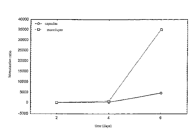
Table 1: Luteinisation index (P4/E2), standard
deviation and sample number of the bovine granulosa
cells cultivated in the capsules.
Days Mean Std. Dev. ~ N
2 5, I 32,0 39
4 567,9 2245,3 35
6 9452,5 18254,4 23
From the results reported in table 1 it is deduced
that cellular vitality is maintained in the encapsulated
cells, with the production of both hormones throughout
the entire culture period: The encapsulated cel 1s
produce low quantities of progesterone as observed in
vivo in the follicle prior to ovulation.
That indicates reduced luteinisation of the
encapsulated cells, which can only occur in follicular
structures very similar to those found in vivo.
The information derived from analysis of the
results underline that the bovine granulosa cell s,
encapsulated according to .the process of the present
invention, have steroidal activity analogous to that in
21
CA 02558306 2006-06-02
WO 2005/041942 PCT/EP2004/012384
vivo and obtainable only with a three-dimensional type
cell culture process.
Reference examp~.e 1
In parallel, cell culture has been carried out in
monolayers, in this case also evaluating the
steroidogenic activity in terms of the production of
progesterone (P4) and 17(3-oestradiol (E2); the
concentrations of such hormones in the samples of medium
withdrawn from the wells have been evaluated using
radioimmuno assay (RTA).
Non-encapsulated cells are seeded and cultivated in
monolayers in welled plates, each containing 600 ~1 of
the culture medium also used for the culture of the
encapsulated cells.
Analogously to that described for the encapsulated
cells, the plates containing the cells in monolayers are
maintained in an incubator for 6 days at 38.5°C, 5% CO~
and 90% humidity.
Every 48 hours, from each well, samples of the
medium containing the cellular metabolic products are
taken; the samples are frozen in Eppendorf tubes, at a
temperature of less than -20°C. From the wells
containing the cells cultivated in monolayers, the
culture medium is completely removed and substituted
22
CA 02558306 2006-06-02
WO 2005/041942 PCT/EP2004/012384
with fresh medium, with the continuation of the culture
on the same sample. The results obtained are reported
in table 2.
Table 2: Luteinisation index (P4/E2), standard
deviation and sample number of the bovine granulosa
cells cultivated in monolayers.
Days Mean Std. Dev. N
2 7,4 38,5 27
4 1700,0 3870,9 23
6 70201,0 131436,5 11
In figure 1 axe reported the luteinisation indices
of the bovine granulosa cells cultivated in monolayers
and in the capsules as a function of culture time.
From the results reported in figure 1 it may be
deduced that cellular vitality is maintained with both
culture techniques, with the production of both hormones
for the entire culture period.
Regarding the progesterone synthesised by the cells
cultivated in monolayers, a significant increase in its
concentration is observed on the 6th day of culture:
this increase is an indication of marked cellular
23
CA 02558306 2006-06-02
WO 2005/041942 PCT/EP2004/012384
luteinisation.
Such increase is less evident for the encapsulated
cells which produce low quantities of progesterone as
observed in vivo in the follicle prior to ovulation.
That indicates reduced luteinisation of the
encapsulated cells and can only occur in follicular-like
structures very similar to those found in vivo.
Example 2: eacapsulatioa ax~.d three-dimensional
culture of porcir~.e graaulosa cells
2a) Preparation of the cells
The ovaries at various stages of the oestrous cycle
are removed from subjects, starting from 6-11 months of
age, during normal slaughter, washed with physiological
solution at 30°C, as known to those skilled in the art.
Follicles having a diameter of 2-6 mm are identified in
the ovaries, from which the follicular liquids,
containing the granulosa cells, are aspirated using
syringes. The cellular suspensions thus obtained are
centrifuged and washed twice with 10 ml of TCM199 medium
+ 10o foetal calf serum + 1% penicillin/streptomycin.
Following centrifugation, a cellular sediment is
obtained, the cellular concentration of which is
determined by direct counting using a Makler chamber.
24
CA 02558306 2006-06-02
WO 2005/041942 PCT/EP2004/012384
2b) Encapsulation
The cellular sediment is diluted in a solution of
xanthan gum (Satiaxane~, SKW Biosystems, France) at 0.5%
in TCM199 culture medium containing Earle salts, L-
glutamine and sodium bicarbonate (Sigma-Aldrich,); the
cellular sediment to xanthan gum solution volume ratio
is 1:3. A cellular suspension is obtained, to which is
then added a saturated barium chloride solution up to a
final concentration of 20 mmol/1 of barium ions. The
resulting suspension is extruded through needles
(26GX1/2", 0.45X13 mm) into a medium viscosity (3500 cP,)
sodium alginate solution at 0.5% w/v in culture medium,
kept stirring using a magnetic stirrer (30 rpm). The
cellular suspension to sodium alginate solution volume
ratio is 1:25. The extrusion takes place dropwise
through the syringe, at a temperature of 25°C. The
barium ions react with the sodium alginate forming a
barium alginate membrane at the interface of the
individual drops of extrudate within 30'. Capsules are
obtained, which are collected by filtration, washed
twice with culture medium and suspended in an aliquot of
the same. Said capsules are subsequently cross-linked
on their external surfaces using a to solution of
protamine sulphate (Sigma-Aldrich, Milan, Italy) in
TCM199 culture medium containing Earle salts, L-
CA 02558306 2006-06-02
WO 2005/041942 PCT/EP2004/012384
glutamine and sodium bicarbonate (Sigma-Aldrich, ) for
30 minutes at a temperature of 25°C.
The population of granulosa cells is found inside
the cross-linked capsule, in an artificial extracellular
matrix.
Spheroidal shaped capsules axe obtained having
dimensions between 2 mm and 10 mm and weights between
20 mg and 100 mg. The capsules thus produced may be
preserved, under normal laboratory conditions, in
specific controlled environment incubators, by
lyophilisation, refrigeration, freezing or
cryopreservation.
2c) Three-dimensional cell culture
A capsule is placed in a sterile cell culture plate
well suspended in 600,1 of culture medium (TCM199
containing foetal calf serum (10%),
penicillin/streptomycin (1%) and 3-l7androstenedione
(100 ng/~1) ) .
The plates containing the capsules are maintained
in an incubator for 6 days at 38.5°C, 5a C02 and 90%
humidity.
Every 48 hours, from each well, samples of the
medium containing the cellular metabolic products are
taken; the samples are promptly frozen in Eppendorf
26
CA 02558306 2006-06-02
WO 2005/041942 PCT/EP2004/012384
tubes, at a temperature of less than -20°C.
In the wells containing the capsules, the culture
medium is substituted with an equal volume of fresh
medium, with the continuation of the culture on the same
sample.
Hence, the steroidogenic activity, in terms of the
production of progesterone (P4) and 17(3-oestradiol (E2),
has been evaluated in each sample of medium removed from
the wells by radioimmuno assay (RIA).
The results are expressed as the ratio between P4
and E2, known to those skilled in the art as the
luteinisation index.
Table 3: Luteinisation index (P4/E2), standard
deviation and sample number of the porcine granulosa
cells cultivated in the capsules.
Days Mean Std. Dev. N
2 14,8 55,3 38
4 124,0 338,2 30
6 43,7 83,8 20
From the results reported in table 3 it may be
deduced that cellular vitality is maintained in the
encapsulated cells, with the production of both hormones
throughout the entire culture period: The encapsulated
27
CA 02558306 2006-06-02
WO 2005/041942 PCT/EP2004/012384
cells produce low quantities of progesterone as observed
in vivo in the follicle prior to ovulation.
That indicates reduced luteinisation of the
encapsulated cells, which can only occur in follicular
structures very similar to those found in vivo.
Reference example 2
In parallel, cell culture has been carried out in
monolayers, in this case also evaluating the
steroidogenic activity in terms of the production of
progesterone (P4) and 17(3-oestradiol (E2); the
concentrations of such hormones in the samples of medium
withdrawn from the wells have been evaluated using
radioimmuno assay (RIA).
Non-encapsulated cells are seeded and cultivated in
monolayers in welled plates, each containing 600 ~,1 of
the culture medium also used for the culture of the
encapsulated cells. Analogously to that described for
the encapsulated cells, the plates containing the cells
in monolayers are maintained in an incubator for 6 days
at 38.5°C, 5o COZ and 90o humidity.
Every 48 hours, from each well, samples of the
medium containing the cellular metabolic products are
taken; the samples are frozen in Eppendorf tubes, at a
temperature of less than -20°C. From the wells
28
CA 02558306 2006-06-02
WO 2005/041942 PCT/EP2004/012384
containing the Bells cultivated in monolayers, the
culture medium is completely removed and substituted
with fresh medium, with the continuation of the culture
on the same sample. The results obtained are reported
in table 4.
Table 4: Luteinisation index (P4/E2), standard
deviation and sample number of the porcine granulosa
cells cultivated in monolayers.
Days Mean Std. Dev. N
2 2,5 2,3 36
4 22,1 23,4 25
6 2160,9 4997,9 24
In figure 2 are reported the luteinisation indices
of the porcine granulosa cells cultivated in monolayers
and in the capsules as a function of culture time.
The information derived from analysis of the
results underline that the porcine granulosa cells,
encapsulated according to the process of the present
invention, have steroidal activity analogous to that in
vivo and obtainable only with a thee-dimensional type
cell culture process.
29