Note : Les descriptions sont présentées dans la langue officielle dans laquelle elles ont été soumises.
CA 02559231 2006-09-11
Attorney Docket Number: ATA-416
PATENT APPLICATfON FOR
SUBCUTANEOUS NEEDLE CONNECTION SYSTEM
S FIELD OF THE INVENTION
The present invention relates to a surgical implant to provide a subcutaneous
connection to a vascular system of a patient. More particularly the present
invention relates
to a subcutaneous needle connection system for providing repeated access to
the vascular
system of a patient.
BACKGROUND OF THE INVENTION
A number of patients today undergo recurring medical procedures requiring
repeated
skin penetration to access the patient's vascular system and internal organs,
including organs
contained within the mediastinal, chest, abdominal and peritoneal cavities.
One such
recurring medical procedure is hemodialysis, which is used to treat kidney
failure by
removing harniful wastes and excess salts and fluid from a patient's blood.
Currently, over one million patients worldwide suffer from End State Renal
Disease
(ESRD) conditions and require some form of daily or thrice weekly dialysis
treatment via
needle or catheter access. Peritoneal Dialysis is one form of dialysis
treatment requiring
needle or catheter access whereby fluids placed into the peritoneal cavity via
a temporary or
permanently placed access catheter provide osmotic transfer of blood
containing toxins into
solutions pumped into and removed from within the peritoneal organ cavity. A
second form
of dialysis treatment is a direct blood filtering process, whereby a needle or
catheter is placed
directly into a vein or artery, and through a series of connecting tubing,
blood is removed,
filtered and re-circulated back into the patient after filtration of the
blood. These two
hemodialysis procedures are the most common means for metabolic toxin removal
from body
fluids when a patient experiences total or bilateral renal failure.
Hemodialysis requires creation and maintenance of vascular access, which is
the site
on the body where blood will be removed and returned to a patient's body
during dialysis. In
some applications, such as dialysis, substantial volumes of fluid are
circulated through the
CA 02559231 2006-09-11
Attorney Docket Number: ATA-416
vascular system of a patient over a multi-year period. Without needle or
indwelling catheter
organ access for dialysis, there is no physical connection means to conduct
dialysis toxin
removal, and the ESRD patients would die within days of total renal failure.
Hence, the
insertion method and form of dialysis connection access relates directly to
the patient's ability
to have body fluids contained within an internal organ communicate and be
safely connected
"externally outside the body" for the purposes of blood hemofiltration.
Generally, some form of needle or indwelling catheter organ access is
typically
required as a physical connection means to conduct dialysis toxin removal. The
insertion
method employed and the form of dialysis access used affects the patient's
ability to have
body fluids contained within an internal organ communicate and be safely
connected
externally outside the body for the purpose of blood hemofiltration.
One approach to accessing the vascular system is the use of two catheters that
are, at
one end, each inserted in blood vessels and are routed out of the body through
the skin,
leaving six or more inches of catheter length outside the body for connection
to a dialysis
machine. However, the skin surrounding the holes where the catheters enter the
body have
trouble sealing around the catheters and can become infected. Another option
for accessing
the vascular system is creating an arteriorvenous (AV) fistula or graft for
connecting an artery
to a vein in the arm. However, the creation of an AV fistula or graft can be
difficult or
impossible in some patients.
In addition, because both types of dialysis treatment techniques discussed
above
require trained medical personnel for needle or catheter access and the
administration of the
actual external dialysis filtration process, there are significant health care
hazards for both the
patient and healthcare worker associated with such traditional needle access
methods. Most
ESRD patients must be transported to a public dialysis treatment center for
treatment.
There are a number of additional complicating issues relating to the process
of
repeatedly sticking and cannulating a patient's circulatory organ system and
removing/returning blood back to the patient. One significant complication is
the need for
maintenance of a sterile installation and connection technique for all
components involved in
establishing dialysis access through the patient's skin and into the hollow
targeted organ, as
-2-
CA 02559231 2006-09-11
Attorney Docket Number: ATA-416
well as connection to the dialysis tubing set, filter, and dialysis pump
apparatus. Another
complication is the inability of a particular artery or vein to be repeatedly
cannulated or
punctured at substantially the same convenient needle or catheter access site,
due to vessel
trauma, exit wound inflammation, dialysis graft complications, and/or enlarged
needle hole
formation resulting in massive needle hole bleeding/ hematoma formation.
Repeated dialysis
needle punctures create unwanted scar tissue formation and infection. Both
conditions are
directly related to repetitive needle and catheter cannulation through the
skin. Such large
gauge needle cannulation complications are uncomfortable for the patient and
the healthcare
worker, due to the associated pain of additional needle sticks. Infection
complications of
these needle access sites are difficult to treat, because of the constant
migratory effects of
nosocomial infections, which have been well documented to routinely originate
from direct
contact with topical skin sourced Staphylococcocus bacteria. These frequently
occurring
needle access complications often require surgical intervention to repair,
reconstruct, or
remove the affected vessel organ, in addition to requiring extended hospital
admission and
costly medication treatment with IV medications.
One approach to the above problems is to provide a subcutaneous vascular
access
system that has a catheter connected to a blood vessel. The system is
configured to receive a
needle to provide access to the catheter. However, prior vascular access
systems used for
percutaneous needle cannulation for removal and return of blood during
dialysis suffer many
clinically devastating problems. Clinical problems include, but are not
limited to,
complications from poor tissue healing around the needle holes in the skin and
subcutaneous
needle tracks under the skin, caused by multiple puncturing of the same
location,
subcutaneous necrosis due to the proximity of the needle entrance to such
devices, fluid
contamination from leakage within the system and outside the needle entry,
poor tissue
coverage due to the shape and location of the indwelling permanent catheter
connections as
they exit the access system device, risk of infection caused by the
compromised skin healing
and leaking fluid during and after needle insertion, infection accumulation
around the catheter
connection and other complications.
One example of such an access system is a port vascular access assembly made
by
Vasca, Inc., described in U.S. Patent Number 6,565,525. The Vasca, Inc.,
vascular access
assembly requires two separate port units, one of which is used for input of
blood back into
-3-
CA 02559231 2006-09-11
Attorney Docket Number: ATA-416
the body and another for output of blood from the body. These ports are
located just under
the skin at a distance not far from each other, anchored to the subcutaneous
tissue under the
skin to inhibit movement, and require a sharp bevel tipped needle to be
inserted in the same
specific skin location and same perpendicular skin orientation every time. Use
of the Vasca,
Inc. vascular access assembly requires repeated punctures to skin, which is
usually found to
be stretched across the top of each port's needle entrance hole. Many
clinicians who have
experience with these devices have observed the patients' skin to be taut over
the tops of
these domed port housings, with minimal subcutaneous tissue found between the
needle hole
of the skin and the port opening which the needle enters the implanted port
device. The
condition of the skin with such devices has been observed to often include a
poor blood
supply condition of the skin because of the stretched condition of the skin,
and the lack of
adequate vascularized subcutaneous tissue to help fight infection and promote
healing of the
injured skin and subdermal tissue. Furthermore, the extremely short path of
the needle
through the skin to the opening of these port devices also provides less
opportunity for the
vascularized tissue to help control and prevent bacterial infection from
occurring in and
around the openings to the port opening. Moreover, because the needle
punctures are
occurring in the same skin location each time, infection and localized fluid
accumulation
abound the skin and device is common. The needle punctures create a clottable
buttonhole
over time, which is a skin injury that is not well controlled or completely
sealed to the
environment and therefore prone to repeatable, if not chronic infection. A
further difficulty
of conventional port access systems is their fluid filling chamber design,
open bevel needle
exposure to flowing blood fluids, causing massive blood cell damage and
turbulent disruption
to the blood by requiring sharp changes in fluid flow direction, such as a
90° change shortly
just prior to fluid entering the exposed beveled edge of the needle, or just
after exiting the
open end of the needle as fluid enters the device fluid chamber.
SUMMARY OF THE INVENTION
The present invention provides a subcutaneous needle connection system for
providing access to a vascular system of a patient that has enhanced
mechanical stability
under the skin, improved sealing, improved fluid flow dynamics, improved
needle hole
healing potential of the skin and improved needle edge interfacing to minimize
flow
disturbance and blood cell trauma during use. The subcutaneous needle
connection system is
-4-
CA 02559231 2006-09-11
Attorney Docket Number: ATA-416
not a port chamber device like other early access systems, as it comprises a
housing having a
shaped bottom surface designed to facilitate stability of the subcutaneous
needle connection
system with a straight bidirectional fluid path. The subcutaneous needle
connection system
is connected to at least one catheter in communication with a vascular system
of a patient and
further includes one or more needle access openings for receiving a needle for
accessing
blood from the patient or returning treated blood to a patient for a procedure
such as
hemodialysis. The needle access openings may include a set of inner valves
configured to
automatically open upon insertion of a needle and a set of outer valves that
may require
manual activation in order to open, thereby ensuring enhanced sealing of the
fluid paths.
The needle access openings may be angled away from each other to facilitate
needle
insertion and reduce operator error by preventing accidental insertion of the
wrong needle in
an opening. In addition, the surgically implanted housing may be manually
rotated within the
body or manipulated by an operator to increase the needle penetration zone are
of the skin,
reducing the need for repeated penetration of one area of skin during
subsequent needle
insertions into the needle connection system. Furthermore, the subcutaneous
needle
connection system may provide a substantially turbulent-free fluid flow path
between one or
both needle access openings and a corresponding catheter to maximize blood
flow through
the subcutaneous connection device.
According to one aspect of the invention, a subcutaneous needle connection
system
for providing access to a vascular system of a patient is provided. The
subcutaneous needle
connection system comprises a housing including a first needle opening
configured to receive
a needle, a second needle opening configured to be placed in communication
with the
vascular system and a first passageway connecting the first needle opening and
the second
needle opening. The system fiirther includes a first valve disposed in the
first needle opening,
the first valve configured to automatically open upon insertion of a needle in
the first needle
opening to provide access to the first passageway. A second valve is also
provided in the first
needle opening that is configured to be mechanically activated to provide
access to the first
valve.
According to another aspect of the invention, a subcutaneous needle connection
system comprises a housing, a first needle opening configured to receive a
needle formed in
-5-
CA 02559231 2006-09-11
Attorney Docket Number: ATA-416
the housing, a second needle opening configured to be placed in communication
with the
vascular system formed in the housing, and a first passageway extending
through the housing
and connecting the first needle opening and the second needle opening. The
housing defines
a bottom surface having three feet configured to rest on a subcutaneous
surface within the
patient.
According to still another aspect of the invention, a subcutaneous needle
connection
system for providing access to a vascular system of a patient comprises a
housing and a
needle mating system. The housing includes a first needle opening configured
to receive a
needle, a second needle opening configured to be placed in communication with
the vascular
system and a first passageway connecting the first needle opening and the
second needle
opening. The needle mating system is provided within the first needle opening
for placing a
needle inserted into the first needle opening in fluid communication with the
first
passageway, the needle mating system including a beveled surface configured to
mate with a
beveled surface on a needle inserted in the first needle opening.
BRIEF DESCRIPTION OF THE DRAWINGS
The invention will be apparent from the description herein and the
accompanying
drawings, in which like reference characters refer to the same parts
throughout the different
views.
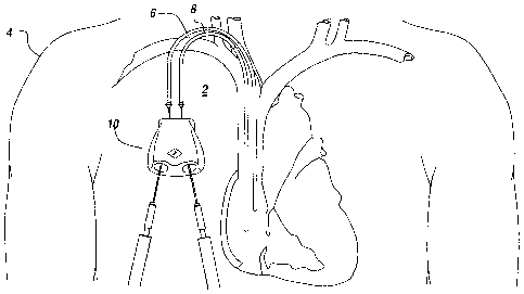
Figure 1 illustrates an embodiment of a subcutaneous needle connection system
implanted in a human body according to one aspect of the present invention;
Figure 2 is a top view of the subcutaneous needle connection system of Figure
1;
Figure 3 is a bottom view of the subcutaneous needle connection system of
Figure 1;
Figure 4 is a perspective view of the subcutaneous needle connection system of
Figure
1;
Figure 5 is a side view of the subcutaneous needle connection system of Figure
l;
Figure 6 is a back side view of the subcutaneous needle connection system of
Figure
1;
Figure 7 is a front side view of the subcutaneous needle connection system of
Figure
1;
_6_
CA 02559231 2006-09-11
Attorney Docket Number: ATA-416
Figure 8 is a top cross-sectional view of the subcutaneous needle connection
system
of Figure 1; and
Figure 9 illustrates the inner and outer set of valves of a needle entry
opening of the
subcutaneous needle connection system of Figure 1.
DETAILED DESCRIPTION OF THE INVENTION
An illustrative embodiment of the present invention provides a subcutaneous
needle
connection system for providing access to a vascular system of a patient that
exhibits
enhanced mechanical stability under the skin, improved fluid flow dynamics,
improved
needle hole healing potential of the skin, improved needle edge interfacing to
minimize flow
turbulence and blood cell trauma during use, and other features. The invention
will be
described below relative to certain illustrative embodiments. Those skilled in
the art will
appreciate that the present invention may be implemented in a number of
different
applications and embodiments and is not specifically limited in its
application to the
particular embodiments depicted herein.
Figures 1-7 illustrates an embodiment of a subcutaneous needle connection
system 10
for providing access to a vascular system of a patient according to an
illustrative embodiment
of the invention. The illustrative subcutaneous needle connection system
ensures smooth,
laminar flow of a body fluid, such as blood, accessed and/or returned to the
body using the
needle connection system, enhanced interfacing between needles, valves and
fluid paths,
leak-free needle connection capability, reduced operator error, minimal pain
to a patient,
minimal skin healing complications, and other improvements over vascular port
access
systems of the prior art.
As shown in Figure 1, the illustrative needle connection system 10 may be
configured to be implanted in a subcutaneous region of a patient, such as the
chest 2 of a
patient 4, as shown in Figure 1. Preferably, the needle connection system 10
is completely
embedded in the skin. When implanted in a patient, the needle connection
system 10 is
placed into communication with the vascular system via cannulas 6, 8 connected
to one or
more cannula openings of the needle connection system 10 and extending into a
blood vessel
3 of the patient. The needle connection system 10 forms needle access
openings, as
CA 02559231 2006-09-11
Attorney Docket Number: ATA-416
described in detail below, for receiving needles that may be placed into
communication with
the cannulas via the needle connection system. A first needle inserted in the
needle
connection system may deliver treated fluid, such as blood treated by a
dialyzer of a
hemodialysis system, to the needle connection system. The first cannula 6
receives the
treated fluid from the needle connection system and passes the treated fluid
back to the
vascular system. The second eannula 8 brings untreated fluid, such as blood
from the
vascular system, to the needle connection system 10. Another needle inserted
in the needle
connection system may receive and convey the untreated fluid provided by the
second
cannula 8 to the hemodialysis system or other treatment system.
According to an illustrative embodiment, the needle connection system may be
used
for receiving and returning treated blood during hemodialysis, though one
skilled in the art
will recognize that the needle connection system may have any suitable
application requiring
removal and return of a body fluid from and/or to a patient.
The illustrative subcutaneous needle connection system 10 preferably provides
for
parallel blood flow with a dual valve guided entry connection system. The
needle connection
system of the illustrative embodiment of the invention creates an ideally
dynamic blood flow
pattern, whereby uniform laminar flow and blood velocity remains continuous
from the
connection system entry to exit. Preferably, the blood follows a substantially
straight path
through the needle connection system 10, reducing disruptions caused by turns
in the flow
path.
The illustrative needle connection system 10 of the illustrative embodiment of
the
invention further facilitates the connection between the cannulas and the
needles for
accessing a body fluid. The needle connection system allows for improved
tactile insertion of
a percutaneous dialysis needle and for tactile confirmation of needle
alignment. The
illustrative needle connection system may include a plurality of sets of
valves for controlling
access to a needle access opening and preventing leaks. In one embodiment, the
system may
facilitate tactile needle alignment following tactile opening of a first
mechanically operated
valve.
_g_
CA 02559231 2006-09-11
Attorney Docket Number: ATA-416
The illustrative needle connection system may also provide a relatively large
needle
penetration area or "cannulation zone" so as avoid repeat needle entrance at
the same location
and to allow adequate repetitive dialysis needle cannulation site healing
between each
treatment. The relatively large penetration area provided by the illustrative
needle connection
system also provides a larger target for the needle, allowing some leeway
during penetration.
Referring to Figures 2-7, the illustrative subcutaneous needle connection
system 10
comprises a housing 12 having a cannula end 12b for interfacing with the
cannulas 6, 8 in
communication with a vascular system. The illustrative cannula end 12b
includes a first
cannula opening 22 configured to be connected to a first cannula 6 connected
to a blood
vessel of a patient and a second cannula opening 24 to be connected to a
second cannula 8
connected to a blood vessel of a patient. One of the cannula openings 22 may
be an outlet for
returning blood to the cannula 6 and vascular system from an associated needle
inserted in the
needle connection system, while the other cannula opening 24 may be an inlet
for receiving
blood from the vascular system and passing the received blood to an associated
needle for
treatment inserted in the needle connection system.
The housing 12 also includes a needle end 12a for interfacing with the needles
inserted through the skin and connected to an outside hemodialysis system or
other suitable
system for treating the body fluid accessed by the needle connection system
10. The
illustrative needle end 12a includes a first needle access opening 32 for
receiving a first
needle and a second needle access opening 34 for receiving a second needle.
Passageways
42, 44 are formed within the housing 12 to selectively connect each needle
access opening
32, 34, to an associated cannula opening 22, 24, respectively.
In the illustrative embodiment, one of the needle access openings 32 is
associated via
passageway 42 with the outlet cannula opening 22, and receives a needle for
providing blood
from a hemodialysis system to the needle connection system, which passes the
treated blood
to an associated cannula 6 for delivery to the vascular system. The other of
the needle access
openings 34 may receive a needle for receiving blood from an associated
cannula 8 via the
needle connection system 10 for delivery to the hemodialysis system for
treatment, which
then returns the treated blood to the body through the needle connection
system 10.
Preferably, the passageways 42, 44 provide for parallel blood flow entering
and exiting the
-9-
CA 02559231 2006-09-11
Attorney Docket Number: ATA-416
vascular system via the needle connection system 10. Valves are also provided
within the
passageways and/or needle access openings for selectively sealing the
passageways and
preventing fluid flow through the system after needle removal from the needle
connection
system, as described below.
The needle connection system 10 is preferably about the size of a pacemaker or
smaller, i.e., less than about two inches across and less than about one-half
an inch in
thickness. The needle connection system 10 is installed surgically under the
skin, in a
location suitable for directed needle cannulation through the skin, and
whereby a catheter can
be inserted into the patient's vascular system and connected in a kink-free
condition to the
needle connection housing. The needle connection system housing preferably has
a
substantially pendulum-shaped configuration that tapers from the needle end
12a and the
needle-connection openings to the cannula-connected end 12b and the cannula
openings.
When surgically installed in the body under the skin in a preferred needle
accessible location,
the needle connection system 10 is preferably located subcutaneously in an
area which allows
the implanted device to be manually rotatable or somewhat movable under the
patients' skin
by an operator to expose new skin areas for piercing by the needles for
connection with the
needle openings of the implanted housing, thereby increasing the needle
cannulation zone of
the skin. For example, one end of the needle connection system, such as the
cannula end 12b
may be stitched or otherwise fixed to the body, while the other end, such as
the needle end
12a may be selectively movable by the operator to expose a new skin area prior
to penetration
of the needle into the tissue 12a. The increased needle penetration or
cannulation zone allows
a new skin area to be manually exposed and pierced by the operator to access
the needle
connection system, allowing for healing of each injured skin location without
re-penetrating
the exact same needle hole location. In this manner, the illustrative needle
connection system
may reduce or avoid repeated trauma to the same skin area each time the
vascular system is
accessed via the implanted needle connection system.
The needle connection system 10 of the illustrative embodiment of the
invention
creates an ideal dynamic blood flow pattern therethrough, whereby uniform
laminar flow and
blood velocity remains substantially continuous from the inlet openings 22, 32
to the outlet
openings 24, 34. The openings may also be configured to facilitate alignment
of a needle axis
with a corresponding passageway axis to enhance blood flow through the needle
access
-10-
CA 02559231 2006-09-11
Attorney Docket Number: ATA-416
device. The alignment ensures uniform continuity of blood flow, blood flow at
a constant
speed as well as smooth physiologic flow. Preferably, the path has a
substantially straight
fluid path through the system to facilitate the smooth non-turbulent and
bidirectional fluid
flow. Taking the example of body fluid flow in a patient's blood vessels, each
vessel runs
generally parallel along the straight line direction of, e.g., the patients
arm, leg, torso, or
internal body cavity. It is desirable when body fluid is to be removed and/or
re-circulated
back into the patient's body for the body fluid within the needle access
system to follow a
generally straight line flow path that angles up and away from the surface of
the cannula, then
returns back into the patient at the same generally straight line flow path
and divergent angle.
As shown, the needle connection system provides a substantially straight fluid
path between
each cannula and needle, with all blood flow remaining substantially parallel
to the direction
of an aligned inserted needle. Thus, the needle connection system 10 directs
the flow of
blood away in a generally straight line flow path from the body fluid organs
or blood vessels,
and returns the blood to back to the blood vessels in the same generally
straight line direction,
without using sharp angles that could cause undue fluid turbulence and blood
cell damage.
Sharp, hard edge angles can adversely affect the natural flow dynamics of the
blood, and
damage fragile blood cell components, in addition to inducing chemical
activation of certain
blood containing components such as platelets, and circulating fibrinogen when
blood
component membranes are challenged by such turbulent forces.
The needle access openings 32, 34 for the needles are preferably formed in a
side
surface 130 forming the needle interface end 12a of the housing and the
openings 22, 24 for
the cannulas are preferably formed on a side surface 136 forming the cannula
interface end
12b of the housing 12. The location of the openings, in particular the needle
access openings
32, 34, on the side surfaces facilitates access to the openings, facilitating
insertion of needles
connected to a hemodialysis system into the needle connection system and also
enables the
substantially parallel, straight fluid flow paths. In addition, as described
below, the provision
of openings for interfacing with instruments outside of the body on the side
surfaces of the
system increases the cannulation zone and results in less stretching of the
skin, thereby
minimizing pain and infection.
The housing 12 may further include a topically recognizable manual actuator,
illustrated as a manual valve activation button 162 formed on a top surface
124 thereof, as
CA 02559231 2006-09-11
Attorney Docket Number: ATA-416
described in detail below, for selectively opening valves in the needle entry
openings 32, 34
by the operator to allow insertion of a needle therein.
The housing 12 may be shaped to facilitate stability of the illustrative
needle access
system 10 when implanted in a patient. For example, the bottom surface 122 of
the housing
may be formed with a tripod-like shape to enhance stability. The illustrative
tripod-shaped
bottom surface 122 may be tapered from the needle interfacing end 12a of the
housing toward
the cannula interfacing end 12b, as described above, to form a substantially
pendulum-shaped
housing. As shown in Figure 3, the tripod-shaped bottom surface 122 may
include a first foot
124 located at the cannula end 12b of the housing, proximate the cannula
openings 22, 24,
and second and third feet 126, 128, respectively, located at the needle end
12a of the housing,
proximate the needle access openings 32, 34. The second and third feet 126,
128 are
disposed on opposite sides of the needle end 12a. The feet 124, 126, 128 may
be in the form
of a line, edge, protrusion or other suitable shape. The edges 123, 125, 127
connecting the
feet 124, 126, 128, respectively, are preferably each formed in the shape of
an arch, so that
the bottom surface 122 may have a substantially concave shape.
The illustrative housing design provides enhanced stability relative to flat
planar
surfaces used in implants of the prior art. The design limits unintentional
movement of the
implanted needle connection system on the irregular surface on which it is
placed, as
movement of the system with the needles inserted therein during a transfer of
blood would be
dangerous. In addition, the tripod bottom surface 122 also withstands forces
without
compressing underlying tissue, thereby minimizing pain to the patient and
minimizing
obstructions to blood circulation. For example, the feet 124, 126, 128 lift
much of the bottom
surface up, off, and away from direct contact with the subcutaneous surface on
which the
device is placed. The illustrative shape also distributes downward loads to
the center of
housing 12, which further increases stability while helping to minimize
subcutaneous tissue
compression and pain to the patient during needle insertion. In contrast, the
flat bottom
surfaces used in implants of the prior art tend to be unstable on irregular
surface, as well as
prone to movement and compression of underlying tissue.
One skilled in the art will recognize that the bottom surface may
alternatively have
four feet, or another suitable shape configured to facilitate stability and is
not limited to the
-12-
CA 02559231 2006-09-11
Attorney Docket Number: ATA-416
illustrative tripod shape. For example, a subcutaneous needle connection
system may have
four or more feet on a bottom surface, a flat surface, a convex surface or any
other suitable
shape.
The side surfaces 132, 134 extending between the needle end 12a and the
cannula end
12b of the housing 12 may be configured with slight depressions 1321, 1341,
respectively, to
enhance the grip of a surgeon when stabilizing the needle connection system 10
to insert the
needles into the needle access openings 32, 34.
The housing 12 may be formed of molded plastic, clear acrylic, titanium, or
another
suitable surgical material known in the art.
According to an illustrative embodiment of the invention, the illustrative
needle
connection system has a needle mating system within each needle access opening
32, 34 that
includes an inner set of valves and an outer set of valves for controlling
access to the needle
access openings 32, 34 and associated passageways 42, 44, respectively. Figure
8 illustrates
the inner set of valves 182, 184 and outer set of valves 192, 194
respectively. Figure 9 is a
detailed view of one of the needle access openings 32 and associated
passageways 42, having
an inner valve 182 and an outer valve 192 for selectively sealing the opening
and blocking
fluid flow therethrough.
The outer set of valves 192, 194 is preferably manually or mechanically
operable by a
user using a valve activator. In the illustrative embodiment, the outer valves
192, 194 require
mechanical activation to open and automatically close in the absence of a
mechanical
activation to maintain the valves in an open position. For example, in an
illustrative
embodiment, the valve activator comprises the valve activation button 162 on
the top surface
of the housing, which may be depressed by a user to manually open the outer
set of valves
192, 194 for the needle access openings 32, 34. The outer valves 192, 194 may
be
independently operated or a single operation may be used to operate both
valves. According
to an illustrative embodiment, each of the outer valves comprises a spring-
loaded stopcock
having a rotating cylinder that can not be opened unless the button 162 is
depressed. After
mechanical opening, the outer valves 192, 194 may be held open by a needle
inserted in an
associated opening 32 or 34 or by holding the button 162 in a depressed
position. The outer
-13-
CA 02559231 2006-09-11
Attorney Docket Number: ATA-416
valves 192, 194 preferably automatically close to maintain a leak-proof
closure means when a
user releases the valve activation button 162 and/or a needle is removed from
the associated
opening. One skilled in the art will recognize that any suitable means for
manually or
mechanically activating the outer valves may be used and that the invention is
not limited to
the illustrative valve activation button 162.
The inner set of valves 182, 184 may automatically open, for example, by
inserting a
needle into the respective opening after opening of the outer valves 192, 194
through
mechanical means. Preferably, the inner set of valves does not automatically
open with the
opening of the outer set of valves, but rather, when a needle is inserted
through the opened
outer valves. In an illustrative embodiment, each inner valve comprises a
sliding cylinder,
opened by the needle tip itself. Upon removal of the needle, the inner valves
automatically
close and seal to form a leak-proof seal within the system 10.
The use of an outer set of valves that can only be manually opened and
automatically
close otherwise further prevents tissue from growing into the needle entrance
when the
dialysis needle is not engaged.
According to another aspect of the invention, each of the needle access
openings 32,
34 is configured to facilitate a uniform, smooth connection with a
corresponding passageway
42 or 44 for laminar flow of blood upon alignment of a needle with a
respective opening.
The needle access openings 32, 34 further provide for tactile feedback to the
operator
regarding the alignment of the needle. For example, as shown in Figure 9, each
needle access
opening may include a beveled surface 196 configured to mate with a beveled
surface of a
needle designed to be inserted therein. The use of mating beveled surfaces
provides
improved tactile insertion of a percutaneous dialysis needle by providing
tactile confirmation
of the needle engagement and alignment to the operator. Preferably, the
passageway 42 has
an inside diameter that matches the diameter of a needle inserted in the
associated needle
access opening 32 to facilitate connection of the needle. The matching beveled
edges when
in alignment to each other, help to minimize blood cell damage when flowing
over the
leading edge of the engaged needle, further improving laminar flow through the
system 10,
with less trauma to the blood. In addition to providing tactile feedback that
the needle has
become fully engaged and aligned properly in the system, the matching beveled
surfaces
-14-
CA 02559231 2006-09-11
Attorney Docket Number: ATA-416
prevent twisting of the dialysis needle after the needle fully engages within
the alignment
area. The mating beveled surfaces further ensure complete alignment of the
axis of the
needle received in the needle access opening when the needle is gently pushed
by the operator
to its fully engaged and aligned stopping point.
The needle connection system may be configured to interface with an extended-
length
access needle. The extended length may enable insertion of the needle a
substantial distance
from the needle insertion openings, proving a substantially larger needle
cannulation area of
skin for spacing out needle holes. The tip of the needle is cut in a bevel
best suited to mate
with the bevel edge of the internal surface formed in the alignment area
within the system,
allowing an operator to feel the mating of the bevel edges when the needle is
gently pushed
forward to engage the beveled surfaces, by reducing rotational freedom about
the axis of the
needle. A needle inserted in the opening may have a slight bending in the tip
to avoid scoring
the inside of the valve. Preferably, when the needle is fully inserted in a
needle access
opening of the needle connection system, the needle cannot be rotated after
the opposing
beveled edges have become completely engaged
In one embodiment, the leading tip edge of a penetrating needle may serve to
activate
the automatically opening of an inner valve in the opening. One skilled in the
art will
recognize that any suitable means for automatically opening the inner valves
may be used in
accordance with the teachings of the invention.
The needle may be locked in place in the opening 32 through friction fit
applied from
the valves 182 and/or 192. In addition, a seal, such as an o-ring 198, may be
disposed at the
interface between the opening 32 and the passageway 42 to engage around the
dialysis needle
to provide a leak-proof connection.
The passageways 42, 44 may be generally tubular and can be further sized,
dimensioned, or formed to either a conical or tapered shape, made generally
smooth surfaced
throughout, or made from several shorter faceted surfaces without sharp edges,
and/or made
coated, covered, or lined with medically purposeful bioactive substances (e.g.
such as a
coating or surface treatment that may contain one or more of an anticoagulant,
antiseptic,
gene therapy medication, anti-inflammatory medication, anti-proliferative
medication, anti-
-15-
CA 02559231 2006-09-11
Attorney Docket Number: ATA-416
biofilm or anti-microbial agent, a biological or lipophilic oil, or a
hydrophilic fluid surface
coating) to reduce needle insertion shear and friction resistance, or to
reduce internal flow
path fluid pressure resistance and flow rate resistance, internal flow path
wall surface shear
force, or to increase wall surface lubricity along all or a portion of
passageways 42, 44, or to
reduce the likelihood of circulating body fluid components and or blood cell
components
from being activated by direct surface contact with any portion of the
passageway 42, 44 and
including any portion of the needle access openings 32, 34 or cannula openings
22, 24. The
coatings can be placed in all or partially in the areas exposed to body
fluids. As described
above, the passageways 42, 44 are preferably straight and aligned with the
needle openings
32, 34 and cannula openings 22, 24 to promote smooth, laminar flow through the
system 10.
According to another embodiment of the invention, as shown in Figure 8, the
needle
access openings 32, 34 of the needle connection system 10 are divergent, i.e.,
angled away
from each other, to facilitate insertion of needles therein. As shown, the
axes 320, 340 of the
openings 32, 34, respectively, are oriented at an angle a with respect to each
other. The
divergent angling of the openings away from each other and from each entrance
site allows
each needle entrance to be adequately located so as to avoid "operator error"
by inadvertently
inserting the wrong flow direction needle in the incorrect needle entrance. In
addition, the
divergent configuration of the openings 32, 34 increases the radius of the
skin area available
for cannulation, minimizing the injury to a needle site. One skilled in the
art will recognize
that the angle a between the axes of the needle access openings may have any
suitable size,
and that the invention is not limited to divergent openings.
Preferable, the needle openings are positioned in the middle or higher of the
back side
surface 130 to inhibit an inserted needle from traveling under the connection
device 10.
According to another aspect of the invention, one or more of the external
needle
housing openings 32,34 can be substantially funnel-shaped to facilitate
directional guidance
of a needle tip into the center of the opening. The funnel shape increases the
cannulation
zone, and also provides a larger target area for guiding needles into the
openings, requiring
less precision when inserting a needle into each valve opening.
-1G-
CA 02559231 2006-09-11
Attorney Docket Number: AT A-416
To use the needle connection system 10 of an illustrative embodiment of the
invention, the needle connection system 10 is first implanted in a
subcutaneous region in
communication with the vascular system or other body fluid region. The system
may be
stitched to tissue to hold the system in a selected position, while allowing
limited rotation or
movement of the system in the subcutaneous region. For example, the cannula
end 12b may
be fixed, while allowing slight rotation of the needle end 12a relative to the
cannula end 12b.
After implantation, needles are inserted into the needle connection system 10
to access the
vascular system.
Prior to insertion of the needles, the system 10 may be angulated or rotated
slightly,
providing access to a wide area of skin that could be used to insert the
needles, allowing for
additional healing time before skin is punctured again. For example, the
needle end 12a of
the system 10 may angulate relative to the cannula end 12b, which may be fixed
to the
patient, in order to expose a new area of skin through which the needle can
puncture in order
to enter the valves.
In use, an operator will insert the first of two needles in the patient's
skin. As the
first needle approaches the associated needle access opening, the surgeon will
depress the top
button 162, opening the outer set of valves. As described above, each outer
valve in the
illustrative embodiment comprises a rotating cylinder that would not be opened
unless the
button is depressed. The insertion of the needle will automatically open the
inner valve on
the needle insertion side. The inner valve of the illustrative embodiment is
formed by a
sliding cylinder, opened by the needle tip itself. The operator continues to
insert the needle
through the second valve optionally rotating the needle slightly about its
axis to detect the
alignment mating of the beveled surfaces.
After insertion of the first needle, the second needle is inserted. The outer
valve
remains open because the first needle is holding it open, or the operator may
optionally
continue to depress the top button. The inner valve on the second side is
opened by the tip of
the second needle. Needle insertion continues until the bevels are mated.
Laminar flow of
blood or other fluids through the needle connection system is provided by a
smooth transition
from the needles to the associated catheter connection means.
-17-
CA 02559231 2006-09-11
Attorney Docket Number: ATA-416
As described above, because the needle access openings are located on a side
of the
device, the subcutaneous and dermal tissue punctured by the needle has
adequate blood
supply to help heal the injured tissue after needle withdrawal, as the tissue
is not overly
stretched and the device is not located next to or directly under the
traumatized skin and
subcutaneous tissue. The needle openings are also angled slightly outward,
further separating
the locations of the needle punctures from the skin, and the needle track
locations of tissue
through which the needle travels.
The illustrative needle connection system provides significant advantages over
prior
vascular access devices. For example, the needle connection system of the
illustrative
embodiment of the invention maintains uniform laminar flow of blood or another
body fluid
from entrance to exit. The system further provides improved tactile insertion
of a
percutaneous dialysis needle for tactile confirmation of complete needle
alignment. The
illustrative system further provides tactile entrance opening of the needle
connection system
with a rotating cylinder valve means and push button activator that
automatically closes upon
needle removal to maintain a leak-proof closure means. The illustrative
connection system
further provides for a permanent indwelling catheter attachment means to be
directed
downward, away from the skin, so as to prevent erosion of skin by the catheter
connection
means to the needle correction system. Another advantage of the invention is
that the
illustrative connection system provides for a large needle skin penetration
area or
"cannulation zone" to avoid repeat needle entrance at the same location,
thereby allowing for
healing between treatments. In addition, the illustrative needle connection
system may
reduce or prevent skin erosion due to contact with a catheter connection means
by directing
the permanent indwelling catheter attachment means downward, away from the
skin. The
illustrative needle connection system has an enhanced grip and feel,
facilitating handling by a
surgeon using the needle connection system to access the vascular system. The
illustrative
connection system also minimizes infection and pain while providing enhanced
blood
handling and other advantages not found or contemplated in vascular access
devices of the
prior art.
The present invention has been described by way of example, and modifications
and
variations of the exemplary embodiments will suggest themselves to skilled
artisans in this
field without departing from the spirit of the invention. Features and
characteristics of the
-18-
CA 02559231 2006-09-11
Attorney Docket Number: ATA-416
above-described embodiments may be used in combination. The preferred
embodiments are
merely illustrative and should not be considered restrictive in any way. The
scope of the
invention is to be measured by the appended claims, rather than the preceding
description,
and all variations and equivalents that fall within the range of the claims
are intended to be
embraced therein.
Having described the invention, what is claimed as new and protected by
Letters
Patent is:
-19-