Note : Les descriptions sont présentées dans la langue officielle dans laquelle elles ont été soumises.
CA 02561684 2006-09-28
WO 2005/108988 PCT/US2005/011914
REAL TIME METHOD OF DETECTING ACUTE INFLAMMATORY
CONDITIONS
CROSS-REFERENCES TO RELATED APPLICATIONS
[0001] This application claims the benefit of U.S. Provisional Application No.
60/560,986,
filed April 9, 2004 and claims the benefit of U.S. Provisional Application No.
60/579,621,
filed June 14, 2004; both of which are herein in incorporated by reference in
their entirety for
all purposes.
STATEMENT AS TO RIGHTS TO INVENTIONS MADE UNDER
0 FEDERALLY SPONSORED RESEARCH AND DEVELOPMENT
[0002] This invention was made with Government support under Grant Nos. R01 HL-
074335, RO1 AR-4676402, RO1 AR-46588, NCRR/GCRC MO1-RR-00056, K24 AR-
02213, K23 AR-051044, awarded by the National Institutes of Health. The
Government has
certain rights in this invention.
FIELD OF THE INVENTION
[0003] This invention relates to the diagnosis and/or monitoring of patients
with
inflammatory diseases or conditions, including systemic lupus erythematosus,
particularly for
diagnosis of the acute stage of the disease, including methods and kits for
carrying out this
activity. This disclosure presents the surprising discovery that levels of
complement pathway
0 components on reticulocytes can be used to diagnose, monitor, or predict the
occurrence of
acute episodes of chronic inflammatory diseases or conditions.
BACKGROUND OF THE 1NVENTION
[0004] This invention relates to the diagnosis and/or monitoring of patients
with an acute
episode of an inflammatory disease or condition. In some embodiments the
inflammatory
:5 disease or condition is systemic lupus erythematosus (SLE). The invention
also provides
means for predicting the onset of an acute episode of an inflammatory disease
or condition,
including SLE.
[0005] Monitoring disease activity is also problematic in caring for patients
with
inflammatory diseases or conditions. Chronic inflammatory diseases or
conditions frequently
SO progress in a series of flares, or periods of acute illness, followed by
remissions. Over time,
CA 02561684 2006-09-28
WO 2005/108988 PCT/US2005/011914
however, these flares can lead to irreversible organ damage. in order to
mmimlze such
damage, earlier and more accurate detection of disease flares would not only
expedite
appropriate treatment, but would reduce the frequency of unnecessary
interventions. From an
investigative standpoint, the ability to uniformly describe the "extent of
inflammation" or
activity of disease in individual organ systems or as a general measure is an
invaluable
research tool. Furthermore, a measure of disease activity can be used as a
response variable
in a therapeutic trial. Thus, there is a need for reliable methods to diagnose
or predict the
acute stage of inflammatory disease or condition, including SLE. The present
invention
meets these and other needs.
0 BRIEF SUMMARY OF THE INVENTION
[0006] This disclosure provides methods for diagnosing or monitoring an acute
inflammatory episode of a chronic inflammatory disease or condition in an
individual by (a)
determining the level of a complement pathway component on a reticulocyte from
the
individual, and (b) comparing the complement pathway component level with a
control level
of complement pathway component, where a difference from the control level of
the
complement pathway component indicates that the individual has the acute
inflarninatory
episode of the chronic inflammatory disease or condition. The level of more
than one
complement component can be determined and compared to a control level. For
example, a
ratio of complement pathway components can be determined and compared to a
ratio of
control complement pathway component levels. In some embodiments, an antibody
specific
for the complement pathway component is used to determine the level of the
complement
pathway component. In one embodiment, the level of the complement pathway
component
C4d is determined.
[0007] The disclosed methods can be used to diagnose or monitor an acute
inflammatory
condition in a number of chronic inflammatory diseases or conditions, e.g.
systemic lupus
erythematosus (SLE), hepatitis C infection, sickle cell anemia, complications
of
transplantation, and complications of pregnancy.
[0008] In one embodiment, an acute episode of SLE is diagnosed. For example,
to
diagnose or monitor an acute episode of SLE, the level of complement pathway
component
C4d on reticulocytes can be determined and compared to a level of complement
component
C4d on reticulocytes from a control. The level of complement component C4d can
be
determined using an antibody specific for C4d. A labeled C4d antibody can be
used and, in
some embodiments the C4d antibody is detected using flow cytometric analysis.
CA 02561684 2006-09-28
WO 2005/108988 PCT/US2005/011914
[0009] This disclosure also provides methods for predicting the occurrence of
an acute
inflammatory episode of a chronic inflammatory disease or condition in an
individual by (a)
determining the level of a complement pathway component on a reticulocyte from
the
individual, and (b) comparing the complement pathway component level with a
control level
of complement pathway component, where a difference from the control level of
the
complement pathway component indicates that the individual has the acute
inflammatory
episode of the chronic inflammatory disease or condition. The level of more
than one
complement component can be determined and compared to a control level. For
example, a
ratio of complement pathway components can be determined and compared to a
ratio of
control complement pathway component levels. In some embodiments, an antibody
specific
for the complement pathway component is used to determine the level of the
complement
pathway component. In one embodiment, the level of the complement pathway
component
C4d is determined.
[0010] The disclosed methods can be used to predict occurrence of an acute
inflammatory
condition in a number of chronic inflammatory diseases or conditions, e.g.
systemic lupus
erythematosus (SLE), hepatitis C infection, sickle cell anemia, complications
of
transplantation, and complications of pregnancy.
[0011] In one embodiment, an acute episode of SLE is predicted. For example,
to predict
an acute episode of SLE, the level of complement pathway component C4d on
reticulocytes
0 can be determined and compared to a level of complement component C4d on
reticulocytes
from a control. The level of complement component C4d can be determined using
an
antibody specific for C4d. A labeled C4d antibody can be used and, in some
embodiments
the C4d antibody is detected using flow cytometric analysis.
[0012] This disclosure describes and enables a kit for diagnosing, monitoring,
or predicting
5 an acute inflammatory episode of a chronic inflammatory disease or condition
in an
individual. The kit can include an antibody specific for a complement pathway
component
and a means for comparing a level of the complement pathway component to a
control level
of complement pathway component. A difference from the control level of the
complement
pathway component indicates that the individual has the acute inflammatory
episode of the
.0 chronic inflammatory disease or condition. In some embodiment an acute
episode of SLE is
diagnosed, monitored, or predicted. The antibody can be fluorescently labeled,
and in some
embodiments, a monoclonal antibody is used.
CA 02561684 2006-09-28
WO 2005/108988 PCT/US2005/011914
[0013] This disclosure also provides a computer readable medium for
diagnosing,
monitoring, or predicting an acute inflammatory episode of a chronic
inflammatory disease or
condition in an individual. The computer readable medium can include (a) code
for
receiving data corresponding to a determination of complement pathway
component on
reticulocytes; (b) code for retrieving a reference value for complement
pathway component
on reticulocytes of individuals; and (c) code for comparing the data in (a)
with the reference
value in (b).
BRIEF DESCRIPTION OF THE DRAWINGS
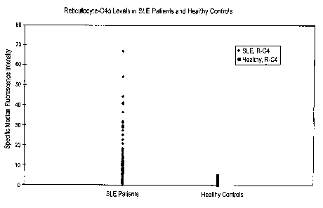
[0014] Figure 1 provides a graph plotting the values of median fluorescence
values for the
0 patients with SLE and healthy controls. R-C4 refers to C4d levels on the
surface of
reticulocytes. Values for patients with SLE are shown on the left; values for
healthy controls
are shown on the right.
[0015] Figure 2 provides tow color flow cytometry data from each of three
individual SLE
patients. C4d levels are shown in the right panels, while matched isotype
controls are shown
in the left panels. Two color flow cytometry was performed using a labeled
anti-Cd4
antibody (~.'-axis) and a labeled anti transfernn receptor antibody (X-axis).
C4d positive
reticulocytes are shown in the upper right quadrant of the panels. Results
from three SLE
patients are shown. 93%, 47.8%, and 14.5% of reticulocytes exhibited C4d
staining. The
control antibodies do not bind to reticulocytes, indicating the C4d antibody
binding is
0 specific.
[0016] Figure 3 provides data showing C4d levels on reticulocytes from an SLE
patient at
three time points: February 2002 (top row), July 2002 (middle row), and August
2002
(bottom row). C4d staining on unfractionated red blood cells, e.g.,
erythrocytes and
reticulocytes, is shown in the left column. The middle column shows two color
flow
;5 cytometry using a labeled anti-Cd4 antibody (Y-axis) and a labeled anti
transferrin receptor
antibody (X-axis) at different time points. C4d positive reticulocytes are
shown in the upper
right quadrant of the panels. The right column shows a comparison of C4d
levels on
erythrocytes and reticulocytes at different time points. Erythrocytes and
reticulocytes were
separated by density gradient centrifugation. The oldest cells elute beginning
in fraction 1.
>0 Reticulocytes are found with the youngest cells eluting in fraction 15. The
vertical axis
shows C4d levels on the surface of the cells.
CA 02561684 2006-09-28
WO 2005/108988 PCT/US2005/011914
[0017] , Figure 4A-D demonstrates that reticulocyte-C4d levels are
significantly elevated in
patients with SLE and fluctuate over time. (A) Reticulocytes from patients
with SLE have
significantly higher levels of C4d than those from patients with other
diseases or healthy
controls. Shown on the Y-axis is the C4d-specific median fluorescence
intensity for
reticulocytes from 156 patients with SLE, 140 patients with other diseases,
and 159 healthy
controls. (B) R-C4d levels remain stable in healthy controls and patients with
other diseases
over time. Shown are R-C4d levels of 7 healthy controls and 16 patients with
non-SLE
autoimmune diseases (1 scleroderma, 7 inflammatory myopathies, 1
Sjorgren°s syndrome, 6
rheumatoid arthritis, and 1 antiphospholipid antibody syndrome) examined at 3
or 4 different
0 study visits. (C and D) R-C4d levels fluctuate in a significant fraction of
patients with SLE.
Shown are R-C4d levels of 64 patients with SLE examined at 3 up to 5 different
study visits.
In 37 patients, R-C4d remained stably low. In 9 patients, R-C4d was elevated
at the first visit,
but decreased in subsequent visits. Remarkable fluctuation of R-C4d was
observed in 18
patients. Representative patients with different patterns of R-C4d were
selected for the case
studies shown in Figure SA-D.
[0018] Figure SA-D demonstrates that R-C4d fluctuates and reflects the
clinical course of
SLE. Shown are serial measurements of Reticulocyte-C4d (R-C4d) and Erythrocyte-
C4d (E-
C4d) from each representative patient with SLE. Numbers shown inside the graph
panel near
each point are values of C4d-specific median fluorescence intensity. See
Example 4 for
,0 additional clinical history and results of other laboratory tests. Normal
lab values are: serum
C4 level is 20-59 mg/dL; ESR 0-20 mmThr; fanti-dsDNA < 2 or < 1:10, depending
on the
type of assay used.
DETAILED DESCRIPTION OF THE INVENTION
Introduction
;5 [0019] The methods of this invention enable the diagnosis and/or monitoring
of acute
episodes of chronic inflammatory diseases or conditions, including SLE. This
disclosure
presents the surprising discovery that levels of complement pathway components
on
reticulocytes can be used to diagnose, monitor, or predict the occurrence of
acute episodes of
chronic inflammatory diseases or conditions. Because acute episodes of chronic
~0 inflammatory diseases and conditions, e.g., SLE, are serious health
problems, there is a need
for relatively accurate and early diagnosis of these conditions. Likewise, the
ability to
monitor or to predict the occurrence of acute episodes inflammatory diseases
or conditions is
of great importance.
CA 02561684 2006-09-28
WO 2005/108988 PCT/US2005/011914
~~ mG m~~il~l~l~ ~~~~mvG~ uwCrmnamons of the level of a complement patnway
component on reticulocytes. In some embodiments, the level of complement
pathway
component C4d is determined.
[0021] In part, the methods of this invention are based on the discovery by
the inventors
that the level of C4d deposited on surfaces of immature red blood cells, l.
e., reticulocytes, can
serve as a diagnostic marker for an acute inflammatory episode resulting from
SLE, a chronic
inflammatory condition.
[0022] In diagnosing the occurrence, or predicted occurrence of an acute
episode of a
chronic inflammatory disease or condition, the level of a complement pathway
component of
reticulocytes in a sample is determined. The determination is then compared
with the
quantities of a complement pathway component found on reticulocytes of
individuals not
having a chronic inflammatory disease or condition, or of individuals who are
not in the acute
phase of a chronic inflammatory disease or condition. For example, a level of
a complement
pathway component such as C4d can be determined on a reticulocyte of a patient
with SLE.
The determination is then compared with the quantities of a complement pathway
component
found on reticulocytes of individuals not having SLE, or of individuals who
are not in the
acute phase of SLE, to diagnose, monitor or predict the occurrence of an acute
episode or
flare of SLE.
[0023] In monitoring disease activity of a patient with an acute episode of a
chronic
inflammatory disease or condition, the same determinations are made in the
patient's blood
sample, and are then compared with determinations of the quantities of a
complement
pathway component present on surfaces of reticulocytes in a sample obtained
from the same
patient in the past.
[0024] Another use of this invention is to monitor complement activation
during the course
of human diseases. Current state-of the-art methods rely on measurement of
serum or plasma
levels of soluble complement C3 and/or C4. However there are known
inadequacies with
tlus approach. For example, C3 and C4 are parent molecules and are precursors
to activation
of the complement cascade. Increased hepatic and extra-hepatic synthesis of C3
and C4 can
balance increased C3 and C4 catabolism during activation of the complement
cascade
resulting in misleading change or lack or change in serum levels. In addition,
genetic
deficiencies of C4 are well documented and result in abnormally low
serum/plasma levels of
C4 due to lack of synthetic capacity that can be misinterpreted as being due
to increased C4
CA 02561684 2006-09-28
WO 2005/108988 PCT/US2005/011914
consumption during complement activation. The invention described herein is
based upon
measurement of protein products of complement activation such as C3d, C4d, and
others that
are attached to surfaces of circulating blood cells such as reticulocytes, and
others. This
enables monitoring levels of activation products as~opposed to reactants, and
eliminates the
weaknesses inherent in measuring soluble C3 and C4 described above. Thus,
levels of
complement pathway components on reticulocytes can be determined and compared
to
control levels of complement pathway components in order to diagnose or
monitor activation
of the complement pathway.
Definitions
0 [0025] As used herein, an "inflammatory disease or condition" refers to any
immune
disease or condition that causes increased inflammation in an individual. An
inflammatory
disease or condition also refers to any infectious disease or condition that
causes increased
inflammation in an individual. In some embodiments the inflammatory disease or
condition
is a '°chronic inflammatory disease or condition." A chronic
inflammatory disease or
5 condition is an inflammatory condition that does not resolve after a period
of weeks, months
or longer. Chronic inflammatory conditions can follow an acute inflammatory
condition, or
for some diseases or conditions can occur in the absence of an acute
inflammatory disease or
condition. An inflammatory disease or condition includes the following:
systemic lupus
erythematosus (lupus or SLE), rheumatoid arthritis, vasculitis (and its
specific forms such as
0 Wegener°s granulomatosis), scleroderma, myositis, serum sickness,
transplant rejection,
sickle cell anemia, gout, complications of pregnancy such as pre-eclampsia,
multiple
sclerosis, cardiovascular disease, infectious disease such as hepatitis C
virus infection, etc.
Each of these diseases or conditions can also be described as chronic
inflammatory diseases
or conditions.
5 [0026] An "acute inflammatory episode" as used herein refers to an increased
irmnune
response. Symptoms of acute inflammation include redness, heat, swelling,
pain, and loss of
function, e.g., loss of joint movement. An acute inflammatory episode of a
chronic
inflammatory disease or condition differs from the typical symptoms of a
chronic
inflammatory disease or condition in the following ways. Frequently, during an
acute
0 inflammatory response the liver synthesizes acute phase proteins or acute
phase reactants that
are detectable in the blood stream. While the presence of acute phase
reactants indicates that
an acute inflammatory condition is occurring in the body, they are not
diagnostic for a
specific acute inflammatory episode. Acute phase reactants include C-reactive
protein
CA 02561684 2006-09-28
WO 2005/108988 PCT/US2005/011914
(CRP); alpha 1-antitrypsm; alpha 1-antichyrnotrypsin; alpha ~-macroglobuhn;
coagulation
factors such as fibrinogen, fibrin, prothrombin, thrombin, factor VIII, and
plasminogen;
complement proteins, and serum amyloid protein. In addition, during an acute
inflammatory
episode, local inflammatory cells, e.g., neutrophils and macrophages, secrete
a number of
cytokines into the bloodstream, most notably IL-1, IL-6, IL-11, and TNF-alpha.
[0027] "Real time diagnosis" refers to diagnosis of an acute inflammatory
episode while the
inflammation or the acute inflammatory symptoms are occurring. Monitoring
markers on
reticulocytes provides real time diagnosis because reticulocytes are present
for only 1-2 days
before maturing into erythrocytes.
0 [0028] As used herein, a "reticulocyte" refers to an immature red blood
cell. Reticulocytes
are usually obtained by taking a blood sample from an individual. In some
embodiment, a
reticulocyte is isolated from a blood sample of an individual.
[0029] As used herein, the "complement pathway or system" refers to a complex
network
of more than 30 functionally linked proteins that interact in a highly
regulated manner to
provide many of the effector functions of humoral immunity and inflammation,
thereby
serving as the major defense mechanism against bacterial and fungal
infections. This system
of proteins acts against invasion by foreign organisms via three distinct
pathways: the
classical pathway (in the presence of antibody) or the alternative pathway (in
the absence of
antibody) and the lectin pathway. Once activated, the proteins within each
pathway form a
:0 cascade involving sequential self assembly into multimolecular complexes
that perform
various functions intended to eradicate the foreign antigens that initiated
the response. For a
review of the complement pathway, see, e.g., Sim and Tsiftsoglou, Biochem.
Soc. Traps.
32:21-27 (2004).
[0030] The classical pathway is usually triggered by an antibody bound to a
foreign
!5 particle. It consists of several components that are specific to the
classical pathway and
designated C1, C4, C2 . Sequentially, binding of Clq to an antigen-antibody
complex results
in activation of C 1 r and C 1 s (both are serine proteases), and activated C
1 s cleaves C4 and
C2 into, respectively, C4a and C4b and C2a and C2b. Fragments C4b and C2a
assemble to
form C4b2a, which cleaves protein C3 into C3a and C3b, which completes
activation of the
SO classical pathway. Fragments C4b and C3b are subject to further degradation
by Factor I.
This factor cleaves C4b to generate C4d and also cleaves C3b, to generate iC3b
followed by
C3d. Thus, activation of the classical pathway of complement can lead to
deposition of a
CA 02561684 2006-09-28
WO 2005/108988 PCT/US2005/011914
number of fragments, such as C4d, iC3b, and C3d, on Immune complexes or other
target
surfaces. Such targets include cells circulating in the blood, e.g.,
lymphocytes and other white
blood cells, erythrocytes and platelets.
[0031] Activation of the alternative complement pathway begins when C3b (or
C3i) binds
to e.g., the cell wall or other surface components of a microbe. Alternative
pathway protein
Factor B then combines with the cell-bound C3b to form C3bB. Factor D then
splits the
bound Factor B into Bb and Ba, forming C3bBb. A serum protein called properdin
then
binds to the Bb to form C3bBbP, which functions as a C3 convertase that lyses
C3 into C3a
and C3b.
0 [0032] The lectin complement pathway is mediated by mannan-binding lectin or
rnamlan-
binding protein (MBP). MBP is a protein that binds to the mannose groups found
in many
microbial carbohydrates. The MBP appears to be functionally equivalent to Clq
in the
classical complement pathway. Activation of the lectin pathway begins when MBP
binds to
the mannose groups of microbial carbohydrates. Two more lectin pathway
proteins called
5 MASP1 and MASP2 (functionally equivalent to Clr and Cls of the classical
pathway) then
bind to the MBP. The MASP1lMASP2lMBL complex forms an enzyme with activity
similar
to C1 of the classical complement pathway that is able to cleave C4 and C2 to
form C4bC2a,
a C3 convertase that lyses C3 into C3a and C3b. The C3 convertase cleaves and
activates
complement pathway components to form a membrane attack complex (MAC) that
forms a
:0 pore in a bacterial cell wall, lysing the bacterial cell.
[0033] As used herein a " complement pathway component" includes proteins from
the
classical, alternative, and lectin complement pathways, e.g., C1, C4, C2, C3
and fragments
thereof, e.g., Clq, Clr, Cls, C4a, C4b, C2a, C2b, C4bC2a, C3a, C3b, C4c, C4d,
iC3b, iC4b,
C3d, C3i, C3dg. Also included are C5, CSb, C6, C7, C~, C9, Clinh, MASPl,
MASP2,
!5 MBL, MAC, CRl, DAF, MCP, C4 binding protein (C4BP), protein factor H,
Factor B,
C3bB, Factor D, Bb, Ba, C3bBb, properdin, C3bBb, CD59, C3aR, CSaR, ClqR, CR2,
CR3,
and CR4, as well as other complement pathway components, receptors and ligands
not listed
specifically herein.
[0034] As used herein, a "control level of the complement pathway component"
refers, in
i0 some embodiments, to a level of a complement pathway component on a cell
from an
individual who does not suffer from a chronic inflammatory disease or
condition. A control
level can also be determined by analysis of a population of individuals. In
other
CA 02561684 2006-09-28
WO 2005/108988 PCT/US2005/011914
embodiments, the control level of a complement pathway component is from an W
dmrluat
who does have a chronic inflammatory disease or condition, but is not
experiencing an acute
phase of the disease. In some embodiments, the control level of a complement
pathway
component is from the same individual for whom a diagnosis is sought or whose
disease is
being monitored, but is obtained at a different time. A control level of a
complement
pathway component can also be used as a reference value for a complement
pathway
component in a computer readable medium.
[0035] As used herein, "a difference from a control level" refers to a
difference that is
statistically significant, as determined by statistical analysis methods used
by those in the art.
A difference from a control level refers to a statistically significant
difference between a
control level of a complement pathway component and a level of a complement
pathway
component from an individual for whom diagnosis or other information is
sought, i. e., an
experimental level. Those of skill will recognize that many methods are
available to
determine whether a difference is statistically significant and the particular
method used is
5 not limiting to the invention.
[0036] As used herein, "systemic lupus erythematosus", "SLE", or "lupus" is
the prototypic
autoimmune disease resulting in multiorgan involvement. This anti-self
response is
characterized by autoantibodies directed against a variety of nuclear and
cytoplasmic cellular
components. These autoantibodies bind to their respective antigens, forming
immune
0 complexes which circulate and eventually deposit in tissues. This immune
complex
deposition and consequential activation of the complement system causes
chronic
inflammation and tissue damage.
[0037] SLE progresses in a series of flares, or periods of acute illness,
followed by
remissions. The symptoms of an SLE flare, which vary considerably between
patients and
,5 even within the same patient, include malaise, fever, symmetric joint pain,
and
photosensitivity (development of rashes after brief sun exposure). Other
symptoms of SLE
include hair loss, ulcers of mucous membranes, inflammation of the lining of
the heart and
lungs which leads to chest pain and synovitis, a painful inflammation of
synovial fluid. Red
blood cells, platelets and white blood cells can be targeted in lupus,
resulting in anemia and
.0 bleeding problems. More seriously, immune complex deposition and chronic
inflammation
in the blood vessels can lead to kidney involvement and occasionally failure
requiring
dialysis or kidney transplantation. Since the blood vessel is a major target
of the autoimmune
CA 02561684 2006-09-28
WO 2005/108988 PCT/US2005/011914
11
response in SLE, premature strokes and heart disease are not uncommon. Over
time,
however, these flares can lead to irreversible organ damage.
[0038] As used herein, "systemic sclerosis or scleroderma" is a chronic
disorder of
connective tissue characterized by inflammation and fibrosis and by
degenerative changes of
the blood vessels, skin, gastrointestinal tract, lung, heart and kidney.
Scleroderma is a
disabling and life-threatening disease. Criteria have been developed for the
classification of
patients with scleroderma (Masi AT, Rodnan GP, Medsger TA Jr, et al.
Preliminary criteria
for the classification of systemic sclerosis (scleroderma). AYth Rheurn 1980;
23:581-590).
These criteria are intended for description of large series of patients in
research studies and
0 not for diagnosis of individual patients. The major criterion is
sclerodermatosus skin changes
(thickening of the skin) in any location proximal to the digits. With the
addition of any two or
three minor criteria [sclerodactyly (skin thickening involving the digits),
digital pitting scars,
bibasilar pulmonary interstitial fibrosis] the sensitivity for the diagnosis
increases. However,
nearly 10% of individuals with definite scleroderma do not satisfy these
criteria (Medsger TA
5 Jr. Comment on scleroderma criteria cooperative study. In: Black CM, . Myers
AR, eds.
Current Topics in Rheumatology: Systemic Sclerosis. New York : Gower Medical
Publishing, 1985:16-17).
[0039] The status of a scleroderma patient or "severity" of his/her disease at
a given time
represents some combination of irreversible changes or "damage" and
potentially reversible
.0 changes or °'activity." Inflammation, early in the course of
disease, leads to fibrosis and
scarring later. If one could accurately detect the inflammatory activity,
early intervention
may prevent future irreversible damage. However, it is often difficult for
clinicians to
distinguish disease damage from disease activity. In part, this may be because
clinical
evidence of activity can be extremely subtle. In addition, there is no
reliable laboratory
;5 marker of inflammation. Cross-sectional and longitudinal assessment of
disease damage and
activity are essential in evaluating the natural history of disease and in
measuring the
effectiveness of interventions, both in individual patients and in clinical
trials. A review of
this disorder can be found in Medsger TA Jr. Systemic sclerosis (scleroderma):
clinical
aspects. In: Koopman WJ, ed. Arthritis and Allied Conditions. 13th ed.
Philadelphia: Lea
.0 and Febiger, 1997: 1433-1464.
[0040] As used herein, the term "hepatitis" relates generally to a disease or
condition
characterized by an inflammation of the liver. The term "hepatitis C" relates
more
specifically to an infection by the hepatitis C virus (HCV). The introduction
of the hepatitis
CA 02561684 2006-09-28
WO 2005/108988 PCT/US2005/011914
12
~ virus mto a nose ~s usuamy ny parenteral means ana ryplcaiiy marKed by blood
to blood
contact. In many instances, the infection by HCV is chronic, and can lead to
severe liver
dysfunction and death. The symptoms of hepatitis C virus infection include,
but are not
limited to: abdominal pain, loss of appetite, liver cirrhosis, autoimmune
complications, liver
cancer, cryoglobulinemia, anxiety, arthritis, ascites (swelling in the stomach
area), blurred
vision, chills, dark urine, decline in sex drive, depression, dizziness, dry
skin, edema
(swelling of the hands, feet & legs), excessive bleeding, excessive gas, eye
or eyesight
problems (blurred vision or dry eyes), fatigue, fever, flu like symptoms,
gallstones, grey,
yellow, white or light colored stools, headaches, hepatalgia (pain or
discomfort in liver area),
0 hot flashes, indigestion, inflammation in the joints, insomnia,
irritability, itching, jaundice
(yellowing of eyes and/or skin), joint pain, kidney disease, lichen planus (a
skin disease),
mood changes or swings, memory loss, mental confusion, menstrual problems,
muscle aches,
nausea, neuropathy, rashes/red spots, red palms, rheumatoid symptoms,
sensitivity to heat or
cold, sleep disturbances, slow healing and recovery, sensitivity to sunlight
(porphyria cutanea
5 tarda), sialadenitis (inflammation of the salivary glands), susceptibility
to illness/flu,
sweating, vertigo, vomiting, water retention, weakness, weight gain, weight
loss.
[0041] As used herein, "autoimmune complications" of HCV infection relate to
activation
of an autoimmune response in a patient and are an acute episode of HCV. This
response
generally is directed at the liver, causing fatigue, low-grade fever and
jaundice, but may also
0 involve extrahepatic tissues, causing, among other symptoms: amenorrhea
(absence of
menstrual period), bloody diarrhea (due to ulcerative colitis), abdominal
pain, arthritis,
rashes, anemia, glomerulonephritis (a form of kidney disease), dry eyes,
keratoconjunctivitis
sicca, Mooren's ulcer and dry mouth.
[0042] As used herein, "cryoglobulinemia", refers generally to the condition
of having the
immunoglobulin, cryoglobulin, in the blood. Cryoglobulinemia is also an acute
episode of
HCV. At cool temperatures, these cryoglobulins turn into a gel, and may cause
inflammation
of the blood vessels.
[0043] Diagnosis of an acute episode of HCV can also direct treatment of the
disease using
specific therapeutics. As used herein, "specific therapy" for hepatitis C
infection includes,
but is not limited to, the administration of antiviral medications, including
interferon,
ribavirin and PEGinterferon.
CA 02561684 2006-09-28
WO 2005/108988 PCT/US2005/011914
13
[0044] As used herein, "sickle cell anemia" refers to an inherited disease
caused by an
abnormality in a hemoglobin protein, e.g., hemoglobin S (sickle hemoglobin);
HbC, HbD,
and Hb0-Arab. The term sickle cell anemia also includes diseases such as
sickle cell-b°
thalassemia, hemoglobin SC disease, or sickle cell-b+ thalassemia. Sickle cell
anemia can be
diagnosed by sequencing the DNA of a patient for the underlying mutation. Red
blood cells
in sickle cell anemia become disc shaped, fragile and inflexible, leading to a
variety of
symptoms of the disease, e.g., joint pain and other bone pain, fatigue,
breathlessness, rapid
heart rate, delayed growth and puberty, susceptibility to infections, ulcers
on the lower legs
(in adolescents and adults), jaundice, bone pain, attacks of abdominal pain,
and fever.
0 [0045] Sickle cell anemia can become life-threatening or acute when damaged
red blood
cells break down (hemolytic crisis), when the spleen enlarges and traps the
blood cells
(splenic sequestration crisis), or when a certain type of infection causes the
bone marrow to
stop producing red blood cells (aplastic crisis). Repeated crises can cause
damage to the
kidneys, lungs, bones, eyes, and central nervous system. Blocked blood vessels
and damaged
5 organs can also cause acute painful episodes. These painful crises, which
occur in almost all
patients at some point in their lives, can last hours to days, affecting the
bones of the back,
the long bones, and the chest.
[0046] As used herein, "transplantation procedure" refers to transfer of an
organ, e.g., heart,
lungs, kidney, cornea, or liver, or of cells from a donor to a recipient. In
preferred
0 embodiments, the donor is a human and the recipient is a human. In some
embodiments, the
transplantation procedure is a bone marrow transplant, in which healthy bone
marrow is
transferred from a donor to a recipient who lacks functioning bone marrow or
has a disease
associated with blood cells, such as leukemia.
[0047] A "complication of a transplantation procedure" includes transplant
rejection, graft
5 versus host disease (GVDH), and infection and is an acute episode of a
transplantation
procedure. Identification of changes in complement pathway components on
erythrocytes
that are associated with complications of transplant procedures can lead to
more effective and
targeted therapeutic intervention or be used to predict the outcome of the
transplantation .
procedure.
.0 [0048] As used herein, the term "pregnancy°' relates generally to
the state of containing
unborn young within the body. Normally, pregnancy progresses smoothly from
conception
to birth. However, pregnancy may include complications which include, but are
not limited
CA 02561684 2006-09-28
WO 2005/108988 PCT/US2005/011914
14
to, one or more of the following: fetal birth defects, ectopic pregnancy,
bleeding,
miscarriage, loss of amniotic fluid, gestational diabetes, toxoplasmosis,
group B strep
association, RH disease, obstetric cholestatis, high blood pressure, uterine
prolapse, morning
sickness, pregnancy induced hypertension, placenta previa, fetal distress,
blighted ovum,
hyperemesis gravidaruxn, dystocia, fibroids and preeclampsia. These
complications are acute
episodes of pregnancy that can be diagnosed, monitored or predicted be
determining levels of
complment pathway components on reticulocytes. The term, "preeclampsia" or
"toxemia" or
"pregnancy-induced hypertension", as used herein, refers to the development of
swelling,
elevated blood pressure, and protein in the urine during pregnancy. Symptoms
of
preeclampsia include, but are not limited to: edema, weight gain in excess of
two pounds per
week, headache, decreased urine output, nausea, vomiting, facial swelling,
high blood
pressure, agitation, vision changes and abdominal pain. Preeclampsia has been
associated
with certain autoimmune disorders including systemic lupus erythematosus (also
known as
"lupus" or "SLE") and anti-phospholipid syndrome (also known as
"antiphospholipid
syndrome" or "APS"). As used herein, the term "anti-phospholipid syndrome" or
"antiphospholipid syndrome" or "APS" refers to an autoimmune disease where the
body
recognizes phospholipids as foreign and produces antibodies against them. APS
is often
associated with fetal loss during pregnancy with antiphospholipid antibodies
present in about
one in five women with recurrent pregnancy losses. The causes of this are
unknown, but may
0 be due to the creation of blood clots in the mother.
(0049] The causes of complications during pregnancy are often difficult to
diagnose,
especially those associated with autoimmune disorders, such as lupus and APS,
as they often
show similar symptoms. Further, complications associated with lupus
pregnancies, in
particular, are often difficult to differentiate from other pregnancy
complications, due to the
vagueness of the disease and the multiple ways the disease presents in
patients. "Antibody"
refers to a polypeptide comprising a framework region from an immunoglobulin
gene or
fragments thereof that specifically binds and recognizes an antigen. The
recognized
immunoglobulin genes include the kappa, lambda, alpha, gamma, delta, epsilon,
and mu
constant region genes, as well as the myriad immunoglobulin variable region
genes. Light
0 chains are classified as either kappa or lambda. Heavy chains are classified
as gamma, mu,
alpha, delta, or epsilon, which in turn define the immunoglobulin classes,
IgG, IgM, IgA, IgD
and IgE, respectively. Typically, the antigen-binding region of an antibody
will be most
critical in specificity and affinity of binding.
CA 02561684 2006-09-28
WO 2005/108988 PCT/US2005/011914
[0050] An exemplary immunoglobulin (antibody) structural unit comprises a
tetramer.
Each tetramer is composed of two identical pairs of polypeptide chains, each
pair having one
"light" (about 25 kD) and one "heavy" chain (about 50-70 kD). The N-terminus
of each
chain defines a variable region of about 100 to 110 or more amino acids
primarily responsible
5 for antigen recognition. The terms variable light chain (VL) and variable
heavy chain (VH)
refer to these light and heavy chains respectively.
[0051] Antibodies exist, e.g., as intact immunoglobulins or as a number of
well-
characterized fragments produced by digestion with various peptidases. Thus,
for example,
pepsin digests an antibody below the disulfide linkages in the hinge region to
produce F
0 (ab)'2 a dimer of Fab which itself is a light chain joined to VH-CH1 by a
disulfide bond.
The F (ab)'2 may be reduced under mild conditions to break the disulfide
linkage in the hinge
region, thereby converting the F (ab)'2 dimer into an Fab' monomer. The Fab'
monomer is
essentially Fab with part of the hinge region (see Fundamental ImTnunology
(Paul ed., 3d ed.
1993). While various antibody fragments are defined in terms of the digestion
of an intact
5 antibody, one of skill will appreciate that such fragments may be
synthesized de novo either
chemically or by using recombinant DNA methodology. Thus, the teen antibody,
as used
herein, also includes antibody fragments either produced by the modification
of whole
antibodies, or those synthesized de novo using recombinant DNA methodologies
(e.g., single
chain Fv) or those identified using phage display libraries (see, e.g.,
McCafferty et al., Nature
,0 348:552-554 (1990))
[0052] For preparation of antibodies, e.g., recombinant, monoclonal, or
polyclonal
antibodies, many techniques known in the art can be used (see, e.g., Kohler &
Milstein,
Nature 256:495-497 (1975); Kozbor et al., Inznaunology Today 4: 72 (1983);
Cole et al., pp.
77-96 in Monoclonal Antibodies and Caracer~ Therapy, Alan R. Liss, Inc.
(1985); Coligan,
;5 Current Protocols in Immunology (1991); Harlow & Lane, Antibodies, A
Laboratory Manual
(1988); and Goding, Monoclonal Antibodies: Principles and Practice (2d ed.
1986)). The
genes encoding the heavy and light chains of an antibody of interest can be
cloned from a
cell, e.g., the genes encoding a monoclonal antibody can be cloned from a
hybridoma and
used to produce a recombinant monoclonal antibody. Gene libraries encoding
heavy and
.0 light chains of monoclonal antibodies can also be made from hybridoma or
plasma cells.
Random combinations of the heavy and light chain gene products generate a
large pool of
antibodies with different antigenic specificity (see, e.g., Kuby, Immunology
(3rd ed. 1997)).
CA 02561684 2006-09-28
WO 2005/108988 PCT/US2005/011914
16
Techniques for the production of single chain antiboclies or recombinant
antiboaies (u.a.
Patent 4,946,778, U.S. Patent No. 4,816,567) can be adapted to produce
antibodies to
polypeptides of this invention. Also, transgenic mice, or other organisms such
as other
mammals, may be used to express humanized or human antibodies (see, e.g., U.S.
Patent
Nos. 5,545,807; 5,545,806; 5,569,825; 5,625,126; 5,633,425; 5,661,016, Marks
et al.,
BiolTechnology 10:779-783 (1992); Lonberg et al., Nature 368:856-859 (1994);
Morrison,
Nature 368:812-13 (1994); Fishwild et al., Nature Biotechnology 14:845-51
(1996);
Neuberger, Nature Biotechnology 14:826 (1996); and Lonberg & Huszar, Intern.
Rev.
Imnzuzzol. 13:65-93 (1995)). Alternatively, phage display technology can be
used to identify
antibodies and heteromeric Fab fragments that specifically bind to selected
antigens (see, e.g.,
McCafferty et al. , Nature 348:552-554 (1990); Marks et al., Biotechnology
10:779-783
(1992)). Antibodies can also be made bispecific, i.e., able to recognize two
different antigens
(see, e.g., WO 93/08829, Traunecker et al., EMBO J. 10:3655-3659 (1991); and
Suresh et al.,
Methods in Enzyznology 121:210 (1986)). Antibodies can also be
heteroconjugates, e.g., two
covalently joined antibodies, or immunotoxins (see, e.g., U.S. Patent No.
4,676,980, WO
91/00360; WO 92/200373; and EP 03089).
[0053] In one embodiment, the antibody is conjugated to an "effector" moiety.
The
effector moiety can be any number of molecules, including labeling moieties
such as
radioactive labels or fluorescent labels for use in diagnostic assays.
0 [0054] The phrase "specifically (or selectively) binds" to an antibody or
"specifically (or
selectively) immunoreactive with," when referring to a protein or peptide,
refers to a binding
reaction that is determinative of the presence of the protein, often in a
heterogeneous
population of proteins and other biologics. Thus, under designated immunoassay
conditions,
the specified antibodies bind to a particular protein at least two times the
background and
more typically more than 10 to 100 times background. Specific binding to an
antibody under
such conditions requires an antibody that is selected for its specificity for
a particular protein.
For example, polyclonal antibodies raised to a component of the complement
pathway or to a
marker of a white blood cell, polymorphic variants, alleles, orthologs, and
conservatively
modified variants, or splice variants, or portions thereof, can be selected to
obtain only those
.0 polyclonal antibodies that are specifically irnmunoreactive with the
component of the
complement pathway or the marker of a white blood cell and not with other
proteins. This
selection may be achieved by subtracting out antibodies that cross-react with
other molecules.
A variety of immunoassay formats may be used to select antibodies specifically
CA 02561684 2006-09-28
WO 2005/108988 PCT/US2005/011914
17
immunoreactive with a particular protein. For example, solid-phase t;Ll~A
immunoassays
are routinely used to select antibodies specifically immunoreactive with a
protein (see, e.g.,
Harlow & Lane, Antibodies, A Laboratory Manual (1988) for a description of
immunoassay
formats and conditions that can be used to determine specific
immunoreactivity).
[0055] An "antigen" is a molecule that is recognized and bound by an antibody,
e.g.,
peptides, carbohydrates, organic molecules, or more complex molecules such as
glycolipids
and glycoproteins. The part of the antigen that is the target of antibody
binding is an
antigenic determinant and a small functional group that corresponds to a
single antigenic
determinant is called a hapten.
[0056] A "label" is a composition detectable by spectroscopic, photochemical,
biochemical, immunochemical, or chemical means. For example, useful labels
include 32p,
1251, fluorescent dyes, electron-dense reagents, enzymes (e.g., as commonly
used in an
ELISA), biotin, digoxigenin, or haptens and proteins for which antisera or
monoclonal
antibodies axe available (e.g., antibody specific for a component of the
complement pathway
or a marlcer of a white blood cell can be made detectable, e.g., by
incorporating a radiolabel
or fluorescent label into the antibody, and used to detect component of the
complement
pathway or the marker of a white blood cell specifically reactive with the
labeled antibody).
A labeled secondary antibody can also be used to detect an antibody specific
for a component
of the complement pathway or a marker of a white blood cell.
0 [0057] The term "contact" or "contacting" is used herein interchangeably
with the
following: combined with, added to, mixed with, passed over, incubated with,
flowed over,
etc.
[0058] The term "immunoassay" is an assay that uses an antibody to
specifically bind an
antigen. The immunoassay is characterized by the use of specific binding
properties of a
;5 particular antibody to isolate, target, and/or quantify the antigen.
[0059] In both instances, when speaking of "determination or determining" and
"quantity,"
we mean to include both an amount or quantity of material. When more than one
complement pathway component is measured, e.g., C4d and C3d "determination or
determining" and "quantity," mean in addition, or alternatively, a ratio of a
first complement
>0 pathway component to a second complement pathway component, e.g., a ratio
of C4d to C3d.
CA 02561684 2006-09-28
WO 2005/108988 PCT/US2005/011914
18
Determination of the level of a complement pathway component on reticulocytes.
[0060] The invention involves conducting assays on blood samples obtained from
patients
to determine the level of a complement pathway component on reticulocytes in
the sample.
Assays for levels of complement pathway components, e.g., C4d levels, are
disclosed in
PCT/LTS02/28910, which is herein incorporated by reference for all purposes.
[0061] Samples of blood are obtained from the patient and are treated with
EDTA
(ethylenediaminetetraacetate) to inhibit complement activation. The samples
are maintained
at room temperature or under cold conditions. Assays are run preferably within
48 hours.
[0062] In some embodiments, FACE is used to isolate reticulocytes. The method
is based
0 on the observation that reticulocytes have a higher RNA content than more
mature
erythrocytes. The term "FAGS" refers to fluorescence activated cell sorting, a
technique used
to separate cells according to their content of particular molecules of
interest. The molecule
of interest can be specific for a type of cell or for particular cell state.
The molecule of
interest can be fluorescently labeled directly by binding to a fluorescent
dye, or by binding to
5 a second molecule, which has been fluorescently labeled, e.g., an antibody
or lectin that has
been fluorescently labeled and that specifically binds to the molecule of
interest. Thus,
reticulocyte specific markers or RNA content can by used to isolate
reticulocytes from other
cells in a blood sample, in particular, from mature red blood cells. In a
preferred
embodiment, RNA is detected by staining with a fluorescent dye, and
reticulocytes are
,0 separated from mature red cells on the basis of fluorescence. Fluorescent
dyes for staining
RNA can include thiazole orange and auramine O. In another preferred
embodiment,
reticulocytes are isolated or detected on the basis of binding to a
transferrin receptor
antibody. Methods for isolating reticulocytes and markers that can be used in
FAGS isolation
of reticulocytes are know to those of skill and are found in Riley et al., J.
Clip. Lab. Ahal.
;5 15:267-294 (2001), which is herein incorporated by reference for all
purposes.
[0063] Reticulocytes can also be isolated using non-FACE methods, for example
by using a
reticulocyte specific cell surface marker, e.g., the transfernn receptor.
Briefly, a blood
sample is obtained from a patient and white blood cells are removed. The
remaining blood
cells are washed and then incubated with transferrin receptor antibody-coated
beads, washed
.0 to remove nonbinding cells, and then displaced from the beads by addition
of the autologous
plasma. The technique is disclosed in Lach-Trifilieff et al., J. Irramuhol.
162:7549-7554
CA 02561684 2006-09-28
WO 2005/108988 PCT/US2005/011914
19
(1999), which is herein incorporated by reference for all purposes.
Keticulocytes can also by
isolated using nephelometry techniques.
[0064] The determination of the level of a complement pathway component may be
done
by a number of methods including flow cytometry, ELISA using reticulocyte
lysates,
radioimmunoassay, and nephelometry. In one embodiment of this invention, the
determination of the level of complement component C4d is made using flow
cytometric
methods, with measurements taken by direct or indirect immunofluorescence
using
polyclonal or monoclonal antibodies specific for each of the two molecules.
Antibodies to
complement components, including C4d are commercially available, e.g., from
Quidel Corp.
0 [0065] Methods to assay the level of a complement pathway component using
antibodies
are known to those of skill in the art. For example, development of an assay
of this type for
CR1 and for C4d is described in Freysdottir, et al., J. Imrnunol. Meth. vol.
135, 2005 (1991).
That assay was a flow cytometric assay for CRl and for protein fragments C4d
and C3d on
erythrocytes, and was described as enabling the identification of individuals
having
5 comparatively high or comparatively low levels of CRl .
Diagnosis or monitoring of an acute episode of a chronic inflammatory disease
or
condition
[0066] Diagnosis of a patient with an acute episode of a chronic inflammatory
disease or
condition is carried out by comparing the determination of a complement
pathway component
;0 with a base value or range of values for the quantities of these entities
typically present on the
surfaces of reticulocytes in control subjects, e.g., normal individuals or
individuals with the
chronic inflammatory disease or condition at time when an acute inflammatory
condition is
not present. A demonstration of diagnosis of an acute episode of an
inflammatory disease or
condition is provided in Example 1. In normal individuals, C4d is present in
relatively low
!5 levels on surfaces of reticulocytes of control individuals compared to
individuals with SLE.
When using flow cytometric measurement with indirect immunofluorescence, the
median
fluorescence intensity (MFI) of C4d on reticulocytes in healthy individuals
ranged from 0 to
4.68, (median 1.08, SD=0.81). In contrast, individuals with SLE had a wide
spectnun of
reticulocyte-bound C4d (R-C4d) levels (median fluorescence intensity
(MFI)=5.05; SD=8.53;
i0 range: 0 to 66.81). Reticulocyte C4d levels fluctuated significantly within
individual patients
with SLE, and increases in reticulocyte C4d levels were accompanied by
increased disease
activity.
CA 02561684 2006-09-28
WO 2005/108988 PCT/US2005/011914
[0067] A particular feature of the methods of this invention is the ability to
monitor the
activity of a patient's disease. The life span of a red blood cell is
approximately 120 days,
and a reticulocyte is an immature red blood cell, e.g., from about 0-2 days
after leaving the
bone marrow. Therefore, a particular feature of this assay or method is to
indicate or reflect
inflammatory disease or condition activity that is occurnng or has occurred
over the previous
0-2 days or at most one week. It is also possible, using this procedure, to
predict the
occurrence of an acute episode of a chronic inflammatory disease or condition
by detecting
increases in complement pathway components on surface of reticulocytes.
Fats
0 [0068] Kits for conducting the assays for diagnosing or monitoring or
predicting disease
activity are a part of the invention. Said kits will use any of the various
reagents needed to
perform the methods described herein. For example using the immunofluorescence
assays,
the kits will generally comprise a conjugate of a monoclonal antibody specific
for
complement pathway component with a fluorescent moiety. Polyclonal antibodies
specific
5 for the complement pathway component can also be used. The kit can also
include a reagent
for detection or isolation of reticulocytes, particularly for use in flow
cytometric or FAGS
methods. The kit can also contain antibody conjugated beads for isolation of
reticulocytes,
e.g., anti-transfernn antibodies. The kit can also include a control level of
complement
pathway component or a means to determine such a control level. Additionally,
the kits will
0 comprise such other material as may be needed in carrying out assays of this
type, for
example, buffers, radiolabelled antibodies, colorimeter reagents, and
instructional materials
etc.
[0069] The antibodies for use in these methods and kits are known. For
example, anti-C4d
antibodies are available from Quidel Corp. in San Diego, California (#A213)
and are
.5 generally described in Rogers, J., N. Cooper, et al. PNAS 89:10016-10020,
1992; Schwab,
C. et al. Brain Res 707(2):196 1996; Gemmell, C. JBiomed Mater Res 37:474-480,
1997;
and, Stoltzner, S.E., et al. Am JPatla 156:489-499, 2000.
[0070] The determination of the complement pathway component values may
alternatively
be conducted using a number of standard measurement techniques such as ELISA.
Instead of
.0 fluorescent labels, there may be used labels of other types, such as
radioactive and
colorimetric labels. If such other types of assays are to be used, the kits
will comprise
monoclonal or polyclonal antibodies specific for complement pathway component
conjugated
CA 02561684 2006-09-28
WO 2005/108988 PCT/US2005/011914
21
with appropriate labels such as radioactive iodine, avidin, biotin or enzymes
such as
peroxidase.
[0071] In some embodiments determinations of more than one complement pathway
component on reticulocytes are made and are used to diagnose or monitor or
predict acute
inflammatory conditions, including acute episodes of SLE.
Automation and computer software
[0072] The determinations of complement pathway components on reticulocytes
and the
diagnostic and disease activity monitoring or predicting methods described
above can be
carried out manually, but often are conveniently carried out using an
automated system
and/or equipment, in which the blood sample is analyzed automatically to male
the necessary
determination or determinations, and the comparison with the base or reference
value, e.g., a
control level, is carried out atuomatically, using computer software
appropriate to that
purpose.
[0073] Thus, in one aspect, the invention comprises a method for diagnosing or
monitoring
an acute episode of a chronic inflammatory disease or condition in an
individual comprising
(a) automatically determining, in a blood sample from the individual
containing reticulocytes,
a complement pathway component deposited on surfaces of reticulocytes in the
sample, and
(b) automatically comparing said determinations with reference values for the
complement
pathway component, respectively, on reticulocytes.
0 [0074] Computer software, or computer-readable media for use in the methods,
e.g., of
diagnosing acute episode of SLE, of this invention include:
(1): a computer readable medium, comprising:
(a) code for receiving data corresponding to a determination of complement
5 pathway component, e.g., C4d, deposited on surfaces of reticulocytes;
(b) code for retrieving a reference value for the complement pathway
component, e.g., C4d, deposited on surfaces of reticulocytes of individuals;
and
(c) code for comparing the data in (a) with the reference value of (b).
~0 [0075] In embodiments of the invention, one or more reference values may be
stored in a
memory associated with a digital computer. After data corresponding to a
determination of
the level of a complement pathway component is obtained (e.g., from an
appropriate
CA 02561684 2006-09-28
WO 2005/108988 PCT/US2005/011914
22
analytical instrument), the digital computer may compare the complement
pathway
component data with one or more appropriate reference values. After this
comparison takes
place, the digital computer can automatically determine if the data
corresponding to the
determination of complement pathway component is associated with an acute
episode of a
chronic inflammatory disease or condition, e.g., SLE.
[0076] Those of skill will recognize that computer programs can be modified to
analyze
levels of more than one complement pathway component on reticulocytes for
diagnosis of an
acute inflammatory episode, including an acute SLE episode. Such analysis can
also be used
to predict occurrence of an acute inflammatory episode, including an acute SLE
episode.
0 [0077] Accordingly, some embodiments of the invention may be embodied by
computer
code that is executed by a digital computer. The digital computer may be a
micro, mini or
large frame computer using any standard or specialized operating system such
as a
WindowsTM based operating system. The code may be stored on any suitable
computer
readable media. Examples of computer readable media include magnetic,
electronic, or
optical disks, tapes, sticks, chips, etc. The code may also be written by
those of ordinary skill
in the art and in any suitable computer programming language including, C,
C++, etc.
[0078] The following examples are provided by way of illustration only and not
by way of
limitation. Those of skill will readily recognize a variety of noncritical
parameters which
could be changed or modified to yield essentially similar results.
EXAMPLES
Example 1' Patients with SLE have increased levels of C4d on the surface of
reticulocytes.
[0079] Systemic lupus erythematosus (SLE) is a disorder characterized by
unpredictable
mufti-organ flares, i.e., an acute phase of the disease. Measurement of serum
C3 and C4 and
soluble complement activation products has been shown to have limited utility
in montoring
;5 the course of SLE. However, significant levels of C4-derived ligands axe
deposited on the
surface of erythrocytes of patients with SLE. Measurement of erythrocyte c4d
(E-C4d) was
determined to be a useful diagnostic test for SLE and fluctuating levels of E-
C4d in a given
patient were found to reflect changes in disease activity.
[0080] Reticulocytes are the youngest form of erythrocytes (0-2 days old) and
when
i0 emerging from the bone marrow during an active disease state, are
immediately be exposed
to and acquire high levels of C4-derived activation products. Therefore,
examination of the
levels of C4-derived activation products on the surface of reticulocytes
circulating at any
CA 02561684 2006-09-28
WO 2005/108988 PCT/US2005/011914
23
given time provides immediate clues to current and nnpendmg disease activity
in patients
with SLE. Two-color flow cytometric analyses was performed to measure C4d on
reticulocytes of SLE patients (n=256), and healthy controls (n=116). The
results, shown
graphically in Figure 1 and in Table 1, indicated that a wide spectrum of
reticulocyte-bound
C4d (R-C4d) levels was detected in SLE patients (median fluorescence intensity
(MFI)=5.05;
SD=8.53; range: 0 to 66.81), but not in patients with other AD (NIF'I=1.51;
SD=1.35; range: 0
to 6.90) or healthy controls (MFI=1.08; SD=0.81; range: 0 to 4.68). In a cross-
sectional
comparison, the mean R-C4d level of SLE patients was higher than that of
patients with other
AD (p<0,0001) or that of healthy controls (p<0.0001).
0 Table 1: Median Fluorescence Intensity, R C4, from SLE patients, patients
with other
diseases and healthy controls.
SLE Patient R-C4 Other Dis R-C4 Control # R-C4
# #
1001 2.11 3002 0.98 2003 0.47
1002 3.81 3003 2.73 2005 1.97
1003 29.52 3014 0.31 2006 0.88
1004 17.76 3021 3.47 2007 2.86
1006 31.86 3022 1.8 2009 2.65
1007 2.08 3028 1.55 2010 1.52
1008 4.28 3029 1.97 2011 1.04
1009 2.32 3030 2.05 2013 0.73
1010 5.5 3031 6.34 2017 0.9
1011 2.51 3032 1.88 2021 0.15
1012 0.9 3034 1.26 2022 1.29
1013 11.58 3035 2.86 2025 0.59
1014 4.21 3036 1.26 2026 0.59
1015 9.04 3037 1.48 2037 0.66
1016 2.06 3038 1.36 2038 1.34
1017 0.7 3039 0.81 2039 1.4
1018 1.94 3040 1.07 2040 1.12
1021 0.72 3041 0.73 2041 1.11
1022 1.41 3042(13015)0.06 2042 1.11
1023 8.99 3043 0.95 2043 0.99
1027 2.43 3044 1.55 2045 1.43
1030 0.26 3045 0.83 2046 0.74
1031 2.93 3046 0.51 2047 1.58
1032 4.47 3047 0.88 2048 1.75
1034 6.45 3048 1.07 2049 2.62
1035 3.66 3049 5.33 2050 0.76
1036 44.39 3050 -0.03 2051 0.64
1037 1.25 3051 2.42 2052 1.67
1038 27.21 3052 1.07 2053 4.68
1039 3.34 3053 0.81 2054 1.45
1041 1.86 3054 3.13 2055 1.01
CA 02561684 2006-09-28
WO 2005/108988 PCT/US2005/011914
24
SLE Patient R C4 Other Dis R-C4 Control # R-C4
# #
1042 6.72 3055 2.08 2056 1.91
1043 -0.38 3056 1.06 2057 1.62
1044 2.77 3057 0.96 2058 2.24
1045 3.58 3058 1.72 2059 1.51
1047 2.32 3059 1.8 2060 1.41
1048 3.19 3060 4.59 2061 1.49
1050 15.11 3061 3.42 2062 1.09
1052 10.65 3062 0.59 2063 3.23
1053 24.9 3063 1.37 2064 0.36
1054 40.66 3064 1.44 2065 0.76
1055 ~ 9.47 3065 1.44 2066 3.32
1056 1.79 3066 1.72 2067 2.81
1057 3.81 3067 0.96 2068 1.74
1059 17.84 4001 0.77 2069 0.81
1060 0.84 4002 0.15 2070 0.15
1061 1.46 4006 0.29 2071 0.66
1062 5.28 4007 0.92 2072 0.21
1063 3.74 4011 0.18 2073 -0.31
1064 3.61 4013 0.35 2074 0.37
1065 3.53 4017 1.53 2075 1.34
1066 36.64 4020 2.52 2076 0.28
1067(1114) 3.04 4021 3.36 2077 0.73
1071 0.78 4025 5.7 2078 0.39
1072 3.39 4026 2.01 2079 1.72
1073 3.18 4027 0.62 2080 0.82
1074 1.05 4028 1.98 2081 -1.24
1075 2.5 4030 2.01 2082 1.11
1078 7.72 4033 0.18 2083 2.17
1079 2.98 4034 1.9 2084 0.93
1080 4.16 4035 0.2 2085 1.21
1082 4 4036(13053)0.56 2086 2.01
1083 3.22 4037 0.63 2087 0.44
1084 6.5 4038 0.65 2088 0.44
1085 1.04 4039 1.45 2089 -0.05
1086 -0.2 4040 0.92 2090 1.92
1089 0.84 4041 1.93 2091 1.79
1090 25.02 4042 0.15 2092 0.99
1091 1.63 4043 0.85 2093 2.1
1092 0.86 4044 2.12 2094 0.34
1093 5.55 4045 1.87 2095 1.1
1094 6.41 4046 7.75 2096 2.92
1095 7.67 4047 1.3 2097 1.47
1096 9.62 4048 2.08 2098 0.73
1097 41.23 5001 1.58 2099 1.13
1098 -0.21 5004 0.37 2100 1.53
1099 -0.11 5005 0.47 2101 1.06
1100 3.16 5006 1.84 2102 0.73
CA 02561684 2006-09-28
WO 2005/108988 PCT/US2005/011914
SLE Patient R-C4 Other Dis R-C4 Control # R-(:4
# #
1101 66.81 5008 4.11 2103 -0.09
1102 29.17 5009 2.83 2104 0.72
1103 1.75 5010 0.37 2105 0.55
1104 1.5 5011 1.64 2106 0.26
1105 14.77 5012 1.21 2107 0.83
1106 1.9 5013 2.68 2108 0.38
1107 1.29 5014 1.72 2109 0.37
1108 1.37 5015 1.21 2110 0.25
1109 1.6 5016 0.66 2111 0.44
1110 10.96 5017 3.6 2112 0.37
1111 1.07 5018 8.45 2113 0.4
1115 0.93 5019 17.6 2114 1.26
1116 1.51 6001 2.47 2115 0.44
1117 -0.87 6002 1.71 2116 0.75
1118 16.92 6003 1.72 2117 0.57
1119 0.42 6004 1.93 2118 0.71
1120 0.01 6005 0.77 2119 0.54
1121 16.92 6008 1.89 2120 0.48
1122 0.53 6009 0.56 2121 0.26
1123 0.55 6011 0.75 2122 0.92
1124 20.73 6012 0.91 2123 0.85
1125 8.97 6013 1.66 2124 0.24
1126 3.58 6014 1.84 2125 0.51
1127 2.1 6015 3.29 2126 0.45
1128 1.34 6017 0.33 2127 1.34
1129 4.84 6018 2.16 2128 1.31
1130 5.76 6019 -0.31 2129 1.31
1131 2.58 6020 0.5 2130 1.7
1132 18.36 6021 0.54 2131 1.17
1133 2.79 6022 0.42 2132 1.5
1136 9.65 6023 0.77 2133 1.28
1137 7.08 6024 10.55 2134 1.07
1138 0.97 6025 1.14 2135 0.84
1139 1.26 6026 1.06 2136 0.76
1140 1.32 6027 1.7 2137 1.5
1141 1.03 6028 0.65 2139 0.56
1142 3.79 6029 2.22 2141 0.81
1143 0.21 6030 3.21 2142 0.67
1145 1.17 6031 1.03 2143 0.76
1146 3.05 6032 1.43 2144 0.54
1147 5.74 6033 0.35 2145 0.36
1148 8.46 6034 1.19 2146 0.03
1149 1.27 6035 1.25 2147 0.44
1150 9.03 6036 0.73 2029 0.67
1152 5.46 7001 1.2 2148 2.01
1153 10.17 7002 0.61 2149 0.55
1154 3.69 7003 0.33 2154 0.84
CA 02561684 2006-09-28
WO 2005/108988 PCT/US2005/011914
26
SLE Patient R-C4 Other Dis R C4 Control R-C4
# # #
1155 1.73 7004 0.61 2156 1.15
1156 1.25 7005 0.41 2152 0.32
1157 5.74 8013 0 2155 0.56
1159 2.29 8021 1.56 2153 0.65
1161 1.19 8035 1.82 2150 0.99
1162 1.03 10001 6.9 2151 -0.02
1163 3.14 10002 1.02 2157 0.98
1164 2.56 10003 0.1 2158 1.29
1165 1.12 15002 1.01 2160 1.41
1166 1.1 15003 1.9 2159 0.65
1167 2.29 15005 0.79 2161 1.09
1168 2.44 15006 0.46 2162 -0.08
1169 0.16 17002 4.72 2163 1.65
1170 2.19 17003 2.02 2164 1.4
1171 1.94 17004 0.97 2165 0.98
1172 2.51 18001 0.91 2166 0.37
1173 3.48 18002 2.77 2167 0.55
1174 0.7 19001 0.31 2168 0.47
1176 1.92 13001 3.87 2032(2169) 0.89
1177 1.02 13003 0.73 2170 1.4
1178 2.97 13007 1.37 2171 0.57
1179 0.52 13008 1.45 2172 1.25
1180 1.63 13010 2.51 2173 0.58
1181 2.44 13011 0.95 2174 1.99
1182 4.86 13012 0.68 2175 0.16
1183 8.72 13015 0.06 2176 1.44
1184 0.18 13016 3.96 2177 1.07
1185(13025) 3.61 13017 1.82 2178 1.38
1186 2.93 13018 1.34 2179 0.78
1187 1.06 13019 3.36 2180 0.71
1188 3.27 13020 0.36 2181 0.83
1189(13037) 10.76 13021(1144)1.56 21$2 0.09
1193 1.21 13022 0.6 2183 0.63
1194 17.05 13023 1.92 2184 1.53
1195 1.56 13024(1151)1.55
1196 1.32 13026 4.36
1197 1.92 13027 0.63
1198 1.8 13028 1.47
1199 1.49 13029 1.05
1200 1.98 13030(2044)0.43
1201 1.47 13031 4.35
1202 1.97 13032 1.51
1203 0.8 13033 1.43
CA 02561684 2006-09-28
WO 2005/108988 PCT/US2005/011914
27
SLE Patient R-C4 Other Dis R-C4 Control R-C4
# # #
1204 6.34 13034 0.77
1205 -0.21 13035 1.29
1206 1.18 13036 2.18
1207 0.9 13038 0.58
1208 2.6 13039 8.96
1209 22.72 13040 1.03
1210 9.66 13041(1160)0.91
1211 0.75 13042 21
1212 2.73 13043 0.36
1213 5.15 13044(1190)1.18
1214 1.97 13045 1.37
1215 3.07 13046(1175)1.29
1216 0.97 13047 0.53
1217 1.9 13048 0.6
1218 31.39 13050 0.53
1219 1.27 13051 0.19
1220 0.08 13054 0.31
1221 1.62 13056 0.77
1222 3.23 13057 2.57
1223 1.2 13058 1.96
1224 2.46 13059 1.45
1225 1.22 13060 0.98
1226 0.32 13061 2.67
1227 0.18 13062 0.39
1228 0.44 13065 0.27
1229 1.26 13066 0.3
1230 2.71 13067 38.3
1231 0.79 13069 1.28
1232 2.64 13070 1.77
1233 0.88 13071 2.54
1234 0.96 13074 1.98
1235 5.59 13075 2.56
CA 02561684 2006-09-28
WO 2005/108988 PCT/US2005/011914
28
SLE Patient R C4 Other Dis R-C4 Control R-C4
# # #
1236 2.71 13076 1.93
1237 0.82 13077 1.2
1238 0.53 13078 1.15
1239 5.07 13079 3.85
1240 0.24 13080 1.17
1241 0.4 13081 1.77
1242 0.54 13082 9.84
1243 2.13 13084 3.51
1244 8.03 13085 0.67
1245 9.55 13086 3.83
1246 0.34 13087(1294)2.22
1247 0.56 13088 7.88
1248 0.41 13089 0.38
1249 2.46 13090 2.51
1250(13052) 0.5 13091 0.13
1251 3.29
1252 1.26
1253 4.65
1254 0.99
1255 2.26
1256 0.39
1257 7.2
1258(13055) 15.81
1259 0.87
1260 -0.52
1261 1.2
1262 1.43
1263 2.58
1264 1.79
1266 11.86
1267 0.9
1268 0.51
CA 02561684 2006-09-28
WO 2005/108988 PCT/US2005/011914
29
~Li'~ 1 eLl.lClll. tt I w-w.~r I I vl.iler lJls ~f I 1C-l~4 I I (:(lntl'nl # I
R_f d
1269 2.69
1270 4.12
1271 5.09
1272(13063) 0.75
1273 0.36
1274(13064) 4.88
1275(13068) 1.17
1276 0.92
1277 1.18
1278 3.08
1280 0.47
1281 0.57
1282 13.38 _
1283 5.31
1284 1.48
1285 0.83
1286 54.27
1287 0.99
1288 1.33
1289 2.01
1290 6.56
1291 1.83
1292 5.51
1293 1.01
1295 1.54
1296 4.14
1297 2.31
1298 1.8
1299 0.41
1300 0.88
1302(13073) 0.91
1303 1.58
CA 02561684 2006-09-28
WO 2005/108988 PCT/US2005/011914
SLE Patient R-C4 Other Dis R C4 Control # R-C4
# #
1304 2.25
1305 1.74
1306 4.15
1307 1.77
1308 3.65
1309 2.4
1310(13049) 1.5
1311 13.09
1312 3.18
1313 7.28
1315 9.55
1316 10.15
1317 1.59
1318 ~ 7.1
[0081] Figure 2 provides examples of FAGS data from individual SLE patients.
C4d levels
are shown in the left panels, while matched isotype controls are shown in the
right panels.
5 Red blood cells were pelleted, washed with PBSB, and aliquotted for anti-C4d
or control
antibody staining. Two-color flow cytometric analyses was performed to measure
C4d on
reticulocytes of SLE patients. Monoclonal antibodies (mAb) were added to red
blood cells
at a concentration of 10 ~,g/ml. An RNA binding dye was added to distinguish
reticulocytes
from erythrocytes. The cells were incubated for 20 min at 4° C, and
washed with cold PBSB
0 + 0.2% sodium azide. A secondary antibody, goat anti-mouse IgG conjugated to
fluorescein
isothyocyanate (FITC) from Jackson Imrnunoresearch Laboratories (# 115-096-
062) was
added to cells at a concentration of 10 ~,g/ml. Cells were incubated and
washed, resuspended
in PBSB + 0.2% sodium azide, and analyzed by flow cytometry using a
FACSCalibur
(Becton Dickinson Immunocytometry Systems, San Jose, CA). Nonspecific binding
of
5 immunoglobulins to cells was determined by performing identical assays in
parallel using the
isotype control antibody MOPC21 (obtained from ATCC). Anti-C4d binding to
reticulocytes
is shown in the upper right quadrant of the panel. Results from three SLE
patients are shown
CA 02561684 2006-09-28
WO 2005/108988 PCT/US2005/011914
31
and have 93%, 47.8%, and 14.5% C4d staining of reticulocytes. The controls do
not show
antibody binding to reticulocytes, indicating the C4d antibody binding is
specific.
[0082] The R-C4 levels were compared to other methods for diagnosing SLE and
results
are shown in Table 2. Two of the most commonly used methods are the Systemic
Lupus
Disease Activity Index (SLEDAI) (Bombardier, et al. Arth R7zeum 35: 630-40
(1992)), and
the Systemic Lupus Activity Measure (SLAM) (Liang, et al. A~th Rheum 32: 1107-
18
(1989)). The SLEDAI includes 24 items representing 9 organ systems. The
variables are
obtained by history, physical examination and laboratory assessment. Each item
is weighted
from 1 to 8 based on the significance of the organ involved. For example,
mouth ulcers are
0 scored as 2, while seizures are scored as 8. The laboratory parameters that
are included in the
SLEDAI include white blood cell count, platelet count, urinalysis, serum C3,
C4 and anti
dsDNA. The total maximum score is 105. The SLAM includes 32 items representing
11
organ systems. The items are scored not only as present/absent, but graded on
a scale of 1 to
3 based on severity. The total possible score for the SLAM is 86. Both the
SLEDAI and the
5 SLAM have been shown to be valid, reliable, and sensitive to change over
time (Liang, et
al.), and are widely used in research protocols and clinical trials. These
indices are
particularly useful for examining the value of newly proposed serologic or
inflammatory
markers of disease activity in SLE.
Table 2: R-C4 and SLE Disease Activity
,0
R-C4 Level SLEDAI value SLAM p value
1St quartile 1.34 4.56
(<l.l)
2"d quartile 2.51 0.030 5.05 N.S.
(1.1-
2.2)
3rd quartile 2.90 0.00003 6.02 0.06
(2.2-
4.5)
4t" quartile 4.32 0.00003 6.93 0.002
(>
4.5)
[0083] Statistical analysis of 164 SLE patients showed that the level of R-C4d
correlated
with clinical disease activity as measured using SLEDAI and SLAM. See, e.g.,
Table 2.
Specifically, patients with R-C4d > 4.5 (the highest quartile), compared to
those with R-C4d
!5 < 1.1 (the lowest quartile), had significantly higher SLEDAI (p = 0.00003)
and SLAM (p =
0.002) scores. Thus, reticulocytes bearing C4-derived higands serve as
"instant messengers"
of disease activity in SLE and can predict impending disease flares.
CA 02561684 2006-09-28
WO 2005/108988 PCT/US2005/011914
32
Example 2~ C4d levels on reticulocytes can predict disease flares.
[0084] Prospective analyses also indicated that R-C4d levels in healthy
controls are
remarkably stable over time, while R-C4d levels fluctuated significantly
within individual
patients with SLE. Moreover, increases in R-C4d were accompanied by increased
disease
activity.
[0085] Patient DC is a 50-year-old Caucasian woman, diagnosed with SLE in
1976. Her
disease has been manifested by arthritis, malar rash, fevers, pleurisy,
leulcopenia,
thrombocytopenia, +ANA, dsDNA, SSA, SSB, Smith and persistently low levels of
serum
C4. On February 13, 2002, she presented to the office complaining of recent
onset of fatigue.
0 She had no other symptoms and her physical examination was normal. Her serum
C3 was
normal and her serum C4 was low, but not significantly different from previous
values.
Although she appeared well, it was unclear whether her increasing fatigue was
due to a viral
infection, or an SLE flare. Her routine laboratory parameters were unhelpful
in making this
distinction. She also had a high level of C4d on the youngest fractions of her
RBCs
5 suggesting current complement activation and a possible SLE flare. See,
e.g., Figure 3. Two
months later, the patient called the office complaining of severe pain and
swelling of her
joints with worsening fatigue and malaise, symptoms consistent with an SLE
flare. Despite
treatment with anti-inflammatory agents, this persisted until her visit on
July 10, 2002. At
this visit, 68% of her reticulocytes had C4d present on the surface and all
fractions of her red
,0 blood cells had high levels of C4d, indicating ongoing and previous (past
120 days) activity
of her lupus. With institution of more aggressive therapy, she responded with
marked
improvement in her joint pain and fatigue. By August l, 2002, she was feeling
well and only
26% of her reticulocytes had surface C4d. This case illustrates how
reticulocytes can serve as
instant messengers of lupus disease activity or for real time diagnosis of
inflammatory
:5 activity.
Example 3 ~ Correlation between R-C4d levels and SLE disease activity, a lame
scale study.
Methods
Study Participants
[0086] All study participants were 18 years of age or older and provided
written informed
~0 consent. No one was excluded based on gender or ethnicity. The University
of Pittsburgh
Institutional Review Board approved this study.
CA 02561684 2006-09-28
WO 2005/108988 PCT/US2005/011914
33
[0087] SLE Patients: Consecutive patients with SLE who met the ly~Z (Tan ~;M
et al.,
Arthritis Rheum, 25:1271-1277 (1982)) or 1997 (Hochberg MC, Arthritis Rheum,
40:1725
(1997)) American College of Rheurnatology (ACR) revised criteria were
recruited for this
study during routine visits to the University of Pittsburgh Lupus Diagnostic
and Treatment
Center. Patients who were pregnant were excluded. As part of their routine
care, all patients
underwent a history and physical examination by one physician (SM or AK), who
was
blinded to the reticulocyte/erythrocyte-bound complement results. Disease
activity was
assessed at the time of the visit using the Systemic Lupus Activity Measure
(SLAM) (Liang
MH et al., Arthritis Rheum, 32:1107-1118 (1989)) and the Safety of Estrogens
in Lupus
0 Erythematosus National Assessment (SELENA) version of the Systemic Lupus
Erythematosus Disease Activity Index (SLEDAI) (Bombardier C, et al.,
Arthf°itis Rheum,
35:630-640 (1992)).
[0088] Patients with Other Diseases: Randomly selected patients with 11 other
rheumatic,
inflammatory/autoimmune, or hematologic diseases, including scleroderma (n =
43), myositis
(n = 30), Sjogren's Syndrome (n =16), rheumatoid arthritis (n = 32), Wegener's
granulomatosis (n = 5), hepatitis C (n = 3), vasculitis (n = 2), primary
Raynaud's
phenomenon (n = 4), hemophilia (n = 1), psoriatic arthritis (n = 2), and
antiphospholipid
syndrome (n =1), were recruited. The diagnoses were confirmed by their
treating
subspecialist physicians from various outpatient facilities at the University
of Pittsburgh
0 Medical Center.
[0089] Healthy Controls: Healthy controls were recruited through local
advertisements
posted on the University of Pittsburgh campus. To confirm their healthy
status, participants
completed a brief questionnaire querying obvious medical conditions.
Flow cytometric characterization of reticulocytes and erythrocytes
[0090] A 3-ml sample of blood was collected for each study participant at the
time of the
visit in VACUTAINER~ tubes containing EDTA as an anticoagulant (Becton
Diclcinson,
Franklin Lakes, NJ), and used for experiments on the same day that it was
collected. Whole
blood cells were washed with phosphate buffered saline (PBS), diluted in PBS,
and aliquotted
for indirect immunofluorescence staining. Mouse monoclonal antibody specific
for human
.0 C4d (reactive with C4d-containing fragments of C4; Quidel, San Diego, CA)
or the isotype
matched control MOPC21 was added to cell suspensions at a concentration of 10
~.g/ml.
Phycoerythrin (PE)-conjugated goat anti-mouse IgG F(ab')Z (Cappel) was used at
a
CA 02561684 2006-09-28
WO 2005/108988 PCT/US2005/011914
34
concentration of 10 ~g/ml. Following antibody staining, cell suspensions were
incubated
with thiazole orange (ReticCount~ reagent; Becton Dickinson) to identify
reticulocytes or
with PBS as the vehicle control. Stained cells were analyzed by flow cytometry
using a
FACSCalibur flow cytometer and CellQuest software (both from Becton Dickinson
Irnrnunocytometry Systems, San Jose, CA). Erythrocytes were electronically
gated based on
forward and side scatter properties. Reticulocytes were electronically gated
based on forward
scatter property and positive staining with ReticCount~. Levels of C4d on the
surface of
reticulocytes or erythrocytes were expressed as specific median fluorescence
intensity (C4d-
specific median fluorescence minus the isotype control median fluorescence
intensity;
SMF~.
Statistical anal
[0091] Descriptive statistics, including means, medians, standard deviations,
and ranges
were computed for continuous data, and frequency distributions were determined
for
categorical variables. Differences in R-C4d and E-C4d levels of the three
groups of study
participants (patients with SLE, patients with other diseases, and healthy
controls) were
compared by I~ruskal-Wallis test, followed by Wilcoxon rank sum tests to
determine
statistical significance of the differences between each of the paired study
groups. Spearman
Rank Correlations were used to determine the association between R-C4d and EC4-
d with
disease activity measured by the SLAM and SELENA-SLEDAI. Wilcoxon rank sum
test
0 was used to analyze significance of differences in SLAM or SELENA-SLEDAI
scores
between the first quartile group and other quartile groups of SLE patients
with different R-
C4d and E-C4d levels. Chi-square test for trend was used to evaluate
associations between
quartiles of R-C4d levels and presence of specific clinical and serologic
manifestations of
SLE. The statistical significance of the various tests was assessed with 2-
sided hypothesis
testing using Intercooled STATA 7.0 for Windows (College Station, TX).
Differences at the
p < 0.05 level were considered significant.
Results
Characteristics of stud~participants
[0092] The study population consisted of 156 patients with SLE, 159 healthy
controls, and
0 140 patients with other immune-mediated, inflammatory or hematologic
diseases. The SLE
participants had a mean age of 43.78 +/- 12.18 years, were 82.7% Caucasian,
and 95.5%
female. Additional demographic and clinical features of the patients with SLE
are shown in
Table 3. The cohort included patients with both new onset as well as
longstanding disease,
CA 02561684 2006-09-28
WO 2005/108988 PCT/US2005/011914
represented a broad range of disease activity as reflected in the SLAM and
SELENA-
SLEDAI scores, and had a wide spectrum of organ involvement. The healthy
control
participants had a mean age of 43.55 +/- 13.45 years, were 85% Caucasian, and
91% female.
The study participants in the other diseases group had a mean age of 52.20 +/-
13.81 years,
> were 93% Caucasian, and 79.6% female.
Table 3. Clinical characteristics of 156 patients with systemic lupus
erythematosus
Characteristic Patients with SLE (n=156)
Ages (yr) 43.7+/- 12.18 (18 - 80)
Race (% Caucasian) 82.7
Sex (% female) 95.5
Disease durationa (yr) 12.21 +/- 9.49 (0.01 - 47.06)
Malar rashb (%) 53.2
Discoid rashb (%) 14.1
Photosensitivityb (%) 52.6
Oral ulcersb (%) 55.8
Arthritisb (%) 89.1
Serositisb (%) 49.4
Renal diseaseb (%) 25.8
Neurological disease (%) 7.7
hematological manifestationsb 57.7
(%)
Anemia (%) 14.1
Leukopenia (%) 42.3
Thrombocytopenia (%) 20.5
Immunological testsb (%) ~ 80.6
Anti Smith (%) (n =154) 13.6
Antiphospholipid antibodies (%) 44.8
(n =154)
Antinuclear antibodies (%) 96.2
SS-A, SS-B, rheumatoid factor, 36.6
etc. (%)
(n =141)
Anti dsDNAb' (%) 69.0b; 39.5
Raynaud's phenomenonb (%) 43.2
SLAMa'a'e 5.79 +/- 3.75 (0 - 20)
SELENA-SLEDAI a'd,e 2.82 +/- 2.91 (0 - 20)
Serum C3 (% below normal) 39.4
Serum C4 (% below normal) 51.0
Erythrocyte sedimentation rates 21.10 +/- 19.80 (0 - 117)
(mm/hr;
n=151)
a Values given as mean ~ SD, range
CA 02561684 2006-09-28
WO 2005/108988 PCT/US2005/011914
36
b A clinical manifestation is recorded positive if ever present since the
diagnosis of ~L~ m
individual patients; % of patients with a positive history of the indicated
manifestation, unless
otherwise specified
of patients with positive anti-dsDNA, or below normal levels of serum C3 or C4
at the
time of the study visit (n=155)
d SLAM (Systemic Lupus Activity Measure) and SELENA-SLEDAI (Safety of
Estrogens in
Lupus Erythematosus National Assessment version of the Systemic Lupus
Erythematosus
Disease Activity Index)
a SLAM or SLEDAI score at the time of the study visit
Comparison of C4d levels on reticulocytes among study participants
[0093] Previous studies by us (Manzi S et al., Arthritis Rlzeum., 50:3596-3604
(2004)) and
others (Tieley CA et al., Nature, 276:713-715 (1978); Atkinson JP et al.,
Complemef2t, 5:65-
> 76 (1988)) have shown the presence of C4d, a complement activation product,
on the surface
of erythrocytes. To evaluate the potential of R-C4d as a biomarker of disease
activity, we
first demonstrate C4d deposition on reticulocytes. Using a 2-color flow
cytometric assay, we
conducted a cross-sectional study to examine and compare the presence of C4d
on
reticulocytes of healthy individuals, patients with SLE, and patients with
other diseases.
0 Initial studies showed that variable yet generally low levels of C4d could
be detected on
reticulocytes (R-C4d) of all healthy controls and most patients with other
diseases (Fig. 4A
and Table 2). In contrast, significantly elevated levels of C4d were detected
on reticulocytes
of many patients with SLE (Fig. 4A). When the R-C4d specific median
fluorescence levels
were compiled for the entire study population, the mean R-C4d level of SLE
patients (5.50
+/- 9.01; range: 0 - 66.81) was significantly higher than those found in
healthy controls (p
<0.0001) or patients with other diseases (p <0.0001) (Table 4).
CA 02561684 2006-09-28
WO 2005/108988 PCT/US2005/011914
37
Table 4. Comparison of Reticulocyte-and Erythrocyte-bounty c:4d leve~s~ m
patients
with SLE, patients with other diseases, and healthy controls
R-C4d E-C4d
mean +/- SD (median; range) mean +/- SD (median; r ange)
p valuea'b p value~'b
SLE 5.50 +/- 9.00 (2.30; 0 - 66.8) 18.36 +/- 27.92 (9.58; 0.5 - 22.7)
(n =156)
Other Diseases 1.79 +/- 2.12 (1.26; 0 - 17.6) 7.21 +/- 7.50 (4.93; 0.4 - 47)
< 0.0001 a < 0.0001 a
(n =140)
Healthy Controls 1.4 +/- 0.72 (0.85; 0 - 4.68) 4.30 +/- 3.51 (3.42; 0 - 27)
< O.OOOIb < 0.0001b
(n =159)
Data shown represent C4d-specific median fluorescence intensity (SMFI).
a Differences between patients with SLE and patients with other diseases
analyzed by
Wilcoxon rank sum test
b Differences between patients with SLE and healthy controls analyzed by
Wilcoxon rank
sum test.
[0094] The R-C4d levels remained relatively constant in the healthy controls
and patients
with other diseases as shown by longitudinal study. Figure 4B summarizes
results obtained
from representative healthy controls and patients with other diseases, with
remarkably similar
R-C4d levels over days, months, and years. However, the R-C4d levels in a
significant
fraction of SLE patients varied considerably over time (Figure 1C and 1D).
Correlation between R-C4d levels and SLE disease activity
[0095] Our cohort of SLE patients representing a wide range of disease
activity enabled us
to determine the capacity of R-C4d to reflect disease activity during a single
clinic visit.
Both disease activity indices were significantly associated with R-C4d,
although the
correlation with the SELENA SLEDAI (r=0.45, p<0.00001) was better than the
SLAM
0 (r=0.23, p=0.003). For an additional analysis, we ranked and sorted the SLE
study
participants into 4 increment groups according to their R-C4d levels, with
patients in the
bottom quartile having the lowest R-C4d levels. The disease activity of each
patient at the
study visit was determined using the SELENA-SLEDAI and the SLAM disease
activity
indices (Table 5). Other laboratory parameters that may reflect lupus disease
activity (e.g.,
anti-dsDNA, complete blood counts, serum complement levels) were also examined
in
relationship to the quartiles of R-C4d.
CA 02561684
2006-09-28
WO 2005/108988 PCT/US2005/011914
38
'fable 5. lation een ALL
Corre betw R-C4d, Disease
E-C4d, activity
and
Thrombo-
Positive cytopenia
Patient SLEDAI SLAM mean Anti- % Anemia(Plt
Group
(n=39/group)mean p value+/- SD p valuedsDNA (Hct <150000)
+/- <35)
SD
1s' quartile1.36 4.64 +/- 24 20 0
+/- 2.41
1.35
(R-C4d 0-1.12)
2"d quartile2.64 0.022 5.26 +/- N.S 30 26 10
+/- 3.48
3.11
(R-C4d 1.12-2.31)
3rd quartile2.85 0.000166.20 +/- N.Sb 54 33 15
+/- 4.28
1.87
(R-C4d 2.31-4.85)
4th quartile4.34 0.00002'7.08 +/- 0.02' 56 50 18
+/- 4.18
3.84
(R-C4d 4.85-66.8)
P <O.OOOld P <O.OOOld p = p = p = 0.0164
0.0074 0.0254
15' quartile1.64 4.97 +/- 29 21 0
+/- 2.92
2.37
(E-C4d 0-
5.1)
2"d quartile2.46 N.Sa 5.48 +/- N.S.a 28 31 15
+/- 3.11
2.27
(E-C4d 5.1-
9.6)
3'a quartile3.23 0.002b 6.26 +/- N.S 54 23 10
+/- 4.23 b
2.92
(E-C4d 9.6
-17.9)
4"' quartile3.95 < 0.001'6.46 +/- N.S.' 53 41 18
+/- 4.44
4.44
(E-C4d 17.9
- 227)
P < O.OOld N.S.d P = P = P= 0.0594
0.024 0.204
a 1 st quartile vs. 2nd quartile; Wilcoxon rank sum test
$ b 1 st quartile vs. 3rd quartile; Wilcoxon rank sum test
' 1 st quartile vs. 4th quartile; Wilcoxon rank sum test
d Significance of trend in increasing analyzed by chi square test for trend
N.S.: not significant
0
[0096] When the disease activity scores of all SLE patients were compiled, the
median
SELENA-SLEDAI scores were significantly different among the 4 groups in pair-
wise
comparison (p =0.022; p =0.0001; p =0.00002) (Table 5). The median SLAM scores
also
differed among the 4 groups, although the differences were statistically
significant only
between the SLE patients in the top quartile with the highest R-C4d levels and
those in the
bottom quartile with the lowest R-C4d levels (p <0.02). R-C4d was also
observed to
correlate significantly with specific disease variables including anti-dsDNA
(p =0.007),
anemia (p <0.025), and thrombocytopenia (p <0.016).
CA 02561684 2006-09-28
WO 2005/108988 PCT/US2005/011914
39
[0097] In comparison, E-C4d was significantly associated with ~r;L~lVA-~L~1~A1
(x=U.~ /,
p<0.00001), although not as strongly as R-C4d (r=0.46, p<0.00001). E-C4d was
not
significantly associated with the SLAM (r=0.14, p=0.07). This is also
demonstrated in Table
5, when the disease activity scores were compiled in SLE patients ranked
according to their
E-C4d levels.
Example 3' Correlation between R-C4d levels and SLE disease activity, case
studies.
[0098] R-C4d levels in healthy controls and in patients with other diseases
were low and
stable over time (Fig. 4B). In contrast, we observed three patterns in
patients with SLE. The
first group of patients had stable low levels of R-C4d (Fig. 4C). The second
group of patients
had a significantly elevated R-C4d level at the first visit, which decreased
in subsequent visits
(Fig. 4C). The third group of patients had R-C4d levels that fluctuated over
time (9-26
months).
[0099] The following four case reports are presented to demonstrate the
capacity of R-C4d
to fluctuate with the clinical course of SLE. In these illustrative examples,
R-C4d
measurement is compared with two "gold standard" laboratory tests used in
clinical care of
patients with SLE: serum C4 and dsDNA antibody titer, with the two disease
activity indices
SLAM and SLEDAI, and with ESR, a non-specific measure of systemic
inflammation. In
addition, we compare the utility of E-C4d with R-C4d.
[0100] Case A: Patient JN is a 19-year-old Caucasian woman diagnosed with SLE
in April
0 of 2002. Her SLE was manifested by inflammatory arthritis, rash, alopecia,
oral ulcers,
fatigue, fevers, presence of anti-nuclear antibody [1:320, homogeneous
pattern], anti-double
stranded DNA antibody [50; normal <2], anti-cardiolipin antibody [IgG, 29],
and slightly
elevated ESR [28 rmn/hr; normal 0-20]. She was noncompliant with her
medications
(hydroxychloroquine and methotrexate) and was taking only prednisone 5 mg/day
at the time
of her first study visit on November 1 ~, 2002 (Fig. 5A). At this visit, she
complained of
fatigue, and had evidence of active arthritis and oral ulcers. Her laboratory
tests showed
undetectable serum C4 [<10], slightly elevated ESR [25 rmn/hr], and anti-dsDNA
[50]; R-
C4d [11] and E-C4d [33] levels were both elevated. The patient was restarted
on
hydroxychloroquine and methotrexate. JN presented to the emergency room with
high fever
0 and headache in December 2002, at which time her serum C4 remained
undetectable and
ESR had decreased to 20 mm/hr. In contrast, R-C4d [29] and E-C4d [47] both
increased
significantly, suggesting an increase in disease activity. Anti-dsDNA was not
determined.
CA 02561684 2006-09-28
WO 2005/108988 PCT/US2005/011914
She was admitted to the hospital. A thorough evaluation for mtection was
negative. the was
discharged with no change in therapy. On January 10, 2003, JN was hospitalized
for newly
developed proteinuria, renal insufficiency, and worsening of constitutional
symptoms. A
diagnosis of mesangial glomerulonephritis was rendered after a renal biopsy.
At this time,
serum C4 was now increased to a detectable level [ 11 ] and anti-dsDNA titer
was decreased to
10, both laboratory tests suggesting possible improvement or no change in
lupus activity. In
contrast, E-C4d [79] and R-C4d [52] had increased markedly by this time,
suggesting a
disease flare that was consistent with the clinical impression and with the
renal biopsy. After
intensive treatment with a 3-day pulse of 1000mg of Solumedrol followed by
oral prednisone,
0 hydroxychloroquine, and mycophenolate mofetil (CellCept), JN's condition
improved, with
decrease in serum creatinine level [ 1.2 to 0.7 mg/dL] and resolution of fever
and arthralgia.
By January 29, 2003, her R-C4d level had decreased from 52 to 24, whereas E-
C4d did not
change significantly [79 to 77]. In contrast, serum C4 remained low and not
significantly
changed [12]. The clinical impression was improvement and response to
intervention, which,
5 despite elevated E-C4d [77], ESR [42], and abnormal C4 [12], was consistent
with marked
decrease in R-C4d. Her prednisone dose was lowered. This impression was
confirmed on
March 31, 2003, at which time the patient returned without symptoms. By this
time, R-C4d
and E-C4d had decreased to 11 and 19, respectively. However, C4 remained low
at 15 and
anti-dsDNA was increased at 10, the same value observed at the peals of the
flare on January
,0 10.
[0101] Case B: Patient MH, is a 40 year-old African American woman who was
diagnosed
with SLE in 1990 and has a history of noncompliance with medications. She was
hospitalized for an ear infection and worsening arthritis a few weeks prior to
her first study
visit in May 2002. By the time of the first study visit, her symptoms had
improved on 40
-5 mg/day prednisone. The detection of markedly elevated E-C4d [90] and
moderately high R-
C4d [29] levels, were consistent with a recent flare. Response to therapy was
confirmed by
significantly reduced E-C4d and R-C4d levels at the next visit in July 2002.
At the third
study visit in April 2003, MH was experiencing worsening arthritis and skin
rash. She
reported noncompliance with all medications during the preceding two months.
At that time,
SO her R-C4d level was significantly elevated as compared to the previous
visit, consistent with
her reported flare, which was successfully treated with an injection of Depo-
Medrol followed
by oral Medrol. She reported marked resolution of all symptoms during the
subsequent
several months, consistent with R-C4d levels in the normal range (Fig. 5B).
CA 02561684 2006-09-28
WO 2005/108988 PCT/US2005/011914
41
[0102] Case C: Patient CM is a 38-year-old African American woman witn aL~
niagnosea
in 1997 and manifested by discoid lesions, arthritis, and anti-phospholipid
antibody
syndrome. In April 2002, CM reported a 2-week history of flare manifested by
polyarthritis.
This was treated with a Medrol dose pack. The patient reported marked
improvement by the
time of her first study visit in May 2002, and she subsequently remained
asymptomatic
(Fig.SC). R-C4d levels decreased with response to therapy and remained low
during the 16-
month asymptomatic interval, despite persistently positive anti-dsDNA and
abnormally low
serum C4.
[0103] Case D: Patient MIA is a 38-year-old Caucasian woman with SLE diagnosed
in
1985 and manifested by malar rash, photosensitivity, arthritis, and pleurisy.
During five
study visits spanning 20 months, she remained asymptomatic. R-C4d levels
remained
normal, despite abnormal levels of serum C4 and fluctuating anti-dsDNA
positivity (Fig.
SD).
[0104] Although the foregoing invention has been described in some detail by
way of
illustration and example for purposes of clarity of understanding, it will be
readily apparent to
those of ordinary skill in the art in light of the teachings of this invention
that certain changes
and modifications may be made thereto without departing from the spirit or
scope of the
appended claims.
[0105] All publications and patent applications cited in this specification
are herein
0 incorporated by reference as if each individual publication or patent
application were
specifically and individually indicated to be incorporated by reference.