Note : Les descriptions sont présentées dans la langue officielle dans laquelle elles ont été soumises.
CA 02566645 2006-11-14
WO 2005/112835 PCT/US2005/017346
Functional Spinal Unit Prosthetic
RELATED DATA
This application claims the benefit of U.S.S.N. 10/3,34,601, filed Dec. 31,
2002,
and entitled" Prosthetic Facet Joint Ligament (Attorney' Docket DEP5014); I
the
specification of which is incorporated by reference in its,entirety.
BACKGROUND OF THE INVENTION
One of the most common surgical 'interventions today is arthrodesis, or spine
fusion, in which two or more adjacent vertebral bodies are fused together in
order to
alleviate pain associated with the disc(s) located between those vertebral
bodies.
Approximately 300,000 such procedures are performed anmually in the United
States
alone. Clinical success varies considerably, depending upon technique and
indications,
and consideration must be given to the concomitant risks and complications.
While spine fusion generally helps to eliminate certain types of pain, it has
also
been shown to decrease function by limiting the range of motion for
patients"in flexion,
extension, rotation and lateral bending. Furthermore, it is =lielieved. that
spine fusion..
creates increased stresses on (and, therefore,,.accelerated degeneration of)
adjacent nbn-
fused motion segments. Additionally, pseudoarthrosis, resulting from an
incomplete or
ineffective fusion, may reduce or even totally eliminate the desired pain
relief for the
patient. Also, the fusion device(s) used to effect fitsion, whether artificial
or biological,
may migrate out of the fusion site, thereby creating significant new problems
for the
patient. Lastly, the recuperation time after a fusion procedure can be
lengthy.
Recently, several attempts have been made to recreate the natural biomechanics
of
the spine through the use of an artificial disc. Artificial discs are intended
to restore
.25 articulation between vertebral bodies so as to recreate the full range of
motion normally
allowed by the elastic properties of the natural disc, which directly connects
two opposed
vertebral bodies. However, the artificial discs developed to date do not fully
address the
mechanics of motion of the spinal column.
In addition to the foregoing, posterior elements called the facet (or
zygapophyseal) joints help to support axial, torsional and shear loads that
act on the
spinal column. Furthermore, the facet joints are diarthroidal joints that
provide both
1
CA 02566645 2006-11-14
WO 2005/112835 PCT/US2005/017346
~ ' ' =
sliding articulation and load transnjission features. The facet's articular
surfaces contact
, . . .
in extension, limiting rotation and increasing compressive load. The articular
surfaces
also contact on one side of the spine in lateral bending and axial rotation,
also limiting
rotation and transferring load.
However, the facet joints can also be a significant source of spinal disorders
and,
'in many cases, debilitating pain. The articular cartilaginous surfaces can
degenerate due
to mechanical or biological factors.and cause pain as with other joint
osteoarthritis, or
enlarge and produce stenosis. For example, a patient may, suffer from
arthritic facet
joints, severe facet joint tropism or otherwise deformed facet joints, facet
joint injuries,
etc. There is currently a lack of suitable intervention procedures for facet
joint disorders.
Facetectomy, or the removal of the facet joints, may provide some relief, but
is also
believed to significantly decrease the stiffness of the spinal colunm (i.e.,
hypermobility)
in all planes of motion: flexion and extension, lateral bending, =and
rotation. Furthermore,
problems with the facet joints can also complicate treatments associated with
other
;portions of the spine. By way of example, contraindications for artificial
discs include
arthritic facet joints, absent facet joints, severe facet joint tropism or
otherwise deformed
' facet joints. Accordingly, there is a need for a facet joint=replacement
that addresses these
concerns.
U.S. Patent No. Re. 36,758 (Fitz I) discloses an artificial facet joint where
the
inferior facet, the mating superior facet, or both, are simply covered with a
cap. Because
placement of the cap requires no preparation of the bone or articular
surfaces; it covers
and, therefore, preserves the bony and articular structures.
However, simple capping of the facet has several potential disadvantages. If
the
facet joint is osteoarthritic, a cap will not remove the source of the pain.
Additioinally, at
least in the case of surface replacements for osteoarthritic femoral heads,
the capping of
articular bone ends has proven to lead to clinical failure due to mechanical
loosening:
This clinical failure is hypothesized to be a consequence of disrupting the
periosteum and
ligamentum teres femoris, both of which play a role in delivering nutrition to
the femoral
head, thereby leading to avascular necrosis of the bony support structure for
the surface
replacement. It is possible that corresponding problems could develop from
capping the
facet. Another potential disadvantage of facet capping is that in order to
accommodate the
2
CA 02566645 2006-11-14
WO 2005/112835 PCT/US2005/017346
wide variability in anatomical morphology~of the facets, not only between
individuals but
also between ]evels within.the spinal column, as well as due to associated
hypertrophic
and degerherative changes, a very wide range of, cap sizes- and shapes is
required, or
significant reshaping.
US Patent No. 6,132,464. ("Martin") describes a replacement of the articular
surfaces and means, for supporting and fixing these, replacements to the
posterior
processes. The articulating surface itself is described as having "the shape,
position, and
orientation of a natural articulqr facet". It discloses a spinal facet joint
prosthesis that is
supported on the lamina (which is sometimes also referred to as the posterior
arch).
Extending from this supporf structure are inferior and/or superior blades that
replace the
cartilage at the facet joint. The prosthesis of U.S. Pat. No. 6,132,464
generally preserves
existing bony structures and therefore does not address pathologies which
affect the bone
of the facets in addition to affecting the associated cartilage. Furthermore,
the prosthesis
of. U.S. Pat. No. 6,132,464 requires a secure mating between the prosthesis
and the
lamina. However, the lamina is a very complex and highly variable anatomical -
surface.
As a result, ' in practice, it is very difficult to design a' prosthesis that
provides..
reproducible positioning against the lamina so as to' correctly locate the'
cartilage-
replacing blades for the facet joirits.
The 6,132,464, patent describes articular surfaces and means of attachment,
but
does not describe a capsular replacement.
US Patent No. 5,571,191 ("Fitz II") describes a facet prosthesis comprising
superior and inferior components, pyramidal or conical in shape, fitting over
the facet
processes, and having low friction mating surfaces. Although this patent
describes
articular surfaces and means of attachment, it does not describe a capsular
replacement.
Gardner et al. Eur. Spine J (2002) (Supp 2): S157-163, discloses Graf
ligamentoplasty as a means of stabilizing and reducing mobility of one or more
severely
symptomatic motion segments associated with degenerative disc disease. Fig. I
shows
Polyester bands wrapped around a pair of pedicle screws extending from,
adjacent
vertebral bodies. This ligament also appears to be disclosed in U.S. Patent
No. 5,092,866
("Breard"). According to Gardner, appropriate Graf bands immobilizes the
motion
segment in lordosis with-the facet joints in a position of full extension, in
which position
3
CA 02566645 2006-11-14
WO 2005/112835 PCT/US2005/017346
' ' =
they are= very stable. See,page S159. Accordingly, Graf ligamentoplasty
essentially
immobilizes the facet joint. Gardner does not disclose a mobile ligament that
traverses a
facet joint.
Senegas et al., 'Eur. Spine J. (2002) 11 (Supp 2): S164-9 discloses= a Wallis
implant system comprising a titanium interspinous blocker and a Dacron
ligament;
Wherein the blocker is placed between two spinous processes and the Dacron
ligament
wraps around spinous processes. See p. S165. Accordingly, Senegas does not
disclose a
=ligament that traverses a facetJoint.
WIPO PCT Published Patent Application No. WO 00/53126 ("Ogun") discloses a
memory metal implant for fixing an articulated joint, including a facet joint.
The Dynesys system is generally used as a replacement for the natural
posterior
longitudinal ligament. The system includes a cable housed inside a plastic
sheath, and is
attached to superior and inferior pedicles. The ligament of the Dynesys system
does not
traverse a facet joint. -
US Published Patent Application No. 2003/0004572 ("Goble") discloses a
prosthesis comprising an intervertebral disc prosthesis and a fact joint
prosthesis. Goble
does not dis'close a facet joint ligament. 'FIG. 12 of- Goble discloses a
facet joint
replacement system wherein each of the superior and inferior components are
fixed
respectively to the upper and lower pedicles. However, the superior component
of the
Goble system has no adjustability (i.e., the screw-articulation surface
distance is fixed).
Second, the articulation surface appears to be set higher than the pedicle
screw securing
the inferior component. In such a case, long term bearing may cause twirling
of the
inferior component about the lower pedicle screw axis.
SUMMARY OF THE INVENTION
The present inventors have appreciated that natural facet joints are true
articulating joints in which the facet joint capsule and surrounding ligaments
play a very
important role. While the articular surface of the joint transfers
compression, the facet
joint capsule transfers tension. In flexion, the joint opens and the facet
joint capsule and
the supraspinous ligament (SSL) is stretched. Several biomechanical in vitro
studies
have demonstrated the contribution of the capsule and surrounding ligaments to
total
4
CA 02566645 2006-11-14
WO 2005/112835 PCT/US2005/017346
motion segment stiffness in flexion. Replacing the articular surface may
relieve pain, but
does not fully restore joint functionality. Accordingly, the present inventors
recognized a
need for stabilizing the facet joint in tension.
In one aspect of the present invention, the patient's, natural intervertebral
disc is
replaced with a prosthetic -rriotion disc, and the patient's natural -facet
joint is replaced
with superior and inferior bearing components that ai-e ~then stabilized in
tension by a
prosthetic ligament having fasteners fixated either in the superior and
inferior, vertebrae
or in superior and inferior prosthetic facet joint components.
Accordingly, the present invention, in'a first part, replaces the natural but
diseased
disc with an artificial motion disc that more fully provides the natural
mechanical
relationship provided by a natural healthy intervertebral disc. Accordingly,
the disc
component of the present invention more closely simulates physiological
contributions of
.the intervertebral disc and so more closely approximates a full natural disc.
Also, the present invention, in a second part, replaces the natural facet
joint
capsule with an artificial construct that more fully provides the
natural'mechariical
relationship provi'ded. by a natural healthy facet joint. In 'particular, by
providing &
ligament that stretches while resisting tension, thus increasing joint
stability, the present
invention more closely simulates physiological contributions of the facet
joint capsule
and so more closely approximates a full natural facet joint.
.20. Therefore, in accordance with the present invention, there is provided a
kit for
providing therapy to a functional spinal unit, the FSU comprising an upper
vertebra Vu
having an upper vertebral body VBu and an upper facet FU, a lower vertebra
having a
lower vertebral body VBL and a lower facet FL, Ahe vertebral bodies defining a
disc space
therebetween, the upper and lower facets defining a facet joint F3, the kit
comprising:
a) a motion disc adapted for insertion into the disc space,
b) a facet joint replacement ("FJR") adapted to replace at least a portion of
a
natural facet.joint comprising first and second facets, and
c) a ligament adapted to constrain relative movement between the facets.
5
CA 02566645 2006-11-14
WO 2005/112835 PCT/US2005/017346
DESCRIPTION OF TIJE FIGURES
FIG. 1 is a'side view of the present invention implaz-ted in a human spine.
FIG. 2 is a posterior view of the present invention implanted in -a hurnan
spine.
FIG. 3 is a 3-piece motion disc component of the.present invention.
~ . .
FIG. 4 is a 2-piece motion disc component of the present invention.
FIG. 5 is a generic articulating facet joint replacement component of the
present
invention.
FIG. 6 is a preferred articulating facet joint replacement component of the
present
invention. , . ,
,. .
FIG. 7 is cushion-type facet joint replacement coinporient of the present
invention
including.
, FIG. 8a, 8b and 8d are generic ligament components of the present invention.
FIG. 8c is a fastener component of the generic ligament component of the
present
invention.
FIG. 9 is a preferred ligament component of the present invention.
FIGS.- l0a and 10b are depictions of a functional spinal unit (FSU) of a human
spine.
FIGS 11 a-11 e are posterior views of a minimally invasive facet joint
replacement system.
FIG. 12 is a side view of a ball-and-socket type facet joint replacement
system.
6
CA 02566645 2006-11-14
WO 2005/112835 PCT/US2005/017346
DETAILED DESCRIPTION OF THE INVENTION
Now referring-to FIGS. l0a and 10b, there is provided an anatomic "functional
,
spinal unit" or FSU comprising an upper vertebrae having an upper vertebral
body Vu
and an upper facet Fu, a lower vertebra having a lower vertebral body VL
having a lower
facet FL, The vertebral bodies lies in.the anterior A portion of the FSU,
while the facets
lie in the posterior portion P of the FSU. Disposed between the vertebral
bodies is a disc.
space DISC. Disposed between the facets is an "facet joint", The supraspinous
ligament
SSL lies posterior to the spinous processes. The Posterior longitudinal
ligament PLL lies
posterior to the vertebral bodies.
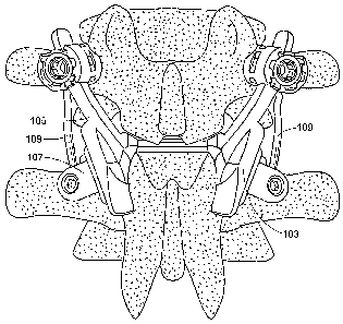
Now referring to FIGS. 1 and 2, there is provided a a kit for providing
therapy to
a functional spinal unit, the FSU comprising an upper vertebra having an upper
vertebral
body Vu and an upper facet Fu, a lower vertebra having a lower vertebral body
VBL and
a lower facet FL, the vertebral bodies defining a disc space therebetween, the
upper and
lower facets defining a facet joint FJ, the kit comprising:
a) a motion disc 101 adapted for insertion into the disc space,
b) a facet joint replacement system 103 comprising superior 105 and inferior
107 components, the system adapted to replace at least a portion of a
natural facet joint comprising first and second facets, and
c) a ligament 109 adapted to constrain relative movement between the facets.
The motion disc component of the present invention can be any prosthetic
capable of at least partially restoring the natural motions of the
intervertebral disc. In preferred
embodiments, the motion disc is selected from the group consisting of an
articulating
disc, a cushion disc and a spring-based disc. Various motion discs are
described by
Stefee et al. in U.S. Pat. No. 5,071,437; Gill et al. in U.S. Pat. No.
6,113,637; Bryan et al.
in U.S. Pat. No. 6,001,130; Hedman et al. in U.S. Pat. No. 4,759,769; Ray in
U.S. Pat.
No. 5,527,312; Ray et al. in U.S. Pat. No. 5,824,093; Buttner-Janz in U.S.
Pat. No.
5,401,269; and Serhan et al. in U.S. Pat. No. 5,824,094; all which documents
are hereby
incorporated herein by reference in their entireties.
7
CA 02566645 2006-11-14
WO 2005/112835 PCT/US2005/017346
Preferred articulating motion devices are disclosed in U.S.. Patent Nos.
5,556,431
and 5,674,296,. the specifications of which are incorporated by reference.
In' some embodiments, the articulating rnotion disc is a three-piece design
comprising two endplates and a core. Now referring to FIG., 3, in some
embodiments, the
articulating three-piece motion disc comprises:
= , , , ~
a) a first prosthetic, vertebral endplate 371 comprising:
= ,
i) an outer,surface 373 adapted to mate with a first vertebral body,
ii) an inner surface 375 having a first articulation surface 377,
iii) a body portion 379 connecting the inner and outer surfaces,
b) a second prosthetic vertebral endplate 381 comprising:
i)= an outer surface 383 adapted to mate with a second vertebral body, and
ii) an inner surface 385 comprising a first articulation surface 387,
c) a core member=391 ,comprising:
i) a first articulation surface 393 adapted for articulation with the first
articulation surface of the first end'plate, and
ii) a second articulation surface 395 adapted for articulation with the first
articulation surface of the second endplate,
.20
wherein the core member is oriented to produce a first articulation interface
between the
first articulation surface of the first endplate and the first articulation
surface of the core
member, and a second articulation interface between the first articulation
surface of the
second endplate and the second articulation surface of the core member.
In some embodiments, the articulating motion disc is a two-piece design
comprising two endplates. Now referring to FIG. 4, in some embodiments, the
articulating two-piece motion disc 401 comprises:
a) a first prosthetic vertebral endplate 431 comprising:
i) an outer surface 433 adapted to mate with a first veriebral body,
ii) an inner surface 435 having a first articulation surface 441,
8
CA 02566645 2006-11-14
WO 2005/112835 PCT/US2005/017346
iii) a body portion 443 connecting the inner and outer surfaces,
b) a second.prosthetic vertebral endplate 411 comprising:
i) an oiuter surface 413 adapted to mate with a second vertebral body, and '
~. . ii) an inner'surface 415 comprising a second articulation surface 417,
wherein the first and second articulation surfaces are oraented produce an
articulation
interface.
The FJR component of the present invention can be any prosthetic capable of at
least partially replacing a natural function of a natural facet joint. As
noted above, the
facet joints are diarthroidal joints that provide both sliding articulation
and load
transmission features. The facet's articular surfaces contact in extension,
limiting rotation
and increasing compressive load.
Now referring to FIG. 5, in some embodiments of :the present invention, the
superior 511 and'inferior 521 facet joint components of the prosthesis are
independent
bodies. In preferred embodiments thereof, the superior facet joint component
511 form's a
15. fixation portion 513 having =an outer surface 515 adapted to -attach to a
first facet and an
,inner articulation surface 517,. while the inferior facet joint component 521
forms a
fixation portion 523 having an outer surface 525 adapted to attach to an
inferior facet and
an inner articulation surface 527. In this embodiment, the inner articulation
surfaces are
adapted to form an articulation interface. For the purposes of the present
invention, this
embodiment is called an "articulation prosthesis". Throughholes 531 are
provided in
each fixation portion for facilitating the reception of a bone screw to
provide bony
fixation.
In some articulation embodiments, the first inner articulation surface is
convex
shaped, while the second inner articulation surface is concave shaped. This
creates =a ball
and socket joint well known in the art.
In some articulation embodiments, each of the first inner articulation surface
and
second inner articulation surface is cylinder-shaped.
In some articulation embodiments, each of the first inner articulation surface
and
second inner articulation surface is plane-shaped.
9
CA 02566645 2006-11-14
WO 2005/112835 PCT/US2005/017346
In some embodiments, the first and second articulation surfaces are
conforming.
In others, the first and second articulation surfaces are non-conforming.
Ndw referring td FIG. 6, in preferred emb,odiments, the facet joint
replacement
component comprises:
a), a superior facet joint component 601 comprising:
i) a longitudinal body 603 having a superior end portion 605 and an
inferior end portion 607, the inferior end portion forming an inner
surface 609, and
ii) a fastener 621 having a, distal threadform adapted to fasten to bone
and a proximal groove 623 adapted to receive the superior end
portion of the longitudinal body, and
iii) a set screw 625 received within the proximal groove of the
, . .
fastener,
b) an inferior facet joint component 631 comprising:
i) a body portion 633,
' ii) ,= outer portion 635 adapted to attach to bone and having a threaded,,.
throughbole 637,
iii) an inner portion having an inner surface 639 adapted to articulate
with the inner surface 609 of the superior facet joint component,
and
iv) a fastener 641 received with the threaded throughhole 637.
Still referring to FIG. 6, in preferred embodiments, the superior component
601
of the FJR includes a fastener 623 adapted to fasten to the pedicle portion of
the superior
vertebral body. The pedicle has been selected as the attachment location
because of its
clinical proven ability to accept pedicle screws under load, its long term
viability and
surgical familiarity. The superior end portion is designed to extend from
facet region to
the upper pedicle, and has a diameter designed to fit approximately within the
threadform
of the fastener, thereby allowing its fixation. The inferior end portion has
an inner
surface 609 adapted for bearing against the lower FJR component.
CA 02566645 2006-11-14
WO 2005/112835 PCT/US2005/017346
Although FIG. 6 shows set'screw 625 as locking onlythe longitudinal body to
the
fastener, . in other embodiments, the set screw is able to lock both a
longitudinal body and
ligament to the fastener.
Still referring to FIG. 6, the lower component 631 of the FJR includes a
fastener,
641 adapted to fasten to the pedicle portion of the lower vertebral body. The
pedicle has
again beeh selected as the attachment location. The= fastener in this case is
a pedicle
screw.
As noted above, conventional FJR systems do not appear to allow the surgeon to
adjust the distance from the pedicle screw component to the articulation
surface
component. The FJR system of FIG. 6 has special advantage because it allows
for intra-
operative adjustability of this distance. In some embodiments, the upper
component of
=
the FJR of FIG. 6 comprises a pedicle screw having a groove adapted to receive
the
superior end portion of the longitudinal body. In use, the pedicle screw is
first=inserted
into the bone, and the superior end portion of the longitudinal body (which in
this case, is
rod-shaped) is laid into the groove. The surgeon can then slide the
longitudinal body
relative to the groove until the appropriate position of the inner surface 609
is set. The
surgeon can then insert set screw 625 into the groove on top of the superior
end portion
of the longitudinal body in order to lock the position of the inner surface.
Therefore, in accordance with the present invention, there is provided a facet
joint
replacement component comprising:
a) a body having an elongate first end portion and a second end portion
forming
an articulation surface,
b) a fastener having a shank having distal threadform thereon adapted to
fasten to=
a bone, and a proximal end having a transverse groove adapted to receive the
elongate first end portion of the longitudinal body,
wherein a portion of the elongate first end portion of the longitudinal body
is slidably
received in the groove of the fastener.
The FJR system of FIG. 6 also has special advantage because it allows for
intra-
operative adjustability of the orientation. In this embodiments, the pedicle
screw is a
polyaxial screw having a substantially groove adapted to receive the superior
end portion
11
CA 02566645 2006-11-14
WO 2005/112835 PCT/US2005/017346
' =
of the longitudinal body.. The polyaxial nature of this screw allows the
surgeon to adjust
the orientation of the. groove component of the screw, thereby allowing for
adjustment of
the orientation of the articulation surface.
Therefore, in accordance with the present invention, there is provided a-facet
joint -
replacement component comprising:
a) ' a body having an elongate first end portion and a second end portion
forming
an articulation surface, and
b) a polyaxial screw adapted for adjustable fixation to the longitudinal body.
The FJR system of FIG. 6 also has special advantage because it reduces or
eliminates any moment upon the lower pedicle screw by insuring the
articulation forces
pas$ substantially through the lower screw. This is achieved by insuring that
the
articulation surface of the upper component substantially bisects the lower
pedicel screw.
Therefore, in accordance with the present invention, there is provided a facet
joint
15.. replac,ement component comprises:
a) a first facet joint component compnsing:
i) a longitudinal body having a first end portion and an second end,
portion, the inferior end portion forming a first inner articulation
surface, and
ii) means for attaching the longitudinal body to bone, and
b) an second facet joint component comprising:
i.) a body portion,
ii) outer portion adapted to attach to bone and having a throughhole,
iii) an inner portion having a second inner articulation surface adapted
to articulate with the inner articulation surface of the first facet joint
component, and
iv) a fastener received with the threaded throughhole,
wherein the first and second inner articulation surfaces are adapted to form
an articulation
interface defining an articulation force vector, and
12
CA 02566645 2006-11-14
WO 2005/112835 PCT/US2005/017346
wherein the articulation force vector passes through the fastener.
, =
The FJR system of FIG. 6 also has special advantage in that both the superior
and
inferior components may attach to=or abut against another bony structure
(e.g., a laxiiina,
pedicle, transverse process or spinous process). The secoipd attachment point
may reduce
or eliminate a moment upon the pedicle screw.
Now referring to FIG. , 7, in other embodiments, the superior and inferior
facet
joint components do not have inner articulation surfaces, but rather are
joined by an
elastic cushion core. In preferred embodiments thereof, the "cushion-type"
prosthesis
comprises:
a) a superior facet joint component 201 forming a superior endplate having an
outer surface 203 adapted to attach to a superior facet and an inner surface
205,
b) an inferior facet joint component 211 forming an inferior endplate havin;g
an
ou'ter surface 213 adapted to attach to an inferior-Tacet and an inner
surface.
215,
c) an elastic core 221 having a superior surface 223 adapted to attach to the
inner
: surface of. the superior facet joint component and an inferior surface 225
adapted to attach to the inner surface of the inferior facet joint component.
For the purposes of the present invention, this embodiment is called a"cushion
prosthesis". In preferred embodiments thereof, the device comprises an
elastomer
adapted to elastically compress during axial loading and relax when the load
is lifted.
In embodiments of the present invention comprising a prosthesis having
superior
and inferior facet joint components, the fixation portions thereof may
comprise an
attachment feature. Preferred attachment features are selected from the group
consisting
of teeth, keels, spikes, pins, holes, and combinations thereof.
In some embodiments, and referring back to FIG. 5, the facet joint replacement
comprises:
13
CA 02566645 2006-11-14
WO 2005/112835 PCT/US2005/017346
" a) a superior facet joint component 511 having a superior fixation portion
513
having an outer surface 515 adapted to attach to a superior.facet, and
b) an'inferior facet joint component 52.1 having an inferior fixation portion
523,
having an outer,surface 525 adapted to attach to an inferior facet. 5 In some
embodiments, the attachment surfaces of the FJR are adapted to attach to
the spinous process. In some embodiments, the attachrnent'surface of the FJR
are adapted
to attach and/or bear against a lamina. In some embodiments, the attachment
surface of
the FJR are adapted to attach to a pedicle. In some embodiments, the
attachment surface
of the FJR are adap'ted to attach to a transverse process. In some
embodiments, the
attachment surface of the FJR are adapted to attach to a native facet.
In some embodiments, bony attachment of the attachment surface of the FJR is
enhanced by the use of an adhesive, such as fibrin glue or bone cement.
In some embodiments, the fastener and/or the bony attachment surface of an FJR
component comprises a material having osteobiologic properties. This material
will help
.15 the osteointegrative process needed for secure attachment of the fastener
and/or the
attachment surface to the bone.
In some embodiments, the fastener ='and/or the attachment surface comprises an
orthoconductive portion. The orthoconductive portion typically has a porosity
(preferably
between about 20 ~tm and 250 m) that is adapted to allow the ingress of the
osteoconductive cells and an internal surface defined by the porosity that is
adapted to
attach these cells. In some embodiments, the fastener has an outer surface
adapted for
bony ingrowth. This. outer surface may have an osteoconductive coating
thereon, such as
a TCP coating or a hydroxyapatite coating.
In some embodiments, the fastener and/or the attachment surface comprises an
orthoinductive portion. The orthoinductive portion is preferably a protein,
and is more
preferably a growth factor. Preferred growth factors include factors from the
TGF-beta,
IGF-, BMP- and CDMP- families. Preferably, MP52 is selected as the CDMP.
In some embodiments, the fastener and/or attachment surface comprises an
orthogenetic portion. The orthogenetic portion preferably comprises
mesenchymal stem
cells. More preferably, the MSCs are present in a concentration greater than
that present
in the patient.'s natural bone marrow.
14
CA 02566645 2006-11-14
WO 2005/112835 PCT/US2005/017346
In some embodiments, the fastener andlor the attachrnent surfaces rpay also be
coated with other desired agents such as antithrombic or antimicrobial
coatings, and pain
relievers such as NSAIDS.
In some embodiments, the fastener component of the FJR system is a pedicle
screw. In some embodiments., the pedicle screw comprises a longitudinal shank
havi'ng an
integral nut thereon., Distal to the nut,.the shank has a first, distal
threadform thereon and a
distal tapered end. Proximal to the integral nut, the shank has a second,
proximal
threadform thereon and a proxitnal attachment, end having a slot.
In some embodiments, the pedicle sct-ew is a polyaxial screw.
In some embodiments, the fastener has a cannulated shank defining a bore that
allows for bony ingrowth into the bore. In some embodiments, this bore defines
an inner
surface adapted for bony ingrowth. This inner surface may have an
osteoconductive
coating thereon, such as a TCP coating or a hydroxyapatite coating.
The superior and inferior facet joint components of the present invention may
be
made from any material appropriate for human surgical implantation,
includingbut not
limited to all' surgi.cally appropriate metals including titanium, titanium
alloy, chrome..
alloys and stainless steel, and 'non-metallic materials 'such as carbon fiber'
inaterials,
resins, plastics and ceramics.
The elastic core, if selected, may comprise polyurethanes, foamed
polyethylene,
silicones, rubbers, copolymers or hydrogels.
In some embodiments, the FJR component is unilateral. A unilateral FJR at
least
partially replaces the function of a single facet joint. In some embodiments,
the FJR
component is bilateral. A bilateral FJR at least partially replaces the
function of both
facet joints of an FSU.
In some embodiments, the FJR component replaces a single articular process of
a
facet joint. This replacement process is adapted to articulate with the
natural articular
process that remains. In some embodiments thereof, the superior process is
replaced,
while in other the inferior process is replaced.
In other embodirnents, the FJR component replaces both articular processes of
the
facet joint, and these replacement process articulate with each other.
CA 02566645 2006-11-14
WO 2005/112835 PCT/US2005/017346
In some embodiments, sub~tantially the entire articular process is replaced
with a
prosthetic FJR. In others, substantially only the articular surface.of the
object process is
replaced, thereby preserving the underlying bony structure. This replacement
surface is.
adapted to articulate. with the natural ai-ticular process surface that
remains: In some.
emb.odiments, the replacement surface comprises a cap.
In 'some embodiments, the FJR component is a single level FJR. A single level
FJR at least partially replaces the function of a single level FSU. In some
embodiments,
the FJR component is a multi-level FJR. A multi-level FJR,at least partially
replaces the
function of facet joints in at least two levels of FSU.
' In some embodiments, the FJR is adapted to replace the natural arch, and so
comprises a transverse arch component. In some embodiments, the FJR is adapted
to
replace a natural process, and so comprises a spinous process component. In
some
embodiments, the FJR is adapted to replace at least one natural transverse
process, and so
comprises a transverse process component. In some embodiments; the FJR is
adapted to
replace at least one pedicle, and so comprises a pedicle component.
Now referring to FIGS. 8a-8d, there is provided a generic ligament 3 of the
present invention:
i) an intermediate portion 5,
ii) first and second end portions 7,9, and
iii) first and second conformable portions 11, 13,
wherein the first conformable portion is disposed between the intermediate
portion and
the first end portion, and the second conformable portion is disposed between
the
intermediate portion and the second end portion, and
iv) first and second fasteners 15,17, and
wherein the first end portion 7 is shaped to cooperatively connect to the
first fastener 15,
and the second end portion 9 is shaped to cooperatively connect to the second
fastener 17.
In some embodiments, the fasteners are selected from the group consisting of
bone screws,. hooks, wires, and pins. In some embodiments, the intermediate
portion of
16
CA 02566645 2006-11-14
WO 2005/112835 PCT/US2005/017346
the ligament is selected from the, group corisisting of a cable, a wires, an
intexconnected
face, and a soft polymer bonded to the fastener and stretching between the
superior and
inferior fa~tener.
In one aspect of the present invention, the facet, joint is stabilized in both
compression and tension by a.prosthetic ligament 'having fast'eners fixated
either iri'the
superior and inferior vertebrae or . insuperior and inferior prosthetic facet
joint
components. In some embodiments, the fasteners are selected from the group
Gonsisting
of bone screws, hooks, wires, and pins. In 'some embodiments, the intermediate
portion
of the ligament is selected from the group consisting of a cable, a wires, an
.10 interconnected face, and a sofl polymer bonded to the fastener and
stretching between the
superior and inferior fastener.
In a preferred embodiment of the present invention, the ligament is shaped as
a
sheath that can prevent debris produced by the facet articulation from
spreading to the
surrounding tissues, in particular to various neural structures. Previous
facet joint
replacement inventions describe resurfacing techniques that replace the
contacting faces
of-the facet joint,with~metals or polymers. Due*to unique variation in motions
of the facet,,.
joint, these resurfaced contacting faces will,inevitalily 'produce wear
debris; which is
likely to irritate tissues. A membrane or sheath that surrounds the contacting
faces and
captures generated particles can reduce tissue irritation and inflammation. '
The
, inembrane or sheath may also have structural integrity in itself and resist
over-stretching
and thereby supply resistance to tension.
In some embodiments, the width of the sheath is much greater. In preferred
sheaths, the sheath is sized to substantially enclose the facet joint. In some
embodiments, the sheath is fluid permeable. This feature permits the ingress
of fluids
that help lubricate the joint, while preventing the egress of wear debris from
the facet
joint articulation surfaces. In some ernbodiments, the sheath contains a
lubricating fluid,
thereby imitating a natural facet joint capsule. In preferred embodiments, the
sheath may
be pre-assembled prior to implantation, or it may be attached via glues,
sutres, wires,
thermally activated coagulation or in situ polymer embedding.
17
CA 02566645 2006-11-14
WO 2005/112835 PCT/US2005/017346
In preferred embodiments, this prosthetic facet joint ligament can be attached
to
anchoring points on, opposing sides of a natural or prosthetic facet joint to
provide a
constraint'against relative movement of the facet joints.
The ligament of the present inventiori can be made of any biocompatible
material
adapted for constraining but not totally eliminating relative -movement
between facet
joints. In this regard, the facet joint ligament of the preserit invention
mimics the natural
facet joint capsule. The ligament of the presentinvention comprises three
features. First
it must be adapted to traverse a facet joint. 'Second, it must allow some
flexion to occur
across the facet joint. Third, it must resist exGessive flexion of the facet
joint.
In preferred embodiments, the ligament comprises a pair of attachment end
portions and an intermediate portion.
The intermediate portion of the ligament may be adapted to have desirable
mechanical qualities found in ligaments, such as elasticity, flexibility,
tensionability, and
extensibility. Combinations of these qualities allows some displacement of the
articular
surfaces, but resists excessive displacement.
Preferably,, the intermediate portion of the facet joint ligament comprises
a,.
nonbioresorbable material including polyesters" (partidularly aromatic esters'
such, as
polyalkylene terephthalates, ' polyamides; polyalkenes; poly(vinyl fluoride);
polyurethanes; polytetrafluoroethylene (PTFE); carbon fibres; silk; rubber,
hydrogels,
, and glass, and mixtures thereof.
Preferably, the intermediate portion of the facet joint ligament is provided
as a
fabric. The fabric may be formed by a flat or circular weaving, knitting,
braiding,
crocheting or embroidery. Preferably, the fabric is braided in order to
provide a high
tensile strength. Preferred materials suitable for use as fabrics include
polyester,
polypropylene, polyethylene, carbon fiber, glass, glass fiber, polyurethane,
polyaramide,
metals, polymers, copolymers, polyactic acid (PLA), polyglycolic acid (PGA),
silk,
cellusoseic acid, and polycaprolactorie fibers.
It is anticipated that, in use, the intermediate portion of the facet joint
ligament
may rub against soft tissue structures and damage not only those structures
but itself as
well. Therefore, in some embodiments, the intermediate portion of the facet
joint
ligament is lubricated. The lubricants lowers the friction coefficient between
the ligament
18
CA 02566645 2006-11-14
WO 2005/112835 PCT/US2005/017346
and the soft tissue, thereby lowering the wear. Preferred lubricants include
hyaluronic
,
acid, proteoglycans,,and hydrogels
In some embodiments, the ligament comprises a material having orthobiologic
properties. This material will help the body's regenerative processes regrow a
natural
ligament to replace the prosthetic ligament of the present invention.
In' some embodiments, the ligament comprises a material having pain relief
properties, such as an NSAID.
In some embodiments, the ligament comprises an orthoconductive portion. The
orthoconductive portion typically-has a poxosity (preferably between about 20
m and
250 m) that is adapted to allow the ingress of the osteoconductive cells and
an internal
surface defined by the porosity that is adapted to attach these cells. In some
embodiments, the orthoconductive portion comprises subintestinal submucosa
(SIS). In
others, it comprises a synthetic polymer.
In some embodiments, the ligament comprises an orthoinductive portion. The
'15 orthoipductive portion is preferably a protein, and is more preferably a
growth factor.
Preferred growth factors include factors from the TGF-beta and IGF- families.
In some embodiments; the ligament comprises an orthogenetic portion. The
orthogenetic portion preferably comprises mesenchymal stem cells. More
preferably, the.
MSCs are present in.a concentration greater than that present in the patient's
natural bone
marrow.
In some embodiments, only the intermediate portion of the ligament comprises
an
orthobiologic material. ln some embodiments, only the attachment end portion
of the
ligament comprises an orthobiologic material. In other embodiments, each of
the
intermediate and attachment end portions of the ligament comprises an
orthobiologic
material.
Preferably, the ligament is provided in a sterile form. In some embodiments,
the
ligment is sterilized, and then placed in a package. Preferably, the inside
surface of the
package is also sterile.
In some embodiments, the intermediate portion of the ligament is tensionable.
A
lensionable ligament sags when the ends of the ligaments are inoved
sufficiently closed
to one another so that length of the ligament is less the distance between its
ends. This
19
CA 02566645 2006-11-14
WO 2005/112835 PCT/US2005/017346
quality allows . the opposing facets to move closer to each -other under loads
without
resistance from- the ligament. A tensionable ligament also becomes taut when
its ends are
moved sufficiently away'from one another so that lejagth of the ligament is
about equal to
the distance between, its ends. This quality constrains relative movement
between the
opposing facets. In some embodiments, the tensibility of the ligament is
between 5 and
=50 N/mm.
In some embodiments, at least a portion of the intermediate portion of the
ligament is extensible. An exteiisible ligament has a first at-rest length
when its ends 4re
.not loaded, and a second larger length when the ligament is subjected to
tensioning. This
quality allows the ligament to "give' a predetermined amount under tension.
This quality
is advantageous because the natural facet joint ligament is also. extensible.
Preferably,
the ligament has an extensibility of between 10% and 30% of "the at-rest
length of the
ligament -when subjected to a load of about 250 N. In some embodiments, the
extensibility of the ligament is between 5 and 50 N/mm. In other embodiments,
the
ligament is not extensible.,
In some embodiments, af least a portion of the intermediate portion of the
ligament is flexible. A flexible; ligament bows under axial' loading/easily
bends under
physiologic flexural loading and easily regains its shape when the loading is
ceased. This
quality allows the ligament to "give' a predetermined amount while
transferring stress
under axial loading. This quality is advantageous because the natural facet
joint ligament
is also flexible. Preferably, the flexible portion of the ligament comprises a
curved
portion.
Preferably, the ligament is adapted to allow restricted motion of the FSU
throughout the life of the patient. However, in the period inunediately after
the
components have been implanted, the human tissue in the wound region has
undergone
considerable damage and so requires a relatively stable environment in order
to heal. In
addition, during this early post-operative time period, both the motion disc
and the FJR
components need to integrate with the bony surfaces to which they are
attached, and so
also appear to require a relatively stable environment.
Therefore, in some embodiments, the ligament is designed to have time-variable
properties. In particular, the ligament is adapted to provide a stiffness that
decreases over
CA 02566645 2006-11-14
WO 2005/112835 PCT/US2005/017346
time. In this condition, the ligament can provide a desirably high stiffness
in the
immediate post-operative period, thereby stabilizing the region and promoting
tissue
repair and osteointegration. Once the wounds have healed the components have
become
integrated, the ligament stiffness decreases, thereby allowing for a desirable
range of
motion of the FSU.
Preferably, the ligament has a final (6-month) stiffness such that the
stiffness of
the FSU at that time is in the range of about 1-2 Nm/degree of flexion.
Similarly, the
ligament preferably has a final (6-month) stiffness such that the stiffness of
the FSU at
that time is in the range of about 2-3 Nm/degree of extension. In some
embodiments, the
ligament has an initial stiffness that is between about 2-4 times its final
(i.e,, 6-month)
stiffness. Without wishing to be tied to a theory, it is believed that if the
initial stiffness
were over about 4 times the final stiffness, fusion would result.
These values provide both the desired high stiffness required for initial
stabilization of the region, and long-term flexibility for the FSU.
.15 In some embodiments, the variability in the stiffness of the ligament is
accomplished by providing a ligament that experiences.sigrnificant creep over
time. In
"preferred einbodiments, the creeping ligament comprises a polymer, preferably
selected
from the group consisting of a polyester, a polyolefin and PTFE..
In preferred embodiments, the variability in the stiffness of the ligament is
accomplished by providing a resorbable material in the ligament. In some
embodiments,
the ligament comprises both non-resorbable and resorbable materials. After
implantation,
each of the non-resorbable and resorbable materials conribute to the initial
high stiffness
of the ligament. Over time, the resorbable materials degrades away, thereby
lowering
the ligament stiffness. In some embodiments, the intermediate portion of the
ligament
comprises non-resorbable fibers and resorbable fibers. In particularly
preferred
embodiments, the intermediate porti'on of the ligament comprises a non-
resorbable fiber
and a resorbable fiber selected from the group consisting PLA, PGA,, PLGA, and
mixtures thereof.
Each attachment end portion of the ligament is adapted to attach to an
anchoring
surface on opposite sides of the facet joint. Typically, the attachment end
portion
comprises a fastener. In other cases, attachment may be provided by sutres or
21
CA 02566645 2006-11-14
WO 2005/112835 PCT/US2005/017346
biologically compatible, glues. However, in other embodiments, an attachment
end
portion can simply be terminus being identical in design to the intermediate
portion. In
such a case, the terminus is inserted into a port located on the anchoring
surface, such as
a port on a prosthetichaving a facet joint articulating surface.,
~
As noted above, in soffie embodiments, the attachment' end'portions of the
facet
joint ligament comprise a pair .of fasteners. The function of the fastener is
to join to
attachment surfaces located on either side of the facet joint in order to
securely fasten the
intermediate portion of the facet joint ligament across the facet joint. The
fastener may=be
adapted to fasten the facet'joint ligament to attachment surfaces located upon
either:
a) a facet joint prosthetic component, or
b) a bony surface located adjacent the facet joint prosthetic component.
The attachment end portions of the prosthetic ligament of the present
invention
may be attached to any two anchoring surfaces on opposite sides of the facet
joint,
provided the ligament traverses the facet joint . These anchoring surfaces may
be located
on a bony surface of the opposing vertebrae, or on other prosthetic facet
joint
components.
In one embodiment, the first attachrnent erid portion of the ligament is
adapted to
attach to a first load-bearing portion of a facet joint prosthesis. This
embodiment is
surgeon friendly in that the attachment can be made by the manufacturer prior
to surgery,
thereby providing ease of use and repeatability.
ln some embodiments in which the ligament is attached to a pedicle screw, at
least one end of the, ligament includes a loop having a diameter slightly
larger than the
shaft diameter of a pedicle screw. In use, a pilot hole is drilled into the
pedicle, the loop is
placed over the pilot hole, and the pedicle screw is inserted into the pilot
hole, thereby
securing the loop therebetween.
In another embodiment, first attachment end portion of the ligament is adapted
to
attach to a portion of the natural vertebra. In some embodiments thereof, the
vertebral
body is used as the anchoring surface. In preferred embodiments thereof, the
pedicle
portion of the vertebral body is used as the anchoring surface. In another,
the facet
portion of the vertebra is the anchoring surface. In others, as shown in FIG.
9, a side wall
of a spinous process is tised as an anchoring surface. In this particular
case, each of the
22
CA 02566645 2006-11-14
WO 2005/112835 PCT/US2005/017346
iipper and lower fasteners 901 di the ligament 903 are respectively attached
to the
respective side walls, 905 of upper and lower spinous processes. In another
embodiment,
the first, end portion of the ligament is adapted to attach to the transverse
process. In
another embodiment, the first end portion of the ligament is adapted to attach
to the
pedicle. In another embodiment, the ligament is wrapped around the facet
joint.
In' embodiments in which at least one attachment end portion of the ligament
is
attached to bone, the methods described in U.S.S.N. 09/822,126, filed March
30, 2001,
the specification of which is incorporated by reference in its entirety, may
be used.
The fastener may be any design known in the art, including winged, barbed or
screw-in mechanisms. Preferably, the fastener is a barbed anchor, as it
prevents pullout
and is easily inserted. When the attachment surface is a bony surface, the
fastener may be
a bone fastener.
Now refemng to Figure 8c, preferably, the fastener 1'9 comprises a
longitudinal
shank 21, an insertion end 23 comprising protrusions 25 laterally extending
from the
shank, and an attachment end 27 having an upper surface 31 for connecting to
the
ligament. Preferably, the lateral protrusions have leading edges 28 which
define an angle
a=of no more than 45 degrees relative to the'axis of the shank. In such
embodiments,-the
bearing of the leading edge against the vertebral body surface will not
substantially,
impede the progress. of the bone fastener into the bone. Preferably, the
leading edges
define an angle of no more than 30 degrees, and more preferably between about
20
degrees and 30 degrees. When the angle a is between 20 and 30 degrees, the
angle is
sufficiently small to not impede the progress of the bone fastener, and yet
sufficiently
large to insure its secure fit. In some embodiments, the height H of the
protrusions on
the bone fastener is no more than 70% of the diameter D of the longitudinal
shank.
When this condition is selected, the risk that any protrusion will act as a
shoulder and
stop further entry of the bone fastener into the vertebra is minimized.
Preferably, the H/D
ratio is no more than 40%, more preferably between about 20% and 40%. Within
this
more preferred window, the protrusion height is sufficiently small to not
impede the
progress of the bone fastener, and yet sufficiently large to insure its secure
fit.
23
CA 02566645 2006-11-14
WO 2005/112835 PCT/US2005/017346
The outer diameter, (2H+D) of the bone fastener is preferably between about 3-
9
mm, more preferably about 4 - 6 mm. The length LBF of the bone fastener is
preferably
between about 3 - 45 mm, more preferably between about 15 - 25 mm.
In some embodiments, the attachment end of the bone fastener is made of a
ceramic material. When the bone fastener is ceramic, it can withstand the,
high impact of
=the driver withou=t failing.
ln some embodiments, at least the end portions of the intermediate portion and
the
attachment end of the bone fastener are made of the same material. When the
materials
are so selected, * these portions may be easily made and pre-connected in an
integral
fashion. This feature also eliminates the need for sutures.
Referring still to FIG 8c, in another aspect of the present 'invention, the
attachment end 27 of the fastener is configured to accept a driver element.
When this
configuration is selected, the bone fastener may be driven ' into the bone by
simply
providing axial force.to the upper surface 31 of the fastener through the
driver. Therefore,
in accordance with the present invention, there is provided a facet joint
ligament
comprising:
.=a)= a ligament comprising first and second end portions, and
b) first and second fasteners,
wherein the first bone fastener is connected to the first end portion of the
ligament, and
the second bone fastener is connected to the second end portion of the
ligament, and
wherein the first bone fastener is configured to accept a driver.
Preferably, the,configurationdefines a recess 29 upon the upper surface 31 of
the
attachment end 27 of the fastener. This recess 29 is configured to accept the
driver (not
shown).
ln some embodiments, the recess 29 of the bone fastener may be configured to
allow insertion of a rescue screw, theieby allowing retrieval of the bone
fastener.
In some embodiments, the fastener comprises a material having orthobiologic
properties. This material will help the osteointegrative process neeed for
secure
attachment of the fastener to the bone.
ln some embodiments, the fastener surface comprises an orthoconductive
portion.
The orthoconductive portion typically has a porosity (preferably between about
20 pm
24
CA 02566645 2006-11-14
WO 2005/112835 PCT/US2005/017346
and 250 m) that is adapted to = allow the, ingress of the osteoconductive
cells and an
internal surface defin,ed by the porosity that is adapted to attach these
cells.
In 'some embodiments, the fastener comprises an orthoinductive portion. The
orthoinductive portion is preferably a protein, and is more, preferably a
growth factor.
Preferred growth factors include factors from the TGF-beta, -IGF-, BMP- and
CDMP-
families. Preferably? MP52 is selected as the CDMP.
In some embodiments, the fastener comprises an orthogenetic portion. The
orthogenetic portion preferably,comprises mesenchymal stem cells. More
preferably, the
MSCs are present in a concentration greater than that present in the patient's
natural bone
marrow.
Preferably, the ligament and fastener components are,pre-connected. That is,
the
components are physically attached prior to their placement upon the spine.
Pre-
connected components are advantageous because their secured attachment to each
other
are guaranteed, and the surgeon need not waste time in making the attachment.
Components may be pre-connected by physical locking, physical connection, or
bonding,
or by making' the, .components 'from the same material and integrally
connecting them.,,.
When the preconnected components are integrally formed (by, for example,
molding or
thermoforming), there is no danger of micromotion. Therefore, in accordance
with the
present invention, there is provided a facet joint ligament comprising:
a) a ligament comprising first and second end portions, and
b) first and second fasteners,
wlierein the first fastener is pre-connected to the first end portion of the
ligament, and the
second fastener is pre-connected to the second end portion of the ligament.
In some embodiments, at least a portion of the ligament comprises a spring.
The
spring quality allows the ligament to initially yield to and eventually resist
an axial
compressive or tension load. In some embodiments, the spring is an expansion
spring. In
other embodiments, the spring is a compression spring.
In some embodiments of the present invention having both a pair of prosthetic
facet joint articulating surfaces and a prosthetic facet joint ligament, the
invention limits
the natural spinal extension. In these embodiments, extension is limited to no
more than
CA 02566645 2006-11-14
WO 2005/112835 PCT/US2005/017346
~ ' ' =
7'degrees, preferably no= more thah 5 degrees. Preferably, the device produces
a spine
stiffness is at least 2 Nm/degrees in order to so limit the extension.
In some embodiments of the present invention having a prosthetic facet joint
ligament, the invention resists flexion. In these embodiments, flexion is
limited to no
more than 15 degrees., and preferably is no more than 12 degrees. Preferably,
the tensile
strength=of the prosthetic capsule is between 50 and 300 N, is preferably at
least 100 N,
and is more preferably at least 200 N.
In some embodiments of the present invention havipg both a pair of prosthetic
facet joint articulating surfaces and a prosthetic facet joint ligament, the
invention resists
axial rotation. In these embodiments, a pair of devices of the present
invention are
preferably used so that each facet joint of a functional spine unit has a
device, whereby a
first device limits the axial rotation while. the ligament of the second
device is put in
tension. This motion tends to produce coupled motion with flexion and lateral
bending. In
some embodiments, the prosthetic articulating surfaces of the first device are
sufficiently
J 5 strong, to withstand compressive forces of at least I OON, and more
preferably at least
150N, and more preferably at least 200N.
In some embodiments of the preseint invention having both a pair of prosthetic
facet joint articulating surfaces and a prosthetic facet joint ligament, the
invention resists
at least anterior-posterior shear. In these embodiments, the prosthetic
articulating surfaces
contact and the prosthetic articulating surfaces are sufficiently strong to
withstand
anterior-posterior contact shear forces of at least 500N, and more preferably
at least
750N, and more preferably at least 1000N.
In some surgical procedures, such as a laminectomy, the patient's superspinous
ligament is often damaged. The SSL is important to the stability of an FSU due
to its
significant role in restraining patient flexion. Because the SSL possesses the
geatest
moment about the axis of rotation of any of the spine-related ligaments,
damage to the
SSL can result in significant instability to the FSU. Therefore, in some
embodiments, the
ligament of the present invention is adapted to at least partially replace the
function of the
SSL. In such embodiments, the ligament of the present invention has high
flexibility and
a high ultimate tensile strength. Preferably, the prosthetic SSL has a tensile
strength of at
26
CA 02566645 2006-11-14
WO 2005/112835 PCT/US2005/017346
least 50 N, preferably at least 100 N, more preferably at least 150 N, most
preferably at,
least 200 N..
Inianother embodiment, the ligament of the,present invention is adapted to
at.least
partially replace the funct.ion of the interspinous ligament (ISL).
In another embodiment, the ligament of the present =invention is adapted 'to
at
least partially replace the function of the'facet joint capsulp (FC).
In another embodiment, the ligament of the present invention is adapted to at
least partially replace the functi,on of the ligamentum flavum (LF). This
embodiment may
be selected when the posterior arch is removed.
In another embodiment, the present invention comprises two ligaments adapted
to
at least partially replace the functions of at least two ligaments selected
from the group
consisting of the superspinous ligament (SSL), the interspinous ligament
(ISL), the facet
joint capsule (FC), and the ligamentum flavum.
In another embodiment, the present invention comprises three ligaments adapted
to at least partially, replace the functions of at least three ligaments
selected frorn the
group consisting of the superspinous ligament (SSL), the interspinous ligament
(ISL), theõ
facet joint capsule (FC) , and the'ligamentum flavum.
In another embodiment, the present invention comprises three ligaments adapted
to at least partially replace the functions of each of the.superspinous
ligament (SSL), the
interspinous ligament (ISL), the facet joint capsule (FC) , and the ligamentum
flavum.
The present invention may be used in therapeutic procedures designed to
alleviate
facet arthritis, stenosis, , spondylolysthesis, post-laminectomy kyphosis, and
scoliosis.
The present invention may also be used in conjunction with the following
surgical
procedures: decompressive laminectomy, facet resection, lamina resection, and
.25 vertebroplasty.
EXAMPLE I
This prophetic example will demonstrate one method of implanting the
components of the present invention:
27
CA 02566645 2006-11-14
WO 2005/112835 PCT/US2005/017346
In some embodiments, thesnotion disc is implanted in -substantial accordance
with
the methods described in US Provisional Application USSN 60/459,280, Hawkins
et al.,
filed March 31, 2004, entitled "Method and Apparatus for Disc Insertion",
Attorney
Docket No. 3518.100-001, the specification of which is incorporated by
reference in its
entirety
First, the surgeon uses a standard posterior approach (either bilateral or
unilateral)
to uncover the facet joint. Next, the surgeon 'resects (excises) the facet
processes, using
standard resection instruments, such as a rongeur or a curette..
Next, the surgeon prepares the surface of each pedicle for insertion of a
pedicle
screw. - This entails locating the appropriate trajectory, probing the pilot
hole, and
preparing the pedicle surface to receiving the screw.
Next, the surgeon implants the, superior pedicle screw into the superior
pedicle,
and places a looped end of a ligament around the screw head.
Next, the surgeon places the longitudinal portion of the superior component
into
the groove of the screw head, and then places a set screw on top of that
longitudinal
portion, effectively securing the longitudinal portion.
The length of the superior component should be such that one surface abuts the
natural superior arch. If the surgeon decides to adjust the length of the
superior
component, the surgeon need only untighten the superior screw and readjust the
length as
desired.
Now that the location of the superior component has been fixed, the superior
component can be used as a template for fixing the location of the inferior
component.
In particular, the articulation surfaces of the two components are aligned in
the desired
positions (typically producing a gap therebetween). The aligmnent should be
such that
the inferior articulating force travels through the axis of the lower pedicle
screw. *Once
alignnent is fixed, the location of the screw hole is marked, and a pilot hole
is drilled.
Next, a looped end of a ligament is placed around the pilot hole and the
inferior
body is oriented to align the pilot hole with the hole in the inferior
component.
Lastly, a pedicle screw is inserted through the inferior component and into
the
pilot hole, thereby fixing the location of the inferior component.
28
CA 02566645 2006-11-14
WO 2005/112835 PCT/US2005/017346
Although sozne' of the facet joint replacement constructs identified above
provide
the necessary limitations on flexion, extension and. rotation of the
functional spinal unit,
the large size of these constructs may also require that,they be implanted
through
.. = = = ~
relatively invasive open prOcedures. In addition, these constructs tend to
=require
complete removal of the facet joint capsule.
Accordingly, in some embodiments of the present invention, the facet joint
replacement constructed is designed to allow fo'r its implantation via a
surgical technique
causing minimal trauma =to the facet joint,capsule and surrounding soft
tissues to the
extent possible.
Therefore, in some erribodiments, these goals are achieved by replacing the
natural facet joint with a construct comprising a bone screw having a head
adapted for
facet-type articulation.
Now refemng to FIG. 11 a, the surgeon selects a posterior approach with slight
lateral-inferior angulation and makes a small incision to access the targeted
facet joint.
Once access is,attairied, the surgeon removes the'superior facet and the
surface of the.=
inferior facet to produce an attachment surface 801 on the remaining portion
of = the
inferior facet.
Now referring= to FIG. I lb, the surgeon inserts the inferior facet
replacement
= component into the facet joint by passing the screw through the pedicle and
into the
vertebral body. In this embodiment, the inferior facet replacement component
comprises
a bone screw having a threaded shaft 803 and a proximal head 805 adapted for
articulation. This approach provides both minimal invasion and a high fixation
strength.
Now referring to FIG. 11 c, a superior facet replacement 807 is implanted
(preferably through the same incision used to implant the inferior component)
and is then
secured to a surface of the superior lamina. In this case, securement is
accomplished by
passing a bone screw '809 through the superior component. In preferred
embodiments,
this superior component comprises a hook (not shown) on its anterior side.. In
other
embodiments, the superior component has a general U-shape to enhance its
security to
the lamina
29
CA 02566645 2006-11-14
WO 2005/112835 PCT/US2005/017346
, ' = .
Now referring to FIG. 1-1 d, in other minimally invasive embodiments, the MIS
superior and inferior components shown above can be connected by at least one
ligament
811 to form a pre-assembled facet joint. The addition of a ligament to this
system
enhances the system's, performance by mimicking the action of facet capsular=
ligaments.
In- some embodiments, this pre-assembled facet joint is inserted as a whole
and fixed as
previously described. In other embodiments, this pre-assembled facet joint is
inserted
piecemeal and assembled in-situ.
Now referring to FIG. 11 e, in some embodiments, the superior facet
replacement
component may be secured to the spine via attachment to a translaminar bolt
813 inserted
into the lamina through a second small incision. As above, the anterior side
of the
superior facet replacement preferably has a hook to grab onto the lamina.
In other minimally invasive embodiments, facet-derived pain is eliminated by
denervation and the facet joint replacement component is replaced with a facet
joint
augmentation component.
15. In one preferred embodiment thereof, there is provided a method of
tieating facet
õjoint pathology wherein a primary therapy, is first applied to the medial
branches and
dorsal rami to denervate the nerves in these regions. In some embodiments, the
therapy
is selected from the group consisting of an energy source (such as pulsed
radiofrequency
(RF) waves, ultrasound, and microwave), chemical treatment, and freezing.
Upon the completion of the primary therapy, the patient will be positioned to
off-
load the facet joints and an injectable material (such as a silicone, a
polymethsiloxane, a
polyurethanes, a hydrogel, and hyaluronic acid) is injected from a syringe
into the facet
joint to produce a facet joint augmentation within the facet joint. In some
embodiments,
the injectable material is loaded with at least one therapeutic agent
including but not
limited: to preservative-free morphine, bupivacaine, tetracaine, opioids,
"tramadol,
ziconotide, betamethasone, clonidine, amitriptyline, fluoxetine,
anticonvulsants (such as
topiramate), carbamezapine, gabapentin, methlprednisolone acetate morphine3,
aminocaproic acid, anti- TNFamol ecul es, growth factors (such as TGF-b3, TGF-
bl, GDF-
5) and Cholinesterase inhibitors (such as neostigmine, and glantamine), and
combinations
thereof. Upon injection, the augmentation of the joint has the effect of
distracting the
CA 02566645 2006-11-14
WO 2005/112835 PCT/US2005/017346
facet joint, re-surfacing the facets' and, providing a cushion, = thereby
reducing the pain
'
.
associated with bone impingement.
=
Accordingly, in some embodiments, ther=e is provided (claim 32) a method of
treating a facet joint, comprising, the steps of:
a) injecting an augmentation material into the facet joint.
In some embodiments, other therapeutic agents such as methlprednisolone
acetate
morphine3, aminocaproic acid or Anti-TNFcx molecules, and/or growth factors
such as
TGF-b3, TGF-bl, GDF-5 could be injected into the facet joint when the
augmentation
material is not used.
Now referring to FIG. 12, therip is provided another embodiment of the present
invention in which a portion of the elongate first end portion bf the
longitudinal.body is
slidably received in the groove of the fastener (as in FIGS.1 and 2). .In FIG.
12, there is
; provided a prosthetic facet joint based upon a ball and socket articulation.
Pedicle screws
are placed superior and inferirorly of the 'involved level,= and=hardware
connected to the
''sc=rews as ball and socket joints-are positioned in the location of the
excised facet joints to
restore posterior intervertebral biomechanics.
31