Note : Les descriptions sont présentées dans la langue officielle dans laquelle elles ont été soumises.
CA 02567299 2006-11-17
WO 2005/112844 PCT/US2005/017991
Attorney Docket No.: 018158-024010
Client Reference No. VX-1203
BINOCULAR OPTICAL TREATMENT FOR PRESBYOPIA
BACKGROUND OF THE INVENTION
[0001] This invention generally relates to methods and systems for providing
optical
correction. More particularly, the invention provides methods and systems for
mitigating or
treating presbyopia and other vision conditions.
[0002] Presbyopia is a condition that affects the accommodation properties of
the eye. As
objects move closer to a young, properly functioning eye, the effects of
ciliary muscle
contraction and zonular relaxation allow the lens of the eye to become rounder
or more
convex, and thus increase its optical power and ability to focus at near
distances.
Accommodation can allow the eye to focus and refocus between near and far
objects.
[0003] Presbyopia normally develops as a person ages, and is associated with a
natural
progressive loss of accommodation, sometimes referred to as "old sight." The
presbyopic
eye often loses the ability to rapidly and easily refocus on objects at
varying distances. There
may also be a loss in the ability to focus on objects at near distances.
Although the condition
progresses over the lifetime of an individual, the effects of presbyopia
usually become
noticeable after the age of 45 years. By the age of 65 years, the crystalline
lens has often lost
almost all elastic properties and has only limited ability to change shape.
Residual
accommodation refers to the amount of accommodation that remains in the eye. A
lower
degree of residual accommodation contributes to more severe presbyopia,
whereas a higher
amount of residual accommodation correlates with less severe presbyopia.
[0004] Known methods and devices for treating presbyopia seek to provide
vision
approaching that of an emmetropic eye. In an emmetropic eye, both distant
objects and near
objects can be seen due to the accommodation properties of the eye. To address
the vision
problems associated with presbyopia, reading glasses have traditionally been
used by
individuals to add plus power diopter to the eye, thus allowing the eye to
focus on near
objects and maintain a clear image. This approach is similar to that of
treating hyperopia, or
farsightedness.
CA 02567299 2006-11-17
WO 2005/112844 PCT/US2005/017991
[0005] Presbyopia has also been treated with bi-focal eyeglasses, where one
portion of the
lens is corrected for distance vision, and another portion of the lens is
corrected for near
vision. When peering down through the bifocals, the individual looks through
the portion of
the lens corrected for near vision. When viewing distant objects, the
individual looks higher,
through the portion of the bi-focals corrected for distance vision. Thus with
little or no
accommodation, the individual can see both far and near objects.
[0006] Contact lenses and intra-ocular lenses (IOLs) have also been used to
treat
presbyopia. One approach is to provide the individual with monovision, where
one eye
(usually the primary eye) is corrected for distance-vision, while the other
eye is corrected for
near-vision. Unfortunately, with monovision the individual may not clearly see
objects that
are intermediately positioned because the object is out-of-focus for both
eyes. Also, an
individual may have trouble seeing with only one eye, or may be unable to
tolerate an
imbalance between their eyes. In addition to monovision, other approaches
include bilateral
correction with either bi-focal or multi-focal lenses. In the case of bi-focal
lenses, the lens is
made so that both a distant point and a near point can be focused. In the
multi-focal case,
there exist many focal points between near targets and far targets.
[0007] Surgical treatments have also been proposed for presbyopia. Anterior
sclerostomy
involves a surgical incision into the sclera that enlarges the ciliary space
and facilitates
movement of the lens. Also, scleral expansion bands (SEBs) have been suggested
for
increasing the ciliary space. Problems remain with such techniques, however,
such as
inconsistent and unpredictable outcomes.
[0008] In the field of refractive surgery, certain ablation profiles have been
suggested to
treat the condition, often with the goal of increasing the range of focus of
the eye, as opposed
to restoring accommodation in the patient's eye. Many of these ablation
profiles can provide
a single excellent focus of the eye, yet they do not provide an increased
depth of focus such
that optimal distance acuity, optimal near acuity, and acceptable intermediate
acuity occur
simultaneously. Shapes have been proposed for providing enhanced distance and
near vision,
yet current approaches do not provide ideal results for all patients. For
example, one profile
may optimize both distance and near vision when pupils are constricted, while
providing only
suboptimal acuity when pupils are dilated.
2
CA 02567299 2012-08-23
[00091 In light of the above, it would be desirable to have improved methods
and systems
for treatment and/or mitigation of presbyopia and other optical defects.
Ideally, such methods
and systems would provide for improved acuity without relying solely on one
eye for distance
vision and one eye for near vision. At least some of these objectives will be
met by various
embodiments of the present invention.
BRIEF SUMMARY OF THE INVENTION
[00101 Systems of the present invention provide for treatment or amelioration
of
presbyopia. In one aspect, there is provided a laser eye surgery system for
treating presbyopia
in a patient, the system comprising: a laser device for emitting a beam of
ablative energy;
delivery system optics coupled to the laser device; and a processor coupled
with the laser
device and the delivery system optics to direct the beam of ablative energy to
ablate a first
ablative shape on a corneal surface of a first eye of the patient and a second
ablative shape on a
corneal surface of a second eye of the patient, wherein the processor includes
a tangible
medium having a treatment table associated with each ablative shape embodied
thereon, and
wherein the first ablative shape has an optical power that varies across the
corneal surface of
the first eye so that the first ablative shape enhances near vision through a
central zone of the
first eye while enhancing far vision through a peripheral zone of the first
eye surrounding the
central zone of the first eye, and the second ablative shape has optical power
that varies across
the corneal surface of the second eye so that the second ablative shape
enhances near vision
through a peripheral zone of the second eye while enhancing far vision through
a central zone
of the second eye surrounded by the peripheral zone of the second eye, the
central zone of each
eye encompassing the pupil center of the eye.
[0010a] The system of the present invention allows for treating presbyopia in
a patient by
ablating a central zone of a corneal surface of a first eye of the patient to
improve the patient's
ability to view near objects through the central zone of the first eye and
ablating a peripheral
zone of a corneal surface of a second eye of the patient to improve the
patient's ability to view
near objects through the peripheral zone of the second eye.
3
CA 02567299 2012-08-23
[00111 The central zone produced during the first ablating step comprises a
substantially
spherical surface. Alternatively, the central zone may comprise a multifocal
aspheric surface.
Optionally, ablating the central zone of the corneal surface of the first eye
may involve leaving
a small central portion of the corneal surface untreated. In some embodiments,
the ablated
central zone may have a diameter scaled to a diameter of a pupil of the first
eye. The ablated
central zone may have any desired optical power, but in some embodiments it
has an optical
power of between about 0.5 and 4.0 Diopters (D), and more preferably between
about 1.0 and
3.0 D, and even more preferably an optical power of about 1.75 D.
[00121 The method further includes ablating a peripheral zone of a corneal
surface of the
first eye to improve the patient's ability to view far objects through the
peripheral zone of the
first eye. For example, in some embodiments the peripheral zone of the first
eye extends
radially outward from an outer boundary of the ablated central zone of the
first eye to a
diameter approximately matching an outer boundary of a pupil of the first eye.
In such
embodiments, the method may optionally further include ablating a transition
zone of the
corneal surface of the first eye, the transition zone extending from an outer
boundary of the
ablated peripheral zone of the first eye.
[00131 Optionally, ablating the peripheral zone of a corneal surface of the
second eye may
involve leaving a central zone of the corneal surface of the second eye
untreated to provide for
vision of distant objects through the central zone. In alternative
embodiments, the method may
include ablating a central zone of the corneal surface of the second eye to
improve the patient's
ability to view distant objects through the central zone.
[00141 The system also allows for a method for performing laser eye surgery on
a patient
to treat presbyopia involving: determining a first ablative shape for a
corneal surface, the first
ablative shape enhancing vision of near objects through a central zone of an
eye; ablating a
corneal surface of a first eye of the patient according to the first ablative
shape; determining a
second ablative shape for a corneal surface, the second ablative shape
enhancing vision of near
objects through a peripheral zone of an eye; and ablating a corneal surface of
a second eye of
the patient according to the second ablative shape.
4
CA 02567299 2012-08-23
[00151 In some embodiments, the first ablative shape comprises a central zone
having a
substantially spherical shape, while in other embodiments the first ablative
shape comprises a
central zone having a multifocal aspheric surface. Optionally, the first
ablative shape may
include a small central portion of the central zone that remains untreated. In
some
embodiments, the central zone of the first ablative shape has a diameter
scaled to a diameter of
a pupil of the first eye.
100161 In some embodiments, the central zone of the eye according to the first
ablative
shape has an optical power of between about 0.5 and 4.0 D, more preferably
between about 1.0
and 3.0 D, and even more preferably about 1.75 D. In some embodiments, the
first ablative
shape includes a peripheral zone shaped to provide for vision of distant
objects. For example,
the peripheral zone in some embodiments extends radially outward from an outer
boundary of
the central zone of the first ablative shape. Optionally, the first ablative
shape may further
include a transition zone extending from an outer boundary of the peripheral
zone.
[00171 In some embodiments, the peripheral zone of the second ablative shape
extends
circumferentially around a center of the corneal surface. In some embodiments,
the second
ablative shape includes an untreated central zone to provide for vision of
distant objects. In
other embodiments, the second ablative shape includes a central zone shaped to
improve the
patient's ability to view distant objects.
[00181
[00191 These and other aspects and embodiments of the present invention are
described in
further detail below, in reference to the attached drawing figures.
4a
CA 02567299 2012-08-23
BRIEF DESCRIPTION OF THE DRAWINGS
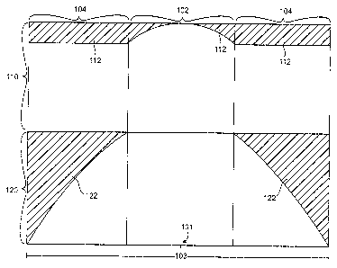
[00201 FIG. 1 is a diagrammatic illustration of two different ablation shapes,
each shape
for a different eye of the same patient, according to one embodiment of the
present invention.
100211 FIGS. 2A and 2B are diagrammatic illustrations of two different power
profiles
resulting from ablation shapes such as those shown in FIG. 1, according to one
embodiment of
the present invention.
[00221 FIG. 3 is a side sectional view of an eye treated to enhance vision of
near objects
through a central zone of the eye, according to one embodiment of the present
invention.
[00231 FIG. 4 illustrates an ablation profile on a corneal surface for
enhancing vision of
near objects through a central zone of the eye, according to one embodiment of
the present
invention.
[00241 FIG. 5 is a side sectional view of an eye treated to enhance vision of
near objects
through a peripheral zone of the eye, according to one embodiment of the
present invention.
[00251 FIG. 6 illustrates an ablation profile on a corneal surface for
enhancing vision of
near objects through a peripheral zone of the eye, according to one embodiment
of the present
invention.
100261 FIG. 7 is a block diagram of an ophthalmic surgery system for
incorporating the
invention.
DETAILED DESCRIPTION OF THE INVENTION
100271 While methods and systems of the present invention are described
primarily in the
context of improving laser eye surgery methods and systems, various
embodiments may also be
adapted for use in alternative eye treatment procedures and systems such as
femtosecond lasers
and laser treatment, infrared lasers and laser treatments, radial keratotomy
(RK), scleral bands,
follow up diagnostic procedures, and the like. In other embodiments,
techniques and systems of
the present invention may be adapted for use in other eye treatment procedures
and systems,
such as contact lenses, intra-ocular lenses, radial keratotomy, collagenous
corneal tissue
thermal remodeling, removable corneal lens structures, glass spectacles and
the like.
CA 02567299 2012-08-23
[00281 The present invention is particularly useful for enhancing laser eye
surgical
procedures such as photorefractive keratectomy (PRK), phototherapeutic
keratectomy (PTK),
laser in situ keratomileusis (LASIK), and the like. Various embodiments
provide enhance
presbyopia correction approaches by using improved combinations of ablation
shapes for a
patient's eyes.
[0029] The techniques of the present invention can be readily adapted for use
with existing
laser systems, including the VISX Excimer laser eye surgery systems
commercially available
from VISX of Santa Clara, California. By utilizing two different corneal
ablation profiles for
two different eyes of a patient, the present invention may enhance treatment
of presbyopia.
[00301 In one embodiment, a first eye of a patient is ablated to have a shape
that enhances
vision of near objects through a central region (or "central zone") of the
first eye. A number of
different ablation shapes and techniques may be used in various embodiments,
such as
shapes/techniques described in U. S. Patent No. 6,280,435, U. S. Patent No.
6,663,619 and/or
U. S. Patent No. 7,293,873, all of which are assigned to the assignee of the
present invention.
[0031] According to the same embodiment, the second eye of the patient is
ablated to have
a shape that enhances vision of near objects through a peripheral region (or
"peripheral
6
CA 02567299 2006-11-17
WO 2005/112844 PCT/US2005/017991
zone") of the second eye. Any suitable ablation techniques or shapes maybe
used, according
to various embodiments. In one embodiment, for example, an ablation technique
and shape
as described in U.S. Patent Application Serial No. 09/841,674 (Publication No.
2002/0156467) may be used.
[0032] By ablating two eyes of a patient to achieve different ablation shapes,
techniques of
the present invention provide for enhanced treatment of presbyopia. The
patient will
typically view both near and distant objects with both eyes. As the patient's
pupils constrict,
one eye will predominate for near vision and the other will predominate for
distance vision.
As the patient's pupils dilate, the predominant near and distance vision eyes
will switch. The
combination of the two ablation shapes enhances the patient's ability to view
near, far and
intermediate objects with an acceptable amount of acuity and without requiring
bifocals or
monovision systems.
[0033] Turning now to the drawings, FIG. 1 illustrates a first ablation
profile 110, which
may be applied to a first eye of a patient, and a second ablation profile 120,
which may be
applied to a second eye of the same patient. In some patients, the first
profile 110 may be
used for the patient's left eye and the second profile 120 may be used for the
right eye, while
in other patients the profiles may be used for the opposite eyes. Furthermore,
the profiles
shown in FIG. 1 are diagrams used solely for illustrative purposes. They are
not drawn to
scale and do not limit actual ablation profiles used in various embodiments of
the invention in
any way.
[0034] That being said, FIG. 1 illustrates ablative shapes 110, 120 along a
pupil 103 of
each of two eyes of a patient, each pupil 103 having a pupil center 101. In
both diagrams of
the ablative shapes 110, 120, the hash-marked areas represent tissue removed
112, 122 from a
corneal surface by ablation, typically by laser. Both ablative shapes 110,
120, include a
central zone 102 and a peripheral zone 104. In the first ablative shape 110,
used for a first
eye of a patient, the removed tissue 112 creates a shape that enhances near
vision through the
central zone 102 and distance vision through the peripheral zone 104. In the
second ablative
shape 120, used for a second eye of the patient, the removed tissue 122
creates a shape that
enhances distance vision through the central zone 102 and near vision through
the peripheral
zone 104. These ablation shapes 110, 120 may be used on the left and right
eyes of the same
patient, so that near and distance vision is enhanced through different
portions of each eye.
7
CA 02567299 2006-11-17
WO 2005/112844 PCT/US2005/017991
[0035] Referring now to FIGS. 2A and 2B, two power diagrams 130, 140
illustrate dioptic
powers of the two ablation shapes in FIG. 1, with a first power diagram 130 of
FIG. 2A
corresponding to the first ablation shape 110, and a second power diagram 140
of FIG. 2B
corresponding to the second ablation shape 120. In FIG. 2A, the first power
diagram 130
shows that power 132 increases toward +2 diopters (+2D) from the outer edge of
the
peripheral zone 104 toward the central zone 102 with the first ablative shape
110. In FIG.
2B, the second power diagram shows that power 132 decreases from +2 diopters
(+2D) from
the outer edge of the peripheral zone 104 toward the central zone 102 with the
second
ablative shape 120.
[0036] Referring now to FIG. 3, a schematic side view of a cornea 200 treated
according to
one.embodiment is illustrated. The cornea 200 has an anterior surface that
provides most of
the refractive power of the eye. The initial anterior surface 205 of the
cornea 200 has been
reshaped to a desired profile. The desired profile includes anterior optical
surface 210 and
anterior transition surface 215. The anterior optical surface 210 has a
multifocal aspheric
shape that corrects for near vision centrally and far vision peripherally.
Such a profile is
similar to the first ablation profile 110 in FIG. 1.
[0037] While the present invention will often be described with reference to
the mitigation
of presbyopia in combination with refractive hyperopia treatment, the benefits
of the present
invention are not limited to these specific procedures. These presbyopia
treatment techniques
may be used when no other refractive correction (other than the correction,
mitigation, and/or
inhibition of presbyopia) is desired, or the present treatment maybe combined
with therapies
for one or more of myopia, astigmatism, irregular refractive aberrations, and
the like, as well
as with hyperopia. Still other aspects of the present invention, including
methods and
systems which accommodate and adjust for re-epithelization, may find uses in a
broad variety
of ophthalmic procedures.
[0038] Anterior transition surface 215 is the anterior surface of the cornea
that provides a
gradual change in shape between anterior optical surface 210 and the portion
of the cornea
retaining the initial anterior surface 205. The outer boundary 212 of the
anterior optical
surface preferably extends entirely across, and is ideally substantially
coextensive with, the
pupil which is bounded by iris 220. The light rays passing through anterior
transition surface
215 do not contribute to the image formed by anterior optical surface 210.
Therefore,
8
CA 02567299 2006-11-17
WO 2005/112844 PCT/US2005/017991
anterior transition surface 215 is desirably positioned outside the pupil.
This positioning of
anterior transition surface 215 causes the light rays passing through anterior
transition surface
215 to be substantially occluded by iris 220. This occlusion improves patient
vision because
the light rays are blocked that do not contribute to image formation, and
which would
otherwise reduce the contrast of the image.
[0039] The optical correction effected by an ablative surgical procedure to
the cornea is
derived from a change in the anterior corneal surface from an initial anterior
surface 205 to
post-operative anterior optical surface 210. The anterior optical correction
is the post-
operative anterior optical surface 210 minus the initial anterior surface 205.
An ablation
profile is a change in an exposed surface profile occurring immediately after
the tissue
removal process. Therefore, the ablation profile is the exposed surface
profile immediately
after the tissue removal process minus the initial exposed surface profile. As
used herein,
"ablated shape" or "ablative shape" can refer either to an ablation-induced
change in a surface
topography on a surface of the cornea, or to the surface topography of the
cornea after
ablation.
[0040] In some instances, it may be desirable to form a central add while
leaving a central
region of the optical zone untreated as illustrated in FIG. 4. A small
untreated zone 500
centered on the optical zone 502 of an ablated cornea has a dimension 504
across the
untreated zone. The untreated zone 504 is smoothed by covering and healing of
the cornea
and contributes to the formation of a central anterior optical surface that
corrects presbyopia.
[0041] Referring now to FIG. 5, a schematic side view of a cornea 300 treated
to achieve
peripheral add, according to one embodiment, is shown. The cornea 300 has an
anterior
surface that provides most of the refractive power of the eye. The initial
anterior surface 305
of the cornea 300 has been reshaped to a desired profile. The desired profile
includes anterior
optical surface 305 that corrects for near-vision peripherally and far-vision
centrally. To
achieve the desired profile, anterior optical surface 305 is ablated lateral
to pupil, which is
bounded by iris 320. In some embodiments, a central zone 312 of the corneal
surface 305 is
not ablated, thus providing for distance vision through central zone 312. In
other
embodiments, central zone 312 may be ablated to enhance distance vision
through central
zone 312. The profile shown here is similar to the second ablative profile 120
illustrated in
FIG. 1.
9
CA 02567299 2006-11-17
WO 2005/112844 PCT/US2005/017991
[0042] FIG. 6 schematically shows an ablation shape for providing peripheral
add as just
described. As can be seen from the figure, a central zone 600, having a radius
of about 5.0
mm, is untreated, while a peripheral zone 610 is ablated to enhance near
vision. The
untreated central zone 600 is then used primarily for distance vision.
[0043] FIG. 7 illustrates a block diagram of an ophthalmic surgery system for
incorporating
the invention. As seen in this Figure, a personal computer (PC) work station
10 is coupled to
an embedded computer 21 of a laser surgery unit 20 by means of a first bus
connection 11.
The PC work station 10 comprises a tangible medium 12 and a treatment table
14. Tangible
medium 12 will typically comprise a memory, magnetic recording media, an
optical disk, or
the like, and will generally comprise machine readable programming instruction
code
implementing the method steps described herein. The laser treatment table 14
includes a
listing of coordinate references of the laser beam during an ablation of the
cornea. The sub-
components of laser surgery unit 20 are known components and preferably
comprise the
elements of the VISX STARTM Excimer Laser Systems, such as the STAR S4TM
System,
available from-VISX, Incorporated of Santa Clara, California. Thus, the laser
surgery system
20 includes a plurality of sensors generally designated with reference numeral
22 which
produce feedback signals from the movable mechanical and optical components in
the laser
optical system, such as the elements driven by an iris motor 23, an image
rotator 24, an
astigmatism motor 25 and an astigmatism angle motor 26. The feedback signals
from sensors
22 arc provided via appropriate signal conductors to the embedded computer 21.
The
embedded computer 21 controls the operation of the motor drivers generally
designated with
reference numeral 27 for operating the elements 23-26. In addition, embedded
computer 21
controls the operation of the excimer laser 28, which is preferably an argon-
fluorine laser
with a 193 nanometer wavelength output designed to provide feedback stabilized
fluence of
160 mJoules per square centimeter at the cornea of the patient's eye 30 via
the delivery
system optics generally designated with reference numeral 29. In addition,
other suitable
laser systems may be utilized in the present invention including, for example,
those
manufactured by Alcon, Bausch & Lomb, Wavelight, Nidek, Schwind, Zeiss-
Meditec,
Lasersight, and the like. Other lasers having a suitable wavelength may be
used to make an
ablative energy for removing a tissue from the eye. For example, solid state
lasers such as a
yttrium aluminum garnet (YAG) laser producing a fifth harmonic of a
fundamental
wavelength may be used to generate an ablative energy. Other ancillary
components of the
CA 02567299 2006-11-17
WO 2005/112844 PCT/US2005/017991
laser surgery system 20 which are not necessary to an understanding of the
invention, such as
a high resolution microscope, a video monitor for the microscope, a patient
eye retention
system, and an ablation effluent evacuator/filter, as well as the gas delivery
system, have
been omitted to avoid prolixity. Similarly, the keyboard, display, and
conventional PC
subsystem components (e.g., flexible and hard disk drives, memory boards and
the like) have
been omitted from the depiction of the PC work station 10. If desired,
embedded computer
21 may be constructed with PC work station components and built into laser
surgery system
20. In this case embedded computer 21 may supplant PC workstation 10.
[0044] While the above provides a full and complete disclosure of the
preferred
embodiments of the invention, various modifications, alternate constructions
and equivalents
may be employed as desired. Therefore, the above description and illustrations
should not be
construed as limiting the invention, which is defined by the appended claims.
11