Note : Les descriptions sont présentées dans la langue officielle dans laquelle elles ont été soumises.
CA 02572406 2009-09-10
28964-133
1
DESCRIPTION
ENDOSCOPE WITH RAISER FOR GUIDING TREATMENT INSTRUMENT
TECHNICAL FIELD
[0001] The present invention relates to an endoscope and
an endoscopic system to which the endoscope is applied, and
more particularly to an endoscope which includes an
insertion portion having a distal end side opening of a
treatment instrument insertion channel in a distal end
portion, and which is employed for a desirable treatment
with a use of a guide wire made to stick out from the
distal end side opening and guided to an affected area, and
an endoscopic system to which the endoscope is applied.
BACKGROUND ART
[0002] In recent years, so called side-looking type
endoscopes provided with an imaging optical system at a
distal end portion of an insertion portion are employed for
treatments of affected areas in alimentary tract system,
pancreaticobiliary duct system, and the like. The side-
looking type endoscope is employed for treatments such as a
preparatory treatment, in which contrast agent is injected
into a bile duct or a pancreatic duct before diagnosis, and
a therapeutic treatment, in which a gallstone present in a
common bile duct or the like is removed with a use of a
balloon, a grasper, or the like.
[0003] When the treatment is performed on the pancreatic
duct, bile duct, hepatic duct, or the like with the use of
the endoscope, the distal end portion of the insertion
portion of the endoscope is inserted into a duct to be
treated such as the pancreatic duct, bile duct, hepatic
duct, or the like. However, since the pancreatic duct,
CA 02572406 2006-12-28
2
bile duct, hepatic duct, and the like are extremely thin
ducts, the insertion of the distal end portion of the
insertion portion of the endoscope is not easy. Therefore,
a following procedure is generally taken.
[0004] First, the distal end portion of the insertion
portion of the side-looking type endoscope is inserted into
an area around a duodenal papilla. Then, while keeping the
area under radioscopy, the operator guides the guide wire
penetrating through the treatment instrument insertion
channel and makes the guide wire stick out from an opening
which opens in a lateral direction in the distal end
portion of the insertion portion of the endoscope, and
further inserts the guide wire into a desired duct to be
treated such as the pancreatic duct, bile duct, hepatic
duct, or the like. Thereafter, the operator inserts a
treatment instrument such as a catheter into the desirable
duct to be treated such as the pancreatic duct, bile duct,
hepatic duct, or the like using the guide wire as a guide.
[0005] Here, the guide wire or the treatment instrument
runs in an axial direction of the insertion portion of the
endoscope. Therefore, the guide wire and the treatment
instrument advance in the same direction. When one desires
to insert the guide wire or the treatment instrument
through the opening of the treatment instrument insertion
channel in the distal end portion of the insertion portion
and into a duct such as the pancreatic duct, bile duct,
hepatic duct, or the like, he/she needs to change the
direction of advance of the guide wire or the treatment
instrument around the opening of the insertion portion.
For this purpose, the side-looking type endoscope has a
treatment instrument raiser in the distal end portion of
the insertion portion. The operator can change the
direction of advance of the guide wire or the treatment
i
CA 02572406 2006-12-28
3
instrument which advances in the axial direction of the
insertion portion to a radial direction by raising the
treatment instrument raiser.
[0006] Thus, once the guide wire is inserted into the
extremely thin duct such as the pancreatic duct, bile duct,
hepatic duct, or the like, the operator can insert and
withdraw various types of treatment instruments into and
out of the duct using the guide wire as a guide.
[0007] When the treatment instrument is withdrawn from
the pancreatic duct, bile duct, or hepatic duct, the guide
wire is sometimes withdrawn together with the treatment
instrument against the will of the operator due to close
contact between the treatment instrument and the guide wire.
Since the pancreatic duct, bile duct, or hepatic duct is an
extremely thin duct, as described above, the insertion of
the guide wire thereinto is difficult to perform.
Therefore, it is extremely cumbersome and troublesome for
the operator to reinsert the guide wire into the pancreatic
duct, bile duct, hepatic duct or the like.
[0008] Hence, the operator needs to devise some ways to
prevent the withdrawal of the guide wire on removing the
treatment instrument from the pancreatic duct, bile duct,
hepatic duct, or the like. For the above purpose,
conventionally, after the operator moves the treatment
instrument to a certain extent in a direction of withdrawal,
an assistant of the operator pushes back the guide wire,
which moves together with the treatment instrument, towards
a direction of the pancreatic duct, bile duct, hepatic duct,
or the like. Alternatively, the guide wire is held so as
not to move and be withdrawn together with the treatment
instrument, for example. Such an operation is extremely
cumbersome and requires plural personnel, i.e., at least
the operator and the assistant. As can be seen from the
CA 02572406 2006-12-28
4
foregoing, the endoscopic diagnosis and treatment take long
time for treatment due to the cumbersome operation, and
places higher financial burden on both the hospital and the
patient since its operation needs many personnel.
[0009] To solve the problems as described above, some
propose an endoscope having a mechanism including a
treatment instrument raiser which can secure the guide wire
at a predetermined position when raised up. For example,
see Japanese Patent Application Laid-Open No. 2002-34905
and Japanese Patent Application Laid-Open No. 2003-116777.
[0010] The endoscope described in Japanese Patent
Application Laid-Open No. 2002-34905 has a slit for
securing the guide wire on a top of a guiding surface of
the treatment instrument raiser. When the treatment
instrument raiser is raised, the guide wire is engaged with
the slit of the treatment instrument raiser, thereby
secured relative to the endoscope.
[0011] On the other hand, the endoscope described in
Japanese Patent Application Laid-Open No. 2003-116777 has a
guide wire engaging groove to secure the guide wire on a
guiding surface of the treatment instrument raiser, and a
.guide wire securing mechanism near a forceps channel
opening in an operation portion.
[0012] The endoscopes according to the documents
mentioned above, secure the guide wire relative to the
endoscope between the treatment instrument raiser and a
predetermined portion of the distal end portion of the
insertion portion of the endoscope while the treatment
instrument is withdrawn. Thus, these endoscopes can
prevent the withdrawal of the guide wire from the
pancreatic duct, bile duct, hepatic duct, or the like at
the time of removal of the treatment instrument.
[0013] Patent Document l:Japanese Patent Application
CA 02572406 2007-03-19
28964-133
Laid-Open No. 2002-34905
Patent Document 2:Japanese Patent Application
Laid-Open No. 2003-116777
DISCLOSURE OF INVENTION
5 PROBLEM TO BE SOLVED BY THE INVENTION
[0014] However, the guide wire, which has an elongated
shape though hard, is not always parallel with the axial
direction of the endoscope at a position right out from the
treatment instrument insertion channel. Therefore, the
axial direction of the guide wire can take any direction on
the guiding surface of the treatment instrument raiser.
[0015] In the endoscopes as described in Japanese Patent
Application Laid-Opens Nos. 2002-34905 and 2003-116777, the
guide wire may be slipped off from the guiding surface when
the treatment instrument raiser is raised depending on the
position of the guide wire at the time the treatment
instrument raiser is raised. Then, the guide wire might not
be engaged with the slit of the treatment instrument raiser.
[0016] In this case, the guide wire might be sandwiched
between the treatment instrument raiser and a side wall of a
housing chamber of the treatment instrument raiser, for
example. Such a state is a so called pseudo-fixed state.
When the pseudo-fixed state occurs, the durability of the
guide wire, for example, may be decreased. Thus, when the
endoscope is configured to secure the guide wire at the
distal end of the insertion portion with the use of the
treatment instrument raiser, it is desirable that the guide
CA 02572406 2007-03-19
28964-133
5a
wire be surely guided to and secured at a predetermined
position, such as a position of the slit in the treatment
instrument raiser, without being mistakenly hooked at an
unintended position (a position between the treatment
CA 02572406 2006-12-28
6
instrument raiser and the side wall of the housing chamber,
for example) and secured (so as to cause pseudo-fixed
state).
[0017] The present invention is made in view of the
foregoing, and an object of the present invention is to
provide an endoscope in which a guide wire can be surely
secured at a predetermined position without being brought
into a pseudo-fixed state at a time the guide wire is to be
secured at a distal end portion of an insertion portion of
the endoscope, and which can guarantee a sufficient
securing strength.
MEANS FOR SOLVING PROBLEM
[0018] To achieve the object as described above, an
endoscope according to the present invention includes an
insertion portion which has a distal end hard portion at a
distal end side and a treatment instrument insertion
channel inside, and is inserted into a body cavity, an
operation portion which is connected to a proximal end side
of the insertion portion, and a treatment instrument raiser
which is arranged near a distal end side opening of the
treatment instrument insertion channel in the insertion
portion, has a treatment instrument guiding surface for
guiding a treatment instrument, and able to rise according
to an operation from the operation portion. The treatment
instrument raiser includes a slit which is formed on a
distal end side of the treatment instrument guiding surface
and with which a guide wire guided toward the distal end
side opening of the treatment instrument insertion channel
can be engaged, and a guide wire guiding unit which is
formed on an outer periphery of the treatment instrument
guiding surface and serves to guide the guide wire into the
slit. The guide wire is configured to be guided into the
CA 02572406 2009-09-10
28964-133
7
slit by the guide wire guiding unit, when the treatment
instrument raiser is raised by the operation from the
operation portion.
[0018a] In one broad aspect, there is provided an
endoscope, comprising: an insertion portion which has a
distal end hard portion at a distal end side and a treatment
instrument insertion channel inside, and is insertable into
a body cavity; an operation portion which is connected to a
proximal end side of the insertion portion; and a treatment
instrument raiser which is arranged near a distal end side
opening of the treatment instrument insertion channel in the
insertion portion, has a treatment instrument guiding
surface for guiding a treatment instrument, and able to rise
according to an operation from the operation portion,
wherein the treatment instrument raiser includes a slit
.which is formed on a distal end side of the treatment
instrument guiding surface and with which a guide wire
guided toward the distal end side opening of the treatment
instrument insertion channel can be engaged,, the guide wire
being for guiding insertion of the treatment instrument into
the treatment instrument insertion channel, and a guide wire
guiding unit which is formed on an outer periphery,
different from the distal end side, of the treatment
instrument guiding surface and serves to guide the guide
wire into the slit, the guide wire guiding unit including a
protrusion formed on a portion of the outer periphery of the
treatment instrument guiding surface, and wherein the guide
wire is configured to be guided into the slit by the guide
wire guiding unit, when the treatment instrument raiser is
raised by the operation from the operation portion.
CA 02572406 2009-09-10
28964-133
7a
EFFECT OF THE INVENTION
[0019] The present invention is advantageous in being
able to provide the endoscope in which the guide wire can be
surely secured at a predetermined position without being
brought into a pseudo-fixed state, i.e., a state in which
the guide wire is sandwiched at an unintended position, at a
time the guide wire is to be secured at the distal end
portion of the insertion portion of the endoscope, and which
can guarantee a sufficient securing strength.
BRIEF DESCRIPTION OF DRAWINGS
[0020) FIG. 1 is a perspective view showing a schematic
structure of an endoscopic system including an endoscope
according to an embodiment of the present invention;
FIG. 2 is an enlarged perspective view of a
relevant portion of a distal end portion of the endoscope of
FIG. 1;
FIG. 3 is a sectional view along line III-III of
FIG. 2;
FIG. 4 is a top view of the distal end portion of
the endoscope of FIG. 1;
FIG. 5 is a perspective view of a treatment
instrument raiser alone of the endoscope of FIG. 1;
FIG. 6 is a perspective view of a portion of an
insulating member of the endoscope of FIG. 1;
FIG. 7 is a diagram of a 'rising range regulating
mechanism in the endoscope of FIG. 1 in a normal state where
a second stopper portion is projected;
CA 02572406 2009-09-10
28964-133
7b
FIG. 8 is a diagram of the rising range regulating
mechanism of FIG. 7 in a state where the second stopper
CA 02572406 2006-12-28
8
portion is housed in an inner wall and a maximum rising
position of the treatment instrument raiser is regulated by
a first stopper portion;
FIG. 9 is an enlarged plan view of a relevant portion
of a stopper driving mechanism which is a part of the
rising range regulating mechanism in the endoscope of FIG.
1, and shows a portion around a position where an operation
knob is arranged;
FIG. 10 is a vertical sectional view of an internal
structure of the portion of FIG. 9;
FIG. 11 is an enlarged perspective view of a cam
member which is a part of the stopper driving mechanism of
FIG. 9;
FIG. 12 is an enlarged view of a relevant portion of a
part of an operation portion of the endoscope of FIG. 1,
and shows an arrangement of an operation knob provided in
the operation portion;
FIG. 13 is a schematic sectional view of a schematic
structure of a treatment instrument which is configured so
as to prevent buckling and which corresponds to the
endoscope of FIG. 1;
FIG. 14 is an enlarged sectional view of a portion
around the distal end portion of the endoscope, which shows
the treatment instrument of FIG. 13 applied to the
endoscope of FIG. 1;
FIG. 15 is a diagram of an example of a display screen
of a monitor in an endoscopic system to which the endoscope
of FIG. 1 is applied;
FIG. 16 is a sectional view along line III-III of FIG.
2, provided to described a function in the endoscope of FIG.
1 during a raising operation, and shows the distal end
portion of the endoscope in an initial state;
FIG. 17 is a top view of the distal end portion of the
CA 02572406 2006-12-28
9
endoscope in the initial state of FIG. 16;
FIG. 18 is a diagram of the distal end portion of the
endoscope in which the treatment instrument raiser is
rotated by a predetermined amount from the state shown in
FIGS. 16 and 17 to raise the guide wire, and a portion of a
guiding surface of the treatment instrument raiser is
brought into contact with the second stopper portion which
regulates the rotation;
FIG. 19 is a diagram of the distal end portion of the
endoscope in which the treatment instrument raiser is
further rotated by a predetermined amount from a state of
FIG. 18 to arrange the treatment instrument raiser at a
maximum rising position thereby securing the guide wire
with a slit;
FIG. 20 is a diagram of another example of the rising
range regulating mechanism in the endoscope of FIG. 1 in a
normal state where the second stopper portion is projected;
and
FIG. 21 is a diagram of the rising range regulating
mechanism of FIG. 20 in a state where the second stopper
portion is housed in an inner wall and a maximum rising
position of the treatment instrument raiser is regulated by
a first stopper portion.
EXPLANATIONS OF LETTERS OR NUMERALS
[0021] 1 Endoscope
2 Light source
3 Video processor
4 Monitor
5 Keyboard
12 Insertion Portion
13 Operation portion
17a, 17Aa First stopper portion
CA 02572406 2006-12-28
17b, 17Ab Second stopper portion
21 Distal end hard portion
22 Distal end cover
23 Treatment instrument insertion channel
5 25 Housing chamber
25b, 25Ab Housing portion
25a Side wall
26 Channel opening
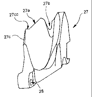
27 Treatment instrument raiser
10 27a Treatment instrument guiding surface
27b Slit
27c Guide wire guiding portion
27cc Edge portion
28 Holder rotation support point
30 Holder wire
35 Bending operation knob
40 Treatment instrument insertion port
47 Stopper driving mechanism
47a, 47Aa Elastic member
48 Operation knob
55 Treatment instrument
56 Guide wire
64 Traction knob
69 Traction wire
77 Insulating member
77a Guiding surface
77b Retraction slit portion
100 Endoscopic system
BEST MODE(S) FOR CARRYING OUT THE INVENTION
[0022] Exemplary embodiments of the present invention
will be described below with reference to the drawings. It
should be noted that the present invention is not limited
CA 02572406 2006-12-28
11
to the embodiments.
[0023] FIG. 1 is a perspective view showing a schematic
structure of an endoscopic system including an endoscope
according to an embodiment of the present invention. FIG.
2 is an enlarged perspective view of a relevant portion of
a distal end portion of the endoscope of FIG. 1. FIG. 3 is
a sectional view along line III-III of FIG. 2. FIG. 4 is a
top view of the distal end portion of the endoscope of FIG.
1. In FIG. 4, a guide wire is not shown. FIG. 5 is a
perspective view of a treatment instrument raiser alone of
the endoscope of FIG. 1. FIG. 6 is a perspective view of a
portion of an insulating member of the endoscope of FIG. 1.
[0024] Prior to a detailed description of the endoscope
of the present embodiment, a schematic overall structure of
the endoscopic system including the endoscope will be
described below mainly with reference to FIG. 1.
[0025] As shown in FIG. 1, an endoscopic system 100
includes an endoscope 1 of the present embodiment and a
peripheral device 50 thereof. The endoscope 1 mainly
includes an operation portion 13, an insertion portion 12,
and a universal cord 14. An insertion portion protecting
member 33 is arranged to protect the insertion portion 12
at a position where the insertion portion 12 and the
operation portion 13 are connected.
[0026] The peripheral device 50 mainly includes various
devices arranged on a counter 9 to which casters 8 are
attached at a bottom portion. The peripheral device 50
includes, for example, a light source 2, a video processor
3, a monitor 4, a keyboard 5, a suction pump device 6, and
a water delivery bottle 7. Further, the light source 2 and
the video processor 3 are electrically connected via a
connecting cable 73. Further, the endoscope 1 and the
peripheral device 50 are connected by a connector 18.
= e
CA 02572406 2006-12-28
12
[0027] The connector 18 is connected to the light source
2 of the peripheral device 50. The connector 18 has a
ferrule (not shown) which forms an end of a fluid pipe, a
light guide ferrule (not shown) which forms an end of a
light guide, and an electrical contact (not shown). The
light guide extends from the universal cord 14, penetrates
through the operation portion 13 and the insertion portion
12, and reaches the distal end portion 17 of the insertion
portion 12. Thus, illumination light emitted from the
light source 2 is emitted from an illumination lens 36 (see
FIGS. 2 and 4) of the distal end portion 17 toward an
interior of a body cavity in a radially expanded manner.
[0028] In the operation portion 13 of the endoscope 1, a
bending operation knob 35, an air/water delivery operation
button 37, a suction operation button 38, a treatment
instrument raiser operation knob (hereinafter simply
referred to as operation knob) 48 which is employed for a
raising operation of a treatment instrument raiser 27
(described in detail later; see FIGS. 3 and 5), and a
treatment instrument insertion port 40 which has an opening
40a through which a predetermined treatment instrument is
inserted into a treatment instrument insertion channel 23
(see FIG. 3) arranged inside the insertion portion 12 of
the endoscope 1 are provided.
[0029] The insertion portion 12 of the endoscope 1
includes a distal end portion 17, a bendable portion 16,
and a flexible tube portion 15. The bendable portion 16 is
manipulated so as to bend via the bending operation knob 35
provided in the operation portion 13, and is arranged
between the distal end portion 17 and the flexible tube
portion 15.
[0030] A portion of an outer circumference of the distal
end portion 17 is removed to form a cut out portion 19
CA 02572406 2006-12-28
13
having a depressed shape, and a channel opening 26 which is
located at a distal end side of the treatment instrument
insertion channel 23 (see FIG. 3) is provided on a surface
of the cut out portion 19.
[0031] Further, an objective lens 34 of an imaging unit
(not shown) housed in the distal end portion 17 and an
illumination lens 36 of an illumination optical system are
arranged near the channel opening 26 in the cut out portion
19 (see FIGS. 2 and 4).
[0032] Further, a nozzle 53 for air and water delivery
is projected from a wall surface 20 at a back end side of
the cut out portion 19 of the distal end portion 17. When
a fluid such as water and air is to be sprayed toward an
outer surface of the objective lens 34 for cleaning of the
objective lens 34 through an operation of the air/water
delivery operation button 37 of the operation portion 13,
the fluid is ejected from the nozzle 53.
[0033] A treatment instrument raiser housing chamber
(hereinafter simply referred to as housing chamber) 25 is
formed near the channel opening 26 in the distal end
portion 17. In the housing chamber 25, the treatment
instrument raiser 27 which serves to raise the treatment
instrument (not shown) or a guide wire 56 is arranged.
[0034] The treatment instrument raiser 27 is driven
according to a rotation operation of the operation knob 48
via a raising wire 30 (see FIGS. 3 and 4) which is driven
in conjunction with a holder engaging/driving mechanism
(not shown) provided inside he operation portion 13. When
the treatment instrument raiser 27 is driven, the direction
of advance (axial direction of the insertion portion 12) of
the treatment instrument or the guide wire 56, which enters
the treatment instrument insertion channel 23 from the
opening 40a of the treatment instrument insertion port 40
CA 02572406 2006-12-28
14
to stick out from the channel opening 26, inside the
treatment instrument insertion channel 23 is changed to a
direction of the channel opening 26. The treatment
instrument raiser 27 is configured to rise to a maximum
extent to secure the guide wire 56 when the treatment
instrument is to be withdrawn from the pancreatic duct,
bile duct, hepatic duct, or the like.
[0035] The guide wire 56 is an elongated linear member
including a core wire of a superelastic allow, for example,
and a soft outer cladding of Teflon or urethane, for
example, which covers the core wire. The guide wire 56 is
inserted into the pancreatic duct, bile duct, hepatic duct,
or the like before the insertion of the treatment
instrument (not shown) when the treatment instrument such
as a forceps and catheter is to be inserted into an
extremely thin duct, such as the pancreatic duct, bile duct,
hepatic duct, or the like, in the body cavity with the use
of the endoscope 1, and thereby the guide wire 56 works as
a guiding member for the insertion of the treatment
instrument.
[0036] In the following, an inner structure of the
distal end portion 17 of the endoscope 1, in particular,
the structure of the treatment instrument raiser 27, will
be described schematically mainly with reference to FIG. 3.
[0037] As shown in FIG. 3, the distal end portion 17 of
the endoscope 1 includes a distal end hard portion 21,
which serves as a main body of the distal end portion, and
a distal end cover 22 which is made of a non-conductive
material such as resin and arranged so as to cover the
distal end hard portion 21. The distal end cover 22 is
bonded and secured to the distal end hard portion 21 at a
distal end side of the distal end hard portion 21 by a
bonding agent or the like.
CA 02572406 2006-12-28
[0038] In the distal end hard portion 21, an elongated
hole 21a is formed along the insertion direction. A
connecting pipe 43 which serves as a guiding path for the
insertion of the treatment instrument (not shown) fits into
5 the elongated hole 21a. A distal end portion of the
treatment instrument insertion channel 23, through which
the treatment instrument is inserted, is fixed around an
outer circumference of the connecting pipe 43 at the back
end side of the connecting pipe 43. At a distal end side
10 of the connecting pipe 43, an introduction guiding path 24,
which guides the treatment instrument or the guide wire 56
inserted into the treatment instrument insertion channel 23
through the connecting pipe 43 to a side of the channel
opening 26, is formed.
15 [0039] At a distal end side of the introduction guiding
path 24, the housing chamber 25 is formed as a space
surrounded by the distal end hard portion 21 and the distal
end cover 22. The housing chamber 25 has an opening at a
top surface side. The opening serves as the channel
opening 26 which forms a distal end opening of the
treatment instrument insertion channel 23.
[0040] In an inside space of the housing chamber 25, the
treatment instrument raiser 27 is arranged. The treatment
instrument raiser 27 is substantially triangular in section,
and one end thereof is rotatably supported at a holder
rotation support point 28 which serves as an axis and is
formed at a position close to a bottom surface of the
distal end hard portion 21 near the distal end opening of
the introduction guiding path 24. Thus, the treatment
instrument raiser 27 can rotate within a predetermined
range within the housing chamber 25 in a direction of arrow
R shown in FIG. 3.
[0041] The treatment instrument raiser 27 has a
CA 02572406 2006-12-28
16
treatment instrument guiding surface 27a in a position
opposite to the channel opening 26. The treatment
instrument guiding surface 27a is a groove with a
substantially V-shaped section communicating with the
introduction guiding path 24 and serves to guide the
treatment instrument toward the channel opening 26.
[0042] On a distal end side of the treatment instrument
guiding surface 27a, a slit 27b which is substantially V
shape (see FIGS. 4 and 5) is formed. When the treatment
instrument raiser 27 is raised by a predetermined operation,
the guide wire 56 fits into the slit 27b and secured
therein.
[0043] In a middle position on a side surface of the
treatment instrument raiser 27, one end of a raising wire
39 is connected. The raising wire 30 extends from the
holder engaging/driving mechanism (not shown) of the
operation portion 13 and penetrates through the insertion
portion 12. An outer circumference of the raising wire 30
is covered with a guide pipe 31, which runs inside a guide
tube 32 penetrating the insertion portion 12.
[0044] The treatment instrument raiser 27 is raised by
rotating around the holder rotation support point 28
according to the traction operation of the raising wire 30.
The treatment instrument raiser 27 is configured so as to
rise up to a position regulated by a first stopper portion
17a described later. In the description, the position
where the treatment instrument raiser 27 is held by the
first stopper portion 17a is referred to as a maximum
rising position.
[0045] An insulating member 77 is arranged at a position
facing the treatment instrument raiser 27 at a distal end
side of the distal end hard portion 21.
[0046] Further, as shown in FIG. 6, a depressed guiding
CA 02572406 2006-12-28
17
surface 77a is formed on a surface of the insulating member
77 at a distal end side so that the guiding surface 77a
opens toward a front side. The guiding surface 77a and the
slit 27b of the treatment instrument raiser 27 sandwich the
guide wire 56 when the treatment instrument raiser 27 is
arranged at the maximum rising position. Then, the guide
wire 56 bites into the slit 27b, and is secured so as not
to move in the axial direction.
[0047] The position of the treatment instrument raiser
27 is regulated by a second stopper portion 17b described
later so that the treatment instrument raiser 27 does not
move farther than a predetermined position slightly forward
from the maximum rising position. The insertion operation
of the treatment instrument is performed while the
treatment instrument raiser 27 is in the above position.
Thus, the insulating member 77 and the treatment instrument
raiser 27 are set and arranged so that the treatment
instrument and the guide wire 56 can move in both the
direction of insertion and the direction of withdrawal
between the guiding surface 77a of the insulating member 77
and the guiding surface 27a of the treatment instrument
raiser 27.
[0048] Further, a retraction slit portion 77b is formed
near the guiding surface 77a of the insulating member 77,
so that a guide wire guiding portion 27c (described later)
of the treatment instrument raiser 27 can fit into the
retraction slit portion 77b to prevent interference when
the treatment instrument raiser 27 is raised to the maximum
rising position. Thanks to the retraction slit portion 77b,
the treatment instrument raiser 27 can surely rise to the
maximum rising position (position of the first stopper
portion 17a) over an angle required for rising.
[0049] Further, a U-shaped groove 77c having a U-shaped
CA 02572406 2006-12-28
18
section and opens upward is formed on a side edge portion
of the insulating member 77. The raising wire 30 is
slidably arranged in the U-shaped groove 77c.
[0050] On an outer periphery of the treatment instrument
guiding surface 27a of the treatment instrument raiser 27,
a guide wire guiding portion 27c is formed as means for
guiding the guide wire. The guide wire guiding portion 27c
holds the guide wire 56 on the treatment instrument guiding
surface 27a so that the guide wire 56 does not fall off
from the treatment instrument guiding surface 27a and
guides the guide wire 56 to the slit 27b when the guide
wire 56 is raised by the treatment instrument raiser 27.
The guide wire guiding portion 27c is a protrusion formed
at a portion of the outer periphery of the treatment
instrument guiding surface 27a adjacent to the housing
chamber 25, and has a substantially trapezoidal section and
is projected outward at a predetermined position on a side
surface of the treatment instrument raiser 27 at a side
adjacent to a fixing member on which the illumination lens
36 or the like is arranged.
[0051] On a predetermined position of the inner wall of
the housing chamber 25, a rising range regulating mechanism
for the treatment instrument raiser 27 is arranged. The
rising range regulating mechanism includes a stopper
driving mechanism 47 which includes the second stopper
portion 17b regulating the rising of the treatment
instrument raiser 27 at a predetermined position, and the
first stopper portion 17a regulating the maximum rising
position of the treatment instrument raiser 27.
[0052] The first stopper portion 17a is projected from a
side wall 25a near the proximal end of the housing chamber
25 so as to protrude inwardly as shown in FIG. 4. Near the
first stopper portion 17a, the second stopper portion 17b
CA 02572406 2006-12-28
19
which is arranged so as to be able to protrude and retract
on the side wall 25a and the stopper driving mechanism 47
which realizes the protruding/retracting operation of the
second stopper portion 17b.
[0053] The first stopper portion 17a and the second
stopper portion 17b are shown in detail in FIGS. 7 and 8.
FIG. 7 shows the second stopper portion 17b in a normal
state, i.e., a protruding state. FIG. 8 shows the second
stopper portion 17b housed in the inner wall in a retracted
state, where the maximum rising position of the treatment
instrument raiser 27 is regulated by the first stopper
portion 17a.
[0054] The second stopper portion 17b is configured so
as to be able to retract and protrude, taking the position
in the side wall 25a (state shown in FIG. 8) or the
position protruding toward inside the housing chamber 25
from the side wall 25a (state shown in FIG. 7).
[0055] A housing portion 25b is formed so as to house
the second stopper portion 17b in the side wall 25b. On a
bottom surface of the housing portion 25b, a tension
elastic member 47a is arranged. The second stopper portion
17b is supported by the elastic member 47a. While the
second stopper portion 17b is in a normal state, the second
stopper potion 17b is constantly biased in a direction of
arrow X2 shown in FIGS. 7 and 8 by the elastic member 47.
[0056] On the other hand, a traction wire channel 74
through which the traction wire 69 is inserted is
communicated with the housing portion 25b. The traction
wire channel 74 is communicated with a predetermined
position inside the operation portion 13 via the interior
of the insertion portion 12 of the endoscope 1. The
traction wire 69 runs through the traction wire channel 74.
On a distal end of the traction wire 69, a distal end
CA 02572406 2006-12-28
member 69a is fixed. The distal end member 69a moves and
makes the second stopper portion 17b protrude in a
direction against the biasing force of the elastic member
47a (direction of arrow X1 in FIGS. 7 and 8) when the
5 traction wire 69 is pushed in a direction of arrow Yl of
FIG. 7 by the stopper driving mechanism 47 described later.
In other words, when the traction wire 69 is pushed, the
distal end member 69a comes inside the housing portion 25b,
thereby pushing out the second stopper portion 17b. To
10 facilitate the above motion, a back end side portion of the
second stopper portion 17b, i.e., an end surface, with
which the distal end member 69a is brought into contact, of
the second stopper portion 17b is formed so as to be
inclined relative to a direction of motion (direction of
15 insertion) of the traction wire 69.
[0057] Thus, the second stopper potion 17b is arranged
so as to protrude toward inside the housing chamber 25 from
the side wall 25a as shown in FIG. 7. The state shown in
FIG. 7 is the normal state of the endoscope 1.
20 [0058] When the second stopper potion 17b is in the
position of FIG. 7, and the treatment instrument raiser 27
rises, a part of the treatment instrument raiser 27 is
brought into contact with the second stopper portion 17b.
Then, a further rotation (in a direction of arrow R1 of FIG.
3) of the treatment instrument raiser 27 is prevented.
[0059] On the other hand, when the traction wire 69 is
pulled by the stopper driving mechanism 47 in a direction
of arrow Y2 of FIG. 8, the distal end member 69a is pulled
out from the housing portion 25b. Then, the second stopper
portion 17b in the state of FIG. 7 (protruding state) moves
in a direction to retract inside the housing portion 25b
(direction of arrow X2 of FIGS. 7 and 8) according to the
biasing force of the elastic member 47a. Thus, the second
CA 02572406 2006-12-28
21
stopper portion 17b comes to be arranged in a retracted
position inside the housing portion 25b in the side wall
25a as shown in FIG. 8.
[0060] While the second stopper portion 17b is at the
position of FIG. 8, a portion of the treatment instrument
raiser 27 passes by the second stopper portion 17b and
rotates further until coming into contact with the first
stopper portion 17a. Thus, the maximum rising position
(see, e.g., position shown by a chain line in FIG. 3) of
the treatment instrument raiser 27 is regulated.
[0061] A part of the stopper driving mechanism 47 is
arranged at a side of the operation portion 13. Among
elements of the stopper driving mechanism 47, elements
(operation members and the like) arranged at the side of
the operation portion 13 will be described with reference
to FIGS. 9 to 11.
[0062] FIG. 9 is an enlarged plan view of a relevant
portion of the stopper driving mechanism, in particular a
portion around the position where the operation knob is
arranged in the stopper driving mechanism which is a part
of the operation portion 13. FIG. 10 is a vertical
sectional view of an internal structure of the portion of
FIG. 9. FIG. 11 is an enlarged perspective view of a cam
member which is a part of the stopper driving mechanism.
[0063] As shown in FIGS. 1 and 9, the operation portion
13 of the endoscope 1 of the present embodiment has a
substantially cylindrical traction knob 64 which is an
operation member for the traction operation of the traction
wire 69. The traction knob 64 is arranged between a grip
62 by which the operator grips the operation portion 13 and
the insertion portion protecting member 33. The traction
knob 64 is rotatably attached to an internal securing
member 46 of the operation portion 13 as shown in FIG. 10.
CA 02572406 2006-12-28
22
Further, an axis of rotation of the traction knob 64 is
arranged so as to be aligned with a central axis of the
insertion portion 12 of the endoscope 1.
[0064] A cylindrical cam member 65 (see FIGS. 10 and 11)
is integrally arranged inside the traction knob 64. On a
circumference of the cam member 65, a cam groove 65a is
curbed askew as shown in FIGS. 10 and 11. A moving pin 66
engages with the cam groove 65a as shown in FIG. 10. Thus,
when the cam member 65 rotates, the moving pin 66 moves in
a direction along the central axis of the insertion portion
12 and the traction knob 64. A proximal end of the
traction wire 69, which is inserted inside the traction
wire channel, is fixed to the moving pin 66. When the
traction knob 64 is rotated, the cam member 65 is rotated
accordingly. Then, the moving pin 66 moves along the cam
groove 65a of the cam member 65. Thus, the traction wire
69 proceeds and retracts along the axial direction of the
insertion portion 12 according to the movement of the
moving pin 66. As described above, the distal end member
69a is fixed to the distal end of the traction wire 69.
When the traction wire 69 proceeds or retracts, the distal
end member 69a follows the movement of the traction wire 69.
[0065] Thus, a position where the rotation of the
treatment instrument raiser 27 can be set at any time by
rotating the traction knob 64 and setting the second
stopper portion 17b at a desired position. For example, if
the operator rotates the traction knob 64 to push the
traction wire 69 to bring it in the state shown in FIG. 7,
the range of rotation of the treatment instrument raiser 27
comes to be regulated by the second stopper portion 17b.
The range of rotation of the treatment instrument raiser 27
is delimited by a position where the treatment instrument
raiser 27 comes into contact with the second stopper
CA 02572406 2006-12-28
23
portion 17b, i.e., a position shown by U in FIG. 7.
[0066] On the other hand, if the operator rotates the
traction knob 64 while the traction wire 69 is in the state
of FIG. 7 so as to pull the traction wire 69 in a direction
of traction and bring the traction wire in the state shown
in FIG. 8, the range of rotation of the treatment
instrument raiser 27 comes to be regulated by the first
stopper portion 17a. Then, the range of rotation of the
treatment instrument raiser 27 is delimited by a position
where the treatment instrument raiser 27 comes into contact
with the first stopper portion 17a, i.e., a position shown
by MAX (maximum rising position) in FIG. 8. In other words,
in this case, the treatment instrument raiser 27 can be
raised further by a predetermined amount from the position
shown by U in FIG. 7.
[0067] The treatment instrument raiser 27 is configured
so that the treatment instrument raiser 27 can be raised
through the traction of the raising wire 30 (see FIGS. 3
and 4) via the holder engaging/driving mechanism (not
shown) provided inside the operation portion 13 when the
operation knob 48 in the operation portion 13 is rotated.
The operation knob 48 is arranged at a predetermined
position in the operation portion 13 as shown in FIG. 12.
[0068] FIG. 12 is an enlarged view of a relevant portion
of a part of the operation portion of the endoscope of the
present embodiment and shows an arrangement of the
operation knob provided in the operation portion. In FIG.
12, members other than the operation knob in the operation
portion are not shown for the simplicity of description.
[0069] The operation knob 48 is arranged so as to be
rotatable around an axial portion 48a arranged
perpendicular to the axial direction of the operation
portion 13, and is arranged on a side surface of the
CA 02572406 2006-12-28
24
operation portion 13. The operation knob 48 is a lever-
like operation member which includes the axial portion 48a,
a proximal end portion 48b fixed to one end of the axial
portion 48a, an arm 48c which extends from the proximal end
portion 48b, and a knob portion 48d which is integrally
arranged at a distal end of the arm 48c. When the operator
puts a finger on the knob portion 48d and moves the knob
portion 48d in a direction of arrow R shown in FIG. 12, the
operator can rotate the operation knob 48. Along with the
rotation of the operation knob 48, the lever portion 48e
formed on an outer circumference of the proximal end
portion 48b rotates. Then, a link member 48f connected to
the lever portion 48e moves in a direction of arrow T of
FIG. 12. To the link member 48f, a raising wire 30 is
connected. Accordingly, the raising wire 30 can be pulled.
[0070] In the endoscope 1 of the present embodiment,
when the treatment instrument raiser 27 is raised to the
maximum rising position, the guide wire 56 is held between
the slit 27b of the treatment instrument raiser 27 and the
guiding surface 77a of the insulating member 77, and at the
same time the slit 27b is made to bite into the guide wire
56, whereby a high securing strength is obtained.
[0071] Here, the securing strength of the guide wire 56
while the treatment instrument raiser 27 is at the maximum
rising position can be adjusted by the amount of rising of
the treatment instrument raiser 27, i.e., a rising stroke.
An easy and effective way to improve the securing strength
of the guide wire 56 by the slit 27b is to increase an
amount of rising angle of the treatment instrument raiser
27. In other words, the increased angle of rotation of the
operation knob 48 is sufficient to increase the rising
angle of the treatment instrument raiser 27 and to increase
the rising range of the treatment instrument raiser 27.
CA 02572406 2006-12-28
[0072] For the above mentioned purpose, in the operation
knob 48 of the present embodiment, a dimension of a length
of the arm of the operation knob 48 (dimension from a
center of the axial portion 48a to a top of the knob
5 portion 48d; also referred to as a height dimension of the
operation knob 48) is made slightly longer than that in a
conventional member. The operation knob 48 is configured
so that the height dimension of the operation knob 48 shown
by a solid line in FIG. 12 is longer than that of the
10 conventional operation knob 48 shown by a chain line in FIG.
12. Specifically, the operation knob 48 of the embodiment
is longer than that of the conventional one by
approximately 1 mm as indicated by character H in FIG. 12.
Thus, the amount of rotation of the operation knob 48 can
15 be made larger than that of the conventional one by an
amount indicated by character S in FIG. 12.
[0073] To increase the height dimension of the operation
knob 48, it is desirable that the position of the top of
the knob portion 48d of the operation knob 48 be placed on
20 a rotation arc of the bending operation knob 35 or within a
radius of rotation of the bending operation knob 35, for
example, so that the operability will not be degraded.
[0074] The increase of the height dimension of the
operation knob 48 does not require a drastic change in
25 design, and still a desirable rising stroke can be obtained.
[0075] The treatment instrument raiser 27 can be
employed also to raise the treatment instrument that has a
tube sheath such as a cannula (not shown in particular) and
to direct a distal end thereof in a desired direction when
such a treatment instrument is to be inserted into a
desired duct such as the pancreatic duct, bile duct, and
hepatic duct. When the treatment instrument raiser 27 is
erroneously raised up to the maximum rising position to
CA 02572406 2006-12-28
26
raise the treatment instrument, the treatment instrument
might be buckled.
[0076] To deal with the above inconveniences, the
treatment instrument may have a following structure.
[0077] FIG. 13 is a schematic sectional view of a
schematic structure of a treatment instrument which is
configured so as to prevent buckling and which corresponds
to the endoscope of the present embodiment. FIG. 14 is an
enlarged sectional view of a portion around the distal end
portion of the endoscope, and shows the treatment
instrument of FIG. 13 applied to the endoscope of the
present embodiment.
[0078] As shown in FIG. 13, a treatment instrument 55
corresponding to the endoscope 1 of the present embodiment
has a tube sheath such as a cannula. The treatment
instrument 55 is supposed to have three regions, i.e., a
distal end region 55a which is a predetermined region near
the distal end, a thick region 55b which is connected to
the distal end region 55b and which is configured to be
slightly thick, and a proximal end region 55c which is
connected to the thick region 55b and arranged near the
proximal end. The distal end region 55a is, for example, a
region having a dimension L1 from a most distal end portion
as shown in FIG. 13 (more specifically, Ll is approximately
20 to 30 mm). The thick region 55b is a region connected
to the distal end region 55a and has a dimension L2
(specifically approximately 200 mm) as shown in FIG. 13,
for example. The proximal end region 55c covers all area
extending from an edge of the thick region 55b to the
proximal end.
[0079] Here, thickness of the tube in the treatment
instrument 55 is substantially the same in the distal end
region 55a and the proximal end region 55c, while the
CA 02572406 2006-12-28
27
thickness in the thick region 55b is slightly increased
than that in the other two regions. An inner diameter of
the treatment instrument 55 is made to be identical from a
distal end up to a proximal end. Thus, the insertability
of the guide wire 56 and the flowablity of the contrast
agent are maintained.
[0080] The thick region 55b of the treatment instrument
55 has a high probability of contacting with the insulating
member 77 during the guiding of the distal end of the
treatment instrument 55 through the channel opening 26 of
the endoscope 1 as shown in FIG. 14. In other words, the
region is a portion where the force is applied when the
treatment instrument raiser 27 raises the treatment
instrument. That is why the portion is made to be thicker.
Thus, the treatment instrument 55 rarely buckles even when
the treatment instrument raiser 27 is raised.
[0081] Meanwhile, in the endoscopic system 100 to which
the endoscope 1 of the present embodiment is applied, a
following display is presented on the screen of the monitor
4 during the rising operation of the treatment instrument
raiser 27 in order to prevent the treatment instrument
raiser 27 from causing the buckling and the damages of the
treatment instrument 55.
[0082] FIG. 15 is a diagram of an example of a display
screen of the monitor in the endoscopic system to which the
endoscope of the present embodiment is applied.
[0083] As shown in FIG. 15, an information display
region 4c is presented on the display screen 4a of the
monitor 4 so as to display various types of information in
addition to an endoscopic image 4b. The information
display region 4c has a predetermined region 4d whose
display indicates a rising state of the treatment
instrument raiser 27 during the raising operation.
CA 02572406 2006-12-28
28
[0084] FIG. 15 is an example of the display. In the
example of FIG. 15, there is a graph-like indication of a
substantially circular arc shape. In the graph-like
indication, a region indicated by character A is shown in
green, while a region indicated by character B is shown in
red. A detecting unit such as a position sensor provided
near the treatment instrument raiser 27 detects an amount
of rising of the treatment instrument raiser 27, and the
display is given in a predetermined manner based on the
detected amount.
[0085] In the example shown in FIG. 15, the graph-like
indication is shown. The present invention is not limited
thereto. Alternatively, a number representing the rising
angle may be displayed together, or the numbers alone may
be displayed.
[0086] The detecting unit is desirably provided near the
treatment instrument raiser 27. The detecting unit,
however, can be provided inside the operation portion 13,
for example. In this case, the detecting unit may be
configured to detect a travel amount of the raising wire 30,
for example, or to detect an amount of rotation of the
operation knob 48 or the like.
[0087] A function of the endoscope 1 of the present
embodiment having the above mentioned structure will be
described below. More specifically, an operation at the
time of raising operation according to which the guide wire
56 is raised via the operation knob 48 (see FIG. 1) and an
operation at a fixing operation according to which the
guide wire 56 is secured at a predetermined position will
be described below.
[0088] FIGS. 16 to 19 are enlarged perspective views of
a relevant portion of the distal end portion of the
endoscope of FIG. 1. Among the drawings, FIG. 16 is a
CA 02572406 2006-12-28
29
sectional view along line III-III of FIG. 2. FIG. 17 is a
top view of the distal end portion of the endoscope in an
initial state of FIG. 16. Here, FIGS. 16 and 17 show the
distal end portion in a state where the treatment
instrument raiser 27 has not been raised and the guide wire
56 sticks out from the channel opening. The state shown in
FIGS. 16 and 17 will be referred to as an initial state.
FIG. 18 is a diagram of the distal end portion of the
endoscope in which the treatment instrument raiser 27 is
rotated by a predetermined amount from the state shown in
FIGS. 16 and 17 to raise the guide wire 56, and a portion
of the guiding surface 27a of the treatment instrument
raiser 27 is brought into contact with the second stopper
portion 17b which regulates the rotation of the treatment
instrument raiser 27. FIG. 19 is a diagram of the distal
end portion of the endoscope in which the treatment
instrument raiser 27 is further rotated by a predetermined
amount from the state of FIG. 18 and arranged at the
maximum rising position, thereby securing the guide wire 56
with the slit 27b.
[0089] After the guide wire 56 is inserted into the
treatment instrument insertion port 40 of the operation
portion 13 from the opening 40a (see FIG. 1) from a back
end located close to the operator of the treatment
instrument (not shown) such as a catheter inserted inside
the treatment instrument insertion channel 23, the distal
end of the guide wire 56 is guided toward the channel
opening 26 as shown in FIG. 16 and the distal end of the
treatment instrument is arranged inside the treatment
instrument insertion channel 23. Here, a portion of the
guide wire 56 is placed on the treatment instrument guiding
surface 27a of the treatment instrument raiser 27.
[0090] While keeping the state as described above, the
CA 02572406 2006-12-28
operator operates the operation knob 48 (see FIG. 1). In
other words, the operator performs the rotation operation
of the operation knob 48 in a predetermined direction so as
to raise the treatment instrument raiser 27. Then, the
5 rotating force of the operation knob 48 is converted into a
force to pull the raising wire 30 via the predetermined
holder engaging/driving mechanism (not shown). When the
raising wire 30 is pulled, the treatment instrument raiser
27 starts to rotate around the holder rotation support
10 point 28 in the direction of arrow R1 (clockwise direction
in FIG. 16) as shown in FIG. 16.
[0091] Once the treatment instrument raiser 27 starts to
rotate in the above mentioned direction, the guide wire 56
which is placed on the treatment instrument guiding surface
15 27a of the treatment instrument raiser 27 starts to be
raised toward a side of the channel opening 26.
[0092] If the guide wire 56 is in the position shown by
a solid line in FIG. 17, in other words, if the guide wire
56 is inside the slit 27b of the treatment instrument
20 guiding surface 27a, the guide wire 56 remains at the
position (predetermined intended position at which the
guide wire 56 is to be placed) while being raised.
[0093] On the other hand, if the guide wire 56 is in a
position (position at which the guide wire 56 is not
25 intended to be placed) shown by a dotted line in FIG. 17,
for example, in other words, if the guide wire 56 is not
inside the slit 27b of the treatment instrument guiding
surface 27a and bent toward a side of the side wall 25a of
the housing chamber 25 and the treatment instrument raiser
30 27, the guide wire 56 is raised as follows.
[0094] The guide wire 56 is raised along with the
rotation of the treatment instrument raiser 27 toward the
rising direction, and moves toward a side of the side wall
CA 02572406 2006-12-28
31
25a of the housing chamber 25 on the treatment instrument
guiding surface 27a of the treatment instrument raiser 27
as if slipping off from the treatment instrument guiding
surface 27a. Here, the guide wire 56 slides over the guide
wire guiding portion 27c in a direction toward the slit 27b
of the treatment instrument raiser 27. The slipping
movement of the guide wire 56, however, is stopped when the
guide wire 56 is brought into contact with an edge portion
27cc of the guide wire guiding portion 27c of the treatment
instrument raiser 27. In other words, when the guide wire
56 comes to be held by the edge portion 27cc of the guide
wire guiding portion 27c, the guide wire 56 does not slip
farther toward the side of the side wall 25a of the housing
chamber 25, and is raised.
[0095] While the above state is maintained (while the
guide wire is held by the edge portion 27cc), the treatment
instrument raiser 27 raises the guide wire 56 to a certain
degree. The guide wire 56 has an elastic tension to return
in a direction to recover a linear state. Hence, when the
guide wire 56 is raised to a certain degree, a force is
applied to the guide wire 56 in a direction of arrow D
shown in FIG. 17. Then, the guide wire 56 moves over the
treatment instrument guiding surface 27a toward the slit
27b of the treatment instrument guiding surface 27a from
the edge portion 27cc of the guide wire guiding portion 27c
while being raised by the treatment instrument raiser 27.
When the treatment instrument raiser 27 rotates up to the
position shown in FIG. 18, the guide wire 56 inevitably
falls inside the slit 27b.
[0096] An outer periphery of the treatment instrument
guiding surface 27a is formed in a smooth shape leading to
the slit 27b. In particular, a region extending from the
guide wire guiding portion 27c to the slit 27b on the outer
CA 02572406 2006-12-28
32
periphery is smoothly inclined starting from the guide wire
guiding portion 27c as a top and through the edge portion
27cc to the slit 27b. The guide wire 56 which moves from
the edge portion 27cc to the slit 27b side smoothly moves
inside the slit 27b without being obstructed by the
presence of the outer periphery. Thus, the guide wire
guiding portion 27c can move the guide wire 56 to the edge
portion 27cc side along with the raising operation of the
treatment instrument raiser 27, and thereafter the guide
wire guiding portion 27c can surely guide the guide wire 56
inside the slit 27b so that the guide wire 56 is brought
into the state shown in FIG. 18.
[0097] On the other hand, when the guide wire 56 is not
inside the slit 27b of the treatment instrument guiding
surface 27a and bent toward a side opposite to the side of
the side wall 25a of the housing chamber 25 and the
treatment instrument raiser 27, i.e., to an outward
direction of the distal end portion 17 of the endoscope 1,
the guide wire 56 moves toward the slit 27b on the
treatment instrument guiding surface 27a of the treatment
instrument raiser 27 along with the raising operation.
Then, the guide wire 56 inevitably comes inside the slit
27b while being raised by the treatment instrument raiser
27 to take the position shown in FIG. 18 similarly to the
case described above.
[0098] Even when the guide wire 56 is off from the slit
27b, the treatment instrument raiser 27 having the above
described structure can surely place the guide wire 56
inside the slit 27b before its rotating movement is stopped
by the second stopper portion 17b by raising the guide wire
56 in the direction of arrow R1 (see FIG. 16) and adjusting
the arranged state of the guide wire 56. Therefore, the
treatment instrument raiser 27 can raise the guide wire 56
CA 02572406 2006-12-28
33
placed inside the slit 27b up to the state shown in FIG. 18
(state facing toward the channel opening 26) without making
the guide wire 56 erroneously sandwiched at an unintended
position (pseudo-fixed state), for example, a position
between the treatment instrument raiser 27 and the side
wall 25a of the housing chamber 25. In the state shown in
FIG. 18, a portion of the treatment instrument guiding
surface 27a of the treatment instrument raiser 27 is
brought into contact with the second stopper portion 17b,
and the rotation of the treatment instrument raiser 27 is
stopped.
[0099] When the guide wire 56 in the state shown in FIG.
18 is moved in the axial direction of the endoscope 1, the
guide wire 56 guided to the channel opening 26 can be
inserted into a desirable duct such as the pancreatic duct,
bile duct, hepatic duct, or the like. If a predetermined
treatment instrument has already been inserted with the use
of the guide wire 56 as a guide, such a treatment
instrument can be withdrawn.
[0100] In the present embodiment, the treatment
instrument raiser 27 can be rotated further from the state
shown in FIG. 18. Prior to the further rotation of the
treatment instrument raiser 27 from the state shown in FIG.
18 in the direction of arrow R1, the restriction on the
rotation of the treatment instrument raiser 27 by the
second stopper portion 17b is removed. For this purpose,
the traction knob 64 (see FIG. 9) of the operation portion
13 is rotated so that the traction wire 69 is pulled in the
direction of traction. Then the second stopper potion 17b
comes to be housed in the side wall 25a of the housing
chamber 25 and takes the position shown in FIG. 8. The
range of rotation of the treatment instrument raiser 27 is
delimited by a position where the treatment instrument
CA 02572406 2006-12-28
34
raiser 27 comes into contact with the first stopper portion
17a, i.e., the maximum rising position shown in FIG. 19.
In this state, the guide wire 56 is sandwiched between the
slit 27b of the treatment instrument raiser 27 and the
guiding surface 77a of the insulating member 77 and also
fits into the slit 27b. Thus, the movement of the guide
wire 56 in the axial direction is restricted and the guide
wire 56 is held at the position. While the guide wire 56
is held at the position, the insertion and the withdrawal
of the treatment instrument into and from the pancreatic
duct, bile duct, hepatic duct, or the like can be easily
performed.
[0101] In order to release the guide wire 56 from the
position where the guide wire 56 is held, the operator
operates the operation knob 48 (see FIG. 1) and rotates the
operation knob 48 in a direction opposite to the direction
of rising of the treatment instrument raiser 27. Then, the
rotating force of the operation knob 48 loosens the raising
wire 30 via the predetermined holder engaging/driving
mechanism (not shown). When the raising wire 30 is
loosened, the treatment instrument raiser 27 starts to
rotate around the holder rotation support point 28 in the
direction of arrow R2 (anticlockwise direction in FIG. 19)
as shown in FIG. 19. Eventually, the treatment instrument
raiser 27 returns to the state of FIG. 16. Thus, the guide
wire 56 is released from the held state. Therefore, the
guide wire 56 can be withdrawn from the pancreatic duct,
bile duct, hepatic duct, or the like.
[0102] As described above, according to the present
embodiment, when the guide wire 56 is raised along with the
raising operation of the treatment instrument raiser 27,
the guide wire 56 is not erroneously sandwiched and held at
an unintended position and the guide wire 56 can be surely
CA 02572406 2006-12-28
guided to the slit 27b of the treatment instrument raiser
27, since the guide wire guiding portion 27c is formed in a
portion of the treatment instrument guiding surface 27a of
the treatment instrument raiser 27. Therefore, the pseudo-
5 fixed state can be surely prevented, and an outer cladding
of the guide wire 56 is not ripped, whereby the security
can be guaranteed and simultaneously a secure fixed state
can be obtained.
[0103] Further, since the rising range of the treatment
10 instrument raiser 27 is set in a two-step manner, according
to which two rising ranges are set, and when the treatment
instrument raiser 27 moves in the normal rising range, the
insertion and the withdrawal of the guide wire 56 are
allowed, whereas when the treatment instrument raiser 27
15 moves in the rising range whose highest position is the
maximum rising range, the guide wire 56 is maintained in a
held state. Therefore, the treatment instrument raiser 27
can be stably raised while the buckling of the treatment
instrument can be prevented. At the same time, the guide
20 wire 56 can be surely brought into the fixed state.
[0104] Further, the rising angle between the upper limit
position within the normal rising range and the maximum
rising position is controlled not at the operation portion
but at a portion near the distal end portion of the
25 endoscope 1, i.e., near the treatment instrument raiser 27.
Therefore, the rising angle does not change in accordance
with the fluctuation in the raising wire 30, difference in
the shape of insertion portion, temporal degradation and
the like, whereby the rising angle can be stably controlled
30 continuously.
[0105] In the endoscope of the present embodiment, two
stopper portions are utilized as appropriate as the rising
range regulating mechanism as shown in FIGS. 7 to 11,
CA 02572406 2006-12-28
36
whereby the rising range of the treatment instrument raiser
27 is controlled in two stages, i.e., a so-called two-stage
raising mechanism is realized. The mechanism that realizes
the two-stage raising mechanism is not limited to the above
mechanism and a following mechanism may be applied, for
example.
[0106] FIGS. 20 and 21 show another example of the
rising range regulating mechanism which realizes the two-
stage raising mechanism of the treatment instrument raiser
of the endoscope according to the present embodiment. FIG.
shows a second stopper portion 17Ab in a normal state,
i.e., protruding state. FIG. 21 shows the second stopper
portion 17Ab housed in the inner wall in a retracted state,
where the maximum rising position of the treatment
15 instrument raiser 27 is regulated by a first stopper
portion 17Aa.
[0107] The second stopper portion 17Ab is configured so
as to be able to protrude and retract taking the position
in the side wall 25a (state shown in FIG. 21) or the
20 position protruding toward inside the housing chamber 25
from the side wall 25a (state shown in FIG. 20).
[0108] A housing portion 25Ab is formed so as to house
the second stopper portion 17Ab at a position in the side
wall 25a. On a bottom surface of the housing portion 25Ab,
a expandable elastic member 47Aa is arranged. The second
stopper portion 17Ab is supported by the elastic member
47Aa. While the second stopper portion 17Ab is in a normal
state, the second stopper potion 17Ab is constantly biases
in a direction of arrow X1 shown in FIGS. 20 and 21, i.e.,
in a direction of protrusion, by the elastic member 47Aa.
The above state (state of FIG. 20) is the normal state in
the present example.
[0109] A forward end portion of the second stopper
CA 02572406 2006-12-28
37
portion 17Ab is an end surface at a side where a part of
the treatment instrument raiser 27 is brought into contact
with. This end surface is formed as an inclined surface
having a predetermined angle corresponding to the contact
surface of the treatment instrument raiser 27. Thus, when
the part of the treatment instrument raiser 27 comes into
contact with the second stopper portion 17Ab, the rising of
the treatment instrument raiser 27 is stopped at this
position temporarily.
[0110] In the state as described above, the raising
operation of the treatment instrument raiser 27 is further
performed, so that a part of the treatment instrument
raiser 27 is pressed against the second stopper portion
17Ab and force of an amount equal to or larger than a
predetermined amount is exerted on the second stopper
portion 17Ab. Then, the second stopper portion 17Ab moves
in the direction of arrow X2 shown in FIGS. 20 and 21
against the force exerted by the elastic member 47Aa. Thus,
the second stopper portion 17Ab comes to be housed inside
the housing portion 25Ab. Then, the treatment instrument
raiser 27 is released from the restriction by the second
stopper portion 17Ab, and becomes able to rise up to the
maximum rising position at which the treatment instrument
raiser 27 is restrained by the first stopper portion 17Aa.
The above state (state of FIG. 21) is a state regulating
the maximum rising position of the present example. When
the treatment instrument raiser 27 stops the rising
operation in the above state and leaves the position where
the treatment instrument raiser 27 presses the second
stopper portion 17Ab, the second stopper portion 17Ab
returns to the protruding state (normal state) as shown in
FIG. 20 due to the pressing force of the elastic member
47Aa.
CA 02572406 2006-12-28
38
[0111] With the above structure, the two-stage raising
mechanism of the treatment instrument raiser 27 can be
realized via the control of the protrusion and depression
of the second stopper portion 17Ab with a more simple
mechanism.
INDUSTRIAL APPLICABILITY
[0112] As can be seen from the foregoing, the endoscope
according to the present invention is useful as an
endoscope employed for a medical treatment on an alimentary
tract, a pancreaticobiliary duct system, or the like, and
in particular, suitable for an endoscope in which a guide
wire guiding a treatment instrument into the
pancreaticobiliary duct system or the like can be secured
at a desirable position.