Note : Les descriptions sont présentées dans la langue officielle dans laquelle elles ont été soumises.
CA 02577281 2007-03-05
WO 2005/025642 PCT/US2004/028003
DEVICE AND METHOD FOR IRRADIATING BLOOD
BACKGROUND OF THE INVENTION
Field of the Invention
This invention relates generally to a method of irradiating blood
and to an irradiation chamber wherein blood is irradiated with various
wavelengths of light for the purpose of altering its immunologic status, and
more particularly to a chamber of this type in which a stationary fixed amount
of
blood is measured and displayed for even exposure to a light source of very
low
heat output, low intensity, and rapid activation and deactivation.
This invention shall also relate to a light source specifically
designed to couple with the chamber for the purpose of irradiating the blood
contained within it, and which has a light source.
Description of the Related Art
The irradiation of blood as a means to alter its immunologic status
has been researched since its inception by Knott in 1928. This has always
included the extraction of blood and passing it continuously through a chamber
while it is irradiated, usually with ultraviolet light. This light is of low
intensity in
the Knott device. In other methods activating chemicals are used, and higher
intensity light is used with devices for clearing the blood of pathogens for
blood
banking. Chambers for this purpose need to have baffles or be constructed of
tubing such that the blood can churn within the chamber so that the greatest
amount of blood is exposed to the UV light. This construction was required
because of the lack of availability of an ultraviolet light source that was of
low
heat output and that could be rapidly turned on and off.
Of prior art interest in regard to such treatment is the blood
irradiation chamber disclosed in an article by E.K. Knott in the August 1948
issue (Vol. LXXVI-No.5) of the American Journal of Surgery, entitled
"Development of Ultraviolet Blood Irradiation." In the disclosed device,
extracorporal blood is pumped through a quartz chamber two inches in
diameter and one inch in thickness. This chamber contains baffles so that the
blood is churned to expose as many elements to the mercury-vapor lamp
source as possible. Patents that show Knott-type blood chambers include U.S.
Pat. Nos. 1,683,877; 2,309,124; 2,308,516; 2,314,281; and 6,312,593.
1
CA 02577281 2007-03-05
WO 2005/025642 PCT/US2004/028003
The failing of the Knott-type devices is that they have light
sources that are hot, noisy, and require warm up before use. This makes
piacebo treatments difficult to accomplish, thus limiting research. This also
leads to inaccuracy in calculating dosages for research purposes.
Another failing of the prior devices is that blood is moved through
an exposure chamber during exposure to the light source. Moving volumes
lead to inaccuracy when dosages are calculated.
Another failing of the prior devices is that they utilize baffles to
churn the blood within their chamber, resulting in uncertainty as to whether
all
elements in the blood have been properly exposed. Unequal exposure leads to
inaccuracy when dosing is calculated.
BRIEF SUMMARY OF THE INVENTION
In view of the foregoing, the disclosed embodiments of the
present invention provide for a chamber whereby light can equally irradiate a
stationary quantified amount of blood extracorporally, and a light source
coupled to the chamber whereby the chamber can be safely irradiated with a
light generator that can be quickly activated and deactivated while remaining
cool and quiet enough to permit placebo treatment.
In accordance with another embodiment of the invention, a
chamber is provided that is configured to be easily sterilized and reused by
the
same patient/subject.
In accordance with another embodiment of the invention, control
over the light source is provided whereby its duration, intensity, and
wavelength
can be easily and quickly adjusted.
In accordance with one embodiment of the invention, a device for
subjecting a stationary quantity of blood to light for the purpose of altering
the
immune function of the patient is provided. The device includes a chamber
formed by a window of a quartz plate and a back formed of hard plastic having
an inlet port and an outlet port that communicate with the chamber, including
a
stopcock valve adjacent the outlet port to retain blood in the chamber and to
selectively permit the entry of fluids into the chamber when treated blood
exits
the chamber back to the patient via the inlet port; and a housing for
directing
light from a light source to the chamber, the housing including a holder made
of
plastic having a slot to receive the chamber, a clamp to hold the chamber in
the
housing, and a mounting with a light source board at an end opposite the
2
CA 02577281 2007-03-05
WO 2005/025642 PCT/US2004/028003
holder, and a reflective inner surface to reflect light from the light source
to the
chamber.
In accordance with another aspect of the foregoing embodiment
of the invention, the light source board includes a printed circuit board
having
an array of light emitting diodes. Preferably at least one of the diodes in an
ultraviolet light emitting diode.
In accordance with another aspect of the foregoing embodiment
of the invention, a microprocessor or computer system is provided that is
coupled to the light source to control the lighting of the diodes such that
the
wavelength of emitted light can be varied or combined to treat various
pathological conditions in the blood.
In accordance with another aspect of the invention, a method of
treating a measured and stationary amount of blood from a patient is provided.
The method includes receiving blood intravenously from a patient at an inlet
port of a chamber by force of the intravenous blood pressure; filling the
chamber with the patient's blood from the inlet port at the bottom of the
chamber to a valve at an outlet port at the top of the chamber; exposing the
blood in the chamber to a light source for the purpose of altering the immune
function of the blood of the patient; opening the valve at the outlet port to
introduce fluids into the chamber through the outlet port with sufficient
force to
return the blood back to the patient intravenously and flushing the chamber
with
the fluid; and repeating the foregoing steps as desired.
In accordance with another embodiment of the invention, a device
for irradiating blood is provided that includes an elongate reflective
chamber,
preferably of a circular cross-sectional configuration, although it may have
other
configurations, such as octagonal, hexagonal, pentagonal, or the like. The
chamber has a hollow interior to which access is provided by an access panel
hingedly attached as part of the chamber wall. An elongate tube sized and
shaped to be received in the interior of the chamber is provided, the tube
having an inside diameter and a reflective core, preferably hollow, placed
therein having an exterior diameter that is smaller than the interior diameter
of
the tube to provide a space for holding the blood stationary; and an array of
light-emitting members mounted on the chamber for providing irradiating light
of
one or more wavelengths to the interior of the chamber for irradiating the
blood.
Ideally, the tube and the hollow reflective core also have circular cross-
sectional
configurations to provide maximum reflectivity.
3
CA 02577281 2007-03-05
WO 2005/025642 PCT/US2004/028003
As wili be readily appreciated from the foregoing, the tube with
hollow' core, referred to as a cassette, is disposable to provide safety to
healthcare providers. It is also detachable from the blood withdrawing
apparatus attached to the patient so as to reduce the risk or eliminate the
risk of
electrocution to the patient. In addition, the designs of the present
invention
maintain the blood in a fixed or stationary condition during irradiation. LEDs
provide instant cooler light and the ability to select wavelengths. A computer
program provides control to the LEDs, allowing double-blind studies and the
transmission of data back to a computer. Energy usage is low enough to
enable portability for disaster relief and field hospitals. The device is
safer
because light cannot escape from the unit and damage the eyes.
BRIEF DESCRIPTION OF THE SEVERAL VIEWS OF THE DRAWINGS
For a better understanding of the invention, as well as features
and advantages thereof, reference is made to the accompanying drawings
wherein;
Figure 1 is an exploded side view illustration of a blood treatment
system that includes an irradiation chamber coupled with its light source in
accordance with the present invention;
Figure 2 is a side view of a chamber;
Figure 3 is a side view of a chamber holder;
Figure 4 is a side view of a housing;
Figure 5 is a side view of a light source board;
Figure 6 is a front view of the chamber of the present invention;
Figure 7 is a front view of the holder of the present invention;
Figure 8 is a side view of another embodiment of the invention;
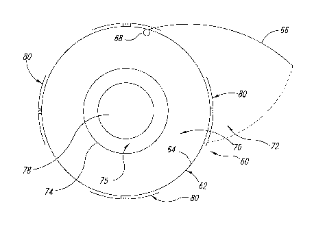
Figure 9 is a cross-sectional view taken along lines 9-9 of the
embodiment of Figure 8; and
Figure 10 is an alternative embodiment of the design of Figures 8
and 9.
DETAILED DESCRIPTION OF THE INVENTION
Referring first to Figure 1, shown therein is a system 10 for
treating immune problems of a patient by affecting the immune elements within
a portion of the patient's blood. The system 10 includes an irradiation
chamber
12 formed in accordance with the invention and a source 14 of light radiation.
4
CA 02577281 2007-03-05
WO 2005/025642 PCT/US2004/028003
An inlet port 16 is formed on the chamber 12 and is coupled by a tube 18 to a
hypodermic needle 20 that is inserted into the arm of a patient for
withdrawing
blood from the patient and returning it after treatment.
The blood of the patient enters into the chamber 12 by gravity
feed and the inherent pressure of the blood stream, whereupon it is stopped
with a stopcock 22 when the chamber 12 is full. The blood in the chamber 12 is
exposed to light emanating from the light source 14 to alter the immune status
of the fixed amount of measured blood within the chamber 12. After exposure,
the blood is then returned to the patient in a reverse direction via the same
pathway. The stopcock 22 is turned to allow fluid from a hung 23 bag or bottle
to enter into the chamber 12, thus forcing the blood back into the patient and
rinsing the chamber 12 of blood.
As illustrated in Figure 2, the chamber 12 has a back plate 24 that
is composed of hard plastic. This back plate 24 has a Luer-type male fitting
26
for connecting the tube 18 from the patient at its lower rear area, and
another
Luer-type female fitting 28 is located at an upper rear area of the back plate
24
for connecting the stopcock 22 and a tube 27 to the intravenous-type fluids to
be delivered to the patient after the blood in the chamber 12 is exposed to
the
UV light.
The chamber 12 has a gasket 30 formed of semisoft plastic,
preferably 2mm thick, and which has an area 32 free within it, preferably 20.0
centimeters by 25.0 centimeters. This free area 32, when a window plate 34 of
quartz or other material is applied, forms a 100.0 cubic centimeter vessel 36
(shown in Figure 6) for measuring the blood to be exposed to the UV light. The
plate 34 and the gasket 30 are held to the back plate 24 with a frame 38 of
hard
plastic through which small screws 39 are fastened into the back plate 24 of
the
chamber 12.
A chamber holder 40 is illustrated in Figure 3 and is configured to
hold the chamber 12 against a housing 42 for the light source 14. Preferably,
the holder 40 is composed of hard plastic. The chamber 12 is placed into a
slot
44 in a lower portion of the holder when a camming clamp is raised. The clamp
46 is hinged to the holder 40 (See Figure 7), and when lowered into position
over the chamber 12, by its wedge shape and its weight it holds the chamber
12 against a frame 41 of the holder 40. The holder 40, being attached to one
end of the light housing 42, thus holds the chamber 42 in place for blood
exposure to the light source 14 at the other end of the housing 42.
5
CA 02577281 2007-03-05
WO 2005/025642 PCT/US2004/028003
The housing 42 illustrated in Figure 4 bears the chamber 12 and
chamber holder 40 at one end and the light source 14 at the other end. The
chamber 12 is composed of reflective metal sheet 48 on one surface, that
surface faced into the center of the housing 42 when the metal sheet 48 is
bent
to form the rectangular tube-shaped housing. The length of the rectangular
tube thus formed is determine by the spread of light from the light source
board
14 , thus aiming to maximize the exposure of the blood in the chamber 12 to
the
light sent from the light source 14.
The light source 4 is mounted to a light source board 50,
illustrated in Figure 5, formed of a printed circuit type board containing an
array
of light emitting diodes 52. In one embodiment of the invention, light source
boards 50 will have ultraviolet light emitting diodes 52. However, other
arrays
can contain a mixture of various wavelength light emitting diodes as the
invention is tailored to treat various diseases more specifically. The board
50
has a USB connection 54 whereby it may be connected to an external
computer of conventional configuration via a cable 56 for powering the light
source 14 and controlling the array of light emitting diodes 52.
Figures 8 and 9 illustrate another embodiment of the invention
wherein a device 60 for irradiating blood is provided. The device 60 includes
a
reflective chamber 62 having in this embodiment a circular cross-sectional
configuration. Ideally, the chamber 62 is formed from a reflective stainiess
steel
wall 64 having an access panel 66 mounted via a hinge 68 thereon. The
access panel 66 opens to provide access to an interior 70 through a
longitudinal opening 72. The device 60 further includes a quartz tube 74
having
a hollow reflective core 78, preferably formed of plastic, that also has a
circular
cross sectional configuration sized and shaped to be received inside the
reflective chamber 62. End caps 76 are placed on each end of the quartz tube
to support the quartz tube inside the chamber 62 and to retain blood in a
space
75 between the core 78 and the tube 74.
A plurality of LED arrays 80 are attached to the outside of the
chamber 62, each having a cover( not shown). Corrugated low-voltage
sheathing (not shown) will pass wires to the array. The LED arrays 80 include
at least one ultraviolet LED. An opening is formed in the chamber 62 at each
LED location to admit light into the chamber 62.
An alternative embodiment of the design shown in Figures 8
and 9 is illustrated in Figure 10 in which the device 90 has a reflective
chamber
6
CA 02577281 2007-03-05
WO 2005/025642 PCT/US2004/028003
72 with an octagonal cross-sectional configuration. An interior-mounted quartz
tube 94 having a circular cross-sectional configuration is shown positioned
inside the chamber 92. In this embodiment irradiation is provided by the LED
arrays 98 arranged around the exterior of the chamber 92. Blood to be treated
will be held stationary in a space 95 between the quartz tube 94 and the core
96.
This tubular design will use components that are readily
commercially available, hence making them extremely inexpensive by
comparison to custom-made components. In addition, this gives the device
disposable characteristics, thus improving contamination safety for the health
care team involved in handling and irradiating the blood.
The advantage of these further embodiments, as with the first
embodiment described above, is that the blood remains fixed or stationary
within the vessel in which it is irradiated. The light source is LED, which
provides instant cooler light and the ability to select wavelengths. A
computer
program is provided that controls the operation of the unit, allowing double-
blind
studies, and data can be transmitted back to a computer for processing. The
program controls intensity, timing, duration, wavelengths of light emission
and
other data to be stored or transmitted to a processor for further processing.
Energy usage from the LEDs is low enough to allow portable units for disaster
relief and field hospitals. Safety is enhanced because light cannot escape
from
the unit and damage eyes.
In operation, the space between the hollow core and the quartz
tube is filled with blood, then disconnected from the patient and the tube is
placed inside the irradiation chamber (62, 92). This offers greater safety to
the
patient by eliminating the chance of electrocution. Alternatively, valves and
hoses may be used as in the first embodiment to couple to the patient and to
the tube 74, as described above in the first embodiment.
Ideally, the quartz tube has a 30 millimeter interior diameter and
contains a 20 millimeter cylindrical core of solid plastic that is reflective,
the
purpose of which is merely to take up space inside the quartz tube and to
provide another reflective surface. The size of the inner reflective core may
be
altered to produce tubular cassettes of varying volumes.
From the foregoing it will be appreciated that, although specific
embodiments of the invention have been described herein for purposes of
illustration, various modifications may be made without deviating from the
spirit
7
CA 02577281 2007-03-05
WO 2005/025642 PCT/US2004/028003
and scope of the invention. Accordingly, the invention is not limited except
as
by the appended claims and the equivalents thereof.
8
